Embed Size (px)
Citation preview

1Wounds UK, 2007, Vol 3, No 1
Wounds UK
The use of Oxyzyme™ sterile wound dressing with
iodine on hard-to-heal wounds:
Case study series
1Front cover.indd 1 2/11/07 13:53:08
The explosion of dressing technology since the discovery by George Winter of the benefi ts of moist wound healing has been dramatic. On a more worrying note is the increasing
number of patients who suffer from wounds that are diffi cult to heal. Despite the abundance of dressings available, and our awareness of this patient group, we are still all too frequently left with patients whose wounds we cannot heal. Is this due to the clinician’s lack of ability to select the correct treatment, or is it more likely to be due to the great diversity of problems that can result in a non-healing wound?
In this supplement, a small series of cases describing clinicians’ experiences with a new dressing, Oxyzyme™ sterile wound dressing with iodine, are presented. The cases demonstrate the use of Oxyzyme™ in patients with a range of diseases, and practical tips that are often required to ensure any new product is used to maximum benefi t are given.
You may feel that yet another dressing will just add to the diffi culty of making the right choice when making wound management decisions, but this unique dressing does offer something new and potentially benefi cial for patients with diffi cult-to-heal wounds. The combination of iodine and oxygen release by a single product provides a means by which two of the factors implicated in non-healing, oxygen imbalance and bioburden, can be addressed, converting such wounds into a healing state.
Oxygen is described by many as the food of life. For many years it has been known that abnormalities in oxygen levels at the surface of the wound exist, which may be the cause of diffi culties in healing.
Two of the most well-respected fi gures in wound research during recent decades — Tom Hunt and Ian Silver — have dedicated their research efforts to discovering how oxygen infl uences healing. It is only now that Oxyzyme™ is available that we have a product that delivers oxygen to the surface of a wound, thus the prospects for this dressing are signifi cant.
In recent years, interest in the use of iodine has waned as more attention has been given to other antiseptics, but it remains a potent agent in treating infection in open wounds. The concept that iodine is released at a rate dependent on the condition of the wound is both novel and relevant.
The real value of Oxyzyme™ is that it combines the delivery of two agents —iodine and oxygen — that are essential to the process of wound healing and provides further opportunities to overcome barriers to healing that may exist in the patient.
This supplement is not able to provide a comprehensive evidence base for this dressing, but the cases illustrated show that it is exciting and has potential for treating the challenging wounds that you and I see in practice. All experiences are evidence and, as such, I would urge you to read this supplement and, when you next see a patient with a wound that is not healing, consider starting your own experiment to see if correction of oxygen or bacterial imbalance can convert the wound to one that is healing.
Keith Harding, Head, Wound Healing Research Unit and Professor of Rehabilitation (Wound Healing), Department of Wound Healing, Cardiff University
Wounds UK
Editorial 3
Product reviewOxyzyme™ sterile wound dressing: a new concept for wound healing 4John Timmons
Case reportsA hard-to-heal venous leg ulcer of 6 months duration 6Andy KerrA hard-to-heal venous leg ulcer of 6 months duration 7Andy KerrThe importance of correctly applying Oxyzyme™ 8Andy Kerr
The importance of selecting the correct secondary dressing 9Andy KerrThe use of Oxyzyme™ to treat a left lateral leg ulcer 10Sylvie HamptonThe need to reassess periwound skin condition when using Oxyzyme™ 12Andy KerrA 6-year-old venous leg ulcer’s progress towards healing 13Andy KerrQuick healing of a chronic wound resulting from a failed peripheral arterial vascular graft (PAVG) 14Johann KollerChronic bilateral leg ulcers at the end of life 15Melvin Bertram
3 Wounds UK
CONTENTS AND EDITORIAL
Editorial
2-15Oxyzyme doc.indd 3 2/11/07 13:56:49

4 Wounds UK
CASE STUDY
Slow wound healing in patients with chronic or complex wounds has a negative impact on the patients’ quality
of life and can result in increased costs of care (Mathias et al, 2000). The aging patient population and the range of associated illnesses such as diabetes and cardiovascular disease can all impact on the wound healing process. Concomitant disease often combines with mechanical and chemical trauma in the skin of the older person, leading to the development of wounds which may then become chronic.
A chronic wound is a wound and its underlying aetiology, which does not respond to treatment (Enoch and Price, 2004). Chronic wounds will often be locked in the inflammatory phase of wound healing, necrotic or sloughy tissue will be difficult to remove due to its adherence to the wound bed and the cycle of events within the wound will prolong the inflammatory process. Excess quantities of proteases, cell senescence, high levels of bacteria and high pH are just a few of the possible factors which can affect the healing process (Falanga, 2002).
Thorough assessment of the tissue types present in the wound in addition to the general history of the patient will enable the practitioner to build up an idea of what may be happening in the wound itself to result in wound healing. For example, the presence of significant amounts of devitalised tissue in the wound will delay healing and predispose it to colonisation and infection by providing a source of attachment and nutrients for bacteria. Therefore, the expedient removal of such tissue is advisable for optimal healing.
Debridement of sloughy/necrotic tissue is one of the cornerstones of wound bed preparation and should be viewed as an ongoing process which is complete only when the wound bed consists of 100% granulation tissue (Vowden and Vowden, 1999). For many patients, debridement can be a long process and some treatments which may speed up the process, such as surgical or sharp debridement, may not be available, due to a lack of qualified personnel. Evidence as to the best methods of debridement is scarce and in clinical practice, there are a large number of alternatives in use which can be confusing for the practitioner. Products which can be used to debride wounds include hydrogels, iodine-based dressings, honey and larval therapy. For a number of years, enzymatic debridement was also a common treatment using streptokinase and streptodornase solution. The use of hydrogels in debridement is a standard treatment for assisting in the autolysis of sloughy tissue, and although this process may be viewed as slow, it is a relatively safe treatment option.
Once the wound environment has been optimised in terms of removing any physical barriers to healing, attention should be turned to maintaining the viability of the healthy tissue and establishing conditions that facilitate the proliferation of new tissues within the wound by providing a moist wound healing environment. Important systemic factors that promote healing include adequate nutrition and blood supply. A common causative factor for delayed healing is interrupted or inadequate blood flow, which leads to hypoxia and tissue death (Grey et al, 2006).
The role of oxygen in healingIt must be remembered that in a wound care context, many patients present with
conditions which result in a reduction in local blood supply to the wounded area. Patients with illnesses such as respiratory disease, impaired cardiac function and anaemia may experience local hypoxia which will negatively impact on the wound healing process (Hunt et al, 2004).
Oxygen is a vital component of almost every part of cellular activity. It is vital for energy metabolism and is used by cells to drive cellular function. In an oxygen poor environment, key cells may not function at normal levels of efficiency. In the wound this could lead to neutrophil failure reducing their ability to destroy bacteria. Furthermore, oxygen also assists in creating an environment which is not suitable for certain anaerobic bacteria, helping to reduce the bacterial load of the wound (Knighton et al, 1986).
Jonsson et al (1991) also discussed the importance of the role of oxygen in collagen synthesis and in angiogenesis, finding that key enzymes involved in these processes are dependent on oxygen and, without its presence, these components of healing are negatively affected and wound healing may be slowed as a result.
Current oxygen therapies include hyperbaric oxygen which has been considered an effective treatment for wound care, although this is expensive and is not available for every patient. The use of oxygen delivery devices, whether topical or systemic, remains controversial but there is a growing body of evidence (Ivins, 2007).
Davis and Sibbald (2007) state that oxygen balance may often become the rate-limiting factor in the healing response, and that every wound should have its oxygen supply balanced with its oxygen demand.
Product REVIEW
Oxyzyme™ sterile wound dressing: a new concept for wound healingJohn Timmons
John Timmons is Clinical Manager, Wounds UK and Tissue Viability Nurse, Grampian NHS Trust
2-15Oxyzyme doc.indd 4 2/11/07 13:56:49

5Wounds UK
CASE STUDY
If local oxygen delivery is to be useful, it should be integrated into good wound bed preparation. Therefore, wound care clinicians should demand new dressings with oxygen accessibility in addition to the standard wound bed preparation pillars (Davis and Sibbald, 2007).
The Oxyzyme™ Sterile Wound Dressing with Iodine (Insense Ltd, UK) is a sheet hydrogel dressing with a debriding action, antimicrobial properties and the unique ability to transport dissolved oxygen to the wound.
Oxyzyme™: a unique wound dressingOxyzyme™ consists of two advanced hydrogels; a wound contact gel and a smaller enzyme-containing gel. The wound contact gel contains glucose and a very small quantity of iodide (less than 0.04%w/w). The enzyme gel contains glucose oxidase, a naturally occurring enzyme. The gels are packed separately. When the two gels are combined the biochemical action action of the dressing is activated.
Mode of actionOxyzyme™ is activated by placing the enzyme layer on top of the wound contact layer. Atmospheric oxygen at the surface of the enzyme layer is used by the glucose oxidase within the dressing to generate a low level of hydrogen peroxide. This creates an oxygen gradient which allows more oxygen to be drawn into the dressing. The hydrogen peroxide is freely soluble and diffuses through the dressing, where it interacts with iodide to liberate iodine and dissolved oxygen (no hydrogen peroxide reaches the wound). The iodine changes the colour of the dressing to amber/gold and helps create an environment hostile to bacteria.
Clinical effectsOxyzyme™ dressing works with the wound on many levels to assist wound healing: 8 The high water content of the dressing
provides a moist wound healing environment to assist in the rehydration of eschar and the autolytic debridement of slough
8 Oxyzyme™ transports dissolved oxygen to the wound interface (Ivins et al, 2007)
It is essential to treat the patients’ underlying/concurrent conditions to maximise the chance of a successful outcome. For example, in patients with venous leg ulcers, the recommended treatment is compression bandaging to help promote venous return. A recent study has demonstrated the dressing’s efficacy beneath compression bandaging (Ivins et al, 2007). Other aspects of care such as nutritional status must also be considered.
ConclusionHard-to-heal wounds present a huge challenge for the clinician. The easy to apply Oxyzyme™ system is a promising new weapon in this field. It is a unique dressing that can be used in wide range of healthcare settings.
.ReferencesDavis P, Sibbald RG (2007) Guest Editorial: Oxygen balance – what’s that? Int Wound J 4(s3): 2–3
Enoch S, Price P (2004) Should alternative endpoints be considered to evaluate outcomes in chronic recalcitrant wounds? www.worldwidewounds.com/2004/October/Enoch-part 2/Alternative-Endpoints. Date last accessed 22 October 2007
Falanga V (2002) Wound Bed Preparation and the role of enzymes: a case for multiple actions of therapeutic agents. Wounds 14(2): 47–57
Grey JE, Enoch S, Harding KG (2006) Venous and arterial leg ulcers. Br Med J 332(7537): 347–50
Hunt TK, Ellison EC, Sen CK (2004) Oxygen the foundation of wound healing-introduction. World J Surg 28(3): 291–3
Ivins N, Simmonds W, Turner A, Harding KG (2007) The use of an oxygenating hydrogel dressing in venous leg ulceration. Wounds UK 3(1): 77–81
Jonsson K, Jensen JA, Goodson WH, et al (1991) Tissue oxygenation anaemia and perfusion in relation to wound healing in surgical patients. Ann Surg 214(5): 605–13
Knighton DR, Halliday B, Hunt TK (1986) Oxygen as an antibiotic: A comparison of the effects of inspired oxygen concentration and antibiotic administration on in-vivo bacterial clearance. Arch Surg 121(2): 191–5
Mathias SD, Prebil LA, Boyko WL, Fastenau J(2000) Health-related quality of life in venous leg ulcer patients successfully treated with apligraf: A pilot study. Advances Skin Wound Care 13: 76–8
Vowden KR, Vowden P (1999a) Wound debridement, Part 1: non-sharp techniques. J Wound Care 8(5): 237–40
Product REVIEW
8 Iodine, generated from iodide, has an antimicrobial effect to help reduce the overall bioburden of the wound
8 The dressing provides a physical barrier to bacteria.
Ivins et al (2007) reported preliminary findings of a multi-centred study of the use of Oxyzyme™. The wounds of five patients with hard-to-heal venous leg ulcers healed within the 6-week trial period. Their findings indicated that Oxyzyme™ can trigger the healing process in hard-to-heal wounds.
Davis and Sibbald (2007) state that oxygen balance may often become the rate-limiting factor in the healing response...If local oxygen delivery is to be useful, it should be integrated into good wound bed preparation. Therefore wound care clinicians should demand new dressings with oxygen accessibility in addition to the standard wound bed preparation pillars.
Indications for useUnder the supervision of a healthcare professional, the Oxyzyme™ system may be used for the treatment of non-infected or mildly-infected wounds. It may be used on moderately exuding, non-exuding and dry wounds. If the wound is dry to low exudate, a breathable polyurethane film may be an appropriate secondary dressing. For moderately exuding wounds the use of a foam secondary dressing may be more appropriate. Due to the action of the Oxyzyme™ therapy, the dressings should be changed every 2–3 days, depending on the nature of the wound, in order to ensure that the dressing remains active in situ. It is also important that the dressing maintains a good contact with the wound surface.
2-15Oxyzyme doc.indd 5 2/11/07 13:56:50

6 Wounds UK
CASE STUDY 1
Andy Kerr, Tissue Viability Nurse, TVCS, Eastbourne
A hard-to-heal venous leg ulcer of 6 months duration
Figure 2. At week 5 of treatment, the wound had healed.
The patientPatient A was a 94-year-old female who presented with two venous leg ulcers on her right leg which had been present for six months. The upper ulcer was a shallow wound on her right knee. The patient had a history of osteoarthritis and mild heart failure, and was taking diuretics for bilateral leg oedema.
The woundAt presentation, the would measured 7.49cm2 , was shallow with an indistinct margin, and consisted of 50% slough and 50% granulation tissue. There was a low volume of clear exudate and the periwound skin was healthy and dry (Figure 1). An OxyzymeTM dressing was applied to the wound and held in position with a Tegaderm® Film dressing (3M Healthcare, Loughborough). Dressings were changed twice a week. On first assessment, following a week of treatment with OxyzymeTM, the wound area had decreased to 3.39cm2, and the wound bed was covered with 100% granulation tissue. Haemoserous exudate was noted and thought to be from the delicate new blood vessels within the wound. The periwound skin remained healthy and dry. At week 2, healthy granulation tissue was apparent and the wound had again significantly reduced in size to 2.32cm2. Wound exudate was still haemoserous and the periwound skin had rehydrated. By week 3 the wound showed very active granulation and measured 1.79cm2. By the fifth week assessment the wound had healed (Figure 2). The patient was satisfied with the outcome and considered the dressing to be comfortable.
Figure 1. The wound at initial assessment.
Conclusions
In this patient, OxyzymeTM successfully debrided the sloughy tissue and supported the formation of granulation tissue in the wound. After 5 weeks of treatment, the wound had healed.
2-15Oxyzyme doc.indd 6 2/11/07 13:56:52

7Wounds UK
CASE STUDY 2
Andy Kerr, Tissue Viability Nurse, TVCS, Eastbourne
A hard-to-heal venous leg ulcer of 6 months duration
Figure 1. The wound at initial assessment.
Figure 2. The wound at the 5 week assessment.
The patientPatient B was a 94-year-old female who presented with two venous leg ulcers on her right leg of six months duration. The lower ulcer was a shallow wound below her right knee. The patient had a history of osteoarthritis and mild heart failure and was taking diuretics for bilateral leg oedema.
The woundAt presentation, the wound measured 6.30cm2 and was assessed as being shallow with an indistinct margin and consisting of 80% slough and 20% granulation tissue. There was a low level of clear exudate and the periwound skin was healthy and dry (Figure 1). Tegaderm® Film (3M Healthcare, Loughborough) was used as a secondary dressing. Dressings were changed twice weekly.
At the first assessment, following a week of treatment with OxyzymeTM, the wound area had decreased significantly to 4.18cm2, the slough had reduced to 50% and the wound presented with ’healthier granulation tissue’ that was rehydrated and more vascular in appearance. The exudate remained clear and the periwound skin was rehydrated. On week 2 examination, the wound consisted of 100% healthy granulation tissue and had again reduced in size to 3.76cm2. Exudate remained clear and the periwound skin remained healthy. By week 3, the wound measured 1.60cm2 with some sersanguinous exudate present. At week 4, the wound was almost closed, following a further reduction in size to 0.41cm2. A small amount of slough was observed (5%). Wound exudate was still haemoserous. By the fifth week of treatment, the wound had almost healed (Figure 2).The patient was satisfied with the outcome and reported finding the dressing comfortable to wear throughout the treatment period and the periwound skin also improved during this time. The wound subsequently went on to heal fully.
Conclusions
In this patient, Oxyzyme™ was used for five weeks and the wound progressed significantly. The wound almost completely healed, a significant reduction in sloughy tissue was noted and the periwound skin also improved during this period.
2-15Oxyzyme doc.indd 7 2/11/07 13:56:54

8 Wounds UK
CASE STUDY 3
Andy Kerr is Tissue Viability Nurse, TVCS, Eastbourne
The importance of correctly applying Oxyzyme™
Figure 1. The wound at first assessment.The patientPatient C was an 87-year-old female who presented with a left lateral leg ulcer proximal to the malleolus that had been present for a year. The patient has a history of rheumatoid arthritis, non-insulin dependent diabetes (NIDDM) and was taking methotrexate, folic acid, oxycodone, iron sulphate, amperozide, glicazide and zopiclone.
The wound Initially, the wound measured 4.13cm2 and was assessed as being static and shallow with a distinct margin. The wound bed consisted of 90% slough and 10% granulation tissue. Exudate was clear and serous, and the initial periwound skin was inflamed with slight erythema (Figure 1). OxyzymeTM was applied to the wound and dressings were changed twice a week (Figure 2). DuoDERM® (ConvaTec, Ickenham) was initially used as a secondary dressing, but when the supply ran out at week 2, Mepilex® Border (MöInlycke, Dunstable) was used. The patient was happy with the OxyzymeTM dressing and described it as comfortable.
At first assessment, following a week of treatment with OxyzymeTM, the wound area had decreased to 3.18cm2 and the slough had decreased to 65% with granulation tissue covering 35% of the wound bed. Haemoserous exudate was noted and the periwound skin was healthy with less erythema. At week 2 assessment, the wound appeared to have worsened, relative to the previous assessment, with an increase in slough comparable to that at the start of treatment. The exudate was still haemoserous.
At week 3, it was noted that the OxyzymeTM dressing had been incorrectly applied at the last dressing change (with the primary dressing applied on top of the secondary). As a result, no records were taken at this assessment, but OxyzymeTM was now correctly applied. At week 4 (Figure 3), the wound measured 2.97cm2, showing a 52% reduction from the size recorded at the week 2 assessment. The wound was covered in 90% epithelial and 10% granulation tissue. The exudate was again haemoserous, and the periwound skin remained healthy. At week 5, despite a small amount of slough being observed, the wound had decreased to 2.48cm2, and the exudate was now clear. At week 6, a further decrease in size was noted with the wound measuring 1.52cm2 and, although the exudate was again haemoserous, it was anticipated that closure was imminent (Figure 4). At week 10 at a final follow-up assessment, the wound remained closed. The patient was satisfied with the outcome and considered the dressing to have been comfortable to wear throughout.
Conclusions
Despite some time with the dressing being inappropriately applied, the wound continued to heal and by week 10 was completely closed. The sloughy tissue was debrided and the Oxyzyme™ dressing continued to support the healing process, until re-epithelialisation had been achieved.
Figure 4. The wound was almost closed at the 6-week assessment.
Figure 2. The wound with the Oxyzyme™ dressing in place.
Figure 3. The wound at week 4 review.
2-15Oxyzyme doc.indd 8 2/11/07 13:56:58

9Wounds UK
CASE STUDY 4
Andy Kerr is Tissue Viability Nurse, TVCS, Eastbourne
The importance of selecting the correct secondary dressing
Figure 2. Wound with the Oxyzyme™ dressing in place at the week one dressing change.
The patientPatient D was a 78-year-old female who presented with a sacral pressure ulcer of one year’s duration. The patient also had Alzheimer’s disease.
The woundThe wound was assessed as being a static, granulating cavity with a distinct margin. It measured 33.32cm2 . Moderate to high levels of purulent exudate were being generated, but the periwound skin was healthy (Figure 1). Oxyzyme™ was applied to the wound and Granuflex® hydrocolloid (ConvaTec, Ickenham) was used as a secondary dressing, as this was available on the local formulary. The dressing was deemed by the patient to be comfortable on first application and was changed 2–3 times per week. At the first assessment, following a week of treatment with Oxyzyme™, the wound area had not changed significantly (Figure 2). At week 2, the wound was considered to be ‘active in areas’ and on a healing trajectory, despite a 6% increase in wound area. The clinician observed that the wound was filling with granulation tissue and its depth was decreasing. By week 3, the wound had reduced to 32.09cm2 returning to approximately the same size as at the outset of the assessment. The exudate was less purulent, but volume remained high. As Oxyzyme™ is contraindicated for heavily exuding wounds, the trial was stopped, with the clinician recording an assessment of ‘some improvement.’
The purpose of the Oxyzyme™ case study programme of which this study was a part is to assess the performance of the dressing in real-life situations. The study protocol indicates that a breathable secondary dressing should be selected from the local formulary. Granuflex®, however, is an occlusive dressing as independent in vitro laboratory testing has shown (Thomas et al, 2005). Oxyzyme™ would therefore have been unable to deliver oxygen to the surface of the wound and so its performance would be expected to be comparable to that of a conventional sheet hydrogel. The biochemical reaction produced in Oxyzyme™ requires gaseous oxygen to be present at the surface of the dressing. Any secondary dressing used should allow oxygen to pass through it, as is indicated in the instructions for use. Typically this can be a breathable polyurethane film, where exudate level is zero to low, or a breathable foam dressing where there is a moderate level of exudate.
Thomas S, Hughes G, Fram P, Hallett A (2005) Comparative ‘in-vitro’ testing of hydrocolloids, hydrogels, alginate/CMC fibrous and foam dressings. Surgical Materials Testing Laboratory, Princess of Wales Hospital, Bridgend: http://www.worldwidewounds.com/
Conclusions
The unsuccessful use of an occlusive dressing in this case demonstrates the importance of the biochemical reaction that produces oxygen and iodine in Oxyzyme™. Without this reaction, the dressing behaves in the same way as any normal sheet hydrogel, so it is essential that the correct secondary dressing is used.
Figure 1. Wound at initial assessment.
2-15Oxyzyme doc.indd 9 2/11/07 13:56:59

10 Wounds UK
CASE STUDY 5
Sylvie Hampton, Tissue Viability Nurse, TVCS, Eastbourne
The patientPatient E was a 90-year-old female who presented with a left lateral leg ulcer which had been present for one year. No relevant medical history was recorded.
The wound On initial assessment, the wound was static and covered in slough, with a distinct margin. There was a moderate amount of green exudate and the periwound skin was inflamed and macerated. No measurement of wound size was made at the initial assessment. Oxyzyme™ was applied and held in position by a breathable polyurethane film dressing. Dressing changes took place twice a week. Flamazine was applied to the periwound skin. The patient considered the dressings to be comfortable.
At the first assessment, following a week of treatment with Oxyzyme™, the wound measured 20.64cm2 and was covered with 50% slough. Exudate was serosanguinous and the periwound skin was not macerated but still inflamed, with less erythema. The use of flamazine on the periwound skin was continued (Figure 1). At week 2, the level of slough had decreased to 10%, with the remainder of the wound bed consisting of healthy granulation tissue. The exudate was haemoserous but the periwound skin was found to be healthy with only very slight erythema (Figure 2). As a result, the application of flamazine was discontinued. No measurement of the wound area was taken.
Figure 3. Week 4.
Figure 2. Week 2.
Figure 1. Week 1.
The use of Oxyzyme™ to treat a left lateral leg ulcer
2-15Oxyzyme doc.indd 10 2/11/07 13:57:02

11Wounds UK
CASE STUDY 5
Conclusions
By week 9 of treatment the wound had almost healed, the exudate had reduced and was no longer green in colour. The surrounding skin was much less inflamed and there were no signs of infection. Treatment continued and the wound progressed to healing in 11 weeks.
Figure 5. Week 9.
Figure 4. Week 5.
At week 3, the wound bed was composed entirely of healthy granulation tissue and had reduced in size by 29% since the week one assessment to 14.58cm2. Exudate remained haemoserous and the periwound skin healthy.
By week 4 (Figure 3), the wound bed was still covered in granulation tissue and had reduced in size by a fur ther 10% to 13.17cm2. By week 5 (Figure 4) the wound continued on a healing trajectory, reducing in size to 12.20cm2. The periwound skin was mildly macerated and the exudate was haemoserous. Although the formal case study had completed the 6-week assessment, it was decided to continue treating the patient with Oxyzyme™ to achieve healing.
Re-examination at week 7 showed that the wound had dramatically decreased in size by 62% during the 2-week interval since the previous assessment and now measured 4.59cm2 . Monitoring and photography of the wound while being treated with Oxyzyme™ continued (Figure 5) and the wound healed fully after 11 weeks.
2-15Oxyzyme doc.indd 11 2/11/07 13:57:04
12 Wounds UK
CASE STUDY 6
Andy Kerr, Tissue Viability Nurse, TVCS, Eastbourne
The need to reassess periwound skin condition when using Oxyzyme™
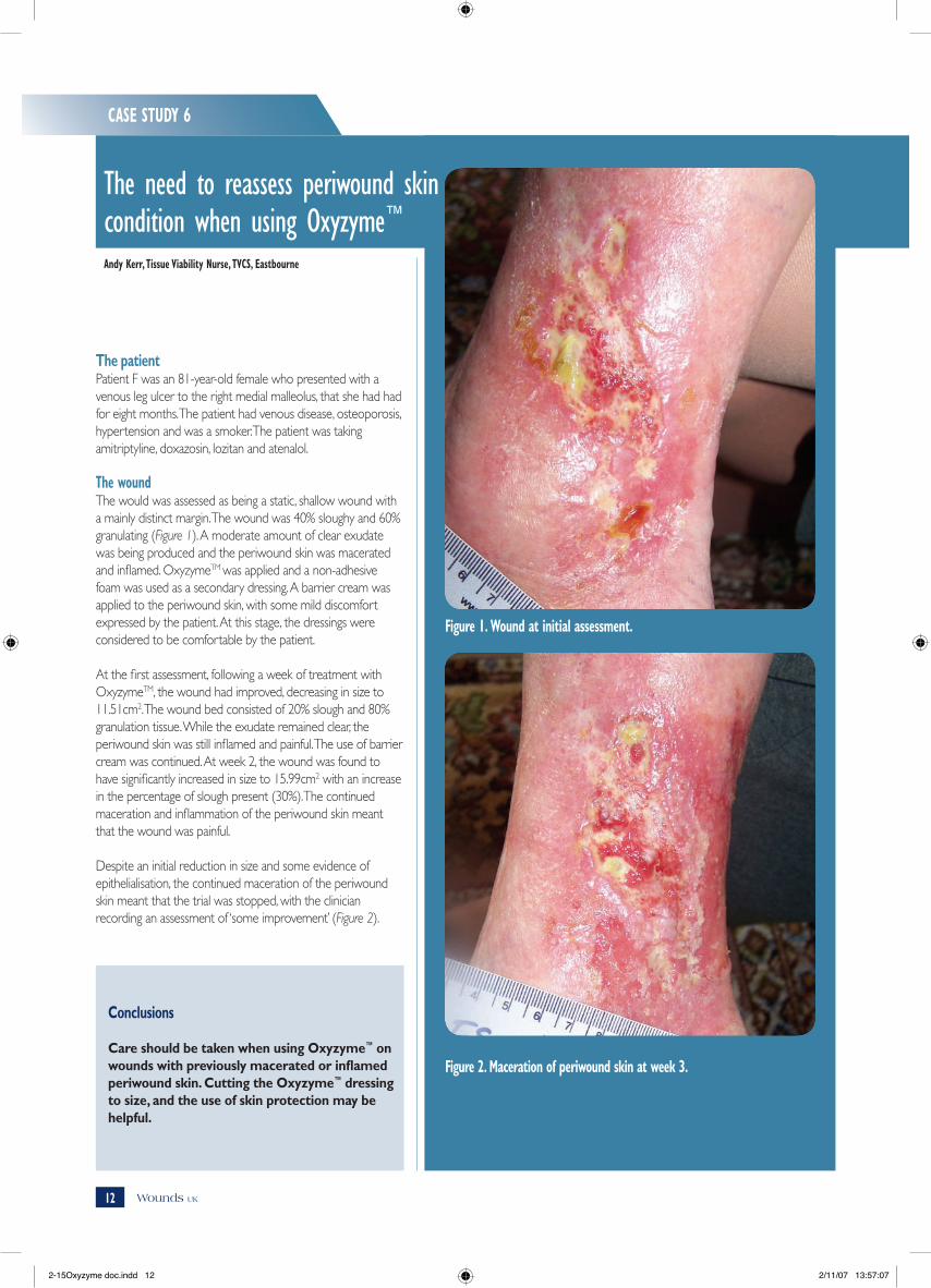
The patientPatient F was an 81-year-old female who presented with a venous leg ulcer to the right medial malleolus, that she had had for eight months. The patient had venous disease, osteoporosis, hypertension and was a smoker. The patient was taking amitriptyline, doxazosin, lozitan and atenalol.
The woundThe would was assessed as being a static, shallow wound with a mainly distinct margin. The wound was 40% sloughy and 60% granulating (Figure 1). A moderate amount of clear exudate was being produced and the periwound skin was macerated and inflamed. OxyzymeTM was applied and a non-adhesive foam was used as a secondary dressing. A barrier cream was applied to the periwound skin, with some mild discomfort expressed by the patient. At this stage, the dressings were considered to be comfortable by the patient.
At the first assessment, following a week of treatment with OxyzymeTM, the wound had improved, decreasing in size to 11.51cm2. The wound bed consisted of 20% slough and 80% granulation tissue. While the exudate remained clear, the periwound skin was still inflamed and painful. The use of barrier cream was continued. At week 2, the wound was found to have significantly increased in size to 15.99cm2 with an increase in the percentage of slough present (30%). The continued maceration and inflammation of the periwound skin meant that the wound was painful.
Despite an initial reduction in size and some evidence of epithelialisation, the continued maceration of the periwound skin meant that the trial was stopped, with the clinician recording an assessment of ‘some improvement’ (Figure 2).
Conclusions
Care should be taken when using Oxyzyme™ on wounds with previously macerated or inflamed periwound skin. Cutting the Oxyzyme™ dressing to size, and the use of skin protection may be helpful.
Figure 2. Maceration of periwound skin at week 3.
Figure 1. Wound at initial assessment.
2-15Oxyzyme doc.indd 12 2/11/07 13:57:07

13Wounds UK
CASE STUDY 7
Andy Kerr, Tissue Viability Nurse, TVCS, Eastbourne
A 6-year-old venous leg ulcer’s progress towards healing
Figure 1. The wound at initial assessment.
The patientPatient G was a 72-year-old female who presented with a venous leg ulcer to the left lateral maleollus that had been present for six years. The patient was taking paracetamol and also had Parkinson’s disease for which she was taking co-careldopa.
The woundOn presentation, the wound was shallow with an indistinct border, as a result of which the wound size was difficult to determine. It was estimated at approximately 5cm2. The wound bed comprised 50% slough and 50% granulation tissue. A moderate amount of clear exudate was produced but the periwound skin was inflamed (Figure 1). The periwound area was treated with an emollient, and an Oxyzyme™ dressing applied to the wound which was held in place with foam and film secondary dressings.
At the first assessment, following a week of treatment with Oxyzyme™, the wound measured 4.47cm2.The wound bed still consisted of 50% slough and 50% granulation tissue but erythema in the surrounding skin had reduced. Following two weeks of treatment, the wound was found to have decreased in size by 12% to 3.93cm2, and consisted of 20% slough and 80% granulation. By week 3, the wound measured 2.47cm2, a reduction of size by 51% since inclusion in the study, but the case was stopped due to patient discomfort (Figure 2).
Conclusions
In this patient, the wound was progressing towards healing, however, the patient was complaining of discomfort and it was therefore decided to stop using the treatment.
Figure 2. The wound at final assessment showing removal of Oxyzyme™ dressing before wound cleaning.
2-15Oxyzyme doc.indd 13 2/11/07 13:57:08
14 Wounds UK
CASE STUDY 8
Dr Johann Koller, Klinikum am Steinenburg, Reutlingen, Germany
Quick healing of a chronic wound resulting from a failed PAVG
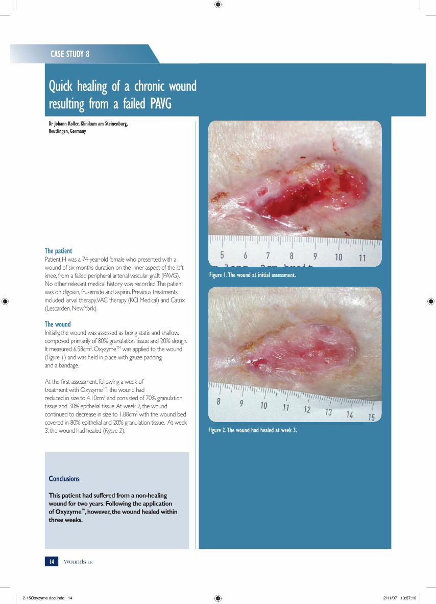
Figure 1. The wound at initial assessment.
The patientPatient H was a 74-year-old female who presented with a wound of six months duration on the inner aspect of the left knee, from a failed peripheral arterial vascular graft (PAVG). No other relevant medical history was recorded. The patient was on digoxin, frusemide and aspirin. Previous treatments included larval therapy, VAC therapy (KCI Medical) and Catrix (Lescarden, New York).
The woundInitially, the wound was assessed as being static and shallow, composed primarily of 80% granulation tissue and 20% slough. It measured 6.58cm2. OxyzymeTM was applied to the wound (Figure 1) and was held in place with gauze padding and a bandage.
At the first assessment, following a week oftreatment with OxyzymeTM, the wound hadreduced in size to 4.10cm2 and consisted of 70% granulation tissue and 30% epithelial tissue. At week 2, the wound continued to decrease in size to 1.88cm2 with the wound bed covered in 80% epithelial and 20% granulation tissue. At week 3, the wound had healed (Figure 2).
Conclusions
This patient had suffered from a non-healing wound for two years. Following the application of Oxyzyme™, however, the wound healed within three weeks.
Figure 2. The wound had healed at week 3.
2-15Oxyzyme doc.indd 14 2/11/07 13:57:10

15Wounds UK
CASE STUDY 9
Melvin Bertram, Tissue Viability Nurse, Aberdeen Royal Infirmary, Aberdeen
Chronic bilateral leg ulcers at the end of life
Figure 1. The wound at presentation.
Figure 2. The wound following honey treatment.
The patientA 92-year-old female presented with right leg cellulitis, chronic bilateral leg ulcers of six months duration and dry gangrenous toes. The patient had recently been diagnosed with critical ischaemia and had undergone an angioplasty on the right leg, following a multi-focal stenosis in the superficial femoral artery and a short popliteal occlusion. Past medical history showed type 2 diabetes, aortic stenosis, peripheral vascular disease and chronic renal failure.
The woundsOn initial assessment (Figure 1), the left leg ulcers were 100% granulating and showing no signs of infection. Conversely, the right leg ulcer, on the outer gaiter region, measured 9cm x 6cm in size; the wound bed was shallow with superficial sloughy areas and red granulating tissue; there was moderate exudate production, and the surrounding skin was well defined, but showed signs of spreading infection and localised oedema. An urgent podiatry review was requested for the ischaemic toes. The patient was commenced on intravenous antibiotics and a topical silver sulphadiazine cream. By day 14, the underlying cellulitic infection on the right leg and subsequent deterioration in the patient’s condition had caused the wound bed to become yellow/black. The previous topical treatment was changed to a honey ointment to facilitate faster autolytic debridement (Figure 2). By day 52, the wound size had reduced to 6.75cm x 2.5cm, exudate levels were low and the surrounding skin showed no signs of infection. The wound bed consisted of 75% red granulating tissue and 25% yellow tissue. At this stage, the honey ointment was discontinued and an Oxyzyme™ dressing was applied, with a secondary Tegaderm® film dressing held in place with a tubular retention stocking. One week after commencing Oxyzyme™, the wound bed was 100% granulating and had reduced in size to 6cm x 2.5cm (Figure 3). The surrounding tissue showed signs of epithelialisation at the margins. Although the removal of the dressing caused no pain, some bleeding of the wound bed was noted. On review one week later, the patient’s condition had deteriorated significantly, so an examination of the wound was inappropriate. However, staff reported that even though the patient’s condition had deteriorated, the wound had continued to progress during the previous week. The patient died several days later.
Conclusions
During the short time the dressing had been in situ, the wound reduced in size, and if the patient’s outcome had been different, it was anticipated that the wound would have progressed to closure.
Figure 3. The wound following one week of Oxyzyme™ treatment.
2-15Oxyzyme doc.indd 15 2/11/07 13:57:12