Embed Size (px)
Citation preview

Exploring Hand Therapy Manual
Wrist SecretsThe Occupational Therapist’s
ApproachPart 2
Explor
ingHand TherapyEx
plor
ingHand Therapy
Treatment 2 Go®
ww
w . e x p l o r i n g h a n d t h e r a p y . co m
Exploring Hand Therapy, Corporationwww.exploringhandtherapy.com
727-341-1674Fax: 727-388-3904

1
Wrist Secrets: Part 2 The Occupational Therapists Approachp p pp
Susan Weiss
This Section is designed
• To enhance what you learned in Part 1
The ExaminationQuick review of the carpal bones and
landmarks
How to Palpate the wrist bones• Trapezium
• Trapezoid
• Capitate
• Hamate
• Lunate
• Scaphoid
• Pisiform
• Triquetrum
Zones ‐ 5
• Radial Dorsal
• Central Dorsal
• Ulnar Dorsal
• Radial Volar
• Ulnar Volar

2
Radial Dorsal
• Radial Styloid palpation
• Tenderness may indicate• Contusion
• Fracture• Fracture
• Radioscaphoid arthritis
• Radial deviation may increase tenderness in this area
Radial Dorsal
• Palate scaphoid– Tubercle on volar wrist
• Tenderness here may indicate:F– Fracture
– Non‐union– Instability
Radial Dorsal
• ST Joint and Trapezium
• Find the trapezium first
• Possible problems– ST arthritis– Instability
– First CMC OA
Radial Dorsal
• Base of 1st metacarpal
• CMC joint
Radial Dorsal
• First extensor compartment (EPB and APL)– Forming the snuffbox
– Finkelsteins test ( de Quervain’s)
Radial Dorsal
• EPL tendon– Check for extension lag
– Palpate for tenderness

3
Radial Dorsal
• Palpate muscle bellies of EPB and APL as they cross the wrist tendons
Radial Dorsal
• DRSN – Irritation here is referred to as Wartenberg’s syndrome
– If this nerve is irritated it will cause numbnessIf this nerve is irritated it will cause numbness, burning and pain
Central Dorsal
• Lister’s Tubercle– Separates the scaphoid and lunate fossa
Central Dorsal
• Lunate– Just distal and ulnar to Listers tubercle
– Forms a rounded prominance
Central Dorsal
• Scapholunate interval
• Tenderness may indicate– Dorsal wrist ganglion
S L li i j– S‐L ligament injury
– Dorsal wrist syndrome
Central Dorsal
• Dorsal wrist syndrome– Described by Watson as a localized SL synovitis
– Positive test produces pain in SL region

4
Central Dorsal
• S‐L ligament injury
– Scaphoid shift test (Watson test)
SL b ll– SL ballottement test
Central Dorsal
• Base of 2nd and 3rd metacarpal pain
• Tenderness may indicate ligament injury
• Linscheid test is performed to assess for i biliinstability
Central Dorsal
• Assess ECRL, ECRB, and EDC for tenderness indicating possible tendonitis
• PIN (motor nerve to the extensors)
Ulnar Dorsal
• DRUJ
• Tenderness here may indicate
• Arthritis or instability
• Prominence of the ulnar head is a sign of instability
• Piano key test – move the distal ulna dorsal and volar – positive if pain and increased mobility
Ulnar Dorsal
• TFCC– Can palpate between the ulnar head and triquetrum
– TFCC loadTFCC load
Ulnar Dorsal
• Hamate
• Triquetrum

5
Ulnar Dorsal
• Midcarpal instablity– Characterized by a volar sag
– Midcarpal shift test
Ulnar Dorsal
• LT interval– Palpate just ulnar to the lunate
– LT instability causes tenderness here
Ulnar Dorsal
• Ballottement test or Shuck Sign – Described by Regan• Kleinman’s LT shear test• Ulnar snuffbox test or Ballotment Test – Described by
Linscheid
Ulnar Dorsal
• 4th and 5th CMC joints– Tenderness may indicate ligament injury or fracture
• ECU tendon• ECU tendon– Tenderness with resisted motion may indicate tendonitis
– Pain and snapping with forearm rotation
Radial Volar
• Radial styloid
• Tenderness may be due to:– Fractures
R di l li i j– Radiocarpal ligament injury
– Wrist extension and radial will accentuate ligament injury
Radial Volar
• Scaphoid tuberosity – Assess in radial deviation
• STT jointC di l l i i d h i i– Can cause radial volar wrist pain due to arthritis here
– Radial deviation is often painful

6
Radial Volar
• Volar wrist ganglion– Arises out of the radiocarpal
– Presents with swelling at the base of the thumb
T i• Trapezium– Located just distal to the pole of the scaphoid– Tenderness may indicate a fracture
Radial Volar
• FCR tendonitis– Swollen and tender with palpation
• Digital flexor tendonsfl i i– flexor tenosynovitis
Radial Volar
• Median Nerve
• Radial Artery
Ulnar Volar
• Pisiform– Tenderness here may indicate:
– Fracture
Pi t i t l th iti• Pisotriquetral arthritis– Shear test for pisotriquetral arthritis
Ulnar Volar
• Hook of the Hamate– Hamate fracture
• Ulnar nerveC i f h G ’ l– Compression of the nerve at Guyon’s canal
Ulnar Volar
• FCU– Palpate at ulnar volar wrist

7
Exam review
• The exam of the wrist is very detailed
Treatment of the wrist by diagnosis
The names below are applied to specific patterns of distal radius
fracture• Colles
• Smith
• Barton’s• Barton’s
• Chauffeur’s
39 40
Physician treatment after fracture of distal radius:
• Closed reduction and casting
• Percutaneous pinning
• Arthroscopic assisted fixation
• ORIF
• Ex‐Fix
Regardless of Method Used for reduction…
• The physician must attempt to restore:– Articular congruency
– Radial length
Volar tilt– Volar tilt
– Radial inclination– Assess DRUJ instability and treat if necessary
42
8
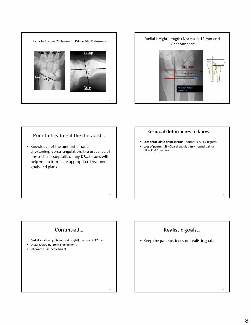
Radial Inclination (22 degrees) Palmar Tilt (11 degrees)
43
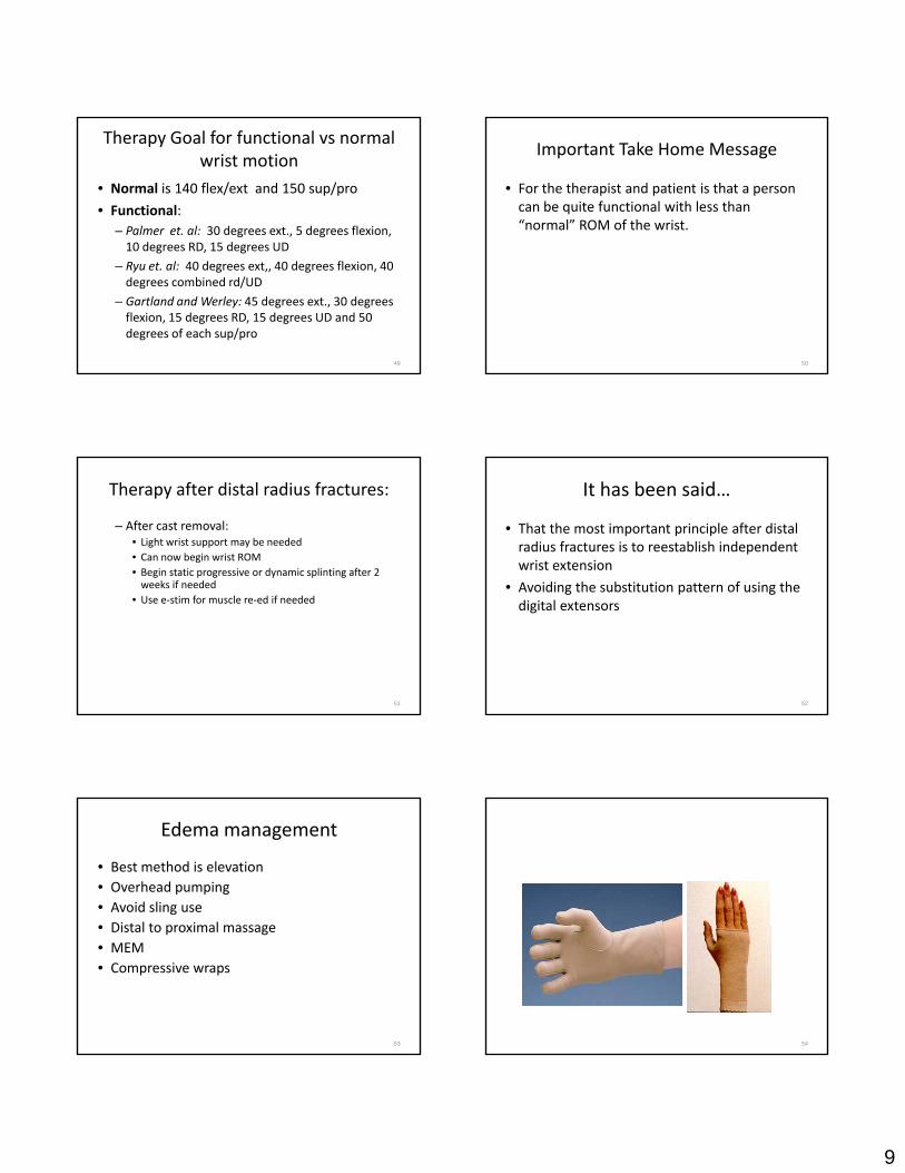
Radial Height (length) Normal is 12 mm and Ulnar Variance
44
Positive Ulnar variance
Prior to Treatment the therapist…
• Knowledge of the amount of radial shortening, dorsal angulation, the presence of any articular step‐offs or any DRUJ issues will help you to formulate appropriate treatmenthelp you to formulate appropriate treatment goals and plans
45
Residual deformities to know
• Loss of radial tilt or inclination –normal is 22‐33 degrees
• Loss of palmer tilt ‐ Dorsal angulation – normal palmar tilt is 11‐12 degrees
46
Continued…
• Radial shortening (decreased height) – normal is 12 mm
• Distal radioulnar joint involvement
• Intra‐articular involvement
47
Realistic goals…
• Keep the patients focus on realistic goals
48
9
Therapy Goal for functional vs normal wrist motion
• Normal is 140 flex/ext and 150 sup/pro
• Functional:– Palmer et. al: 30 degrees ext., 5 degrees flexion, 10 degrees RD 15 degrees UD10 degrees RD, 15 degrees UD
– Ryu et. al: 40 degrees ext,, 40 degrees flexion, 40 degrees combined rd/UD
– Gartland and Werley: 45 degrees ext., 30 degrees flexion, 15 degrees RD, 15 degrees UD and 50 degrees of each sup/pro
49
Important Take Home Message
• For the therapist and patient is that a person can be quite functional with less than “normal” ROM of the wrist.
50
Therapy after distal radius fractures:
– After cast removal:• Light wrist support may be needed• Can now begin wrist ROM• Begin static progressive or dynamic splinting after 2 weeks if neededweeks if needed
• Use e‐stim for muscle re‐ed if needed
51
It has been said…
• That the most important principle after distal radius fractures is to reestablish independent wrist extension
• Avoiding the substitution pattern of using the• Avoiding the substitution pattern of using the digital extensors
52
Edema management
• Best method is elevation• Overhead pumping• Avoid sling use• Distal to proximal massage• Distal to proximal massage• MEM• Compressive wraps
53 54
10
Pain management
• Watch for CRPS
• High volt electro‐mesh glove can h l h dhelp the edema and pain
• TENS
55
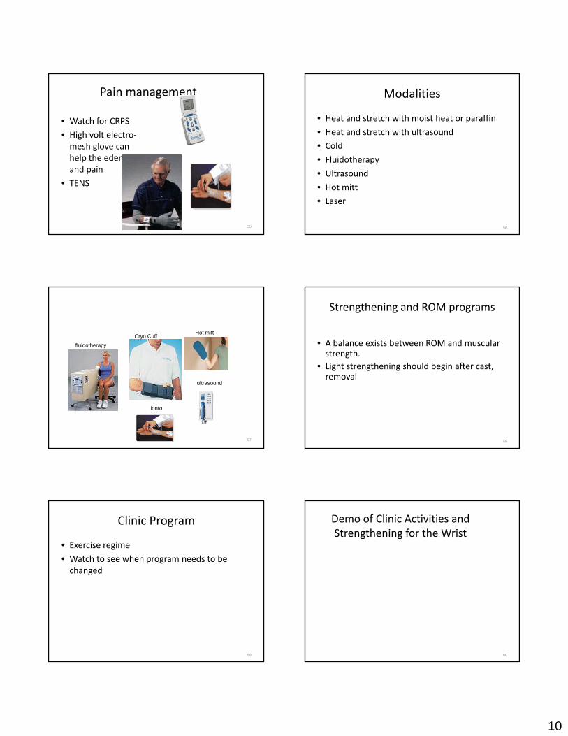
Modalities
• Heat and stretch with moist heat or paraffin
• Heat and stretch with ultrasound
• Cold
• Fluidotherapy
• Ultrasound
• Hot mitt
• Laser
56
Cryo Cufffluidotherapy
Hot mitt
57
ionto
ultrasound
Strengthening and ROM programs
• A balance exists between ROM and muscular strength.
• Light strengthening should begin after castLight strengthening should begin after cast, removal
58
Clinic Program
• Exercise regime
• Watch to see when program needs to be changed
59
Demo of Clinic Activities and Strengthening for the Wrist
60

11
More demos…
61
the Stiff Wrist
• PROM
• Heat and stretch
• US with a stretch
• CPM
• Serial static splint
• Dynamic/static progressive splints
62
Static progressive
63
JAS
Dynasplint for wrist Extension Flexion
64
CPM
• They are good to use at night
• Good when stiff in both directions
• Can be portable or table‐top
65 66

12
Stiff Wrist Tx. Continued
• Joint Mobilization
• Soft tissue mob.
• Graston Technique
67
Frequent presentation after wrist fracture
68
Distal Radius Malunion
What can we as therapists do clinically for malunions?
69
Malunions result in many problems
• Patients with shortening and dorsal angulation are likely to have TFCC damage and instability
• Patients with step‐offs will likely end up with DRUJ arthritis
70
Corrective OsteotomyBefore After
71
Darrach
• Distal ulna resection
• Can have problems with the ulnar stump
72

13
Sauve‐Kapandji
• Fusion of the DRUJ and creation of a pseudoarthrosis in the distal ulna
73
Bowers
• Hemiresection with interposition arthroplasty
74
Salvage procedures continued
• One bone forearm – will create one bone to provide stability and eliminate pain but sacrifices all rotation
75
Total Wrist Fusion
76
Wrist Arthroplasty
• Video of Total Wrist Implant
77
But it hurts on the other side!

14
79
TFCC testing
• Look for instability of the DRUJ• TFCC load test • Piano key sign• Ulnar Carpal SagLT t d• LT tenderness
80
TFCC tears
• If tear is detected acutely can treat with immobilization
• If the tear is on the periphery initial treatment is often immobilization
• If ulnar‐positive variance is present an ulnar shortening i i di dosteotomy is indicated at any stage.
81
central tear Peripheral tear
82
Case Study
83 84
15
85 86
87
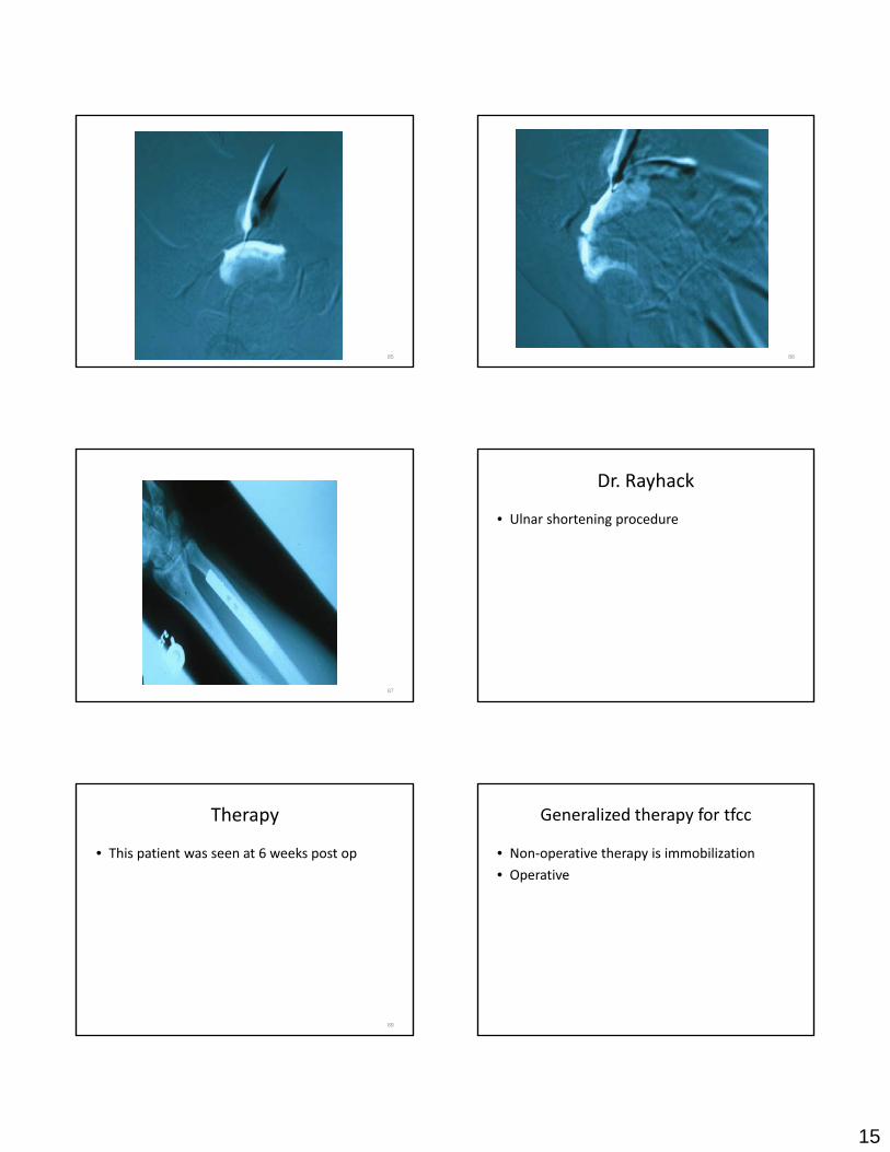
Dr. Rayhack
• Ulnar shortening procedure
Therapy
• This patient was seen at 6 weeks post op
89
Generalized therapy for tfcc
• Non‐operative therapy is immobilization
• Operative

16
Debridement – central tear ‐ therapy
• Volar wrist splint
• AROM at 1 week
• No impact loading
• Light strengthening at 4‐6 weeks
• Gradually resume ADL’s
Peripheral repair
• Week 1 Long arm cast• Week 2‐4 long arm splint• Week 4‐6 short arm splint• Week 6‐10
– AROM– Avoid extremes of rotation– Continue use of splint except for bathing and exercise– Light ADL’s– Week 10 – begin gentle PROM– Light strengthening
• 12 weeks continue and upgrade strength program– Begin dynamic/static progressive splinting
Management of Carpal fractures
93
Scaphoid
• Accounts for 60‐70% of carpal fractures
• Cast is short arm with thumb included
• Therapy begins when the fracture is clinically healed
94
Why this fracture is missed
• The patient frequently overlooks this fracture because it feels like "just a sprain.“
• The fracture may occasionally be invisible on the first x‐ray, only to show up on an x‐ray examination taken weeks or months later
95
Suspect a scaphoid fracture?
• In cases with clinically suspected scaphoid fracture and negative or inconclusive findings on radiography, the wrist is usually immobilized and the radiographic examinationimmobilized and the radiographic examination is repeated 10–12 days later.
96

17
Healing time
• Expected time to union for acute fractures:– (1) Distal third = 6‐8 weeks
– (2) Middle third = 8‐12 weeks
– (3) Proximal third = 12‐24 weeks( )
97
Surgical intervention
• There has been a trend away from the extremes of conservative management
98
Method of Fixation Pros/ Cons
• Cast • Kirschner wires• Bone screws• Vascularized distal radius bone graft.
99
Scaphoid Fracture
Treatment: compression screw with radial bone graft
Photo Courtesy of Deborah A. SchwartzPhoto Courtesy of Deborah A. Schwartz OTR/L, CHTOTR/L, CHT
101 102
18
103
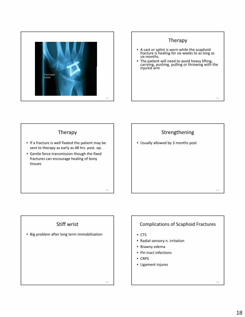
Four bone fusion
Therapy
• A cast or splint is worn while the scaphoid fracture is healing for six weeks to as long as six months.
• The patient will need to avoid heavy lifting, carrying, pushing, pulling or throwing with the y g p g p g ginjured arm
104
Therapy
• If a fracture is well fixated the patient may be sent to therapy as early as 48 hrs. post. op.
• Gentle force transmission though the fixed fractures can encourage healing of bonyfractures can encourage healing of bony tissues
105
Strengthening
• Usually allowed by 3 months post
106
Stiff wrist
• Big problem after long term immobilization
107
Complications of Scaphoid Fractures
• CTS
• Radial sensory n. irritation
• Brawny edema
• Pin tract infections
• CRPS
• Ligament injures
108
19
Lunate Fractures
• Lunate injuries are generally associated with a fall on an outstretched wrist; a compression force may also be involved.
b k d f h l• Kienböck disease, or AVN of the lunate, is believed by some to be a chronic manifestation of lunate fractures in which collapse of the lunate causes the late development of symptoms
109
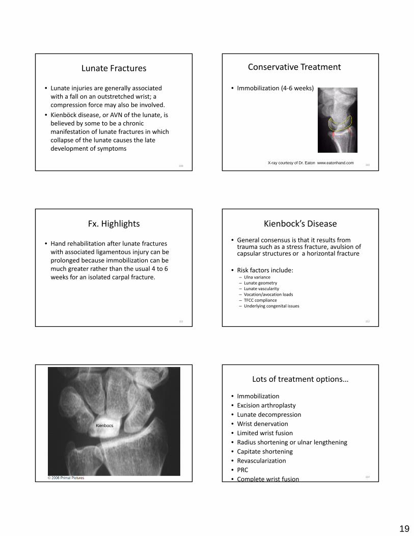
Conservative Treatment
• Immobilization (4‐6 weeks)
110X-ray courtesy of Dr. Eaton www.eatonhand.com
Fx. Highlights
• Hand rehabilitation after lunate fractures with associated ligamentous injury can be prolonged because immobilization can be much greater rather than the usual 4 to 6much greater rather than the usual 4 to 6 weeks for an isolated carpal fracture.
111
Kienbock’s Disease
• General consensus is that it results from trauma such as a stress fracture, avulsion of capsular structures or a horizontal fracture
k f l d• Risk factors include:– Ulna variance– Lunate geometry– Lunate vascularity– Vocation/avocation loads– TFCC compliance– Underlying congenital issues
112
Kienbocs
Lots of treatment options…
• Immobilization • Excision arthroplasty• Lunate decompression• Wrist denervation• Wrist denervation• Limited wrist fusion• Radius shortening or ulnar lengthening• Capitate shortening• Revascularization• PRC• Complete wrist fusion 114

20
115
Kienbock’sDisease
116
Video Dr. Rayhack
• Radial Shortening /
117
Therapy
• Mobilization after casting
118
Triquetrum Fractures
• Triquetral fractures generally occur on the dorsal surface or, less commonly, involve the body of the triquetrum.
119
Triquetrum
120
21
Fx. Highlights
• Stable injuries and usually are immobilized with a short arm cast for 4 weeks.
121
Pisiform Fractures
• The pisiform is a sesamoid bone within the insertion of the flexor carpi ulnaris tendon and is subjected to large stresses.
• Pisiform bone injury often occurs in the setting of a direct blow.
122
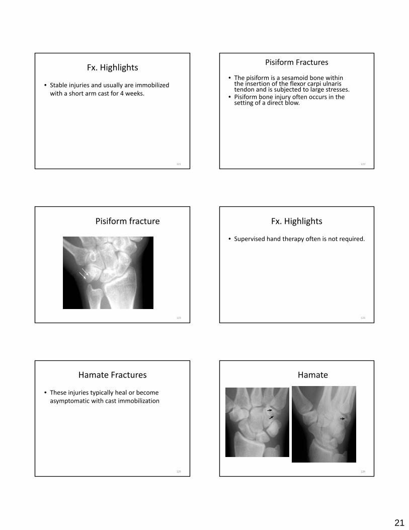
Pisiform fracture
123
Fx. Highlights
• Supervised hand therapy often is not required.
124
Hamate Fractures
• These injuries typically heal or become asymptomatic with cast immobilization
125
Hamate
126
22
Hook of Hamate – these can be troublesome
CT scan
127
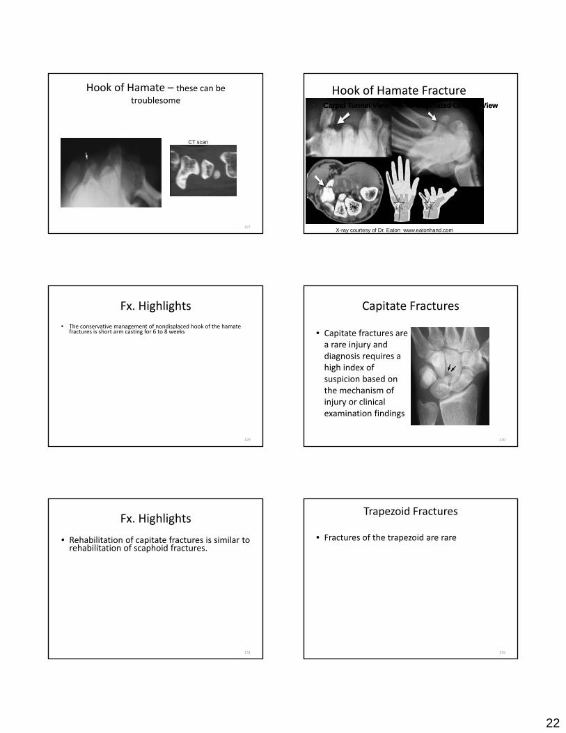
Hook of Hamate FractureCarpal Tunnel ViewCarpal Tunnel View Supinated Oblique ViewSupinated Oblique View
X-ray courtesy of Dr. Eaton www.eatonhand.com
Fx. Highlights• The conservative management of nondisplaced hook of the hamate
fractures is short arm casting for 6 to 8 weeks
129
Capitate Fractures
• Capitate fractures are a rare injury and diagnosis requires a high index ofhigh index of suspicion based on the mechanism of injury or clinical examination findings
130
Fx. Highlights
• Rehabilitation of capitate fractures is similar to rehabilitation of scaphoid fractures.
131
Trapezoid Fractures
• Fractures of the trapezoid are rare
132

23
Trapezoid
133
Fx. Highlights
• Because of the significant trauma often associated with trapezoid fractures, the patient may experience a fair amount of soft tissue injury, edema, and scarring.
134
Trapezium Fractures
• Fractures of the trapezium are most commonly transverse loading injuries in the setting of an adducted thumb in which the first metacarpal is driven into the trapeziumthe trapezium
135
Trapezium
CT scan
136
Fx Highlights
• Conservative management of trapezial fractures is typically with a thumb spica cast.
137
Wrist Instabilities
• Instability of the carpal bones results in weakness, stiffness, chronic pain, and often arthritis if not treated appropriately.
• Carpal instability results from an injury to one• Carpal instability results from an injury to one or more ligamentous or bony constraints in the wrist.
24
Generalized treatment
• If these injuries are caught early within 4‐6 weeks they can be treated with closed reduction, pinning or open repair
• If they are caught 6 weeks to 6 mths they can• If they are caught 6 weeks to 6 mths they can be treated by ligament repair
• 6 – 12 mths ligament reconstrution or intercarpal arthrodesis
• Longer than 12 mths usually requires intercarpal arthrodesis
Nonenclature for Instabilities
• By Severity: Dynamic, static, subluxation, dislocation
• Direction: DISI, VISI, Dorsal, volar, radial, ulnarulnar
• Location: Proximal, distal, radial, ulnar, dorsal, volar, mid‐carpal, radio carpal, specific bone, specific ligament
• Ligament Type: CID, CIND,
• Acuity: Acute, subacute, chronic
CID/CIND
• Carpal Instability Dissociative (CID) – Scapho‐lunate dissociation
– Luno‐triquetral dissocation
C l I t bilit N Di i ti (CIND)• Carpal Instability Non‐Dissociative (CIND)
VISI/DISI Facts
• DISI: dorsiflexion instability is more common
• VISI: volar flexion (palmar flexion) instability
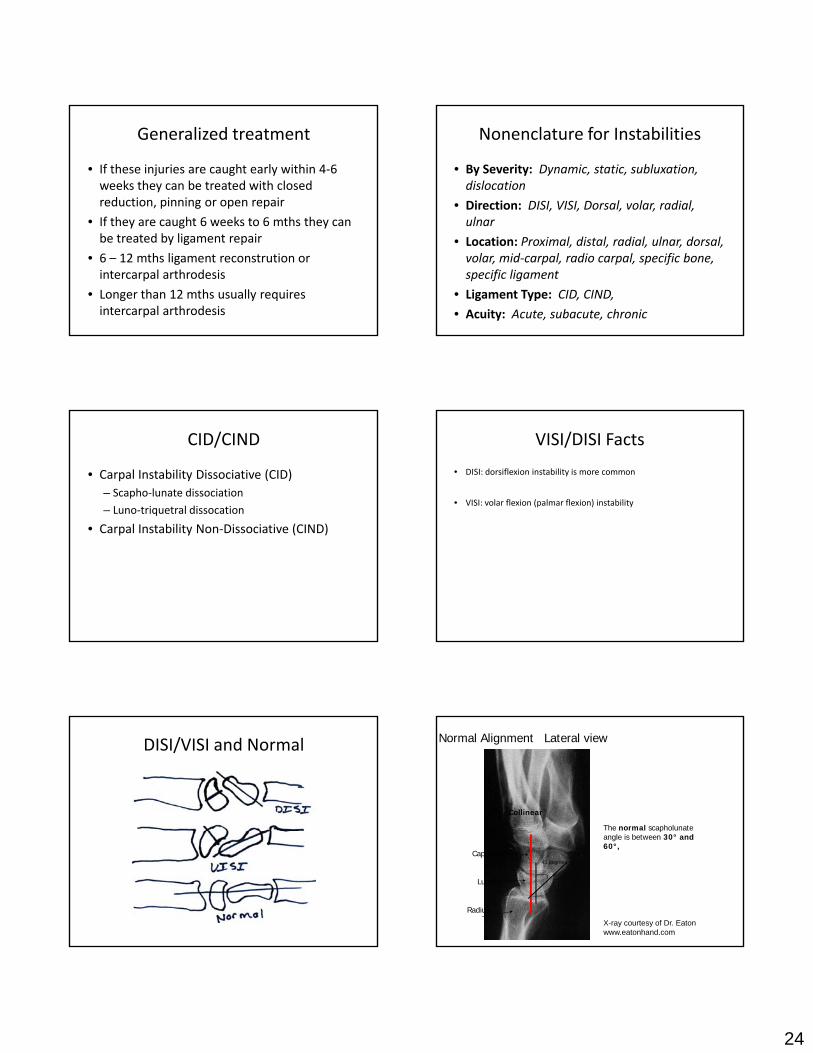
DISI/VISI and Normal Lateral viewNormal Alignment
Collinear
The normal scapholunate
Lunate
Capitate
Radius
pangle is between 30° and 60°,
45 degrees
X-ray courtesy of Dr. Eaton www.eatonhand.com
25
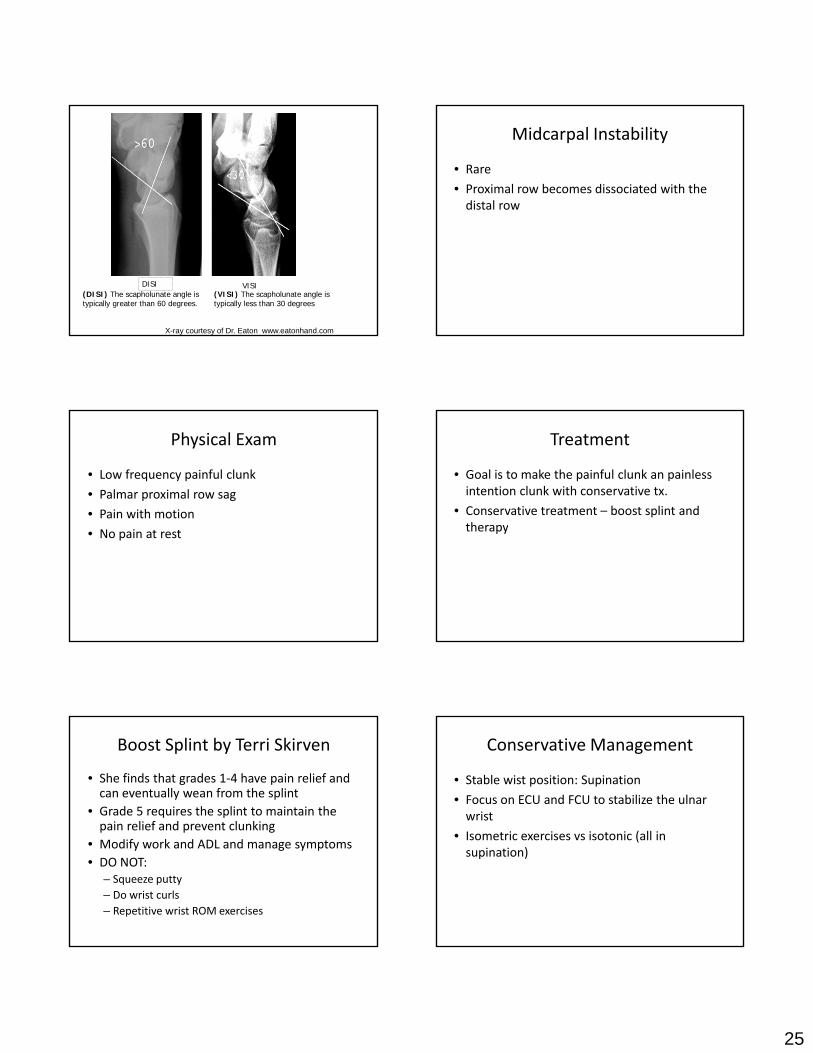
DISI VISI(DISI) The scapholunate angle is typically greater than 60 degrees.
(VISI) The scapholunate angle is typically less than 30 degrees
X-ray courtesy of Dr. Eaton www.eatonhand.com
Midcarpal Instability
• Rare
• Proximal row becomes dissociated with the distal row
Physical Exam
• Low frequency painful clunk
• Palmar proximal row sag
• Pain with motion
• No pain at rest
Treatment
• Goal is to make the painful clunk an painless intention clunk with conservative tx.
• Conservative treatment – boost splint and therapytherapy
Boost Splint by Terri Skirven
• She finds that grades 1‐4 have pain relief and can eventually wean from the splint
• Grade 5 requires the splint to maintain the pain relief and prevent clunkingp p g
• Modify work and ADL and manage symptoms• DO NOT:
– Squeeze putty– Do wrist curls– Repetitive wrist ROM exercises
Conservative Management
• Stable wist position: Supination
• Focus on ECU and FCU to stabilize the ulnar wrist
I i i i i ( ll i• Isometric exercises vs isotonic (all in supination)

26
LT Instability
• LT instability presents as a VISI when advanced• Symptoms include
– Ulnar sided wrist pain– Click or catchUlnar nerve symptoms– Ulnar nerve symptoms
• Testing includes:– Shear test– Shuck Sign– Ballotment Test– Tender at lt interval
VISI Carpal Instability Advanced
Narrow Scapholunate Angle
Palmar Flexed Lunate
AP View Lateral ViewX-ray courtesy of Dr. Eaton www.eatonhand.com
Treatment
• There is no consensus on the appropriate treatment of lunotriquetral instability.
LT Instability after stabilization
• Immobilized 6‐8 weeks
• Avoid impact loading and forceful rotation for 4‐6 mths
SL Instability
• Associated with DISI
• Lunate extends
Scapholunate Dissociation
Radial DeviationRadial Deviation Ulnar DeviationUlnar Deviation
X-ray courtesy of Dr. Eaton www.eatonhand.com

27
Photo Courtesy of Deborah A. SchwartzPhoto Courtesy of Deborah A. Schwartz OTR/L, CHTOTR/L, CHT
Scaphoid non-union leading to Scapholunate advanced collapse of wrist
4 bone fusion
SLAC
Treated with a PRC – the capitate now articulates with the lunate fossa
XX--ray Courtesy of Jim W Roderique MDray Courtesy of Jim W Roderique MD
Assessing for SL tears
• Scaphoid Shift test – pos. when the clunk occurs after removal of pressure
• SL tenderness
P i d l i i i i h• Patient reports dorsal wrist pain, pain with extension and weakness of grip
Treatment
• Multitudes of soft tissue techniques ie: Dorsal Capsular Advancement (Blatt) and ligament reconstruction
• If the instability is advanced in seen in static yfilms then STT fusion is a good option
• If advanced to a complete SLAC options include: STT or SC fusion with radial styloidectomy, PRC,four corner fusion or arthrodesis

28
Generalized therapy after wrist arthroscopy
• Promote function
• Maintain Stability
• Protect joint integrity
• Achieve functional ROM
• Wrist splint 5‐7 days
Debridements or synovectomy
• Therapy will progress as tolerated after immobilization
SL instability with stabilization
• Immobilized 6‐8 weeks
• Avoid weigh bearing for 3‐6 mths
• Expect a loss of ROM around a 12‐ 25% loss of fl i / i d 12 30% l f RD/UDflexion/extenion and a 12 ‐30% loss of RD/UD
Carpal Dislocations
• Are often the result of hyperextension injuries on he out‐stretched hand
Lunate vs. perilunate dislocation
• The key to differentiation between these is what is centered over the radius.
Lunate centers over the distal radiusLunate centers over the distal radius
perilunate dislocation

29
Lunate Dislocations
capitate is centered over the radius and the lunate is tilted outcapitate is centered over the radius and the lunate is tilted outcapitate is centered over the radius and the lunate is tilted outcapitate is centered over the radius and the lunate is tilted out
X-ray courtesy of Dr. Eaton www.eatonhand.com
Arcs (Mayfield)
• Lesser arc– Lesser arc injuries are pure ligamentous injuries
• Greater arcF di l i f h b d h– Fracture dislocation of the bones around the lunate
Greater Lesser Lesser Arc Stages
• Stage I includes scaphoid dissociation from tearing of the scapholunate interosseous and volar displacement of the radioscaphoid joint.
• Stage II includes dorsal dislocation of the capitate with dissociation at the lunocapitate joint.
• Stage III includes lunotriquetral ligament disruption. The lunate remains aligned with the radius, while the rest of the carpus is displaced, usually dorsally.
• Stage IV is complete ligament disruption. The capitate remains aligned with the radius, while the lunate is squeezed out in a volar direction.
Therapy after Lunate or Perilunate Dislocations – lesser arc
• Ligament repair
• Immobilize in a LAC for 4 week and then a SAC for 4 weeks
Pi 8 k• Pins out at 8 weeks
• Gentle ROM
• Thumb spica splint for 2‐4 more weeks
• 2‐4 weeks later can start strengthening
Greater Arc Stages:Trans ‐ (fractured bone ‐ one or more)
• Stage I: transradial styloid perilunate fracture‐dislocation; Stage II: transscaphoid perilunate fracture‐dislocation; Stage III: transscaphoid, transcapitate perilunate g p , p pfracture‐dislocation; Stage IV: transscaphoid, transcapitate, transtriquetral perilunate fracture‐dislocation; and Stage V: complete palmar lunate dislocation associated with carpal fractures
30
Therapy after Perilunate Fracture Dislocations – Greater Arc Injury’s
• Ligaments and bone injured
Treatment
• Numerous combinations of lesser‐ and greater‐arc disruptions are observed
• Similar to most carpal injuries
h d d d l l• The most dreaded complication is carpal instability
The End of One Chapter and the Beginning of another…
REFERENCES Taleisnik, Julio, The Wrist , New York, 1985, Churchill Livingston Cooney, WP., Linscheid RL, Dobyns JH: The Wrist: diagnosis and operative treatment , St Louis 1998, Mosby Mackin EJ, Callahan AD, Skirven TM, et al: eds, Hunter Macin & Callahan’s Rehabilitation of the hand and upper extremity, 5th ed, St. Louis, 2002, Mosby Freeland, Alan E. Jabaley ME, Hughes JL: Stable fixation of the hand and wrist, New York, 1986, Springer-Verlag. Andrews,et al Physical Rehabilitation of the Injured Athlete 3rd Edition, Saunders Philadelphia 2004 Plancher, Kevin , Atlas of the Hand Clinics Sports Injuries, Philadelphia March 2006, Saunders