Embed Size (px)
Citation preview
Novel Polymorphism of Fc
g
RIIIa (CD16) and Autoimmune Disease
1059
J. Clin. Invest.© The American Society for Clinical Investigation, Inc.0021-9738/97/09/1059/12 $2.00Volume 100, Number 5, September 1997, 1059–1070http://www.jci.org
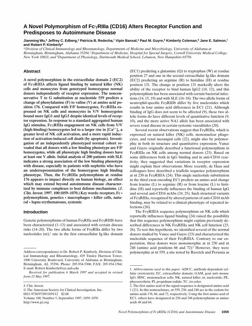
A Novel Polymorphism of Fc
g
RIIIa (CD16) Alters Receptor Function andPredisposes to Autoimmune Disease
Jianming Wu,* Jeffrey C. Edberg,* Patricia B. Redecha,
‡
Vipin Bansal,* Paul M. Guyre,
§
Kimberly Coleman,
§
Jane E. Salmon,
‡
and Robert P. Kimberly*
*
Division of Clinical Immunology and Rheumatology, Departments of Medicine and Microbiology, University of Alabama at Birmingham, Birmingham, Alabama 35294;
‡
Department of Medicine, Hospital for Special Surgery, Cornell University Medical College, New York 10021; and
§
Department of Physiology, Dartmouth Medical School, Lebanon, New Hampshire 03756
Abstract
A novel polymorphism in the extracellular domain 2 (EC2)of Fc
g
RIIIA affects ligand binding by natural killer (NK)cells and monocytes from genotyped homozygous normaldonors independently of receptor expression. The noncon-servative T to G substitution at nucleotide 559 predicts achange of phenylalanine (F) to valine (V) at amino acid po-sition 176. Compared with F/F homozygotes, Fc
g
RIIIa ex-pressed on NK cells and monocytes in V/V homozygotesbound more IgG1 and IgG3 despite identical levels of recep-tor expression. In response to a standard aggregated humanIgG stimulus, Fc
g
RIIIa engagement on NK cells from V/V(high-binding) homozygotes led to a larger rise in [Ca
2
1
]
i
, agreater level of NK cell activation, and a more rapid induc-tion of activation-induced cell death (by apoptosis). Investi-gation of an independently phenotyped normal cohort re-vealed that all donors with a low binding phenotype are F/Fhomozygotes, while all phenotypic high binding donors haveat least one V allele. Initial analysis of 200 patients with SLEindicates a strong association of the low binding phenotypewith disease, especially in patients with nephritis who havean underrepresentation of the homozygous high bindingphenotype. Thus, the Fc
g
RIIIa polymorphism at residue176 appears to impact directly on human biology, an effectwhich may extend beyond autoimmune disease character-ized by immune complexes to host defense mechanisms. (
J.Clin. Invest.
1997. 100:1059–1070.) Key words: receptors, Fc
•
polymorphism, genetics
•
macrophages
•
killer cells, natu-ral
•
lupus erythematosus, systemic
Introduction
Genetic polymorphisms of human Fc
g
RIIa and Fc
g
RIIIb havebeen characterized (1–13) and associated with certain diseaserisks (14–20). The two allelic forms of Fc
g
RIIa differ by twonucleotides (nt),
1
one in the first extracellular Ig-like domain
(EC1) predicting a glutamine (Q) to tryptophan (W) at residueposition 27 and one in the second extracellular Ig-like domain(EC2) predicting an arginine (R) to histidine (H) at residueposition 131. The change at position 131 markedly alters theability of the receptor to bind human IgG2 (10, 12), and thispolymorphism has been associated with certain bacterial infec-tions (14, 15, 19) and with SLE (16–18). The two allelic forms ofneutrophil-specific Fc
g
RIIIb differ by five nucleotides whichresults in four amino acid differences in EC1 (21). Althoughbinding of IgG does not seem to be affected (9), these two al-lelic forms do have different levels of quantitative function (9,10), and the more active NA1 allele has been associated withsevere renal disease in certain systemic vasculitides (20).
Several recent observations suggest that Fc
g
RIIIa, which isexpressed on natural killer (NK) cells, mononuclear phago-cytes, and renal mesangial cells (22), might also be polymor-phic in both its structure and quantitative expression. Vanceand Guyre originally described a functional polymorphism inFc
g
RIIIa on NK cells among normal donors (23). Based onsome differences both in IgG binding and in anti-CD16 reac-tivity, they suggested that variations in receptor expressionmight explain their observations. More recently, de Haas andcolleagues have described a triallelic sequence polymorphismat nt 230 in Fc
g
RIIIA (24). This single nucleotide substitutionin the third exon encoding EC1 predicts an amino acid changefrom leucine (L) to arginine (R) or from leucine (L) to histi-dine (H) and reportedly influences the binding of human IgGand several anti-CD16 mAbs (24, 25). Such structural variantsof Fc
g
RIIIa, recognized by altered patterns of anti-CD16 mAbbinding, may be related to a clinical phenotype of repeated in-fections (26).
The Fc
g
RIIIA sequence polymorphism on NK cells whichreportedly influences ligand binding (24) raised the possibilitythat this sequence polymorphism might explain previously de-scribed differences in NK Fc
g
RIIIa and NK cell function (23,26). To test this hypothesis, we identified several of the normaldonors studied by Vance and Guyre (23) and characterized thenucleotide sequence of their Fc
g
RIIIA. Contrary to our ex-pectation, these donors were monomorphic at nt 230 and nt248 (amino acid positions 66 and 72).
2
However, they werepolymorphic at nt 559, a site noted by Ravetch and Perussia as
Address correspondence to Dr. Robert P. Kimberly, Division of Clin-ical Immunology and Rheumatology, 429 Tinsley Harrison Tower,1900 University Boulevard, University of Alabama at Birmingham,Birmingham, AL 35294. Phone: 205-934-5306; FAX: 205-934-1564;E-mail: [email protected]
Received for publication 6 March 1997 and accepted in revisedform 23 May 1997.
1.
Abbreviations used in this paper:
ADCC, antibody-dependent cel-lular cytotoxicity; EC, extracellular domain; GAM, goat anti–mouseIgG; MNC, mononuclear cells; NK, natural killer; nt, nucleotide; PE,phycoerythrin; PI, propidium iodide; TC, tri-color.2. The first amino acid of the signal sequence is designated amino acid1 (21). In this nomenclature, nt 559, 230, and 248 are in the codons foramino acids 176, 66, and 72, respectively. Using the first amino acid ofEC1, others have designated nt 230 and 248 polymorphisms as aminoacids 48 and 64.
1060
Wu et al.
potentially polymorphic (21). This nonconservative T to Gsubstitution predicts a change of phenylalanine (F) into valine(V) at position 176 in the membrane-proximal EC2. Since sev-eral studies suggest that the second Ig-like domain strongly in-fluences ligand binding (27–32), we pursued further character-ization of this 176F/V polymorphism by identifying normaldonors homozygous at position 176 (and homozygous at posi-tions 66 and 72). Compared with F/F homozygotes, Fc
g
RIIIaexpressed in V/V homozygotes bound more IgG1 and IgG3despite identical levels of receptor expression. These observa-tions indicate that the sequence polymorphism at nt position559 alters the apparent affinity of Fc
g
RIIIa on both NK cellsand monocytes for IgG. This difference affects the ability ofthe receptor to initiate a range of cell programs in response toa standard stimulus and underlies the previously describedvariation in NK Fc
g
RIIIa function (23). Initial analysis of 200patients with SLE indicates a strong association of the lowbinding phenotype with disease, especially nephritis, and a cor-responding underrepresentation of the homozygous high bind-ing phenotype. Thus, this polymorphism appears to impact di-rectly on human biology, an effect which may well extendbeyond autoimmune disease characterized by circulating im-mune complexes.
Methods
Donors.
Anticoagulated peripheral blood was obtained from healthynormal volunteers and from 200 patients fulfilling the revised criteriaof the American College of Rheumatology for SLE (33). All studieswere reviewed and approved by each Institutional Review Board andall donors provided written informed consent.
Reagents.
Human IgG (hIgG) subclass proteins were obtainedfrom The Binding Site (San Diego, CA) or Sigma Chemical Co. (St.Louis, MO). All mAbs used were murine origin. Anti–human CD56-phycoerythrin (PE), anti–human CD14-tri-color (TC), anti–humanCD3-FITC, anti-CD25-FITC, and anti-CD33-FITC were from CaltagLaboratories (Burlingame, CA). Anti-Fc
g
RI (mAb 197, mIgG2a andmAb 22.2, mIgG1), anti-Fc
g
RII (mAb IV.3, mIgG2b), and anti-Fc
g
RIII (mAb 3G8-FITC, mIgG1) were from Medarex Inc. (Annan-dale, NJ). Other anti-Fc
g
RIII mAbs used in this study were CLBFcRgran1 (mIgG2a), B73.1 (mIgG1), 1D3 (mIgM), MEM154 (mIgG1),30.2 (mIgG1), 214.1 (mIgG1), 135.9 (mIgG1), GRM1 (mIgG2a), andLeu11a (mIgG1). 1D3, MEM154, and CLBFcRgran1 were obtainedthrough the 5
th
Leukocyte Typing Workshop. mAbs 30.2, 214.1, and135.9 were generously provided by Dr. Howard Fleit (SUNY, StonyBrook, NY) (34); mAb GRM1 was from Research Diagnostics Inc.(Flanders, NJ). B73.1 (Leu11a) and Leu11a-FITC were obtained fromBecton Dickinson Immunocytometry Systems (San Jose, CA). FITC-conjugated and unconjugated goat anti–mouse IgG(H
1
L) (GAM),which recognizes mIgG1, IgG2a, and mIgM, was obtained from Boeh-ringer Mannheim (Indianapolis, IN) and Jackson ImmunoResearch(West Grove, PA). Heat-aggregated human IgG was prepared by in-cubating the hIgG (Sigma), 20 mg/ml, at 63
8
C for 20 min.
Mononuclear cell (MNC) and NK cell preparation.
Fresh antico-agulated blood was diluted 1:1 in Hanks’ buffer (GIBCO BRL,Gaithersburg, MD) and centrifuged through a discontinuous two-stepFicoll-Hypaque gradient in 50-ml conical tubes (35). MNC were har-vested from the upper and neutrophils from the lower Ficoll-Hypaqueinterface and washed three times with PBS, pH 7.4. After the lastwash, the cells were resuspended, counted, and used either formRNA preparations, for quantitative flow cytometry, or for furtherpurification of NK cells.
NK cells were purified with the NK Cell Isolation Kit (MiltenyiBiotec Inc., Auburn, CA) which depletes human T cells, B cells, andmyeloid cells from MNC by magnetic separation. Isolated NK cells
were washed with Ca
2
1
- and Mg
2
1
-free PBS, pH 7.4, and assayed forpurity by flow cytometry with either the combination of anti-CD3-FITC, anti-CD14-TC, and anti-CD56-PE or the combination of 3G8-FITC, anti-CD14-TC, and anti-CD56-PE. Starting with 5
3
10
7
MNC,the yield of NK was typically 4–5
3
10
6
total cells with a purity of
.
85%. The efficiency of NK cell recovery was
z
75%.
Nucleic acid isolation.
Total RNA was isolated from 10
7
MNC byusing TRIzol™ total RNA isolation reagent (GIBCO BRL). 5
m
g oftotal MNC RNA was used to synthesize cDNA with the SuperScript™preamplification system (GIBCO BRL). For sequencing of genomicDNA and for allele-specific PCR, genomic DNA was isolated usingthe Puregene DNA isolation kit (Gentra Systems, Minneapolis, MN).
RT-PCR and cDNA sequencing.
To facilitate heterozygote de-tection, a dye primer strategy was used for fluorescence-based auto-mated cycle sequencing of PCR product on an ABI 377 (ABI PRISM™Dye Primer Cycle Sequencing -21M13 FS and M13REV FS ReadyReaction Kits; Applied Biosystems, Inc., Foster City, CA). Two over-lapping sets of primers, with either M13 universal or reverse primersequences at the appropriate 5
9
ends, were designed for the Fc
g
RIIIAcDNA. Sequencing set 4 (Fig. 1) was used to amplify position 50 toposition 414: forward 5
9
-CAG GAA ACA GCT ATG ACC TCCCAA CTG CTC TGC TAC TT-3
9
and reverse 5
9
-TGT AAA ACGACG GCC AGT CCT CAG GTG AAT AGG GTC TTC-3
9
. Se-quencing set 3 (Fig. 1) was used to amplify position 328 to position869: forward 5
9
-TGT AAA ACG ACG GCC AGT CCG GTG CAGCTA GAA GTC CA-3
9
and reverse 5
9
-CAG GAA ACA GCT ATGACC GGG GTT GCA AAT CCA GAG AA-3
9
. The PCR productswere purified with the QIAquick Gel Extraction Kit (QIAGEN Inc.,Chatsworth, CA).
Allele-specific PCR.
Three primers were designed for allele-spe-cific PCR for genotyping genomic DNA at position 559 in Fc
g
RIIIA(PCR set 1, Fig. 1
A
). The Fc
g
RIIIA-specific forward primer (5
9
-TCA CAT ATT TAC AGA ATG GCA ATG G-3
9
) corresponds tothe Fc
g
RIIIA sequence between position 449 and position 473 andwas used in both T allele–specific and G allele–specific PCR assays.The reverse primers, corresponding to nt 586–559, provided allelespecificity. The nt 559 G-specific reverse primer (5
9
-TCT CTG AAGACA CAT TTC TAC TCC CTA C-3
9
) differs in one nucleotide fromT-specific reverse primer (5
9
-TCT CTG AAG ACA CAT TTC TACTCC CTA A-3
9
) at the 3
9
end. The allele-specific PCR product of 138 bpwas assayed on a 3% agarose gel. The PCR reaction was performedin a GeneAmp 2400 PCR System with 360 ng of DNA, 200 nM ofeach primer, 200
m
M of dNTPs, 1.5 mM of MgCl, and 2.5 U of
Taq
polymerase (Boehringer-Mannheim Biochemicals) in a 50-
m
l reactionvolume starting with 95
8
C for 5 min, 35 cycles of denaturing at 94
8
Cfor 30 s, annealing at 51
8
C for 45 s, and extension at 72
8
C for 20 s witha final extension at 72
8
C for 7 min. The appearance of the 138-bpPCR products in the T or G allele–specific reaction indicates thepresence of that allele.
Genomic DNA sequencing.
To confirm Fc
g
RIIIA genomic se-quence, primers were designed to amplify a portion of exon 4 ofFc
g
RIIIA which corresponds to EC2 (sequencing set 1, Fig. 1
A
).The forward primer (5
9
-TGT AAA ACG ACG GCC AGT TCATCA TAA TTC TGT CTT CT-3
9
, corresponding to nt 486–505) in-cludes an intentional mismatch six nucleotides from the 3
9
end to pro-vide Fc
g
RIIIA-specific priming. The reverse primer (5
9
-CAG GAAACA GCT ATG ACC CTT GAG TGA TGG TGA TGT TCA-3
9
)corresponds to nt 610–590. The 162-bp PCR product containing thent 559 polymorphic site was purified from a 3% agarose gel with theQIAquick Gel Extraction Kit. Fluorescence-based automated cyclesequencing of PCR product was performed on an ABI 377 (ABIPRISM™ Dye Primer Cycle Sequencing -21M13 FS and M13REV FSReady Reaction Kits).
To determine the Fc
g
RIIIA genomic sequence of EC1, primerswere designed to amplify a portion of exon 3 encompassing nt posi-tions 230 and 248 (sequencing set 2, Fig. 1
A
). The forward primer(5
9
-CAG GAA ACA GCT ATG ACC CTC TTT CTG TAG CTTGGT TC-3
9
) anneals to the intron region between S1 and S2 of the
Novel Polymorphism of Fc
g
RIIIa (CD16) and Autoimmune Disease
1061
Fc
g
RIIIA gene. The reverse primer (5
9
-TGT AAA ACG ACG GCCAGT ATG GAC TTC TAG CTG CAC-3
9
) corresponds to nt 348 to331 in exon 3. The PCR product was purified and sequenced as de-scribed above.
Flow cytometric assay for human IgG binding.
Human IgG bind-ing assays were performed using anticoagulated, washed whole blood.Mouse mAbs were used either for direct immunofluorescence (Leu11a-FITC, 3G8-FITC) or for indirect immunofluorescence (3G8, Gran1,B73.1, 1D3, MEM154, 30.2, 214.1, 135.9, GRM1) in conjunction withFITC-labeled F(ab
9
)
2
GAM. Human IgG subclass myeloma proteins(IgG1, IgG2, IgG3, and IgG4) were directly conjugated with FITC ac-cording to standard techniques (36). Aggregates were removed by ul-tracentrifugation and removal was confirmed by the lack of bindingof the FITC-labeled myelomas to human neutrophils. Before each ex-periment, the flow cytometer (FACScan
®
; Becton-Dickinson Immu-nocytometry) was calibrated with quantitative fluorescein microbeads(Flow Cytometry Standards Corp., Research Triangle Park, NC). Iden-tification of individual cell populations was based on forward andright angle light scattering in combination with three-color immuno-fluorescence using TC, PE, and FITC.
For each IgG binding assay, 3 ml of heparinized whole blood waswashed and cytophilic IgG was removed by incubation in 45 ml ofPBS at 37
8
C for 20 min. For direct immunofluorescence, 100-
m
l ali-quots of the washed whole blood were incubated at 4
8
C for 1 h withan hIgG-FITC myeloma protein (final concentrations of 15 and 30
m
g/ml) or with anti-CD16 mAb [either directly FITC-conjugated orindirectly with F(ab
9
)
2
GAM IgG-FITC, see below] and with CD14-TC and CD56-PE at 5
m
g/ml. NK cells were identified as CD56-PEpositive, CD14-TC negative cells within the lymphocyte light scattergate; the binding of the different human IgG subclasses or the differ-ent anti-CD16 mAbs was assessed by the intensity of FITC fluores-cence. Blood monocytes were identified as CD14-TC positive, CD56-negative cells within the typical blood monocyte light scatter gate. Formyeloma protein binding to monocyte Fc
g
RIIIa, washed whole bloodwas preincubated at 4
8
C for 10 min with mAb 197 IgG (10
m
g/ml) toblock the ligand binding site of Fc
g
RI (12).For the anti-CD16 mAb panel assay, washed whole blood cells
were incubated with a saturating concentration of primary mAb for30 min, washed twice with PBS, incubated with FITC-conjugated goatF(ab
9
)
2
anti–mouse IgG at 4
8
C for 30 min, and washed twice withPBS. After blocking remaining GAM binding sites with controlmIgG1 and mIgG2a (10
m
g/ml final concentration), CD14-TC and
CD56-PE were added for phenotypic identification. After further in-cubation and washes, cells were analyzed on the FACScan
®
.
Measurement of change in [Ca
2
1
]
i
.
Changes in intracellular [Ca
2
1
]
i
,induced by cross-linking of purified NK cell Fc
g
RIIIa with heat-aggregated human IgG or with anti-FcgRIIIa mAb, were determinedin purified indo-1-AM–loaded NK cells using an SLM 8000 spectro-fluorometer and the simultaneous 405/490 nm fluorescence emis-sion ratio as described previously (37, 38). Briefly, cells in suspensionat 107 cells/ml in Ca21- and Mg21-free PBS, pH 7.4, were incubatedwith 5 mM indo-1-AM at 378C for 15 min and washed in PBS. Cellpreparations to be opsonized with mAb 3G8 were resuspended inCa21- and Mg21-free PBS at 107 cells/ml, incubated with saturatingconcentrations of 3G8 (10 mg/ml) at 378C for 5 min, and washed inPBS. All cells were resuspended in 1.1 mM Ca21, 1.6 mM Mg21 PBSat 378C for 5 min and then immediately transferred to a continuouslystirring cell cuvette maintained at 378C in the SLM 8000. With excita-tion at 355 nm, the simultaneous fluorescence emission at 405 and 490nm was measured, integrated, and recorded each second. After estab-lishing a base line for 60 s, either aggregated human IgG or goatF(ab9)2 anti–mouse IgG was added at final concentrations of 20 or 35mg/ml, respectively, and data acquisition was continued for an addi-tional 3.5 min. Each sample was individually calibrated by lysing cellsin 1% Triton X-100 to determine the maximal emission ratio and byadding EDTA (20 mM final concentration) to determine the minimalratio. The indo-1 fluorescence emission ratio was converted to [Ca21]i
by the method of Grynkiewicz (39).Induction of NK cell IL-2 receptor (CD25) expression and cell via-
bility. Purified NK cells were cultured in RPMI/10% FCS and rIL-2(100 U/ml) with or without a defined stimulus for varying periods oftime in 96-well plates. FcgRIIIa-mediated stimulation of NK cells waseffected either with the anti-FcgRIIIa mAb 3G8 IgG (10 mg/ml) orwith heat-aggregated human IgG (20 mg/ml). For mAb stimulation,wells were precoated with 10 mg/ml F(ab9)2 GAM (Jackson Immu-noResearch) for 2 h at 378C, rinsed, and 105 NK cells (106 cells/ml)were added with mAb and incubated for various periods of time. Ag-gregated human IgG was used either in solution as a soluble stimulusor immobilized to the tissue culture plated (precoating for 2 h at378C) before the addition of cells. Since induction of CD25 expres-sion was observed within 1 h of stimulation as previously reported(40, 41), we examined incubation periods ranging from 1 to 48 h.CD25 expression was determined by flow cytometry using anti-CD25-FITC (Caltag Laboratories).
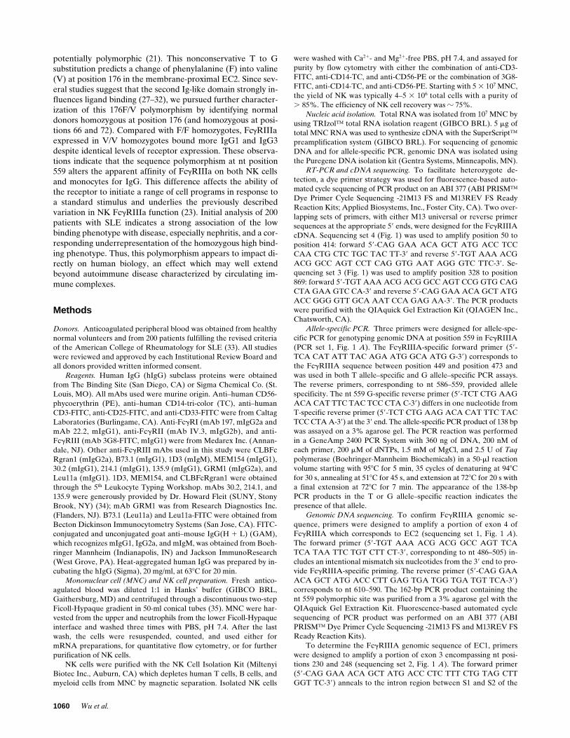
Figure 1. Schematic representation of the FcgRIIIA genomic structure and cDNA structure showing the relative location of primers used for PCR-based sequencing (sequencing sets 1–4) and allele-specific PCR (PCR set 1). For sequencing analysis, an M13-based dye-primer sequencing strategy was used. FcgRIIIa encoding cDNA was prepared from purified MNC. Relative positions of nt 230 and 559 are shown. S, Signal sequence (encoded in two exons); TM/CY, transmembrane/cytoplas-mic domains; 39-UT, 39 untranslated se-quence.
1062 Wu et al.
The viability of purified and 24-h IL-2–primed (100 U/ml) NK cellsafter FcgRIIIa-mediated stimulation was determined by quantitationof propidium iodide (PI) uptake in the FACScan® and by direct vi-sual assessment of trypan blue exclusion. Significant changes in cellviability could be detected within 1 h of stimulation with mAb or ag-gregated IgG (42–44). To determine if FcgRIIIa-mediated stimula-tion was inducing cell death via apoptosis, in selected experimentscells were fixed, permeabilized, and analyzed for quantitative DNAcontent. Subdiploid uptake of PI reflects cell death via apoptosis (45).
Additionally, we examined stimulated cells for apoptotic morphology(chromatin condensation and nuclear fragmentation) (43) after fixa-tion (2% paraformaldehyde plus 0.5% glutaraldehyde in 0.05 M ca-codylate buffer, pH 7.2, followed by post-fixation with 1% osmic acidin cacodylate buffer, pH 7.2). Cells were suspended in 1.5% agar, em-bedded in Spurr’s resin, and the thin sections were viewed in a Phil-lips CM-12 electron microscope.
Statistical analysis. Differences in ligand binding and mAb bind-ing were analyzed by Student’s t test. The x2 test was used to analyze
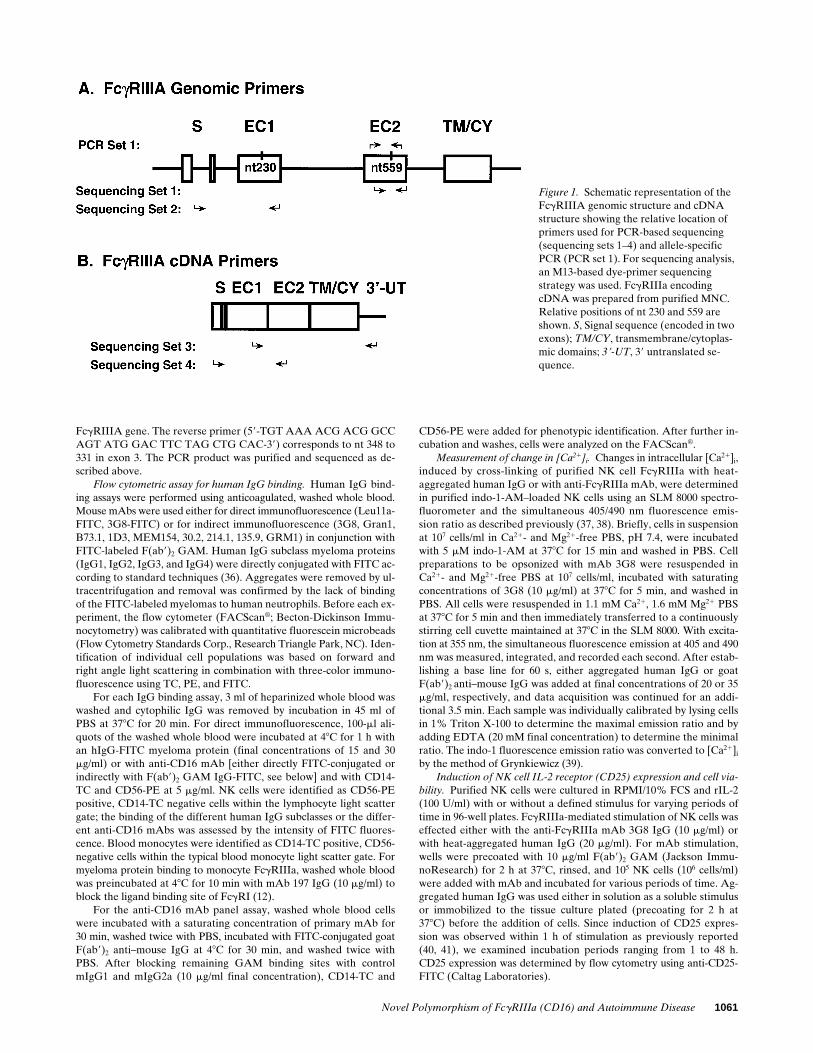
Figure 2. Sequence analysis of a portion of FcgRIIIa cDNA from three normal do-nors. FcgRIIIa encoding cDNA was pre-pared from purified MNC and an M13-based dye-primer sequencing strategy was used (see Methods). Donors homozygous for nt 559-T (A), homozygous for nt 559-G (C), and a donor heterozygous for nt559-T/G (B) are shown. In each tracing, nt 531 (*) is shown to indicate the presence of cDNA encoding the FcgRIIIA gene (C at nt 531) and not the FcgRIIIB gene (which is T at nt 531).
Novel Polymorphism of FcgRIIIa (CD16) and Autoimmune Disease 1063
the distribution of FcgRIIIA genotypes (corresponding to 176-V/V,176-F/F, and 176F/V) in SLE and non-SLE controls. The null hypoth-esis was rejected at the 95% confidence level (P , 0.05).
Results
FcgRIIIA sequence polymorphisms. The recent observation ofan FcgRIIIA sequence polymorphism on NK cells which influ-ences ligand binding (24) raised the possibility that this se-quence polymorphism might explain previously described dif-ferences in NK FcgRIIIa and NK cell function (23). To testthis hypothesis we identified several of these normal donorsand characterized the nucleotide sequence of their FcgRIIIA.The cell type specific expression of FcgRIIIA and FcgRIIIB inNK cells/mononuclear phagocytes and in neutrophils, respec-tively, provides a strategy for selective sequencing of cDNAsderived from these two highly homologous genes. Further-more, within the coding region of FcgRIII, there are 10 nucle-otide differences between FcgRIIIA and FcgRIIIB that can beused to confirm the presence of only FcgRIIIA or FcgRIIIBsequence. Using this approach, two normal donors, one withthe low binding FcgRIIIa phenotype and one with a high bind-ing FcgRIIIa phenotype (23), were both shown to be T/ T230
homozygotes. Interestingly, however, while the low FcgRIIIaphenotype showed no differences from the conventional se-quence, the donor characterized phenotypically as high bindingFcgRIIIa was heterozygous T/G at nt 559 (Fig. 2). This nucle-otide difference, previously mentioned by Ravetch and Perus-sia (21), raised the possibility that this nonconservative nucle-otide polymorphism encoding a phenylalanine to valine atamino acid residue 1762 in the membrane proximal EC2 ofFcgRIIIa might affect ligand binding and receptor function.
Sequence analysis of the entire coding region for MNC
FcgRIIIA cDNA from a total of 30 normal donors revealedvariation in nt position 559 (T or G). In this group, cDNAsfrom three individuals contained only G559 while six donorscontained only T559. The remaining 21 donors were found tocontain both T559 and G559 (Fig. 2). All 30 normal donors werehomozygous T at nt 230 and homozygous C at nt 248 (24, 25).There were no other sequence differences throughout thewhole FcgRIIIA gene except that two donors were heterozy-gous at position 249 for a conservative G249 to A249 substitu-tion, a silent variation at the third position of the codon forserine. These data demonstrate that the sequence variation inthe FcgRIIIA gene at nt 559 (amino acid 176) is not a rare mu-tation, but rather a common polymorphism.
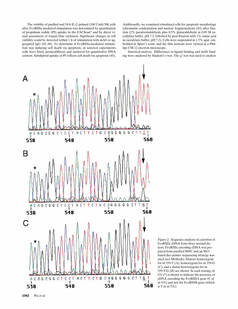
Characterization of CD16 epitopes. To determine if the 176Fto V change affects the binding of anti-CD16 mAb whichmight explain previously reported variations in anti-CD16mAb reactivity (28, 30, 31, 46, 47), the reactivity of FcgRIIIaon peripheral blood NK cells was characterized using a panelof anti-CD16 mAb. Donors homozygous for 176F or 176V andhomozygous for 66L2 and 72S2 were examined by flow cytome-try. Using the well characterized anti-CD16 mAb CLB-Gran1,identical CD16 fluorescence intensities were observed onCD56 positive NK cells from donors of both genotypes (Fig. 3A and Table I). Similar results were evident with six additionalanti-CD16 mAbs (Table I) including mAb B73.1 which is af-fected by the polymorphism at nt 230 (24, 25). mAbs 1D3 andMEM154 showed differential binding to NK cells from donorshomozygous for F compared with V. In both instances, thesemAb bound well to 176V/V donors but only poorly to 176F/Fdonors (Fig. 3, C and D, and Table I). mAb 3G8 showed subtledifferences which did not reach statistical significance with oursample size (Fig. 3 B). These data indicate that although do-nors homozygous for either the F or the V alleles express the
Figure 3. CD16 mAbs MEM154 and 1D3 show differential reactivity with FcgRIIIa-176V and FcgRIIIa-176F alleles. Lympho-cytes and NK cells were identified in washed whole blood by characteristic light scatter properties and lack of reactivity with anti-CD14. Anti-CD16 staining on the CD142 lymphocytes is shown. Blood from donors homozygous for FcgRIIIa-176V/V and FcgRIIIa-176F/F was examined. Iden-tical Gran1 reactivity (A) confirms identi-cal receptor density between the two do-nors. As previously reported, mAb 3G8 binds slightly less to the FcgRIIIa-176F al-lele than the FcgRIIIa-176V allele (the functional high binding phenotype, see Re-sults) (B). mAbs 1D3 (C) and MEM154 (D) bound well to NK cells from theFcgRIIIa-176V/V donor but reacted poorly with NK cells from the FcgRIIIa-176F/F donor. Data represent a single do-nor pair out of a total of five different do-nor pairs examined.
1064 Wu et al.
same level of CD16 protein on the surface of NK cells, the re-activities for some anti-CD16 mAbs differ, suggesting that thethese allelic proteins have different three-dimensional struc-tural characteristics.
We then examined the mAb epitopes expressed on CD16positive circulating monocytes. This population, typically a smallpercentage of circulating monocytes (35), was identified bymulticolor fluorescence. Because FcgRIIIa expression by mono-cytes is variable among donors, we sought donors homozygousfor 176F or 176V (and homozygous for both 66L and 72S) ex-pressing comparable levels of mAb CLB-Gran1 reactivity ontheir peripheral blood monocytes. In paired experiments, B73.1showed identical reactivity while both 1D3 and MEM154showed less reactivity with the 176 F/F donor (results notshown).
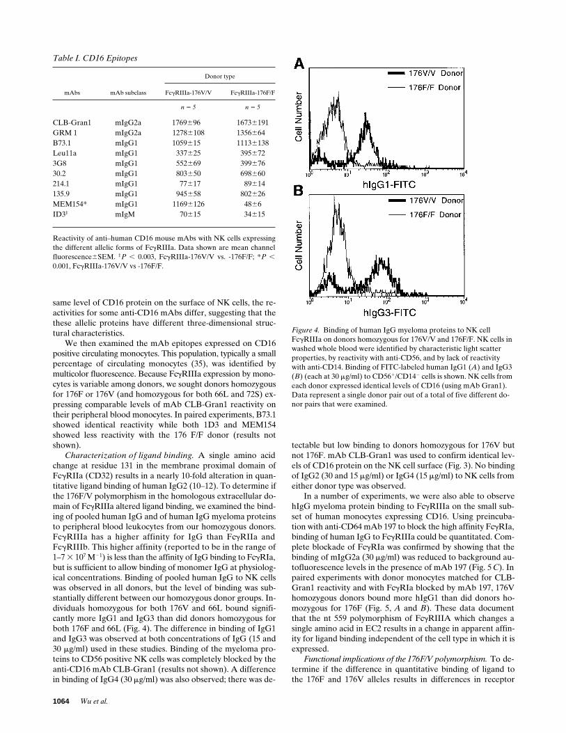
Characterization of ligand binding. A single amino acidchange at residue 131 in the membrane proximal domain ofFcgRIIa (CD32) results in a nearly 10-fold alteration in quan-titative ligand binding of human IgG2 (10–12). To determine ifthe 176F/V polymorphism in the homologous extracellular do-main of FcgRIIIa altered ligand binding, we examined the bind-ing of pooled human IgG and of human IgG myeloma proteinsto peripheral blood leukocytes from our homozygous donors.FcgRIIIa has a higher affinity for IgG than FcgRIIa andFcgRIIIb. This higher affinity (reported to be in the range of1–7 3 107 M21) is less than the affinity of IgG binding to FcgRIa,but is sufficient to allow binding of monomer IgG at physiolog-ical concentrations. Binding of pooled human IgG to NK cellswas observed in all donors, but the level of binding was sub-stantially different between our homozygous donor groups. In-dividuals homozygous for both 176V and 66L bound signifi-cantly more IgG1 and IgG3 than did donors homozygous forboth 176F and 66L (Fig. 4). The difference in binding of IgG1and IgG3 was observed at both concentrations of IgG (15 and30 mg/ml) used in these studies. Binding of the myeloma pro-teins to CD56 positive NK cells was completely blocked by theanti-CD16 mAb CLB-Gran1 (results not shown). A differencein binding of IgG4 (30 mg/ml) was also observed; there was de-
tectable but low binding to donors homozygous for 176V butnot 176F. mAb CLB-Gran1 was used to confirm identical lev-els of CD16 protein on the NK cell surface (Fig. 3). No bindingof IgG2 (30 and 15 mg/ml) or IgG4 (15 mg/ml) to NK cells fromeither donor type was observed.
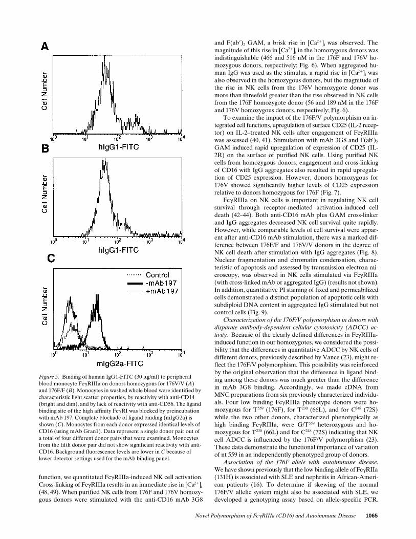
In a number of experiments, we were also able to observehIgG myeloma protein binding to FcgRIIIa on the small sub-set of human monocytes expressing CD16. Using preincuba-tion with anti-CD64 mAb 197 to block the high affinity FcgRIa,binding of human IgG to FcgRIIIa could be quantitated. Com-plete blockade of FcgRIa was confirmed by showing that thebinding of mIgG2a (30 mg/ml) was reduced to background au-tofluorescence levels in the presence of mAb 197 (Fig. 5 C). Inpaired experiments with donor monocytes matched for CLB-Gran1 reactivity and with FcgRIa blocked by mAb 197, 176Vhomozygous donors bound more hIgG1 than did donors ho-mozygous for 176F (Fig. 5, A and B). These data documentthat the nt 559 polymorphism of FcgRIIIA which changes asingle amino acid in EC2 results in a change in apparent affin-ity for ligand binding independent of the cell type in which it isexpressed.
Functional implications of the 176F/V polymorphism. To de-termine if the difference in quantitative binding of ligand tothe 176F and 176V alleles results in differences in receptor
Table I. CD16 Epitopes
mAbs mAb subclass
Donor type
FcgRIIIa-176V/V FcgRIIIa-176F/F
n 5 5 n 5 5
CLB-Gran1 mIgG2a 1769696 16736191GRM 1 mIgG2a 12786108 1356664B73.1 mIgG1 1059615 11136138Leu11a mIgG1 337625 3956723G8 mIgG1 552669 39967630.2 mIgG1 803650 698660214.1 mIgG1 77617 89614135.9 mIgG1 945658 802626MEM154* mIgG1 11696126 4866ID3‡ mIgM 70615 34615
Reactivity of anti–human CD16 mouse mAbs with NK cells expressingthe different allelic forms of FcgRIIIa. Data shown are mean channelfluorescence6SEM. ‡P , 0.003, FcgRIIIa-176V/V vs. -176F/F; *P ,
0.001, FcgRIIIa-176V/V vs -176F/F.
Figure 4. Binding of human IgG myeloma proteins to NK cellFcgRIIIa on donors homozygous for 176V/V and 176F/F. NK cells in washed whole blood were identified by characteristic light scatter properties, by reactivity with anti-CD56, and by lack of reactivity with anti-CD14. Binding of FITC-labeled human IgG1 (A) and IgG3 (B) (each at 30 mg/ml) to CD561/CD142 cells is shown. NK cells from each donor expressed identical levels of CD16 (using mAb Gran1). Data represent a single donor pair out of a total of five different do-nor pairs that were examined.
Novel Polymorphism of FcgRIIIa (CD16) and Autoimmune Disease 1065
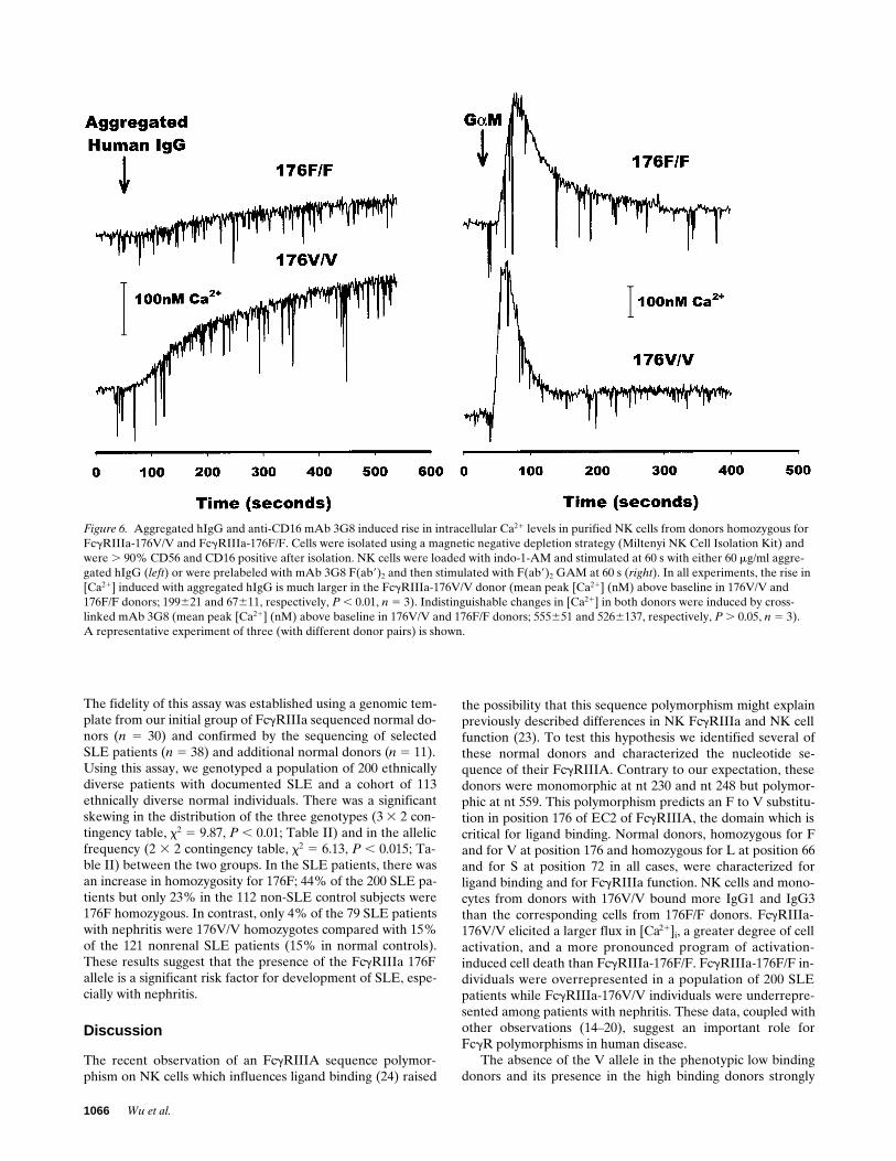
function, we quantitated FcgRIIIa-induced NK cell activation.Cross-linking of FcgRIIIa results in an immediate rise in [Ca21]i
(48, 49). When purified NK cells from 176F and 176V homozy-gous donors were stimulated with the anti-CD16 mAb 3G8
and F(ab9)2 GAM, a brisk rise in [Ca21]i was observed. Themagnitude of this rise in [Ca21]i in the homozygous donors wasindistinguishable (466 and 516 nM in the 176F and 176V ho-mozygous donors, respectively; Fig. 6). When aggregated hu-man IgG was used as the stimulus, a rapid rise in [Ca21]i wasalso observed in the homozygous donors, but the magnitude ofthe rise in NK cells from the 176V homozygote donor wasmore than threefold greater than the rise observed in NK cellsfrom the 176F homozygote donor (56 and 189 nM in the 176Fand 176V homozygous donors, respectively; Fig. 6).
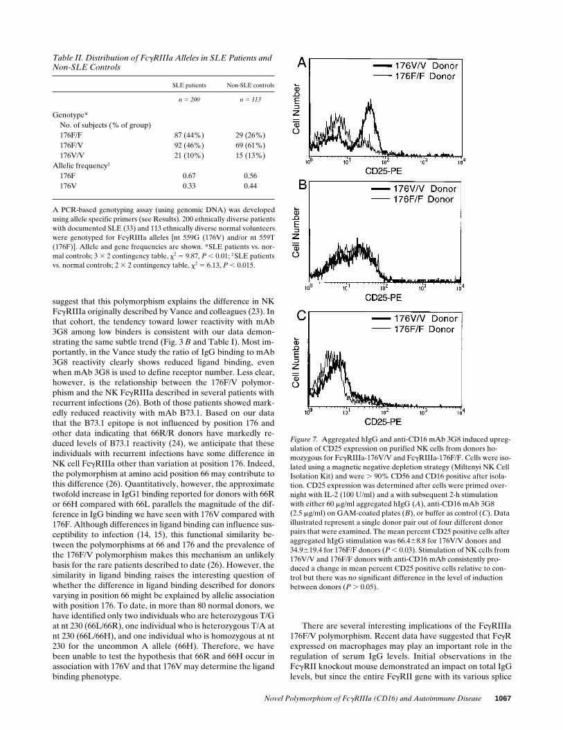
To examine the impact of the 176F/V polymorphism on in-tegrated cell functions, upregulation of surface CD25 (IL-2 recep-tor) on IL-2–treated NK cells after engagement of FcgRIIIawas assessed (40, 41). Stimulation with mAb 3G8 and F(ab9)2
GAM induced rapid upregulation of expression of CD25 (IL-2R) on the surface of purified NK cells. Using purified NKcells from homozygous donors, engagement and cross-linkingof CD16 with IgG aggregates also resulted in rapid upregula-tion of CD25 expression. However, donors homozygous for176V showed significantly higher levels of CD25 expressionrelative to donors homozygous for 176F (Fig. 7).
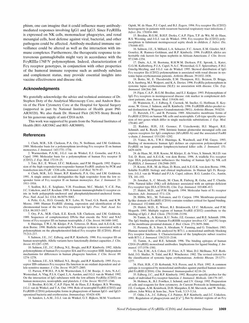
FcgRIIIa on NK cells is important in regulating NK cellsurvival through receptor-mediated activation-induced celldeath (42–44). Both anti-CD16 mAb plus GAM cross-linkerand IgG aggregates decreased NK cell survival quite rapidly.However, while comparable levels of cell survival were appar-ent after anti-CD16 mAb stimulation, there was a marked dif-ference between 176F/F and 176V/V donors in the degree ofNK cell death after stimulation with IgG aggregates (Fig. 8).Nuclear fragmentation and chromatin condensation, charac-teristic of apoptosis and assessed by transmission electron mi-croscopy, was observed in NK cells stimulated via FcgRIIIa(with cross-linked mAb or aggregated IgG) (results not shown).In addition, quantitative PI staining of fixed and permeabilizedcells demonstrated a distinct population of apoptotic cells withsubdiploid DNA content in aggregated IgG stimulated but notcontrol cells (Fig. 9).
Characterization of the 176F/V polymorphism in donors withdisparate antibody-dependent cellular cytotoxicity (ADCC) ac-tivity. Because of the clearly defined differences in FcgRIIIa-induced function in our homozygotes, we considered the possi-bility that the differences in quantitative ADCC by NK cells ofdifferent donors, previously described by Vance (23), might re-flect the 176F/V polymorphism. This possibility was reinforcedby the original observation that the difference in ligand bind-ing among these donors was much greater than the differencein mAb 3G8 binding. Accordingly, we made cDNA fromMNC preparations from six previously characterized individu-als. Four low binding FcgRIIIa phenotype donors were ho-mozygous for T559 (176F), for T230 (66L), and for C248 (72S)while the two other donors, characterized phenotypically ashigh binding FcgRIIIa, were G/T559 heterozygous and ho-mozygous for T230 (66L) and for C248 (72S) indicating that NKcell ADCC is influenced by the 176F/V polymorphism (23).These data demonstrate the functional importance of variationof nt 559 in an independently phenotyped group of donors.
Association of the 176F allele with autoimmune disease.We have shown previously that the low binding allele of FcgRIIa(131H) is associated with SLE and nephritis in African-Ameri-can patients (16). To determine if skewing of the normal176F/V allelic system might also be associated with SLE, wedeveloped a genotyping assay based on allele-specific PCR.
Figure 5. Binding of human IgG1-FITC (30 mg/ml) to peripheral blood monocyte FcgRIIIa on donors homozygous for 176V/V (A) and 176F/F (B). Monocytes in washed whole blood were identified by characteristic light scatter properties, by reactivity with anti-CD14 (bright and dim), and by lack of reactivity with anti-CD56. The ligand binding site of the high affinity FcgRI was blocked by preincubation with mAb 197. Complete blockade of ligand binding (mIgG2a) is shown (C). Monocytes from each donor expressed identical levels of CD16 (using mAb Gran1). Data represent a single donor pair out of a total of four different donor pairs that were examined. Monocytes from the fifth donor pair did not show significant reactivity with anti-CD16. Background fluorescence levels are lower in C because of lower detector settings used for the mAb binding panel.
1066 Wu et al.
The fidelity of this assay was established using a genomic tem-plate from our initial group of FcgRIIIa sequenced normal do-nors (n 5 30) and confirmed by the sequencing of selectedSLE patients (n 5 38) and additional normal donors (n 5 11).Using this assay, we genotyped a population of 200 ethnicallydiverse patients with documented SLE and a cohort of 113ethnically diverse normal individuals. There was a significantskewing in the distribution of the three genotypes (3 3 2 con-tingency table, x2 5 9.87, P , 0.01; Table II) and in the allelicfrequency (2 3 2 contingency table, x2 5 6.13, P , 0.015; Ta-ble II) between the two groups. In the SLE patients, there wasan increase in homozygosity for 176F; 44% of the 200 SLE pa-tients but only 23% in the 112 non-SLE control subjects were176F homozygous. In contrast, only 4% of the 79 SLE patientswith nephritis were 176V/V homozygotes compared with 15%of the 121 nonrenal SLE patients (15% in normal controls).These results suggest that the presence of the FcgRIIIa 176Fallele is a significant risk factor for development of SLE, espe-cially with nephritis.
Discussion
The recent observation of an FcgRIIIA sequence polymor-phism on NK cells which influences ligand binding (24) raised
the possibility that this sequence polymorphism might explainpreviously described differences in NK FcgRIIIa and NK cellfunction (23). To test this hypothesis we identified several ofthese normal donors and characterized the nucleotide se-quence of their FcgRIIIA. Contrary to our expectation, thesedonors were monomorphic at nt 230 and nt 248 but polymor-phic at nt 559. This polymorphism predicts an F to V substitu-tion in position 176 of EC2 of FcgRIIIA, the domain which iscritical for ligand binding. Normal donors, homozygous for Fand for V at position 176 and homozygous for L at position 66and for S at position 72 in all cases, were characterized forligand binding and for FcgRIIIa function. NK cells and mono-cytes from donors with 176V/V bound more IgG1 and IgG3than the corresponding cells from 176F/F donors. FcgRIIIa-176V/V elicited a larger flux in [Ca21]i, a greater degree of cellactivation, and a more pronounced program of activation-induced cell death than FcgRIIIa-176F/F. FcgRIIIa-176F/F in-dividuals were overrepresented in a population of 200 SLEpatients while FcgRIIIa-176V/V individuals were underrepre-sented among patients with nephritis. These data, coupled withother observations (14–20), suggest an important role forFcgR polymorphisms in human disease.
The absence of the V allele in the phenotypic low bindingdonors and its presence in the high binding donors strongly
Figure 6. Aggregated hIgG and anti-CD16 mAb 3G8 induced rise in intracellular Ca21 levels in purified NK cells from donors homozygous for FcgRIIIa-176V/V and FcgRIIIa-176F/F. Cells were isolated using a magnetic negative depletion strategy (Miltenyi NK Cell Isolation Kit) and were . 90% CD56 and CD16 positive after isolation. NK cells were loaded with indo-1-AM and stimulated at 60 s with either 60 mg/ml aggre-gated hIgG (left) or were prelabeled with mAb 3G8 F(ab9)2 and then stimulated with F(ab9)2 GAM at 60 s (right). In all experiments, the rise in [Ca21] induced with aggregated hIgG is much larger in the FcgRIIIa-176V/V donor (mean peak [Ca21] (nM) above baseline in 176V/V and176F/F donors; 199621 and 67611, respectively, P , 0.01, n 5 3). Indistinguishable changes in [Ca21] in both donors were induced by cross-linked mAb 3G8 (mean peak [Ca21] (nM) above baseline in 176V/V and 176F/F donors; 555651 and 5266137, respectively, P . 0.05, n 5 3).A representative experiment of three (with different donor pairs) is shown.
Novel Polymorphism of FcgRIIIa (CD16) and Autoimmune Disease 1067
suggest that this polymorphism explains the difference in NKFcgRIIIa originally described by Vance and colleagues (23). Inthat cohort, the tendency toward lower reactivity with mAb3G8 among low binders is consistent with our data demon-strating the same subtle trend (Fig. 3 B and Table I). Most im-portantly, in the Vance study the ratio of IgG binding to mAb3G8 reactivity clearly shows reduced ligand binding, evenwhen mAb 3G8 is used to define receptor number. Less clear,however, is the relationship between the 176F/V polymor-phism and the NK FcgRIIIa described in several patients withrecurrent infections (26). Both of those patients showed mark-edly reduced reactivity with mAb B73.1. Based on our datathat the B73.1 epitope is not influenced by position 176 andother data indicating that 66R/R donors have markedly re-duced levels of B73.1 reactivity (24), we anticipate that theseindividuals with recurrent infections have some difference inNK cell FcgRIIIa other than variation at position 176. Indeed,the polymorphism at amino acid position 66 may contribute tothis difference (26). Quantitatively, however, the approximatetwofold increase in IgG1 binding reported for donors with 66Ror 66H compared with 66L parallels the magnitude of the dif-ference in IgG binding we have seen with 176V compared with176F. Although differences in ligand binding can influence sus-ceptibility to infection (14, 15), this functional similarity be-tween the polymorphisms at 66 and 176 and the prevalence ofthe 176F/V polymorphism makes this mechanism an unlikelybasis for the rare patients described to date (26). However, thesimilarity in ligand binding raises the interesting question ofwhether the difference in ligand binding described for donorsvarying in position 66 might be explained by allelic associationwith position 176. To date, in more than 80 normal donors, wehave identified only two individuals who are heterozygous T/Gat nt 230 (66L/66R), one individual who is heterozygous T/A atnt 230 (66L/66H), and one individual who is homozygous at nt230 for the uncommon A allele (66H). Therefore, we havebeen unable to test the hypothesis that 66R and 66H occur inassociation with 176V and that 176V may determine the ligandbinding phenotype.
There are several interesting implications of the FcgRIIIa176F/V polymorphism. Recent data have suggested that FcgRexpressed on macrophages may play an important role in theregulation of serum IgG levels. Initial observations in theFcgRII knockout mouse demonstrated an impact on total IgGlevels, but since the entire FcgRII gene with its various splice
Table II. Distribution of FcgRIIIa Alleles in SLE Patients and Non-SLE Controls
SLE patients Non-SLE controls
n 5 200 n 5 113
Genotype*No. of subjects (% of group)176F/F 87 (44%) 29 (26%)176F/V 92 (46%) 69 (61%)176V/V 21 (10%) 15 (13%)
Allelic frequency‡
176F 0.67 0.56176V 0.33 0.44
A PCR-based genotyping assay (using genomic DNA) was developedusing allele specific primers (see Results). 200 ethnically diverse patientswith documented SLE (33) and 113 ethnically diverse normal volunteerswere genotyped for FcgRIIIa alleles [nt 559G (176V) and/or nt 559T(176F)]. Allele and gene frequencies are shown. *SLE patients vs. nor-mal controls; 3 3 2 contingency table, x2 5 9.87, P , 0.01; ‡SLE patientsvs. normal controls; 2 3 2 contingency table, x2 5 6.13, P , 0.015.
Figure 7. Aggregated hIgG and anti-CD16 mAb 3G8 induced upreg-ulation of CD25 expression on purified NK cells from donors ho-mozygous for FcgRIIIa-176V/V and FcgRIIIa-176F/F. Cells were iso-lated using a magnetic negative depletion strategy (Miltenyi NK Cell Isolation Kit) and were . 90% CD56 and CD16 positive after isola-tion. CD25 expression was determined after cells were primed over-night with IL-2 (100 U/ml) and a with subsequent 2-h stimulation with either 60 mg/ml aggregated hIgG (A), anti-CD16 mAb 3G8(2.5 mg/ml) on GAM-coated plates (B), or buffer as control (C). Data illustrated represent a single donor pair out of four different donor pairs that were examined. The mean percent CD25 positive cells after aggregated hIgG stimulation was 66.468.8 for 176V/V donors and 34.9619.4 for 176F/F donors (P , 0.03). Stimulation of NK cells from 176V/V and 176F/F donors with anti-CD16 mAb consistently pro-duced a change in mean percent CD25 positive cells relative to con-trol but there was no significant difference in the level of induction between donors (P . 0.05).
1068 Wu et al.
isoforms was disrupted, the relative roles of FcgRIIb1 ex-pressed on B cells as opposed to FcgRIIb2 expressed on mac-rophages could not be determined (50). Somewhat surpris-ingly, the naturally occurring disruption of the expression ofthe FcgRIIb2 isoform expressed on macrophages of NODmice is strongly associated with upregulation of both IgG1 andIgG2b serum levels despite relatively normal expression of theB cell specific FcgRIIb1 (51). The possibility that macrophageFcgR may be playing an important role in the regulation ofIgG levels is further underscored by the observation in hu-mans that different alleles of FcgRIIa are associated with dif-ferent serum levels of IgG2 (14). This observation is particu-larly important because in humans, FcgRIIa is expressed onmacrophages but not on B cells. Furthermore, this observationemphasizes that alleles with different capacities to bind humanIgG2, not just presence or absence of receptor, are associatedwith different levels of IgG. Thus, it seems reasonable to ex-trapolate to the prediction that the FcgRIIIa 176F/V allelesmay influence the level of IgG1 and IgG3. How they influencespecific responses to vaccination and net effective humoral im-munity remains to be determined.
Of course the implications of the FcgRIIIa 176F/V poly-
morphism extend beyond the regulation of serum IgG levels.Soluble FcgRIIIa is clearly present in the circulation (52–54).In a number of systems, soluble receptor can influence thelevel of B cell activation presumably through binding surfaceimmunoglobulin (55, 56). Since FcgRIIIa binds ligand withhigher affinity than FcgRIIIb which is unable to bind ligand inmonomeric form, FcgRIIIa may play a particularly importantrole in mediating these effects. FcgRIIIa may also play a criti-cal role in the first-dose cytokine-release syndrome seen withsome therapeutic monoclonal antibodies (57). Furthermore,our earlier studies in a primate model of immune complexhandling demonstrated an essential role for FcgRIIIa (58, 59),and more recent observations in mice with targeted disruptionof murine FcgRIII also support an important role in immunecomplex–mediated triggering of inflammatory reactions (60).Since each of these effects is dependent on binding of IgG, thepotential for FcgRIIIa 176F/V alleles to influence the biologicpotential of both receptor and ligand is clearly evident.
To directly test this potential in human biology in vivo, weinvestigated the possibility that FcgRIIIa 176F/V alleles mightbe abnormally represented in patients with SLE, a prototypicimmune complex disease. A role for abnormal FcgR functionin SLE has been described (61), and the skewing of FcgRIIaalleles in SLE has supported the hypothesis that the FcgRIIaallele with a low binding phenotype for human IgG2 would beoverrepresented in SLE (16, 17). In most immune complexes,however, autoantibodies are not of the IgG2 isotype but ratherof the IgG1 and IgG3 isotypes. Thus, one might anticipate anoverrepresentation of 176F and an underrepresentation of176V in immune complex disease. In our study of 200 ethni-cally diverse SLE patients, this skewing was very apparent(Table II). Indeed, in patients with SLE and nephritis, the ho-mozygous 176V was underrepresented by more than fourfoldcompared with those without nephritis. We recognize thatthese observations need confirmation in large independentpopulations, that further stratification by clinical phenotypeand ethnicity may be insightful, and that studies of multiplexfamilies will be informative. We also recognize that this asso-ciation may result from linkage to a different gene at anotherlocus. However, the biology of this polymorphism, its rele-vance to the pathophysiology of SLE, and the coincidence ofFcgRIIIa’s chromosomal location with a region of high inter-est in the microsatellite-based scanning of the genome in SLEpatients (62, 63), all make FcgRIIIa a likely gene for SLE dis-ease risk.
Based on the biology of the FcgRIIIa-176F/V polymor-
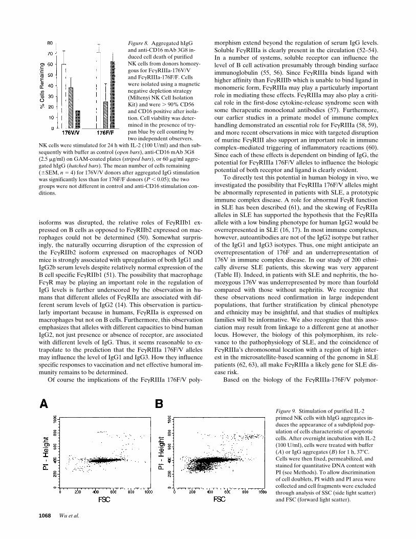
Figure 8. Aggregated hIgG and anti-CD16 mAb 3G8 in-duced cell death of purifiedNK cells from donors homozy-gous for FcgRIIIa-176V/V and FcgRIIIa-176F/F. Cells were isolated using a magnetic negative depletion strategy (Miltenyi NK Cell Isolation Kit) and were . 90% CD56 and CD16 positive after isola-tion. Cell viability was deter-mined in the presence of try-pan blue by cell counting by two independent observers.
NK cells were stimulated for 24 h with IL-2 (100 U/ml) and then sub-sequently with buffer as control (open bars), anti-CD16 mAb 3G8 (2.5 mg/ml) on GAM-coated plates (striped bars), or 60 mg/ml aggre-gated hIgG (hatched bars). The mean number of cells remaining (6SEM, n 5 4) for 176V/V donors after aggregated IgG stimulation was significantly less than for 176F/F donors (P , 0.05); the two groups were not different in control and anti-CD16 stimulation con-ditions.
Figure 9. Stimulation of purified IL-2 primed NK cells with hIgG aggregates in-duces the appearance of a subdiploid pop-ulation of cells characteristic of apoptotic cells. After overnight incubation with IL-2 (100 U/ml), cells were treated with buffer (A) or IgG aggregates (B) for 1 h, 378C. Cells were then fixed, permeabilized, and stained for quantitative DNA content with PI (see Methods). To allow discrimination of cell doublets, PI width and PI area were collected and cell fragments were excluded through analysis of SSC (side light scatter) and FSC (forward light scatter).
Novel Polymorphism of FcgRIIIa (CD16) and Autoimmune Disease 1069
phism, one can imagine that it could influence many antibody-mediated responses involving IgG1 and IgG3. Since FcgRIIIais expressed on NK cells, mononuclear phagocytes, and renalmesangial cells, host defense against viral, bacterial, and otherpathogens could be affected. Antibody-mediated immune sur-veillance could be altered as well as the interaction with im-mune complexes. Furthermore, the therapeutic response to in-travenous gammaglobulin might vary in accordance with theFcgRIIIa-176F/V polymorphism. Indeed, characterization ofFcg receptor genotypes, in conjunction with other propertiesof the humoral immune response such as antibody subclassand complement status, may provide essential insights intovaccine effectiveness and disease risk.
Acknowledgments
We gratefully acknowledge the advice and technical assistance of Dr.Stephen Doty of the Analytical Microscopy Core, and Andrew Bea-vis of the Flow Cytometry Core at the Hospital for Special Surgery(supported in part by National Institutes of Health grant P60-AR38320). We also thank Dr. Howard Fleit (SUNY-Stony Brook)for his generous supply of anti-CD16 mAb.
This work was supported by grants from the National Institutes ofHealth (R01-AR33062 and R01-AR38889).
References
1. Clark, M.R., S.B. Clarkson, P.A. Ory, N. Stollman, and I.M. Goldstein.1989. Molecular basis for a polymorphism involving Fcg receptor II on humanmonocytes. J. Immunol. 143:1731–1734.
2. Warmerdam, P.A.M., J.G.J. van de Winkel, E.J. Gosselin, and P.J.A.Capel. 1990. Molecular basis for a polymorphism of human Fcg receptor II(CD32). J. Exp. Med. 172:19–25.
3. Tate, B.J., E. Witort, I.F.C. McKenzie, and P.M. Hogarth. 1992. Expres-sion of the high responder/non-responder human FcgRII: analysis by PCR andtransfection into FcgR2 COS cells. Immunol. Cell Biol. 70:79–87.
4. Clark, M.R., S.G. Stuart, R.P. Kimberly, P.A. Ory, and I.M. Goldstein.1991. A single amino acid distinguishes the high responder from the low re-sponder form of Fcg receptor II on human monocytes. Eur. J. Immunol. 21:1911–1916.
5. Scallon, B.J., E. Scigliano, V.H. Freedman, M.C. Miedel, Y.-C.E. Pan,J.C. Unkeless, and J.P. Kochan. 1989. A human immunoglobulin G receptor ex-ists in both polypeptide-anchored and phosphatidylinositol-anchored forms.Proc. Natl. Acad. Sci. USA. 86:5079–5083.
6. Peltz, G.A., H.O. Grundy, R.V. Lebo, H. Yssel, G.S. Barsh, and K.W.Moore. 1989. Human FcgRIII: cloning, expression and identification of thechromosomal locus of the two Fcg receptors for IgG. Proc. Natl. Acad. Sci.USA. 86:1013–1017.
7. Ory, P.A., M.R. Clark, E.E. Kwoh, S.B. Clarkson, and I.M. Goldstein.1989. Sequences of complementary DNAs that encode the NA1 and NA2forms of Fcg receptor III on human neutrophils. J. Clin. Invest. 84:1688–1692.
8. Huizinga, T.W.J., M. Kleijer, P.A. Tetteroo, D. Roos, and A.E.G.K. vondem Borne. 1990. Biallelic neutrophil NA-antigen system is associated with apolymorphism on the phosphoinositol-linked Fcg receptor III (CD16). Blood.75:213–217.
9. Salmon, J.E., J.C. Edberg, and R.P. Kimberly. 1990. Fcg receptor III onhuman neutrophils. Allelic variants have functionally distinct capacities. J. Clin.Invest. 85:1287–1295.
10. Salmon, J.E., J.C. Edberg, N.L. Brogle, and R.P. Kimberly. 1992. Allelicpolymorphisms of human Fcg receptor IIA and Fcg receptor IIIB. Independentmechanisms for differences in human phagocyte function. J. Clin. Invest. 89:1274–1278.
11. Salmon, J.E., S.S. Millard, N.L. Brogle, and R.P. Kimberly. 1995. Fcg re-ceptor IIIb enhances Fcg receptor IIa function in an oxidant dependent and al-lele-sensitive manner. J. Clin. Invest. 95:2877–2885.
12. Parren, P.W.H.I., P.A.M. Warmerdam, L.C.M. Boeije, J. Arts, N.A.C.Westerdaal, A. Vlug, P.J.A. Capel, L.A. Aarden, and J.G.J. van de Winkel. 1992.On the interaction of IgG subclasses with the low-affinity FcgRIIa (CD32) onhuman monocytes, neutrophils, and platelets. J. Clin. Invest. 90:1537–1546.
13. Bredius, R.G.M., C.A.P. Fijen, M. de Haas, E.J. Kuijper, R.S. Weening,J.G.J. van de Winkel, and T.A. Out. 1994. Role of neutrophil FcgRII (CD32) andFcgRIIIb (CD16) polymorphic forms in phagocytosis of human IgG1- and IgG3-opsonized bacteria and erythrocytes. Immunology. 83:624–630.
14. Sanders, L.A.M., J.G.J. van de Winkel, G.T. Rijkers, M.M. Voorhorst-
Ogink, M. de Haas, P.J. Capel, and B.J. Zegers. 1994. Fcg receptor IIa (CD32)heterogeneity in patients with recurrent bacterial respiratory tract infections. J.Infect. Dis. 170:854–860.
15. Bredius, R.G.M., B.H.F. Derkx, C.A.P. Fijen, T.P. de Wit, M. de Haas,R.S. Weening, and J.G.J. van de Winkel. 1994. Fcg receptor IIa (CD32) poly-morphism in fulminant meningococcal septic shock in children. J. Infect. Dis.170:848–853.
16. Salmon, J.E., S. Millard, L.A. Schacter, F.C. Arnett, E.M. Ginzler, M.F.Gourley, R. Ramsey-Goldman, and R.P. Kimberly. 1996. FcgRIIA alleles areheritable risk factors for lupus nephritis in African-Americans. J. Clin. Invest.97:1348–1354.
17. Duits, A.J., H. Bootsma, R.H.W.M. Derksen, P.E. Spronk, L. Kater,G.G.M. Kallenberg, P.J.A. Capel, N.A.C. Westerdaal, G.T. Spierenbury, F.H.J.Gmelig-Meyling, and J.G.J. van de Winkel. 1995. Skewed distribution of IgGFcg receptor IIa (CD32) polymorphism is associated with renal disease in sys-temic lupus erythematosus patients. Arthritis Rheum. 39:1832–1836.
18. Botto, M., E. Theodoridis, E.M. Thompson, H.L. Beynon, D. Briggs,D.A. Isenberg, M.J. Walport, and K.A. Davies. 1996. FcgRIIa polymorphism insystemic lupus erythematosus (SLE): no association with disease. Clin. Exp.Immunol. 104:264–268.
19. Fijen, C.A.P., R.G.M. Bredius, and E.J. Kuijper. 1993. Polymorphism ofIgG Fcg receptors in meningococcal disease: risk marker in complement defi-cient patients. Ann. Intern. Med. 119:636–641.
20. Wainstein, E., J. Edberg, E. Csernok, M. Sneller, G. Hoffman, E. Key-stone, W. Gross, J. Salmon, and R. Kimberly. 1996. FcgRIIIB alleles predict re-nal dysfunction in Wegeners Granulomatosis (WG). Arthritis Rheum. 39:S210.
21. Ravetch, J.V., and B. Perussia. 1989. Alternative membrane forms ofFcgRIII (CD16) on human NK cells and neutrophils. Cell-type specific expres-sion of two genes which differ in single nucleotide substitutions. J. Exp. Med.170:481–497.
22. Radeke, H.H., J.E. Gessner, P. Uciechowski, H.J. Magert, R.E.Schmidt, and K. Resch. 1994. Intrinsic human glomerular mesangial cells canexpress receptors for IgG complexes (hFcgRIII-A) and the associated FceRIg-chain. J. Immunol. 153:1281–1292.
23. Vance, B.A., T.W.J. Huizinga, K. Wardwell, and P.M. Guyre. 1993.Binding of monomeric human IgG defines an expression polymorphism ofFcgRIII on large granular lymphocyte/natural killer cells. J. Immunol. 151:6429–6439.
24. de Haas, M., H.R. Koene, M. Kleijer, E. de Vries, S. Simsek, M.J.D. vanTol, D. Roos, and A.E.G.K. von dem Borne. 1996. A triallelic Fcg receptortype IIIA polymorphism influences the binding of human IgG by NK cellFcgRIIIA. J. Immunol. 156:2948–2955.
25. Koene, H.R., M. de Haas, D. Roos, and A.E.G.K. von dem Borne. 1996.Soluble FcgRIII: biology and clinical implications. In Human IgG Fc Recep-tors. J.G.J. van de Winkel and P.J.A. Capel, editors. R.G. Landes Co., Austin,TX. 181–193.
26. Jawahar, S., C. Moody, M. Chan, R. Finberg, R. Geha, and T. Chatila.1996. Natural killer (NK) cell deficiency associated with an epitope-deficientFcg receptor type IIIA (CD16-II). Clin. Exp. Immunol. 103:408–413.
27. Hulett, M.D., and P.M. Hogarth. 1994. Molecular basis of Fc receptorfunction. Adv. Immunol. 57:1–124.
28. Hibbs, M.L., M. Tolvanen, and O. Carpen. 1994. Membrane-proximalIg-like domain of FcgRIII (CD16) contains residues critical for ligand binding.J. Immunol. 152:4466–4474.
29. Hulett, M.D., E. Witort, R.I. Brinkworth, I.F.C. McKenzie, and P.M.Hogarth. 1995. Multiple regions of human FcgRII (CD32) contribute to thebinding of IgG. J. Biol. Chem. 270:21188–21194.
30. Tamm, A., A. Kister, K.U. Nolte, J.E. Gessner, and R.E. Schmidt. 1996.The IgG binding site of human FcgRIIIB receptor involves CC9 and FG loopsof the membrane-proximal domain. J. Biol. Chem. 271:3659–3666.
31. Perussia, B., S. Starr, S. Abraham, V. Fanning, and G. Trinchieri. 1983.Human natural killer cells analyzed by B73.1, a monoclonal antibody blockingFcg receptor functions. I. Characterization of the lymphocyte subset reactivewith B73.1. J. Immunol. 130:2133–2144.
32. Tamm, A., and R.E. Schmidt. 1996. The binding epitopes of humanCD16 (FcgRIII) monoclonal antibodies. Implications for ligand binding. J. Im-munol. 157:1576–1581.
33. Tan, E.M., A.S. Cohen, J.F. Fries, A.T. Masi, D.J. McShane, N.F. Roth-field, J.G. Shaller, N. Talal, and R.J. Winchester. 1982. The revised criteria forthe classification of systemic lupus erythematosus. Arthritis Rheum. 25:1271–1277.
34. Fleit, H.B., C.D. Kobasiuk, N.S. Peress, and A. Fleit. 1992. A commonepitope is recognized by monoclonal antibodies against purified human neutro-phil FcgRIII (CD16). Clin. Immunol. Immunopathol. 62:16–24.
35. Edberg, J.C., and R.P. Kimberly. 1992. Receptor specific probes for thestudy of individual Fcg receptor function. J. Immunol. Methods. 148:179–187.
36. Holmes, K., B.J. Fowlkes, I. Schmid, and J.V. Giorgi. 1992. Preparationof cells and reagents for flow cytometry. In Current Protocols in Immunology.J.E. Cooligan, A.M. Kruisbeek, D.H. Margulies, E.M. Shevatch, and W. Strobe,editors. John Wiley & Sons Inc., New York. 5.3.5–5.3.6.
37. Odin, J.A., J.C. Edberg, C.J. Painter, R.P. Kimberly, and J.C. Unkeless.1991. Regulation of phagocytosis and [Ca21]i flux by distinct regions of an Fcg
1070 Wu et al.
receptor. Science (Wash. DC). 254:1785–1788.38. Kimberly, R.P., J.W. Ahlstrom, M.E. Click, and J.C. Edberg. 1990. The
glycosyl phosphatidylinositol-linked FcgRIIIPMN mediates transmembrane sig-naling events distinct from FcgRII. J. Exp. Med. 171:1239–1255.
39. Grynkiewicz, G., M. Poenie, and R.Y. Tsien. 1985. A new generation ofCa21 indicators with greatly improved fluorescence properties. J. Biol. Chem.260:3440–3450.
40. Anegon, I., M.C. Cuturi, G. Trinchieri, and B. Perussia. 1988. Interac-tion of Fcg receptor (CD16) ligands induces transcription of interleukin 2 re-ceptor (CD25) and lymphokine genes and expression of their products in hu-man natural killer cells. J. Exp. Med. 167:452–461.
41. Harris, D.T., W.W. Travis, and H.S. Koren. 1989. Induction of activa-tion antigens on human natural killer cells mediated through the Fcg receptor.J. Immunol. 143:2401–2406.
42. Ortaldo, J.R., A.T. Mason, and J.J. O’Shea. 1995. Receptor-induceddeath in human natural killer cells: involvement of CD16. J. Exp. Med. 181:339–344.
43. Azzoni, L., I. Anegon, B. Calabretta, and B. Perussia. 1995. Ligandbinding to FcgR induces c-myc-dependent apoptosis in IL-2 stimulated NKcells. J. Immunol. 154:491–499.
44. Eischen, C.M., J.D. Schilling, D.H. Lynch, P.H. Krammer, and P.J.Leibson. 1996. Fcg receptor-induced expression of Fas ligand on activated NKcells facilitates cell-mediated cytotoxicity and subsequent autocrine NK cell apop-tosis. J. Immunol. 156:2693–2699.
45. Darzynkiewicz, Z., X. Li, J. Gong, and F. Traganos. 1997. Methods foranalysis of apoptosis by flow cytometry. In Manual of Clinical Laboratory Im-munology. 5th edition. N.R. Rose, editor. ASM Press, Washington, DC. 334–343.
46. de Haas, M., M. Kleijer, D. Roos, and A.E.G.K. von dem Borne. 1994.Characterization of mAbs of the CD16 cluster and six newly generated CD16mAbs. In Leukocyte Typing V: White Cell Differentiation Antigens. S.F. Schloss-man, L. Boumsell, and W. Gilks, editors. Oxford University Press. 811–814.
47. Trounstine, M.L., G.A. Peltz, H. Yssel, T.W. Huizinga, A.E.G.K. vondem Borne, H. Spits, and K.W. Moore. 1990. Reactivity of cloned, expressedhuman FcgRIII isoforms with monoclonal antibodies which distinguish cell-type-specific and allelic forms of FcgRIII. Int. Immunol. 2:303–310.
48. Cassatella, M.A., I. Anegnon, M. Cuturi, P. Griskey, G. Trinchieri, andB. Perussia. 1989. FcgR (CD16) interaction with ligand induces Ca21 mobiliza-tion and phosphoinositide turnover in human natural killer cells. J. Exp. Med.169:459–467.
49. Gismondi, A., F. Mainiero, S. Morrone, G. Palmieri, M. Piccoli, L. Frati,and A. Santoni. 1992. Triggering through CD16 or phorbol esters enhances ad-hesion of NK cells to laminin via very late antigen 6. J. Exp. Med. 176:1251–1257.
50. Takai, T., M. Ono, M. Hikida, H. Ohmori, and J.V. Ravetch. 1996. Aug-mented humoral and anaphylactic responses in FcgRII-deficient mice. Nature(Lond.). 379:346–349.
51. Luan, J.J., R.C. Monteiro, C. Sautes, G. Fluteau, L. Eloy, W.H. Frid-man, J.-F. Bach, and H.-J. Garchon. 1996. Defective FcgRII gene expression inmacrophages of NOD mice. Genetic linkage with up-regulation of IgG1 andIgG2b in serum. J. Immunol. 157:4707–4716.
52. de Haas, M., M. Kleijer, R.M. Minchinton, D. Roos, and A.E.G.K. vondem Borne. 1994. Soluble FcgRIIIa is present in plasma and is derived fromnatural killer cells. J. Immunol. 152:900–907.
53. Harrison, D., J.H. Phillips, and L.L. Lanier. 1991. Involvement of a me-talloprotease in spontaneous and phorbol ester-induced release of natural killercell-associated FcgRIII (CD16-II). J. Immunol. 147:3459–3465.
54. Fleit, H.B., C.D. Kobasiuk, C. Daly, R. Furie, P.C. Levy, and R.O. Web-ster. 1992. A soluble form of FcgRIII is present in human serum and other bodyfluids and is elevated at sites of inflammation. Blood. 79:2721–2728.
55. Teillaud, C., J. Galon, M.-T. Zilber, N. Mazieres, R. Sasgnoli, R. Kurrle,W.H. Fridman, and C. Sautes. 1993. Soluble CD16 binds peripheral bloodmononuclear cells and inhibits pokeweed-mitogen-induced responses. Blood.82:3081–3090.
56. Hoover, R.G., C. Lary, R. Page, P. Travis, R. Ownes, J. Flick, J. Korn-bluth, and B. Baslogie. 1995. Autoregulatory circuits in myeloma. Tumor cellcytotoxicity mediated by soluble CD16. J. Clin. Invest. 95:241–247.
57. Wing, M.G., T. Moreau, J. Greenwood, R.M. Smith, G. Hale, J. Isaacs,H. Waldmann, P.J. Lachmann, and A. Compstom. 1996. Mechanism of first-dose cytokine-release syndrome by CAMPATH 1-H. Involvement of CD16(FcgRIII) and CD11a/CD18 (LFA-1) on NK cells. J. Clin. Invest. 98:2819–2826.
58. Clarkson, S.B., R.P. Kimberly, J.E. Valinsky, M.D. Witmer, J.B. Bussel,R.L. Nachman, and J.C. Unkeless. 1986. Blockade of clearance of immune com-plexes by an anti-Fcg receptor monoclonal antibody. J. Exp. Med. 164:474–489.
59. Kimberly, R.P., J.C. Edberg, L.T. Merriam, S.B. Clarkson, J.C. Unke-less, and R.P. Taylor. 1989. The in vivo handling of soluble complement fixingAb/dsDNA immune complexes in chimpanzees. J. Clin. Invest. 84:962–970.
60. Hazenbos, W.L.W., J.G.E. Gessner, F.M.A. Hofhuis, H. Kuipers, D.Meyer, I.A.F.M. Heijnen, R.E. Schmidt, M. Sandor, P.J.A. Capel, M. Daëron,et al. 1996. Impaired IgG-dependent anaphylaxis and arthus reaction in FcgRIII(CD16) deficient mice. Immunity. 5:181–188.
61. Kimberly, R.P., J.E. Salmon, and J.C. Edberg. 1995. Receptors for im-munoglobulin G. Molecular diversity and implications for disease. ArthritisRheum. 38:306–314.
62. Harley, J.B., P. Sheldon, B. Neas, S. Murphy, D.H. Wallace, R.H.Scofield, T.S. Shaver, and K.L. Hardgrave-Moser. 1994. Systemic lupus erythe-matosus. Considerations for a genetic approach. J. Invest. Dermatol. 103:144–149.
63. Tsao, B.P., R.M. Cantor, K.C. Kalunian, C.-J. Chen, H. Badsha, R.Singh, D.J. Wallace, R.C. Kitridou, S.-l. Chen, N. Shen, Y.W. Song, D.A. Isen-berg, C.-L. Yu, B.H. Hahn, and J.I. Rotter. 1997. Evidence for linkage of a can-didate chromosome 1 region to human systemic lupus erythematosus. J. Clin.Invest. 99:725–731.


![RIII[1]ROCAS EXOGENAS](https://img.pdfslide.net/doc/110x75/552494994a795934498b47ab/riii1rocas-exogenas.jpg)