Embed Size (px)
Citation preview
NURSE ANESTHESIOLOGY PROGRAMFLORIDA INTERNATIONAL UNIVERSITY
LINDA WUNDER, CRNA MSN
Objectives
� 1. Recognize the pain pathways from
peripheral stimulus to supraspinal processing.
� 2. Understand the differences between pain
fibers.
� 3. Discuss pain modulation from supraspinal
processing to substantia gelantinosa.
� 4. Explain the physiology of nociception.
PAIN PATHWAYS
� 1. Peripheral stimulus
� 2. Receptor (transduction)
� 3. Peripheral transmission
� 4. Spinal transmission
� 5. Ascending tracts
� 6. Supraspinal processing
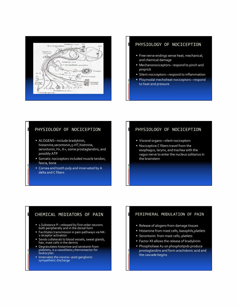
� Pain is conducted along three neuron pathways (first, second and third order neurons) that
transmit noxious stimuli from the periphery to the cerebral cortex.
� Primary afferent neurons are located in the dorsal root ganglion which lie in the vertebral
foramina at each spinal cord level. Each neuron has a single axon that bifurcates, sending one end to the peripheral tissues it innervates and
the other into the dorsal horn of the spinal cord.
PAIN PATHWAYS
� First order neurons
� The majority, enter the dorsal spinal root at each cervical, thoracic, lumbar, and sacral level
� Pain fibers originating in the head are carried by the trigeminal (V), facial (VII), glossopharyngeal (IX), and vagal (X)
� Each have specific ganglion which hold cell bodies of these nerves. The first order neurons in the ganglia (head) reach the brainstem and synapse with the second order neuron
PAIN PATHWAYS
� Second order neurons
� Spinal cord gray matter is divided by Rexed into 10 lamina
� First six makeup the dorsal horn, receive all afferent neural activity, represent the principle site for modulation of pain
� Second order neurons are either nociceptive-specific or wide dynamic range (WDR)
PAIN PATHWAYS
� Lamina I responds to nociceptive stimuli from cutaneous and deep somatic tissues
� Lamina II (substancia gelatinosa), contains many interneurons responsible for processing and modulating nociceptive input from cutaneous tissue. Major site of action for opoids
� Lamina VII contains preganglionic sympathetic neurons
� Lamina V and I contains visceral afferents
� Lamina V responds to both noxious and non noxious stimuli and receives both somatic and visceral inputs
� Thus referred pain
PAIN PATHWAYS
� Spinothalamic tract
� Cross the midline to the level of origin to the contralateral side of the spinal cord
� Divided into lateral and medial
� Lateral spinothalmic tract projects—location,density,duration of pain in the ventral posteriorlateral nucleus of the thalamus
� Medial spinothalamic tract projects—unpleasant emotional perception of pain in the medial thalamus
PAIN PATHWAYS
� Spinalreticular pain pathway- arousal and
autonomic responses to pain
� Spinalmesencephalic--anti-nociceptive
descending pathways because of its projections in the periductal gray area
PAIN PATHWAYS
� THIRD ORDER NEURONS
� Located in the thalamus
� Send fibers to the somatosensory areas I and
II in the post central gyrus of the parietal cortex and superior wall of the sylvian fissure
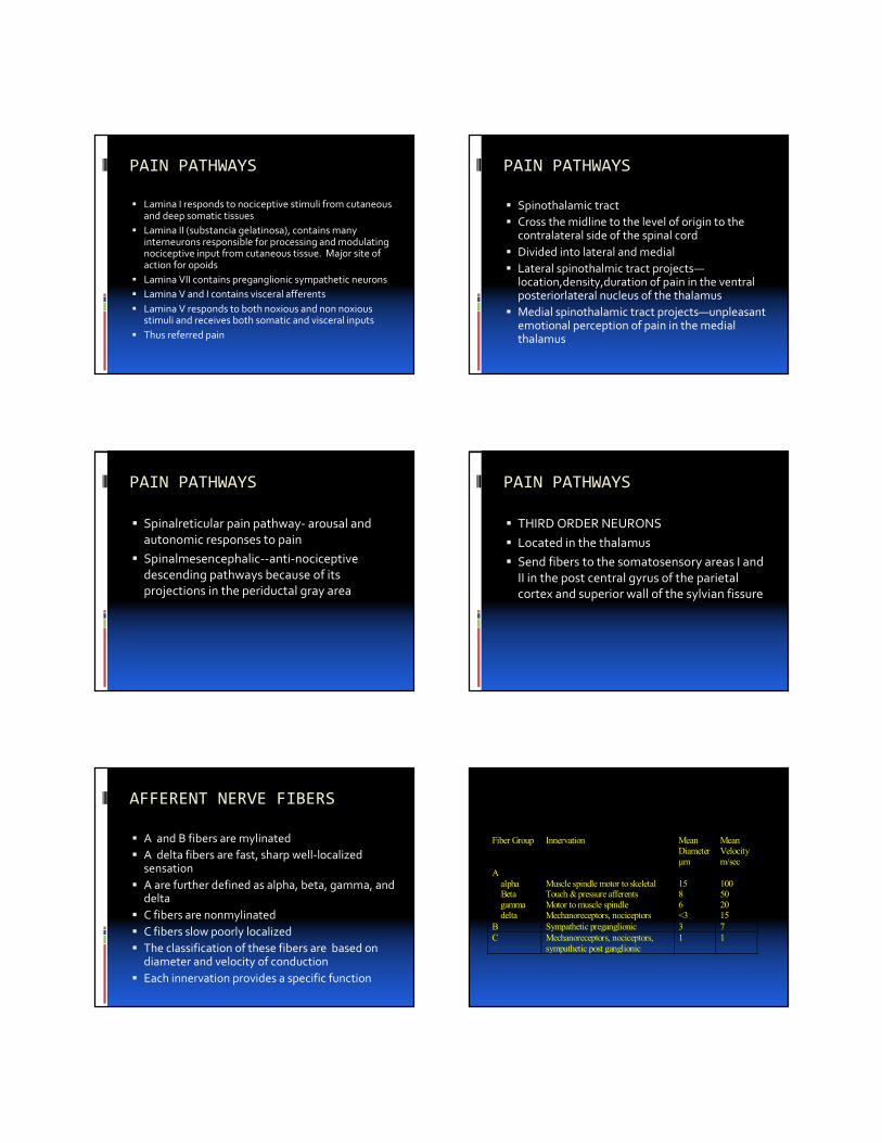
AFFERENT NERVE FIBERS
� A and B fibers are mylinated
� A delta fibers are fast, sharp well-localized sensation
� A are further defined as alpha, beta, gamma, and delta
� C fibers are nonmylinated
� C fibers slow poorly localized
� The classification of these fibers are based on diameter and velocity of conduction
� Each innervation provides a specific function
Fiber Group Innervation MeanDiameter
µm
MeanVelocity
m/sec
A
alpha Beta
gamma delta
Muscle spindle motor to skeletalTouch & pressure afferents
Motor to muscle spindleMechanoreceptors, nociceptors
158
6<3
10050
2015
B Sympathetic preganglionic 3 7
C Mechanoreceptors, nociceptors,
sympathetic post ganglionic
1 1
PHYSIOLOGY OF NOCICEPTION
� Free nerve endings sense heat, mechanical,
and chemical damage
� Mechanonociceptors– respond to pinch and
pinprick
� Silent nociceptors—respond to inflammation
� Ploymodal mechoheat nociceptors—respond to heat and pressure
PHYSIOLOGY OF NOCICEPTION
� ALOGENS—include bradykinin,
histamine,serontonin,5-HT,histmine, serontonin, H+, K+, some prostaglandins, and
possibly ATP
� Somatic nociceptors included muscle tendon,
fascia, bone
� Cornea and tooth pulp and innervated by A delta and C fibers
PHYSIOLOGY OF NOCICEPTION
� Visceral organs—silent nociceptors
� Nociceptive C fibers travel from the esophagus, larynx, and trachea with the
vagus nerve to enter the nucleus solitarius in the brainstem
CHEMICAL MEDIATORS OF PAIN
� 1.Substance P—released by first order neurons both peripherally and in the dorsal horn
� Facilitates transmission in pain pathways via NK-1 receptor activation
� Sends collaterals to blood vessels, sweat glands, hair, mast cells in the dermis
� Degranulates histamine and serotonin from platelets, is a vasodilator,chemoreactor for leukocytes
� Innervates the viscera—post ganglionic sympathetic discharge
PERIPHERAL MODULATION OF PAIN
� Release of alogens from damage tissues
� Histamine from mast cells, basophils,platlets
� Serontonin from mast cells, platlets
� Factor XII allows the release of bradykinin
� Phospholiase A2 on phospholipids produce
prostaglandins and form arachidonic acid and the cascade begins
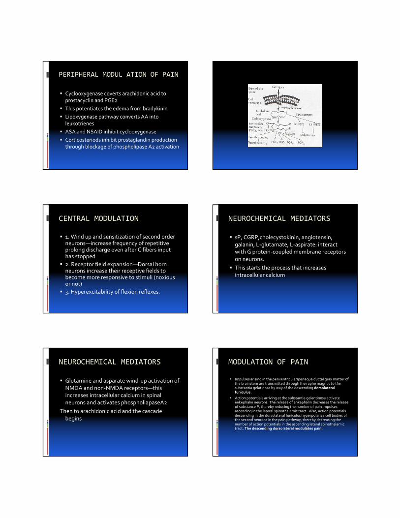
PERIPHERAL MODUL ATION OF PAIN
� Cyclooxygenase coverts arachidonic acid to
prostacyclin and PGE2
� This potentiates the edema from bradykinin
� Lipoxygenase pathway converts AA into
leukotrienes
� ASA and NSAID inhibit cyclooxygenase
� Corticosteriods inhibit prostaglandin production
through blockage of phospholipase A2 activation
CENTRAL MODULATION
� 1. Wind up and sensitization of second order neurons—increase frequency of repetitive prolong discharge even after C fibers input has stopped
� 2. Receptor field expansion—Dorsal horn neurons increase their receptive fields to become more responsive to stimuli (noxious or not)
� 3. Hyperexcitability of flexion reflexes.
NEUROCHEMICAL MEDIATORS
� sP, CGRP,cholecystokinin, angiotensin,
galanin, L-glutamate, L-aspirate: interact with G protein-coupled membrane receptors
on neurons.
� This starts the process that increases
intracellular calcium
NEUROCHEMICAL MEDIATORS
� Glutamine and asparate wind-up activation of
NMDA and non-NMDA receptors—this increases intracellular calcium in spinal
neurons and activates phospholiapaseA2
Then to arachidonic acid and the cascade
begins
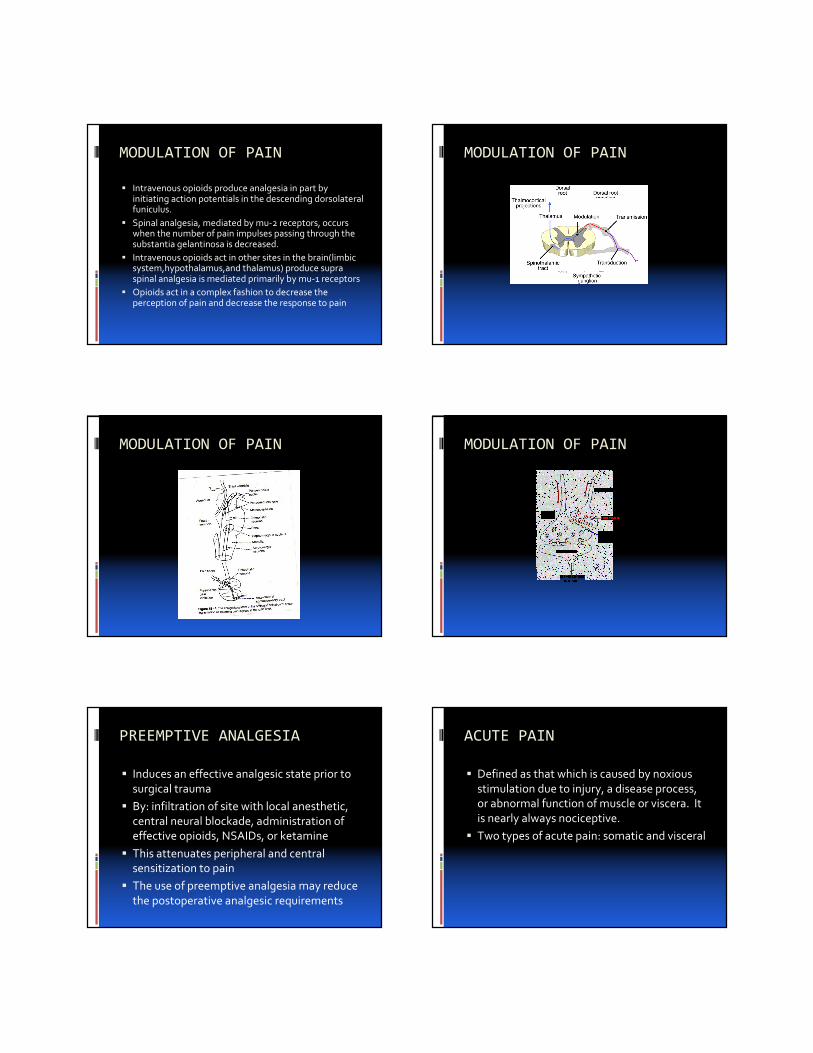
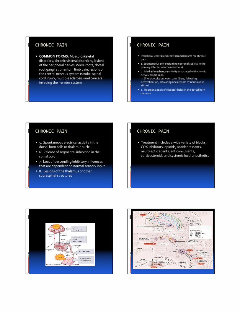
MODULATION OF PAIN
� Impulses arising in the periventricular/periaquaiductal gray matter of the brainstem are transmitted through the raphe magnus to the substantia gelatinosa by way of the descending dorsolateral funiculus.
� Action potentials arriving at the substantia gelantinosa activate enkephalin neurons. The release of enkephalin decreases the release of substance P, thereby reducing the number of pain impulses ascending in the lateral spinothalamic tract. Also, action potentials descending in the dorsolateral funiculus hyperpolarize cell bodies of the second neurons in the pain pathway, thereby decreasing the number of action potentials in the ascending lateral spinothalamic tract. The descending dorsolateral modulates pain.
MODULATION OF PAIN
� Intravenous opioids produce analgesia in part by initiating action potentials in the descending dorsolateral funiculus.
� Spinal analgesia, mediated by mu-2 receptors, occurs when the number of pain impulses passing through the substantia gelantinosa is decreased.
� Intravenous opioids act in other sites in the brain(limbic system,hypothalamus,and thalamus) produce supra spinal analgesia is mediated primarily by mu-1 receptors
� Opioids act in a complex fashion to decrease the perception of pain and decrease the response to pain
MODULATION OF PAIN
MODULATION OF PAIN MODULATION OF PAIN
PREEMPTIVE ANALGESIA
� Induces an effective analgesic state prior to
surgical trauma
� By: infiltration of site with local anesthetic,
central neural blockade, administration of effective opioids, NSAIDs, or ketamine
� This attenuates peripheral and central sensitization to pain
� The use of preemptive analgesia may reduce
the postoperative analgesic requirements
ACUTE PAIN
� Defined as that which is caused by noxious
stimulation due to injury, a disease process, or abnormal function of muscle or viscera. It
is nearly always nociceptive.
� Two types of acute pain: somatic and visceral
ACUTE PAIN: SOMATIC
� Superficial somatic -skin, subcutaneous,
mucous membranes
� Well localized-sharp, pricking, throbbing,
burning
� Deep somatic – muscles, tendons, joints, bones
� Less well localized, dull, aching
ACUTE PAIN: VISCERAL
� Disease process or abnormal function of an internal organ or itscovering (eg, parietal pleura, pericardium, or peritoneum).
� Four types: true localized visceral, true localized parietal, referred visceral, referred parietal
� True visceral is dull, diffuse, midline and is associated with abnormal sympathetic or parasympathetic activity (N/V, sweating, changes in B/P and HR )
� True parietal is sharp and localized
� Referred-disease process involving the peritoneum or pleura over the central diaphragm is referred to the neck and shoulder whereas disease affecting the parietal surfaces of the peripheral diaphragm is referred to the chest or upper abdominal wall
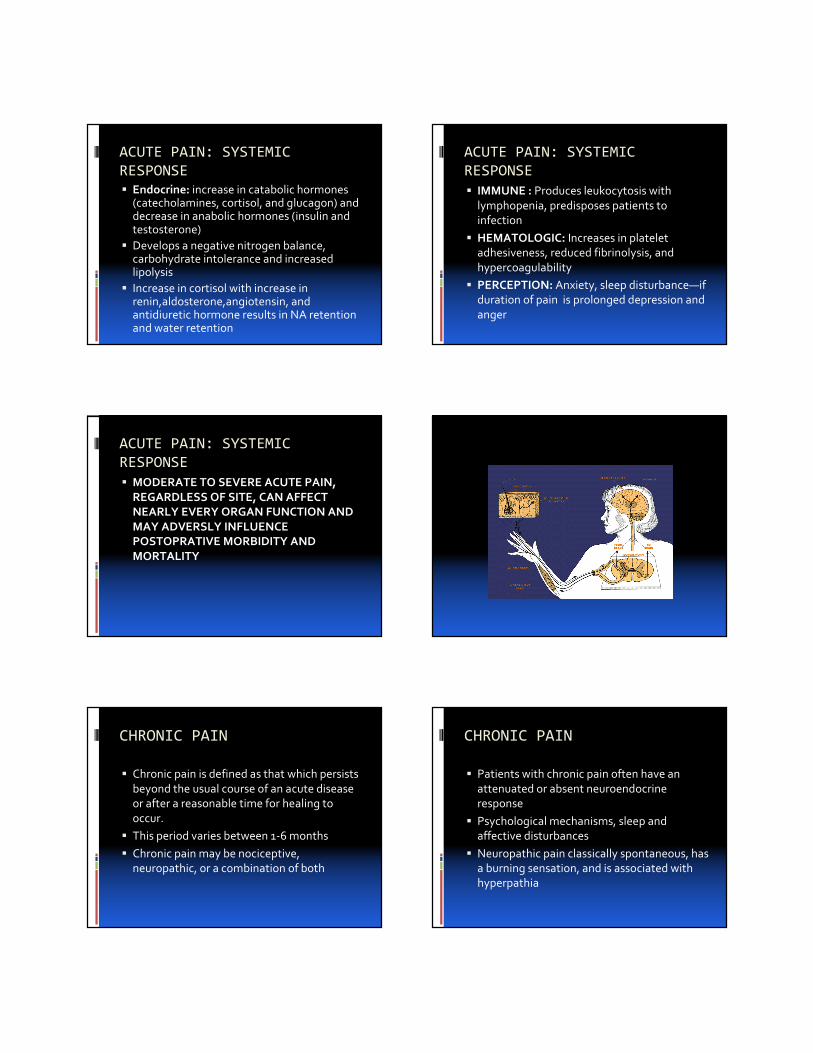
ACUTE PAIN: SYSTEMIC
RESPONSE
� Sympathetic activation increases efferent
sympathetic tone to all viscera and releases catecholamines from the adrenal medulla.
� The hormonal response results from increased sympathetic tone and
hypothalamically mediated reflexes
ACUTE PAIN: SYSTEMIC
RESPONSE
� CARDIOVASCULAR: hypertension,
tachycardia, enhanced myocardial irritability, increased SVR
� Increased CO, may be decrease with patients who have compromised ventricular
function
� Increased myocardial oxygen demand, therefore, pain can aggravate or precipitate
myocardial ischemia
ACUTE PAIN: SYSTEMIC RESONSE
� RESPIRATORY: Increase in total body O2
consumption and CO2 production increases minute ventilation
ACUTE PAIN: SYSTEMIC
RESPONSE
� GASTROINTESTINAL & URINARY:
� Enhanced sympathetic tone increases sphincter tone and decreases intestinal and
urinary motility, promoting ileus and urinary retention
� Hyper-secretion of gastric acid promotes stress ulceration, together with deceased motility, predisposes the patients to severe
aspiration pneumonitis
ACUTE PAIN: SYSTEMIC
RESPONSE
� Endocrine: increase in catabolic hormones (catecholamines, cortisol, and glucagon) and decrease in anabolic hormones (insulin and testosterone)
� Develops a negative nitrogen balance, carbohydrate intolerance and increased lipolysis
� Increase in cortisol with increase in renin,aldosterone,angiotensin, and antidiuretic hormone results in NA retention and water retention
ACUTE PAIN: SYSTEMIC
RESPONSE
� IMMUNE : Produces leukocytosis with
lymphopenia, predisposes patients to infection
� HEMATOLOGIC: Increases in platelet adhesiveness, reduced fibrinolysis, and
hypercoagulability
� PERCEPTION: Anxiety, sleep disturbance—if duration of pain is prolonged depression and
anger
ACUTE PAIN: SYSTEMIC
RESPONSE
� MODERATE TO SEVERE ACUTE PAIN,
REGARDLESS OF SITE, CAN AFFECT NEARLY EVERY ORGAN FUNCTION AND
MAY ADVERSLY INFLUENCE POSTOPRATIVE MORBIDITY AND
MORTALITY
CHRONIC PAIN
� Chronic pain is defined as that which persists
beyond the usual course of an acute disease or after a reasonable time for healing to
occur.
� This period varies between 1-6 months
� Chronic pain may be nociceptive, neuropathic, or a combination of both
CHRONIC PAIN
� Patients with chronic pain often have an
attenuated or absent neuroendocrine response
� Psychological mechanisms, sleep and affective disturbances
� Neuropathic pain classically spontaneous, has a burning sensation, and is associated with hyperpathia
CHRONIC PAIN
� COMMON FORMS: Musculoskeletal
disorders, chronic visceral disorders, lesions of the peripheral nerves, nerve roots, dorsal
root ganglia , phantom limb pain, lesions of the central nervous system (stroke, spinal
cord injury, multiple sclerosis) and cancers invading the nervous system
CHRONIC PAIN
� Peripheral-central and central mechanisms for chronic pain
� 1. Spontaneous self-sustaining neuronal activity in the primary afferent neuron (neuroma)
� 2. Marked mechanosensitivity associated with chronic nerve compression3. Short circuits between pain fibers, following demyelination, activating nociceptors by nonnoxious stimuli
� 4. Reorganization of receptor fields in the dorsal horn neurons
CHRONIC PAIN
� 5. Spontaneous electrical activity in the
dorsal horn cells or thalamic nuclei
� 6. Release of segmental inhibition in the
spinal cord
� 7. Loss of descending inhibitory influences
that are dependent on normal sensory input
� 8. Lesions of the thalamus or other supraspinal structures
CHRONIC PAIN
� Treatment includes a wide variety of blocks,
COX inhibitors, opioids, antidepressants, neuroleptic agents, anticonvulsants,
corticosteroids and systemic local anesthetics

� http://www.youtube.com/watch?v=n2Jzt3zd8
vQ
References
� http://www.youtube.com/watch?v=n2Jzt3zd8
vQ
� Nagelhout, J., & Plaus, K. (2010) Nurse
anesthesia (4th ed.). St. Louis: Elsevier.
Stoelting, R., & Miller, R. (2007). Basics of
anesthesia (5th ed.). Philadelphia: Elsevier.