Embed Size (px)
Citation preview

X-ray crystal structure of a reiterative transcriptioncomplex reveals an atypical RNA extension pathwayKatsuhiko S. Murakamia,1,2, Yeonoh Shina,1, Charles L. Turnbough Jr.b, and Vadim Molodtsova,2
aDepartment of Biochemistry and Molecular Biology, Center for RNA Molecular Biology, Pennsylvania State University, University Park, PA 16802;and bDepartment of Microbiology, University of Alabama at Birmingham, Birmingham, AL 35294-2170
Edited by Carol A. Gross, University of California, San Francisco, CA, and approved May 30, 2017 (received for review February 16, 2017)
Reiterative transcription is a noncanonical form of RNA synthesisin which a nucleotide specified by a single base in the DNAtemplate is repetitively added to the nascent transcript. Here wedetermined the crystal structure of an RNA polymerase, thebacterial enzyme from Thermus thermophilus, engaged in reitera-tive transcription during transcription initiation at a promoter re-sembling the pyrG promoter of Bacillus subtilis. The structurereveals that the reiterative transcript detours from the dedicatedRNA exit channel and extends toward the main channel of theenzyme, thereby allowing RNA extension without displacementof the promoter recognition σ-factor. Nascent transcripts contain-ing reiteratively added G residues are eventually extended by non-reiterative transcription, revealing an atypical pathway for theformation of a transcription elongation complex.
reiterative transcription | RNA polymerase | X-ray crystal structure |transcription initiation | transcript slippage
In the canonical form of transcription, RNA polymerase (RNAP)reads the sequence of a template strand DNA one base at a time
to produce a complementary strand of RNA. However, in someinstances of transcription, a single base in the template DNA canspecify multiple bases in the RNA product (1–4). This process isdue to multiple rounds of upstream slippage of the RNA withouttranslocation of the template DNA within the active site of RNAP(5). This unconventional reaction is called “reiterative transcrip-tion,” and it occurs primarily within a homopolymeric tract in thetemplate DNA. Reiterative transcription can involve the repetitiveaddition of any nucleotide, and it can occur during all phases of thetranscription cycle. During initiation, a homopolymeric tract as shortas three residues can enable reiterative transcription (6–8), whereasa significantly longer homopolymeric tract is required during elon-gation and termination (9–12). This difference reflects the length ofthe obligatory DNA/RNA hybrid that forms within all transcriptioncomplexes, which during initiation can be shorter than the ∼9-bphybrid that forms during elongation and termination (10, 13).Reiterative transcription plays key roles in gene expression in
eukaryotes, viruses, and especially bacteria, where it has beenshown to control gene expression through a variety of mechanisms(1, 3). The earliest example was UTP-sensitive regulation of tran-scription initiation of the pyrBI operon of Escherichia coli, whichencodes two subunits of the pyrimidine nucleotide biosyntheticenzyme aspartate transcarbamylase (14). In this case, reiterativetranscription occurs within a T3 tract located in the initially tran-scribed region 5′-AATTTG (nontemplate strand sequence) of thepyrBI promoter. Reiterative transcription produces transcripts withthe sequence 5′-AAUUUUn (where n = 1 to >100), essentially allof which are released from the transcription initiation complex. Theextent of this nonproductive reiterative transcription is directlyproportional to the intracellular level of UTP, so that pyrBI geneexpression is reduced when UTP levels are high. Several otherregulatory mechanisms were subsequently discovered that weresimilar to the pyrBI mechanism in that they relied on variable re-iterative transcription at TTTn tracts in initially transcribed regionsto produce transcripts that were released immediately after re-petitive UMP addition, thereby repressing gene expression (2).
Another well-studied example is CTP-mediated control ofpyrG expression in Bacillus subtilis, which produces the pyrimi-dine biosynthetic enzyme CTP synthetase (15). The nontemplateDNA sequence of the pyrG initially transcribed region is5′-GGGCT, with transcription initiated predominantly at thefirst G residue (Fig. 1A). Low intracellular levels of CTP causetranscription pausing at position +4C, which provides time neededfor upstream transcript slippage and reiterative transcription tooccur at the end of the 5′-CCC tract of the DNA template. Thisreaction results in the addition of up to nine extra G residues at the5′ end of the pyrG transcript, after which the transcript is extendedby nonreiterative transcription through the pyrG leader region. Theextra G tract then forms an antiterminator hairpin with a down-stream segment of the nascent transcript that precludes intrinsictranscription termination at an attenuator preceding the pyrGgene. As a result, transcription proceeds through the pyrG geneleading to the production of more CTP synthetase and increasedlevels of CTP.Although reiterative transcription clearly plays important roles
in controlling gene expression, much remains to be learnedabout the mechanism of this reaction and how it differs fromcanonical transcription. This is particularly true for the posi-tioning of the nascent RNA inside RNAP. In this study, we haveexplored this question by preparing a reiterative transcriptioncomplex by in crystallo transcription and determining the X-raycrystal structure of a bacterial RNAP in the process of reiterativetranscription at a promoter with the same critical features as thepyrG promoter of B. subtilis.
Significance
Under certain conditions during transcription, a single base of thetemplate DNA specifies multiple bases in the RNA transcript due toslippage between the transcript and template. This noncanonicalform of RNA synthesis is called “reiterative transcription,” and itplays key regulatory roles in bacteria, eukaryotes, and viruses. Inthis study, we determined the crystal structure of a bacterial RNApolymerase engaged in reiterative transcription. Our study found acompletely unexpected RNA extension pathway during reiterativetranscription and uncovered an atypical mechanism for the transi-tion from the open promoter complex to the transcription elon-gation complex. These findings represent a major advancement inunderstanding the mechanics and flexibility of transcription.
Author contributions: K.S.M., C.L.T., and V.M. designed research; K.S.M., Y.S., and V.M.performed research; Y.S. and V.M. analyzed data; and K.S.M., C.L.T., and V.M. wrotethe paper.
The authors declare no conflict of interest.
This article is a PNAS Direct Submission.
Data deposition: The atomic coordinates and structure factors have been deposited in theProtein Data Bank, www.pdb.org (PDB ID codes 5VOI and 5VO8).1K.S.M. and Y.S. contributed equally to this work.2To whom correspondence may be addressed. Email: [email protected] or [email protected].
This article contains supporting information online at www.pnas.org/lookup/suppl/doi:10.1073/pnas.1702741114/-/DCSupplemental.
www.pnas.org/cgi/doi/10.1073/pnas.1702741114 PNAS Early Edition | 1 of 6
BIOCH
EMISTR
Y
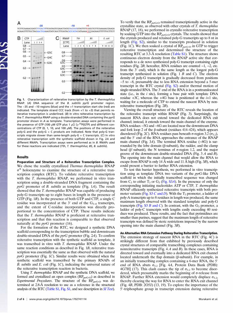
ResultsPreparation and Structure of a Reiterative Transcription Complex.We chose the readily crystallized Thermus thermophilus RNAPσA holoenzyme to examine the structure of a reiterative tran-scription complex (RTC). To validate reiterative transcriptionwith the T. thermophilus RNAP, we performed in vitro tran-scription using a double-stranded DNA fragment containing thepyrG promoter of B. subtilis as template (Fig. 1A). The resultshowed that the T. thermophilus RNAP was capable of producingpoly-G transcripts up to eight bases in length in the presence ofGTP (Fig. 1B). In the presence of both GTP and CTP, a single Cresidue was incorporated at the 3′ end of the G3–8 transcripts,and the extent of C-residue incorporation was directly pro-portional to the concentration of CTP. These results indicatethat the T. thermophilus RNAP is proficient at reiterative tran-scription and that this reaction is comparable to that observednaturally at the pyrG promoter (16).For the formation of the RTC, we designed a synthetic DNA
scaffold corresponding to the transcription bubble and downstreamdouble-stranded DNA of the pyrG promoter (Fig. 2A). To confirmreiterative transcription with the synthetic scaffold as template, itwas transcribed in vitro with T. thermophilus RNAP. Under thesame reaction conditions as described in Fig. 1B, reiterative tran-scription was essentially the same as that observed with the naturalpyrG promoter (Fig. 1C). Similar results were obtained when thesynthetic scaffold was transcribed by the primary RNAPs ofB. subtilis and E. coli (Fig. 1C), indicating the universal nature ofthe reiterative transcription reaction in bacteria.Using T. thermophilus RNAP and the synthetic DNA scaffold, we
formed and crystallized an open complex (RPpyrG-O) as described inExperimental Procedures. The structure of the RPpyrG-O was de-termined at 2.8-Å resolution to use as a reference in the structuralanalysis of the RTC (Table S1, Fig. S1, and see description in SI Text).
To verify that the RPpyrG-O remained transcriptionally active in thecrystalline state, as observed with other crystals of T. thermophilusRNAP (17, 18), we performed in crystallo reiterative transcriptionby soaking GTP into the RPpyrG-O crystals. The results showed thatthe crystals produced and retained poly-G transcripts up to 8 nt inlength (Fig. S2), similar to the transcripts produced in solution(Fig. 1C). We then soaked a crystal of RPpyrG-O in GTP to triggerreiterative transcription and determined the structure of theresulting RTC at 3.3-Å resolution (Table S1). The structure showscontinuous electron density from the RNAP active site that cor-responds to a de novo synthesized poly-G transcript containing eightresidues (Fig. 2B; hereafter, RNA residues are counted −1, −2, etc.from the 3′ end), which is the same length as the longest poly-Gtranscript synthesized in solution (Fig. 1 B and C). The electrondensity of poly-G transcript is gradually decreased from positions−5 to −8, presumably due to less RNA extension beyond a 5-mertranscript in the RTC crystal (Fig. S2) and/or thermal motion ofsingle-stranded RNA. The 3′ end of the RNA is in a posttranslocatedstate (i.e., in the i site), forming a base pair with template DNAresidue +3C, whereas the +4G base is positioned at the i+1 site,waiting for a molecule of CTP to extend the nascent RNA by non-reiterative transcription (Fig. 2B).Viewing the overall structure of the RTC reveals the location of
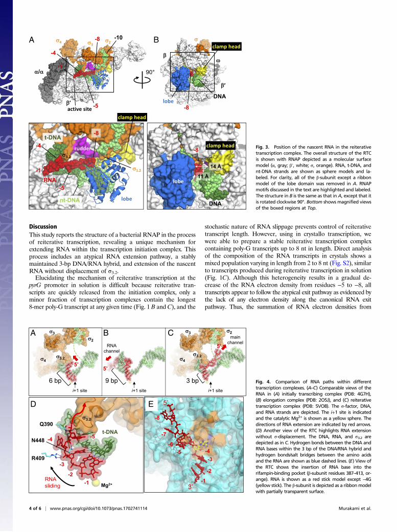
the reiterative transcript within RNAP (Fig. 3). Surprisingly, thenascent RNA does not extend toward the dedicated RNA exitchannel; instead, it extends toward the main channel of the enzyme.RNA residues−5G and −6G are located near the downstreamDNAand fork loop 2 of the β-subunit (residues 414–424), which appearsdisordered (Fig. 2C). RNA residues pass beneath σ-region 3.2 (σ3.2),and the 5′ end of the RNA approaches the entrance of the RNAPmain channel (Fig. 3A). The terminal RNA residue (−8G) is sur-rounded by the lobe domain (β-subunit), the rudder, and the clamphead (β′-subunit), the N terminus of σ-region 1.2, and the majorgroove of the downstream double-stranded DNA (Fig. 3 A and B).The opening into the main channel that would allow the RNA toescape from RNAP is only 14 Å wide and 11 Å high (Fig. 3B), whichcould present a barrier to further RNA extension.To test this barrier hypothesis, we performed in vitro transcrip-
tion using as template DNA two variants of the pyrG-like DNAscaffold in which the initially transcribed sequence was changedfrom C3 to either T3 or G3 (Fig. S3A). In the presence of only thecorresponding initiating nucleotides ATP or CTP, T. thermophilusRNAP efficiently synthesized reiterative transcripts with both pro-moter variants (Fig. S3 C andD). With the T3 promoter, a ladder ofpoly-A transcripts up to 8 residues in length was produced, the samemaximum length observed with the standard template and poly-Gtranscripts (Fig. S3 B and C). In contrast, with the G3 promoter, aladder of poly-C transcripts with lengths easily exceeding 40 resi-dues was produced. These results, and the fact that pyrimidines aresmaller than purines, suggest that the maximum length of reiterativetranscripts was defined by steric restrictions imposed by the narrowopening into the main channel (Fig. 3B).
An Alternative RNA Extension Pathway During Reiterative Transcription.The extension strategy of nascent RNA in the RTC (Fig. 4C) isstrikingly different from that exhibited by previously describedcrystal structures of comparable transcribing complexes containingnonreiterative transcripts (Fig. 4 A and B). In these cases, RNA isdirected toward and eventually into a dedicated RNA exit channellocated underneath the flap domain (β-subunit). For example, inan initially transcribing complex containing a 6-mer RNA, the 5′end of RNA abuts σ3.2 [Fig. 4A, Protein Data Bank (PDB):4G7H] (17). This clash causes the tip of σ3.2 to become disor-dered, which presumably marks the beginning of σ-release fromRNAP. Further RNA extension would completely displace σ3.2,thereby clearing the way for RNA to enter the RNA exit channel(Fig. 4B, PDB: 2O5J) (13, 19). To explore the importance of the5′-triphosphate group in transcript extension during reiterative
A
B C
Fig. 1. Characterization of reiterative transcription by the T. thermophilusRNAP. (A) DNA sequence of the B. subtilis pyrG promoter region.The −35 and −10 regions (blue) and the +1 transcription start site (red) areindicated. The template strand CCC track (from +1 to +3) that permits re-iterative transcription is underlined. (B) In vitro reiterative transcription bythe T. thermophilus RNAP using a double-stranded DNA containing the pyrGpromoter shown in A as template. Transcription assays were performed inthe presence of GTP (100 μM GTP plus 1 μCi [γ-32P]GTP) and different con-centrations of CTP (0, 1, 10, and 100 μM). The positions of the reiterativepoly-G and the poly-G + C products are indicated. Note that poly-G tran-scripts migrate slower than same-length poly-G + C transcripts. (C) In vitroreiterative transcription with the synthetic scaffold shown in Fig. 2A anddifferent RNAPs. Transcription assays were performed as in B. RNAPs usedfor these reactions are indicated (Tth, T. thermophilus; BS, B. subtilis).
2 of 6 | www.pnas.org/cgi/doi/10.1073/pnas.1702741114 Murakami et al.

transcription, we used the dinucleotide pGpG, which contains a5′-monophosphate group, to initiate reiterative transcription atthe scaffold pyrG promoter. The transcript ladder produced wasidentical to that obtained by transcription with GTP as solesubstrate (Fig. S4).In both the initially transcribing (Fig. 4A) and elongation
complexes (Fig. 4B), the 5′ end of RNA is guided toward the RNAexit channel by an extended DNA/RNA hybrid (i.e., 6 bp and an∼9 bp, respectively). However, in the case of a RTC containing thesynthetic pyrG promoter, the DNA/RNA hybrid is only 3 bp long(Fig. 4C). The remaining nonbase-pairing residues of the RNAare turned perpendicular to the DNA/RNA hybrid and directedtoward the main channel, without displacement of σ3.2. RNAresidue −4G is inserted into a pocket on the surface of theβ-subunit known as the rifampin-binding pocket (also known asthe rifampin-resistance determining region I) (Fig. 4E). To de-termine the importance of the interaction between RNA and therifampin-binding pocket during reiterative transcription, we mea-sured reiterative transcription from the pyrG promoter in solutionwith three rifampin-resistant mutants of E. coli RNAP havingamino acid substitutions either at D516V, H526Y, or S531Lof the β-subunit. The D516V and H526Y mutants alter the
electrostatic potential and the shape of the rifampin-binding pocket,respectively, whereas the S531L mutant replaces a hydrophilic witha hydrophobic side chain in the rifampin-binding pocket (20). Wefound that all mutants synthesize equal amounts of reiterativetranscripts and that the efficiencies of CMP incorporation to the 3′end of poly-G transcripts are equally dependent on CTP concen-tration in mutant and wild-type RNAPs (Fig. S5). These resultssuggest that the rifampin-binding pocket functions merely to shelteran unpaired RNA base after the transcript changes its directionfrom the dedicated RNA exit to the main channels of RNAP.The typical transcription elongation complex is optimally sta-
bilized by an ∼9-bp DNA/RNA hybrid (13, 19). In contrast, theRTC at the pyrG promoter demonstrates that a 3-bp hybrid, witha calculated Tm of 17 °C (OligoCalc, in the presence of 100 mMNa+) (21), is sufficient to maintain the RNA in the transcriptioncomplex and to allow its extension one base at a time. The 3-bphybrid does this while still being weak enough to allow the up-stream slippage of the RNA, which is a central feature of re-iterative transcription. RNA residues from −3G to −5G interactwith amino acid residues Q390, R409, and N448 of the β-subunit,which presumably would limit diffusion of the nonbase-pairedregion of the RNA (Fig. 4D).
B
A
C
Fig. 2. Structure of the reiterative transcription com-plex. (A) Schematic representation of the RTC formedby the RPpyrG-O in the presence of GTP. The DNA scaf-fold used for crystallization and reiterative transcriptare depicted in boxes [template DNA (t-DNA)], darkgreen; nontemplate DNA (nt-DNA), light green; RNA,red; the same colors are used in all other figures). DNAbases disordered in the structures are indicated bydashed boxes. Positions of DNA and RNA bases areindicated by numbers. (B) Ribbon models of RNAPmotifs (BH, bridge helix; TL, trigger loop) are shownwith transparent surfaces of the σ-factor and the β-flapdomain. RNA and DNA are shown as stick models. Thedisordered region of the TL is shown as a dashed line.The nucleotide binding sites (i and i+1) are indicated,and the active site catalytic Mg2+ is shown as a yellowsphere. The 2Fo–Fc electron density for RNA is shown(gray mesh, 1.5 σ). The RNAP orientation in this figureis similar to Fig. 3A. (C) Stick models of the DNA, RNA,and fork loop 2 in the RTC are shown with 2Fo–Fcelectron densities (gray mesh, 1.5 σ) of the DNA andfork loop 2. The disordered part of fork loop 2 (whitestick model) is modeled from fork loop 2 of the RPpyrG-Oafter superimposing these structures.
Murakami et al. PNAS Early Edition | 3 of 6
BIOCH
EMISTR
Y
DiscussionThis study reports the structure of a bacterial RNAP in the processof reiterative transcription, revealing a unique mechanism forextending RNA within the transcription initiation complex. Thisprocess includes an atypical RNA extension pathway, a stablymaintained 3-bp DNA/RNA hybrid, and extension of the nascentRNA without displacement of σ3.2.Elucidating the mechanism of reiterative transcription at the
pyrG promoter in solution is difficult because reiterative tran-scripts are quickly released from the initiation complex, only aminor fraction of transcription complexes contain the longest8-mer poly-G transcript at any given time (Fig. 1 B and C), and the
stochastic nature of RNA slippage prevents control of reiterativetranscript length. However, using in crystallo transcription, wewere able to prepare a stable reiterative transcription complexcontaining poly-G transcripts up to 8 nt in length. Direct analysisof the composition of the RNA transcripts in crystals shows amixed population varying in length from 2 to 8 nt (Fig. S2), similarto transcripts produced during reiterative transcription in solution(Fig. 1C). Although this heterogeneity results in a gradual de-crease of the RNA electron density from residues −5 to −8, alltranscripts appear to follow the atypical exit pathway as evidenced bythe lack of any electron density along the canonical RNA exitpathway. Thus, the summation of RNA electron densities from
A B
Fig. 3. Position of the nascent RNA in the reiterativetranscription complex. The overall structure of the RTCis shown with RNAP depicted as a molecular surfacemodel (α, gray; β′, white; σ, orange). RNA, t-DNA, andnt-DNA strands are shown as sphere models and la-beled. For clarity, all of the β-subunit except a ribbonmodel of the lobe domain was removed in A. RNAPmotifs discussed in the text are highlighted and labeled.The structure in B is the same as that in A, except that itis rotated clockwise 90°. Bottom shows magnified viewsof the boxed regions at Top.
A
D E
B C
Fig. 4. Comparison of RNA paths within differenttranscription complexes. (A–C) Comparable views of theRNA in (A) initially transcribing complex (PDB: 4G7H),(B) elongation complex (PDB: 2O5J), and (C) reiterativetranscription complex (PDB: 5VO8). The σ-factor, DNA,and RNA strands are depicted. The i+1 site is indicatedand the catalytic Mg2+ is shown as a yellow sphere. Thedirections of RNA extension are indicated by red arrows.(D) Another view of the RTC highlights RNA extensionwithout σ-displacement. The DNA, RNA, and σ3.2 aredepicted as in C. Hydrogen bonds between the DNA andRNA bases within the 3 bp of the DNA/RNA hybrid andhydrogen bonds/salt bridges between the amino acidsand the RNA are shown as blue dashed lines. (E) View ofthe RTC shows the insertion of RNA base into therifampin-binding pocket (β-subunit residues 387–413, or-ange). RNA is shown as a red stick model except −4G(yellow stick). The β-subunit is depicted as a ribbonmodelwith partially transparent surface.
4 of 6 | www.pnas.org/cgi/doi/10.1073/pnas.1702741114 Murakami et al.

somewhat heterogeneous reiterative transcription complexes in asingle crystal provides a means for establishing the location ofan otherwise unstable reiterative RNA. The crystalline state ofT. thermophilus RNAP–promoter DNA complex has a high sol-vent content (∼60%), is fully hydrated to allow nucleotide accessto the enzyme active site, and can produce either a canonicalRNA transcript with 6-bp DNA/RNA hybrid (17) or a reiterativetranscript with a 3-bp DNA/RNA hybrid (this study) dependingon only the initially transcribed region of the DNA template andthe nucleotide(s) soaked into the crystal. The unique, ideal, andnear physiological environment of RNAP in crystallo captures ahitherto undetected and unstable intermediate by freezing crys-tals, the structure of which reveals an atypical RNA extensionpathway at atomic resolution.
Alternative Pathway for Transcription Elongation Complex Formation.Because pyrG transcripts containing extra G residues at their 5′ends are eventually fully extended by canonical transcription inB. subtilis, our results reveal an alternative pathway for the for-mation of a transcription elongation complex (Fig. 5). Duringreiterative transcription at the pyrG promoter, only a 3-bp DNA/RNA hybrid forms, and the first 5′-end RNA base not included inthe DNA/RNA hybrid fills the rifampin-binding pocket (Fig. 5E).The 5′-end RNA is redirected away from σ3.2 and toward the mainchannel. At some point during further RNA extension up to ap-proximately eight bases, there can be a switch to nonreiterative
transcription (Fig. 5F). This switch is accompanied by an increasein the length of the DNA/RNA hybrid, which would displace σ3.2to expose the dedicated RNA exit channel of the core enzyme(Fig. 5F). Possibly, the 5′ end of the nascent RNA transcript isthen redirected toward the RNA exit channel. We speculate thatreiterative transcription pauses when the 5′-end RNA reaches thenarrow opening of the main channel of RNAP (Fig. 3C and Fig.S3), providing time for CTP to be incorporated at the 3′ end ofRNA and the switch from reiterative to canonical transcriptions.Increasing the length of the DNA/RNA hybrid triggers theeventual displacement of σ from core enzyme (Fig. 5F). Withoutσ-factor, the main and RNA exit channels are separated only by aweak interaction between the lid region (β′-subunit) and the innersurface of the flap domain (β-subunit) (22), which can be dis-rupted by opening of the RNAP clamp and/or flap (23). There-fore, disruption of this interaction during the transition fromreiterative to nonreiterative transcription might be involved inredirecting the RNA toward the dedicated RNA exit channel.Our studies also suggest that the maximum length of reiterative
pyrG transcripts is established when an 8-mer RNA collides with anarrow opening into the main channel of RNAP (Fig. 3 and Fig.S3). Apparently, this opening is too small for further extension ofpoly-G (and poly-A) transcripts, but not for poly-C transcripts.Establishing the general nature of this restriction will requirestructural studies of RTCs, including other promoters and RNAPs.
B
A
EF
C
D Fig. 5. Alternative pathways for transcription elon-gation complex formation. Two distinct pathways areshown for elongation complex formation followingcanonical transcription (B and C) or reiterative tran-scription (E and F) during initiation at the pyrG pro-moter. Cross-sectional views of the RNAP holoenzyme(β-flap, blue; σ, orange; rest of RNAP, gray; catalyticMg2+, yellow sphere), promoter DNA (templatestrand, dark green; nontemplate strand, light green)and the RNA transcript (red) are shown. Base pairsbetween the DNA and RNA are depicted as blacklines. (A and D) Open and the elongation complexes,respectively. In the canonical transcription pathway(Top), the initially transcribing complex (B) and thecomplex at the stage of promoter clearance (C) areshown. In the reiterative transcription pathway, theRTC (E) and the transcription complex switched rightafter from the reiterative to nonreiterative tran-scriptions (F) are depicted.
Murakami et al. PNAS Early Edition | 5 of 6
BIOCH
EMISTR
Y

Although our study provides a detailed view of reiterativetranscription at the molecular level, much remains to be knownabout the mechanism of RNA slippage. In the future, we intendto apply time-dependent soak–trigger–freeze X-ray crystallog-raphy (24) to visualize RNA slippage during reiterative tran-scription. It is also important to note that the fates of reiterativetranscripts from other promoters are fundamentally differentfrom that of the pyrG transcript. Most notably, during tran-scription initiation at some bacterial promoters (e.g., the pyrBIpromoter of E. coli), once extra nucleotides are added to a na-scent RNA by reiterative transcription, there is no switch tocanonical transcription (2). As a consequence, these reiterativetranscripts are released from the transcription initiation com-plex, resulting in reduced gene expression.Interestingly, in all known examples of reiterative transcription
leading to release of transcripts during initiation, the promotersinvolved differ from the pyrG promoter in a similar way. All ofthese promoters include an initially transcribed region in whichthe homopolymeric tract at which reiterative transcription occursis located one or two bases downstream from the transcriptionstart site (2). Thus, at the start of reiterative transcription, theDNA/RNA hybrid will be one or two base pairs longer than thatobserved at the pyrG promoter. This small difference might besufficient to prevent a switch from reiterative to canonical tran-scription. Although the RNAP structural features controllingthese alternative fates are not known, it is possible that the pres-ence of the 5′ end base(s) preceding the homopolymeric tractcauses nascent transcripts to be directed into a pathway withinRNAP that precludes transcribing downstream DNA sequence.Confirming such a model will require structural comparisons ofreiterative transcription complexes that differ in transcript fate.
Experimental ProceduresPreparation and Purification of T. thermophilus, E. coli, and B. subtilis RNAPs.T. thermophilus, E. coli, and B. subtilis RNAP holoenzymes were purified asdescribed previously (25–27). The rifampin-resistant E. coli RNAPs were pu-rified as described (20).
Preparation of pyrG Promoter DNA Scaffold for the Crystallization. The promoterDNA scaffold that resembles B. subtilis pyrG promoter region was constructed by
using two oligodeoxynucleotides and used for the crystallization. The sequencesof the nontemplate strand and template strand were 5′-TATAATGGGAG-CTGGCTCTGATGCAGG -3′ and 5′- CCTGCATCAGAGCCCAAAATAC-3′, respec-tively. The two oligonucleotides were annealed in 40 μL containing 5 mMTris·HCl (pH 7.7), 200 mMNaCl, and 10 mMMgCl2 to the final concentration of0.5 mM. The solution was heated at 95 °C for 10min, and the temperature wasgradually decreased to 22 °C.
Crystallization of the T. thermophilus RNAP Promoter DNA Complex. The crystalswere prepared by mixing the purified T. thermophilus RNAP holoenzyme(18 μM) and the pyrG promoter DNA scaffold (27 μM) in the crystallizationbuffer [20 mM Tris·HCl (pH 8 at 4 °C), 100 mM NaCl, 0.1 mM EDTA, 1% glycerol,1 mM DTT]. The full-sized crystal in the hanging drop containing 0.1 M Tris·HCl(pH 8 at 22 °C), 0.2 M KCl, 50 mM MgCl2, 9.5% PEG4000 was harvested andcryoprotected by stepwise transferring in the solutions 0.1 M Tris·HCl (pH 8 at22 °C), 0.2 M KCl, 50 mMMgCl2, 15% PEG4000, and 15% butanediol. The crystalswere frozen by liquid nitrogen. To prepare the crystals of the RTC, the RNAP andpyrG promoter complex crystals were transferred to the cryosolution containing2 mM GTP and incubated for 30 min at room temperature.
X-Ray Data Collections and Structure Determinations. The X-ray dataset wascollected and structure was determined as previously described (17).
Reiterative in Vitro Transcription Assay with CTP Concentration Titration. The sameDNA scaffold used in crystal formation was used as template DNA. In vitrotranscription assays were performed in 10 μL containing 250 nM RNAP holoen-zyme, 250 nM DNA scaffold, 40 mM Tris·HCl (pH 8.0 at 25 °C), 30 mM KCl, 10 mMMgCl2, 15 μMacetylated BSA, 1mMDTT, 100 μMGTP, 1 μCi (1 Ci= 37GBq) [γ-32P]GTP,and various concentrations of CTP. The samples were incubated for 10 min at37 °C (B. subtilis and E. coli) or 55 °C (T. thermophilus), and the reactions werestopped by adding 10 μL of 2× stop buffer (90% formamide, 50 mM EDTA,xylene cyanol, and bromophenol blue). The reaction products were electro-phoretically separated on a denaturing 24% polyacrylamide/7 M urea gel andvisualized with a phosphorimager (Typhoon 9410; GE Healthcare).
ACKNOWLEDGMENTS. We thank Dr. Ritwika Basu for the T. thermophilusRNAP preparation, Dr. Shoko Murakami for critical advice for crystallo-graphic data collection, Dr. Masaya Fujita for providing the B. subtilis strainfor the RNAP preparation, Dr. Paul Babitzke for critically reading themanuscript, and the staff at the Cornell High Energy Synchrotron Source(CHESS)/Macromolecular Diffraction Facility at CHESS for support of crystal-lographic data collection. This work was supported by NIH Grants GM087350(to K.S.M.) and GM94466 (to C.L.T.).
1. Turnbough CL, Jr (2011) Regulation of gene expression by reiterative transcription.
Curr Opin Microbiol 14:142–147.2. Turnbough CL, Jr, Switzer RL (2008) Regulation of pyrimidine biosynthetic gene ex-
pression in bacteria: Repression without repressors. Microbiol Mol Biol Rev 72:266–300.3. Jacques JP, Kolakofsky D (1991) Pseudo-templated transcription in prokaryotic and
eukaryotic organisms. Genes Dev 5:707–713.4. Chamberlin M, Berg P (1962) Deoxyribonucleic acid-directed synthesis of ribonucleic
acid by an enzyme from Escherichia coli. Proc Natl Acad Sci USA 48:81–94.5. Anikin M, Molodtsov V, Temiakov D, McAllister WT (2010) Transcript Slippage and
Recoding. Nucleic Acids andMolecular Biology (Springer, New York), Vol 24, pp 409–432.6. Cheng Y, Dylla SM, Turnbough CL, Jr (2001) A long T. A tract in the upp initially
transcribed region is required for regulation of upp expression by UTP-dependent
reiterative transcription in Escherichia coli. J Bacteriol 183:221–228.7. Guo HC, Roberts JW (1990) Heterogeneous initiation due to slippage at the bacte-
riophage 82 late gene promoter in vitro. Biochemistry 29:10702–10709.8. Xiong XF, Reznikoff WS (1993) Transcriptional slippage during the transcription ini-
tiation process at a mutant lac promoter in vivo. J Mol Biol 231:569–580.9. Barr JN, Wertz GW (2001) Polymerase slippage at vesicular stomatitis virus gene junc-
tions to generate poly(A) is regulated by the upstream 3′-AUAC-5′ tetranucleotide:
Implications for the mechanism of transcription termination. J Virol 75:6901–6913.10. Zhou YN, et al. (2013) Isolation and characterization of RNA polymerase rpoB mu-
tations that alter transcription slippage during elongation in Escherichia coli. J Biol
Chem 288:2700–2710.11. Molodtsov V, Anikin M, McAllister WT (2014) The presence of an RNA:DNA hybrid that is
prone to slippage promotes termination by T7 RNA polymerase. J Mol Biol 426:3095–3107.12. Wagner LA, Weiss RB, Driscoll R, Dunn DS, Gesteland RF (1990) Transcriptional slip-
page occurs during elongation at runs of adenine or thymine in Escherichia coli.
Nucleic Acids Res 18:3529–3535.13. Vassylyev DG, Vassylyeva MN, Perederina A, Tahirov TH, Artsimovitch I (2007) Structural
basis for transcription elongation by bacterial RNA polymerase. Nature 448:157–162.
14. Liu C, Heath LS, Turnbough CL, Jr (1994) Regulation of pyrBI operon expression inEscherichia coli by UTP-sensitive reiterative RNA synthesis during transcriptional ini-tiation. Genes Dev 8:2904–2912.
15. Meng Q, Turnbough CL, Jr, Switzer RL (2004) Attenuation control of pyrG expressionin Bacillus subtilis is mediated by CTP-sensitive reiterative transcription. Proc NatlAcad Sci USA 101:10943–10948.
16. Jensen-MacAllister IE, Meng Q, Switzer RL (2007) Regulation of pyrG expression inBacillus subtilis: CTP-regulated antitermination and reiterative transcription withpyrG templates in vitro. Mol Microbiol 63:1440–1452.
17. Basu RS, et al. (2014) Structural basis of transcription initiation by bacterial RNA po-lymerase holoenzyme. J Biol Chem 289:24549–24559.
18. Bird JG, et al. (2016) The mechanism of RNA 5′ capping with NAD+, NADH and de-sphospho-CoA. Nature 535:444–447.
19. Korzheva N, et al. (2000) A structural model of transcription elongation. Science 289:619–625.20. Molodtsov V, Scharf NT, Stefan MA, Garcia GA, Murakami KS (2017) Structural basis for
rifamycin resistance of bacterial RNA polymerase by the three most clinically importantRpoB mutations found in Mycobacterium tuberculosis. Mol Microbiol 103:1034–1045.
21. Kibbe WA (2007) OligoCalc: An online oligonucleotide properties calculator. NucleicAcids Res 35:W43–46.
22. Zhang G, et al. (1999) Crystal structure of Thermus aquaticus core RNA polymerase at3.3 Å resolution. Cell 98:811–824.
23. Darst SA, et al. (2002) Conformational flexibility of bacterial RNA polymerase. ProcNatl Acad Sci USA 99:4296–4301.
24. Basu RS, Murakami KS (2013) Watching the bacteriophage N4 RNA polymerasetranscription by time-dependent soak-trigger-freeze X-ray crystallography. J BiolChem 288:3305–3311.
25. Murakami KS (2013) X-ray crystal structure of Escherichia coli RNA polymerase σ70
holoenzyme. J Biol Chem 288:9126–9134.26. Fujita M, Sadaie Y (1998) Rapid isolation of RNA polymerase from sporulating cells of
Bacillus subtilis. Gene 221:185–190.27. Zhang Y, et al. (2012) Structural basis of transcription initiation. Science 338:
1076–1080.
6 of 6 | www.pnas.org/cgi/doi/10.1073/pnas.1702741114 Murakami et al.