Embed Size (px)
Citation preview

CSA-NASA-JSC Wyle Aerospace Medicine Clerkship Rotation Summary Dr. Sean PetersonOctober 30, 2009
AEROSPACE MEDICINE CLERKSHIP
All of these aims pertained to the following risks
from the NASA Bioastronautics Road Map:
ROTATION SUMMARY
SMART U LTRASOUND REMOTE G UIDANCE
EXPERIMENT (SURGE)
PRELIMINARY FINDINGS
PRECEPTORS
• Scott Dulchavsky, principle investigator• Victor Hurst, co-investigator• Kathleen Garcia, co-investigator• Ashot Sargsyan, co-investigator
• Doug Ebert, co-investigator
I NTRODUCTION
To date, diagnostic quality ultrasound images were
obtained aboard the International Space Station (ISS)
using the ultrasound of the Human Research Facility
(HRF) rack in the Laboratory module. Through the
Advanced Diagnostic Ultrasound in Microgravity
(ADUM) and the Braslet-M Occlusion Cuffs (BRASLET
SDTO) studies, non-expert ultrasound operators
aboard the ISS have performed cardiac, thoracic,
abdominal, vascular, ocular, and musculoskeletal
ultrasound assessments using remote guidance from
ground-based ultrasound experts.
With exploration class missions to the lunar and
Martian surfaces on the horizon, crew medical
officers will necessarily need to operate with greater
autonomy given communication delays (round trip
times of up to 5 seconds for the Moon and 90
minutes for Mars) and longer periods of
communication blackouts (due to orbital constraints
of communication assets). The SURGE project
explored the feasibility and training requirements of
having non-expert ultrasound operators perform
autonomous ultrasound assessments in a simulated
exploration mission outpost. The project aimed to
identify experience, training, and human factors
requirements for crew medical officers to perform
autonomous ultrasonography.
• Risk 18: Major Illness and Trauma• Risk 20: Ambulatory Care
• Risk 22: Medical Informatics, Technologies,and Support Systems
• Risk 23: Medical Skill Training andMaintenance
M ETHODS
SURGE explored the use of a “just-in-time”
computer-based learning tool, called the Onboard
Proficiency Enhancer Light (OPEL) as an aid to a
hypothetical crew medical officer working
autonomously. Subjects were randomized into one
of three groups. Each subject received standardized
training before the experiment. The experiment
consisted of two parts: 1) Ultrasound fracture
assessment; and, 2) Focused Assessment with
Sonography in Trauma (FAST) assessment of a
simulated patient’s abdomen. A post-experiment
questionnaire was completed by the subjects.
SUBJECTS
Subjects were selected from available medical and
non-medical staff associated with Wyle and the
NASA Space Medicine group. Exclusion criteria
included having taken a formal ultrasound course or
having completed more than two hours of hands-on
ultrasound use. From the twenty-two (22) subjects,
six (6) had more than three years of medical school
training with one of the six being a physician
astronaut.
RANDOMIZED GROUPS
The subjects were randomized into three groups for
the entirety of the experiment.
• Group A — Remote Guidance: subjects hadaccess to an expert ultrasound remote
guider (radiologist or emergency medicine
physician with FAST ultrasound
certification). There was a 5-second round-
trip communication delay in both the audio
and video communication between the
1
https://ntrs.nasa.gov/search.jsp?R=20100017681 2018-05-10T23:23:32+00:00Z

CSA-NASA-JSC Wyle Aerospace Medicine Clerkship Rotation Summary Dr. Sean PetersonOctober 30, 2009
subject and the remote guider. The remote UNO'-L• IMBF~ RACTVR@- ..rr.. al . - Isar • nr u^gt.
guider had a video link from the ultrasound
machine but no other cabin views of >^iq^srcmro^+
subject. Additionally, the subjects had an d^'-mr±snsdusaGro!I^e
Ultrasound Cue Card affixed next to the^e^oraalp
ultrasound screen. 9ear.sn^ssni^uscoi^4penlracwei+,HE Wrq raige oT^mglOR, a 4
• Group B — Autonomous operation withOPEL: subjects had access to the computer-
based training tool, OPEL, to review
techniques and guidance before performing
the ultrasound assessment. Additionally,
the subjects had an Ultrasound Cue Card
affixed next to the ultrasound screen.
• Group C — Remote Guidance with OPEL:subjects had access to the same resources
as Group A with additional access to the
computer-based training tool, OPEL.
Additionally, the subjects had an Ultrasound
Cue Card affixed next to the ultrasound
screen.
O N -BOARD PROFICIENCY ENHANCER LIGHT (OPEL)
The OPEL system comprised of a multimedia
presentation including a line-by-line written
procedural description, reference ultrasound
images, and an illustrative video of the procedure to
follow to complete a given ultrasound scan. OPEL
was separated into two parts, one for each of the
parts of the experiment. The duration of the videos
were 2 minutes and 32 seconds for the fracture
assessment and 1 minute and 0 seconds for the FAST
abdomen assessment. The following figures
illustrate the OPEL system.
Figure 1. OPEL fracture assessment. The actual
procedure is at the top. An example of the
procedure being executed is seen in the lower left
while a video of what is being seen through the
ultrasound probe is on the right.
2

CSA-NASA-JSC Wyle Aerospace Medicine Clerkship Rotation Summary
Dr. Sean PetersonOctober 30, 2009
Figure 2. OPEL FAST abdomen assessment. The
actual procedure is at the top. An computer
generated example of the procedure being executed
is seen below the procedure with the inset picture
showing video of what is being seen through the
ultrasound probe.
Pre-experiment training
All of the subjects received a standardized 10-minute
training session that included ultrasound
familiarization, principles of image generation, probe
orientation conventions, use of ultrasound interface,
and communicating with a time delay. If time
permitted, subjects were introduced to ultrasound
use on a living person by scanning either a thyroid or
an antecubital fossa.
EXPERIMENT TASKS
The experiment was divided into two parts. The first
part tasked the subject to complete an ultrasound
assessment of two phantom limbs to determine if a
bone fracture was present in either of the two limbs.
Subjects of group A (remote guidance) were guided
through the assessment by the remote guider
operating through a 5-second communication delay.
Subjects of group B (autonomous operation with
OPEL) reviewed OPEL’s video and written procedure
prior to initiating their assessment. Subjects of C
(remote guidance and OPEL) were instructed to
review the video component of OPEL and then given
the option of reviewing the written procedure or
proceeding with the remote guider providing
guidance through the procedure. All subjects were
presented with phantoms limbs that had a fracture
of the right limb, but no fracture of the left limb.
Task completion time was recorded and four
ultrasound images, a longitudinal and a transverse
view of each limb (at the site of fracture, if
applicable), were stored for later review.
Additionally, the subject was asked to record
whether or not each limb was fractured and the
confidence of their diagnosis.
The second part of the experiment tasked the
subject to complete a FAST abdomen ultrasound
assessment of a simulated patient. The experiment
used three different simulated patients all holding
NASA Human Test Subject certification. In an
identical manner to part one, subjects reviewed
OPEL and/or had remote guidance depending upon
their experiment group. None of the simulated
patients had free fluid within their abdomen and all
were asked not to void before the experiment to
improve ultrasound visibility of their bladder. Task
completion time was recorded and four ultrasound
images, right-upper-quadrant hepatorenal interface
view, left-upper-quadrant splenorenal interface
view, suprapubic bladder view, and a sub-xyphoid
3
CSA-NASA-JSC Wyle Aerospace Medicine Clerkship Rotation Summary
Dr. Sean PetersonOctober 30, 2009
pericardial view, were stored for later review.
Subjects were not asked to interpret the ultrasound
images.
The images from each of the two parts of the
experiment were reviewed by a non-blinded, FAST
certified, family physician and provided with an
image quality rating. Each of the four views for each
part of the experiment was provided a rating of
either 0 meaning “non-diagnostic” or 1 meaning
“diagnostic”. The overall image quality for a
particular part of the experiment was formed by
summing the ratings for each of the four views such
that the ratings ranged from 0 to 4.
POST-EXPERIMENT QUESTIONNAIRE.
All subjects completed a 22-question questionnaire
that assessed the subjects’ perceived effectiveness
of the pre-experiment training, the cue card, the
OPEL computer-based training, and the remote
guidance. Furthermore, the questionnaire
specifically assessed the subjects’ perceived level of
difficulty and frustration in completing the two
experimental tasks.
FINDINGS
The results are presented in three sections with the
first two corresponding to the respective two parts
of the experiment and the third corresponding to
the results of the post-experiment questionnaire.
An analysis of variance (ANOVA) with Tukey’s
Honestly Significant Difference (HSD) test was used
to compare the three groups. This test controlled
for the multiple comparisons and provided a
pairwise comparison of groups.
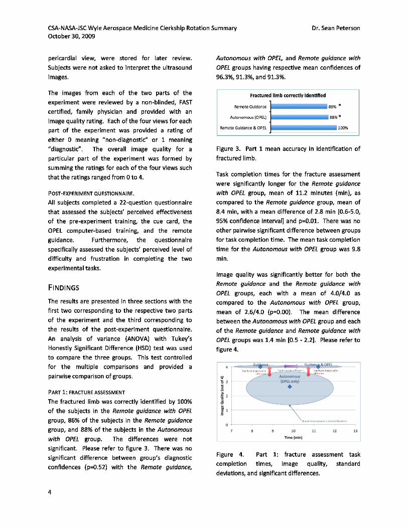
PART 1: FRACTURE ASSESSMENT
The fractured limb was correctly identified by 100%
of the subjects in the Remote guidance with OPEL
group, 86% of the subjects in the Remote guidance
group, and 88% of the subjects in the Autonomous
with OPEL group. The differences were not
significant. Please refer to figure 3. There was no
significant difference between group’s diagnostic
confidences (p=0.52) with the Remote guidance,
Autonomous with OPEL, and Remote guidance with
OPEL groups having respective mean confidences of
96.3%, 91.3%, and 91.3%.
Figure 3. Part 1 mean accuracy in identification of
fractured limb.
Task completion times for the fracture assessment
were significantly longer for the Remote guidance
with OPEL group, mean of 11.2 minutes (min), as
compared to the Remote guidance group, mean of
8.4 min, with a mean difference of 2.8 min [0.6-5.0,
95% confidence interval] and p=0.01. There was no
other pairwise significant difference between groups
for task completion time. The mean task completion
time for the Autonomous with OPEL group was 9.8
min.
Image quality was significantly better for both the
Remote guidance and the Remote guidance with
OPEL groups, each with a mean of 4.0/4.0 as
compared to the Autonomous with OPEL group,
mean of 2.6/4.0 (p=0.00). The mean difference
between the Autonomous with OPEL group and each
of the Remote guidance and Remote guidance with
OPEL groups was 1.4 min [0.5 - 2.2]. Please refer to
figure 4.
Figure 4. Part 1: fracture assessment task
completion times, image quality, standard
deviations, and significant differences.
4

CSA-NASA-JSC Wyle Aerospace Medicine Clerkship Rotation Summary
Dr. Sean PetersonOctober 30, 2009
PART 2: FAST ABDOMEN ASSESSMENT
For the second part of the experiment, FAST
abdomen assessment, the standard deviations for
both task completion time and image quality were
greater than for the first part of the experiment. As
such, the only significant difference was in the image
quality between the Remote guidance with OPEL,
mean of 3.1/4.0, compared to the Autonomous with
OPEL group, mean of 1.6/4.0 (p=0.03). The mean
difference was 1.5 min [0.1 – 2.9]. The Remote
guidance group mean image quality was 2.9. Of all
groups, subjects with previous medical training
obtained a significantly higher image quality, mean
of 3.3/4.0, compared to those without medical
training, mean of 2.2/4.0 (p=0.05). Please refer to
figure 6. The mean task completion times were 21.1
min for the Remote guidance group, 21.2 min for the
Autonomous with OPEL group, and 23.5 min for the
Remote guidance with OPEL group. Please refer to
figure 5.
Finally, an examination of the results of the post-
experiment questionnaire revealed that there were
no questions with statistically significant differences
across the three groups. There was a trend towards
those in the Autonomous with OPEL group finding
the experiment more difficult and more frustrating
as compared to the other two groups. Please refer
to figures 7 and 8.
Figure 7. Difficulty rating on post-experiment
questionnaire with 1 being “Not difficult at all” and
7 being “Very difficult”.
Figure 5. Part 2: FAST abdomen assessment task
completion times, image quality, standard
deviations, and significant differences.
Figure 6. Comparison of FAST image quality
between medically trained vs. non-medically trained
subjects.
POST-EXPERIMENT QUESTIONNAIRE
Figure 8. Frustration rating on post-experiment
questionnaire with 1 being “Not frustrated at all”
and 7 being “Completely frustrated”.
Key qualitative suggestions for improvement
included the following:
Overall task
• Maintain consistent plain language• Reinforce firmer pressure to improve image
quality
Pre-experiment training
• Include a “tour” through the human body
showing appearance of specific organs
5

CSA-NASA-JSC Wyle Aerospace Medicine Clerkship Rotation Summary
Dr. Sean PetersonOctober 30, 2009
Cue-card
• Add instructions on how to capture a STILL
and a VIDEO LOOP
• Include a description of “SWEEP” = tilting
probe one way and then the other to
visualize an organ or interface
• Change position of A4 to be more posterior
in mid-axillary line
Remote guidance
• Limit instructions to 3 steps so as to not getahead of ultrasound operator
• Provide positive feedback when properimages obtained to aid ultrasound operator
confidence
• Share with ultrasound operator what a“positive” scan would show
FAST abdomen procedure
• Remove medical language
• Better describe orientation of probe andinclude pictures of orientation
• Better describe how to locate the kidney
• Describe how to manage with rib shadows
• Better describe procedure to visualize heartfrom sub-xyphoid approach
• Reset depth setting after each position to
avoid missing far-field structures
• Include a “problem-solving” section that
describes potential maneuvers to attempt
to gain the desired image
• Embed videos in word document atrelevant line items
FAST abdomen video
• Remove medical language
• Expand video to include more still pictures
of the desired views with labels describing
the target organs and where “free fluid”
would appear
• Better describe how to do a SWEEP or “tilt”
to visualize an interface
• Emphasize need to have probe nearlyparallel with abdomen and tucked under
ribs with firm pressure to visualize heart
• Provide examples of “positive” free fluidultrasound images in video
• Include a “problem-solving” section thatdescribes potential maneuvers to attempt
to gain the desired image
(i.e. breath holds, bending knees, rotating
probe, panning probe)
DISCUSSION
With no more than ten (10) minutes of ultrasound
training, all subjects were able to use the ultrasound
to obtain relevant images. This speaks to the
benefits of a focused teaching session and to the
intrinsic ability of humans to adapt to new
situations. As was expected, those with previous
medical training, and by virtue of this training
greater anatomy knowledge, produced better
quality images.
For both the fracture assessment and the FAST
abdomen assessment, subjects with remote
guidance produced better quality images than those
operating autonomously. This was primarily due to
near-instant feedback on the quality of images
provided by the remote guider. As the
communication time delay expands, the capability to
provide this feedback greatly diminishes. As such,
successful autonomous ultrasound operation
becomes a greater necessity.
Subjects provided feedback that they would have
preferred more reference images placed directly
next to their ultrasound screen so as to provide a
degree of quality feedback through the subjects’
own pattern-recognition capabilities. Subjects
theorized that by having a tool that advanced
through the ultrasound assessment in a stepwise
manner and presented relevant images and
technique aids to obtain these images, they would
have better captured the images necessary to
achieve higher image quality in the study.
Interestingly, subjects of the autonomous with OPEL
group rated the task as being neither more difficult
6

CSA-NASA-JSC Wyle Aerospace Medicine Clerkship Rotation Summary
Dr. Sean PetersonOctober 30, 2009
nor more frustrating than those with remote
guidance.
NEXT STEPS
Much has been learned in the first phase of the
SURGE project. By implementing both the
suggestions obtained from subjects and observed
areas for augmentation obtained from
experimenters, the OPEL product will be
substantially improved. Further testing of the
autonomous operation of ultrasound with the
assistance of OPEL in the microgravity environment
is the next step. Patient restraint systems,
ultrasound operator restraint systems, ultrasound
operator stress management, workstation set-up
and securing, and gel containment are but some of
the issues to be addressed for successful completion
of autonomous ultrasound in a microgravity
environment.
CONCLUSION
Remote guidance continues to produce higher
quality ultrasound images than autonomous
ultrasound operation. The OPEL has potential to
provide an excellent training and coaching tool for
both remotely guided and autonomous operation.
With the implementation of some of the many
suggestions for improvement obtained during the
experiment OPEL has potential to become an
essential component of future exploration class
medical operations.
ACKNOWLEDGEMENTS
• Ms. Corrine Williams for coordinating a vastnumber of exciting experiences
• Wyle for hosting us and providing logisticalsupport
• NASA-JSC and flight docs for excellentteaching and outstanding experiences
• Canadian Space Agency for funding andproviding the opportunity to attend the
clerkship
• Mary Carvalho for rapid statistical analysis• David Ham for technical support
• Victor, Kat, Ashot, and Doug for providing
me an opportunity to participate in such a
neat project!
7