Embed Size (px)
Citation preview

CONTENTSINTRODUCTIONBRAGG EQUATION INSTRUMENTATIONX-RAY DIFFRACTION METHODSAPPLICATIONSANALYSIS AVAILABILITY
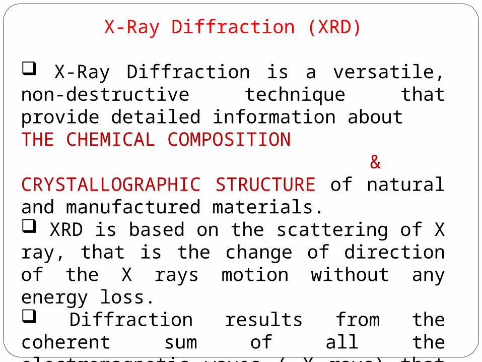
X-Ray Diffraction (XRD)
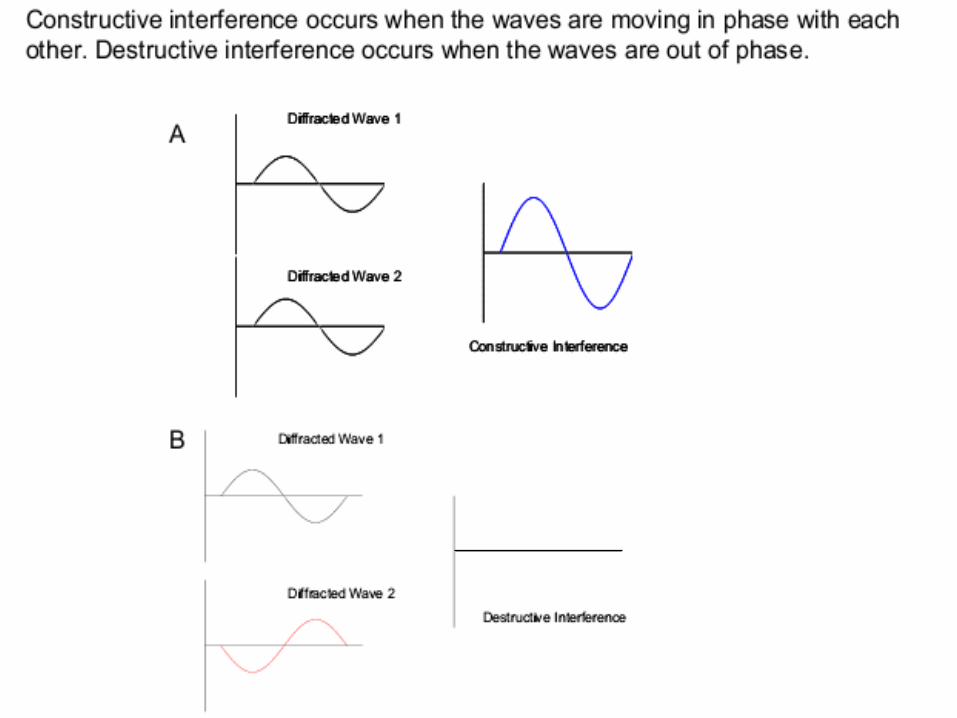
X-Ray Diffraction is a versatile, non-destructive technique that provide detailed information about THE CHEMICAL COMPOSITION &CRYSTALLOGRAPHIC STRUCTURE of natural and manufactured materials. XRD is based on the scattering of X ray, that is the change of direction of the X rays motion without any energy loss. Diffraction results from the coherent sum of all the electromagnetic waves ( X rays) that are diffused from the atoms belonging to the same family/planes.
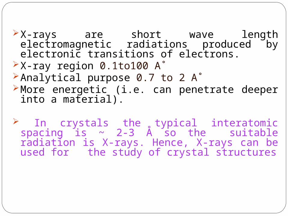
X-rays are short wave length electromagnetic radiations produced by electronic transitions of electrons.
X-ray region 0.1to100 A˚Analytical purpose 0.7 to 2 A˚More energetic (i.e. can penetrate deeper into a
material).
In crystals the typical interatomic spacing is ~ 2-3 Å so the suitable radiation is X-rays. Hence, X-rays can be used for the study of crystal structures

GENERATION OF X-RAYS
Generated by bombarding a metal target with an energetic beam of electrons.
When high energy electrons strike an anode in a sealed vacuum, x-rays are generated. Anodes are often made of radioactive source like copper, iron or molybdenum.
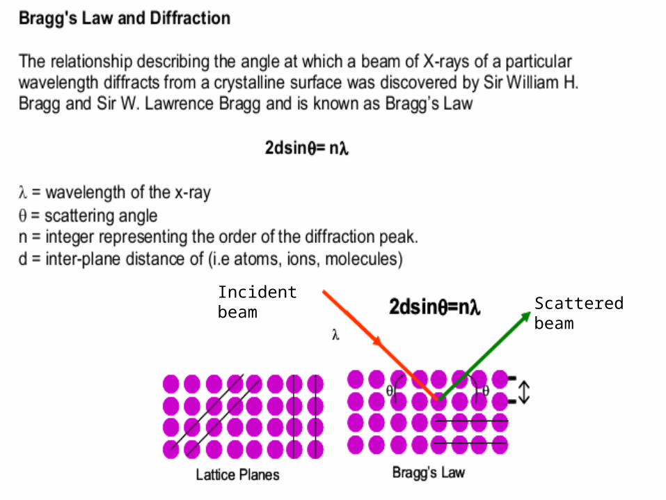
Incident beamScattered beam
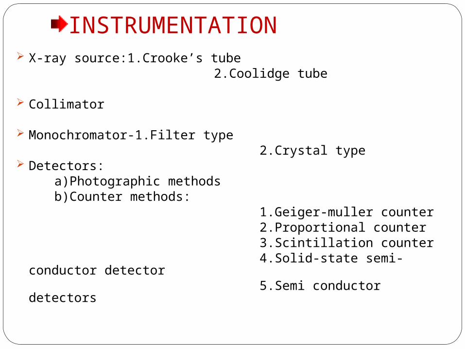
INSTRUMENTATION X-ray source:1.Crooke’s tube 2.Coolidge tube
Collimator
Monochromator-1.Filter type 2.Crystal type Detectors: a)Photographic methods b)Counter methods: 1.Geiger-muller counter 2.Proportional counter 3.Scintillation counter 4.Solid-state semi-conductor detector 5.Semi conductor detectors
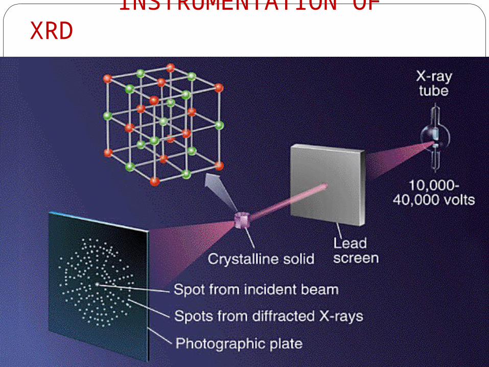
INSTRUMENTATION OF XRD

X-ray source;
Crookes tubeCalled as cold cathode tube.Electrons are generated by ionization of the residual air in the tube, instead of heated filament.An aluminum cathode plate at one end of the tube created a beam of electrons, which struck a platinum anode target at the center generating X-rays.
Advantagesharper images are obtained.
Disadvantage :UnreliableCrookes tube
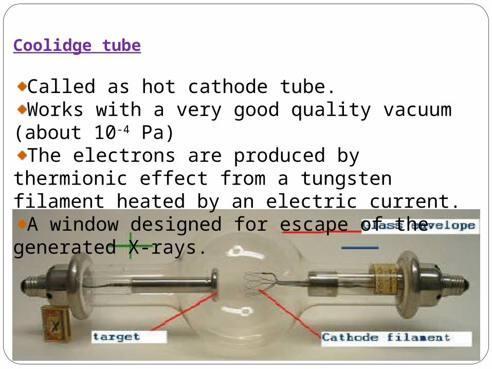
Coolidge tube
Called as hot cathode tube.Works with a very good quality vacuum (about 10-4 Pa)The electrons are produced by thermionic effect from a
tungsten filament heated by an electric current.A window designed for escape of the generated X-rays.
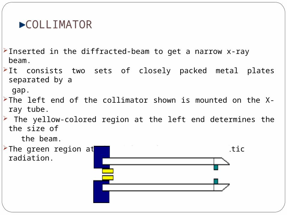
COLLIMATOR
Inserted in the diffracted-beam to get a narrow x-ray beam.It consists two sets of closely packed metal plates separated by a gap.The left end of the collimator shown is mounted on the X-ray tube. The yellow-colored region at the left end determines the the size of the beam.The green region at the right end removes parasitic radiation.

Materials used:Nacl,LiF,quartz etc,.Filter type
A window that absorbs undesirable radiation and allows required wavelength to pass.Crystal type
Curved crystal are used so that the angle of the reflecting planes satisfied the Bragg’s equation for the required wave length.
Characteristics of a crystal:
Mechanically strong and stable
Resolution of the crystal, should be small.
MONOCHROMATOR

DETECTORS Photographic methods
Contains photographic plate Blackening of developed film is expressed in terms of density ,D
D=Log Io/I.Io- is the intensity of measured X-rays without sample and I- is the intensity measured X rays with sample.
Photostimulable phosphors (luminesent material)• Common used method•Photostimulable phosphor plate (PSP plate) is used in place of the photographic plate. •After the plate is X-rayed, excited electrons in the phosphor material remain ‘trapped' in'colour centres' in the crystal. •The light given off during laser stimulation is collected by a photomultiplier tube ADVANTAGE:The PSP plate can be reused
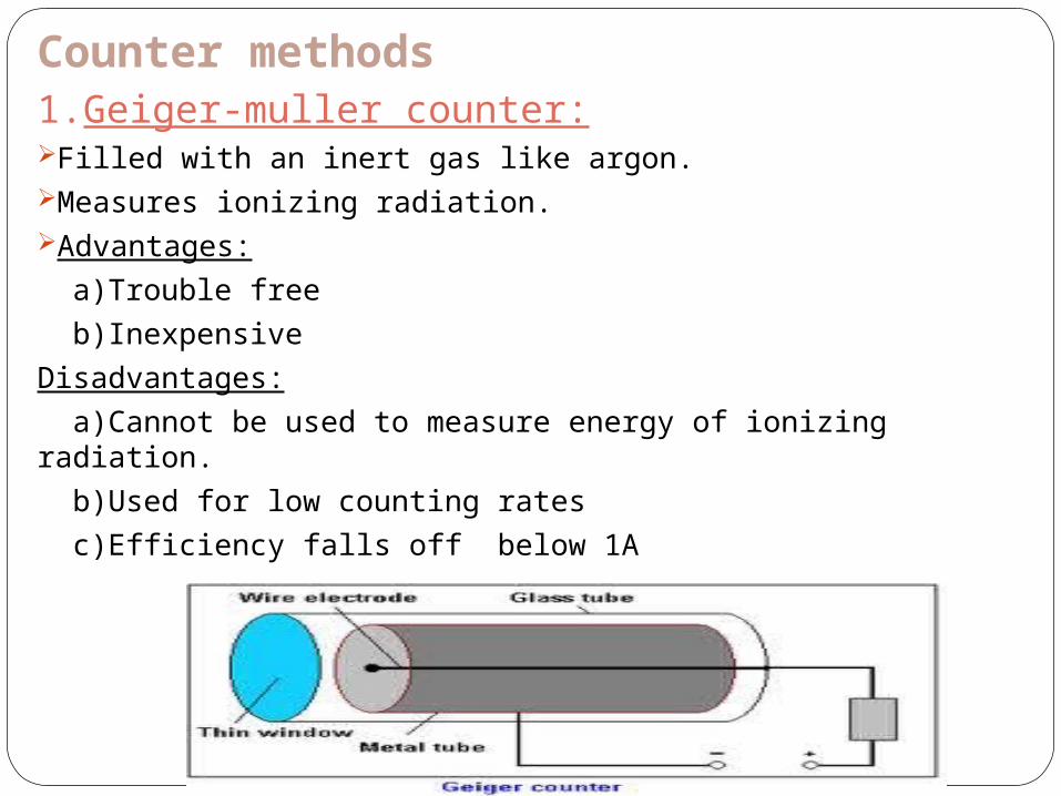
Counter methods 1.Geiger-muller counter:Filled with an inert gas like argon.Measures ionizing radiation. Advantages:
a)Trouble free
b)Inexpensive
Disadvantages:
a)Cannot be used to measure energy of ionizing radiation.
b)Used for low counting rates
c)Efficiency falls off below 1A
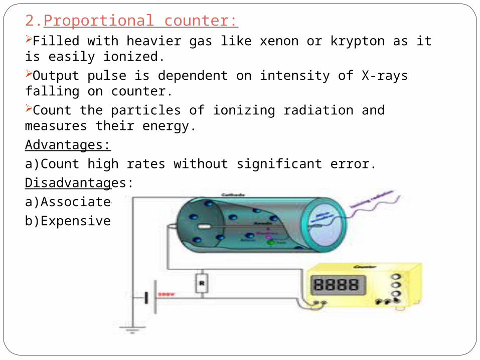
2.Proportional counter:Filled with heavier gas like xenon or krypton as it is easily ionized.Output pulse is dependent on intensity of X-rays falling on counter. Count the particles of ionizing radiation and measures their energy.
Advantages:
a)Count high rates without significant error.
Disadvantages:
a)Associated electronic circuit is complex.
b)Expensive.
3.Scintillation counter:Measures X-rays of shorter wavelengths.The sensor, called a scintillator, consists of a transparent crystal, usually phosphor, plastic (usually containing anthracene), or organic liquid that fluoresces when struck by ionizing radiation. The PMT is attached to an electronic amplifier to count and possibly quantify the amplitude of the signals. Advantages:a)Count high rates.
4.Solid state semi-conductor detector:The electrons produced by X-ray beam are promoted
into conduction bands and the current which flows is directly proportional to the incident X-ray energy.
advantage:Maintainted at very low Temp to minimise the noise
and prevent deterioration of the detector. 5.Semi-conductor detectors: Silicon-lithium drifted detector. The principle is similar to gas ionization detector. Voltage of pulse = Q/CQ= Charge C= capicitor Application: In neutron activation analysis ( used to
determine the concentration of trace and major elements in a variety of matrics)
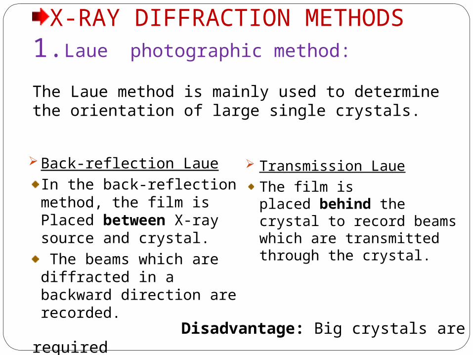
X-RAY DIFFRACTION METHODS1.Laue photographic method:
The Laue method is mainly used to determine the orientation of large single crystals.
Back-reflection LaueIn the back-reflection method, the film is Placed between X-ray source and crystal. The beams which are diffracted in a backward direction are recorded.
Transmission Laue
The film is placed behind the crystal to record beams which are transmitted through the crystal.
Disadvantage: Big crystals are required
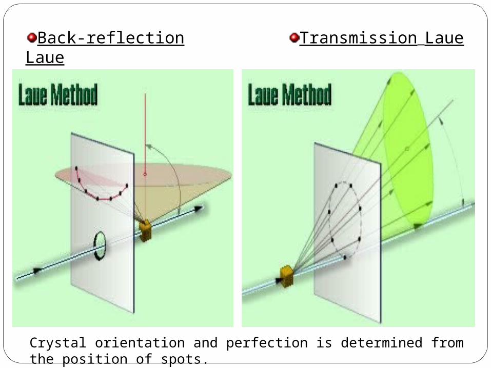
Back-reflection Laue Transmission Laue
Crystal orientation and perfection is determined from the position of spots.
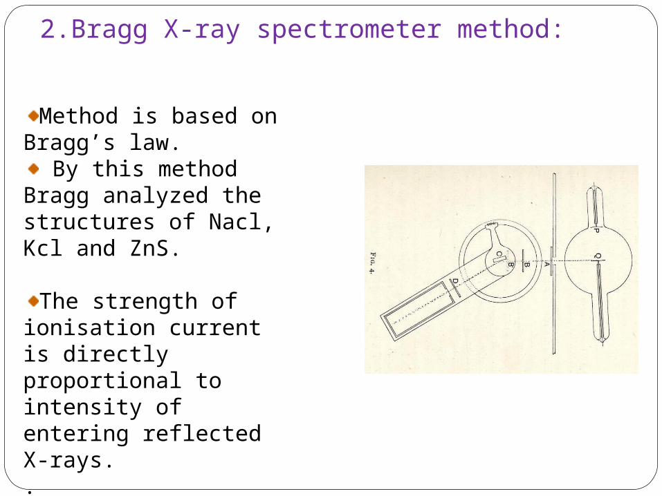
2.Bragg X-ray spectrometer method:
Method is based on Bragg’s law.
By this method Bragg analyzed the structures of Nacl, Kcl and ZnS.
The strength of ionisation current is directly proportional to intensity of entering reflected X-rays..
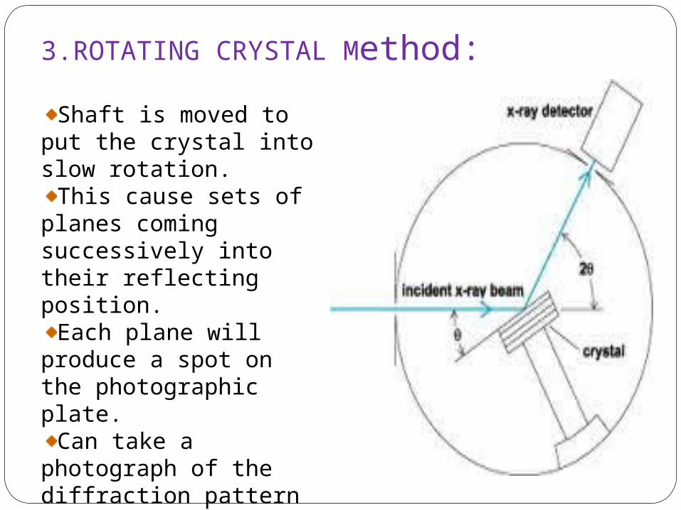
3.ROTATING CRYSTAL Method:
Shaft is moved to put the crystal into slow rotation.
This cause sets of planes coming successively into their reflecting position.
Each plane will produce a spot on the photographic plate.
Can take a photograph of the diffraction pattern in two ways1.complete rotation method 2.oscillation method
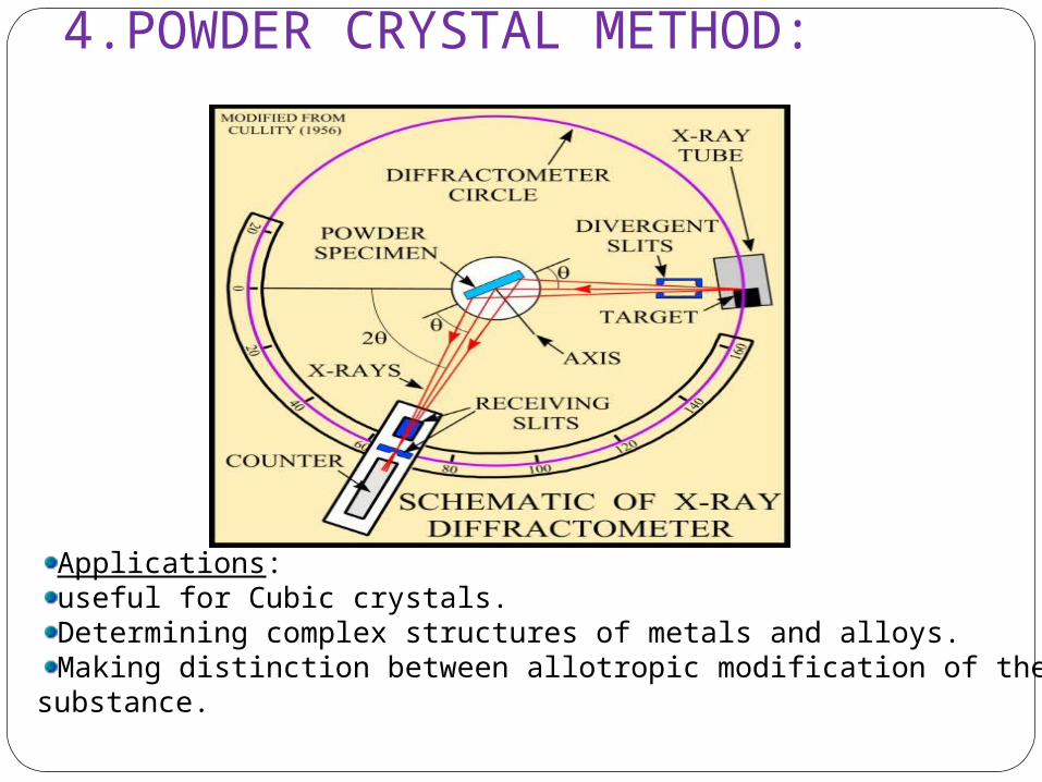
4.POWDER CRYSTAL METHOD:
Applications: useful for Cubic crystals.Determining complex structures of metals and alloys.Making distinction between allotropic modification of the same substance.
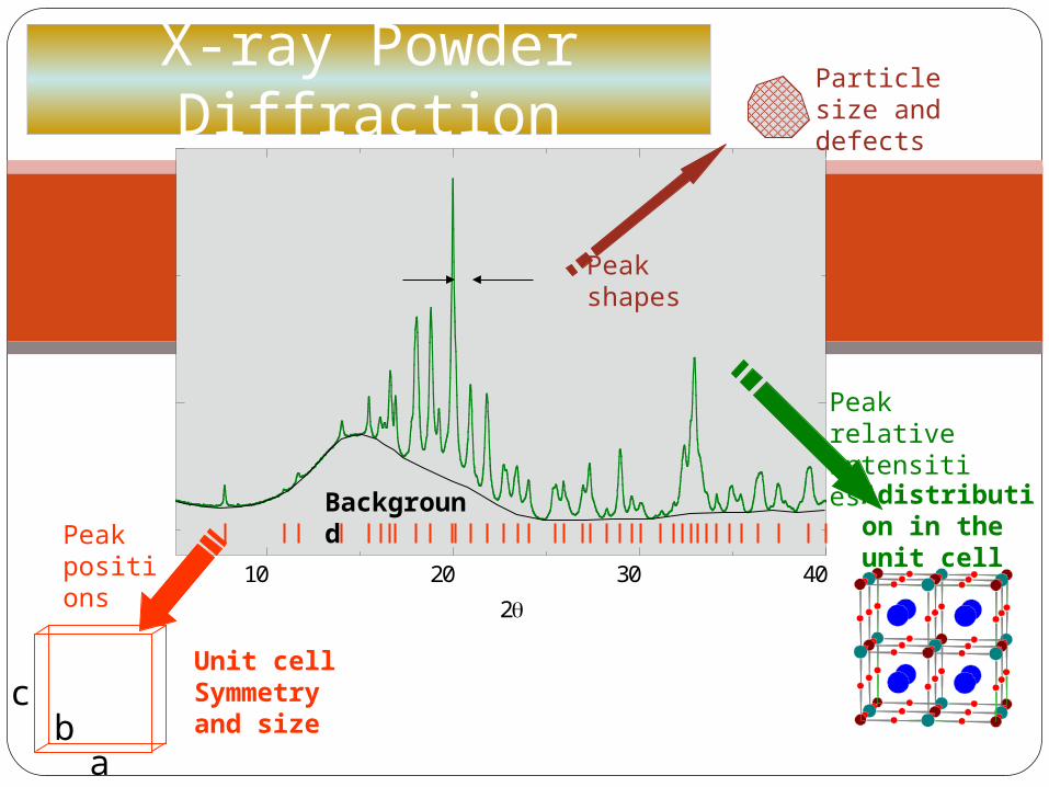
10 20 30 40
2
PowderCell 2 .0
Adistribution in the unit cell
Peak relative intensities
Unit cell Symmetry and size
Peak positions
a
cb
Peak shapes
Particle size and defects
Background
X-ray Powder Diffraction


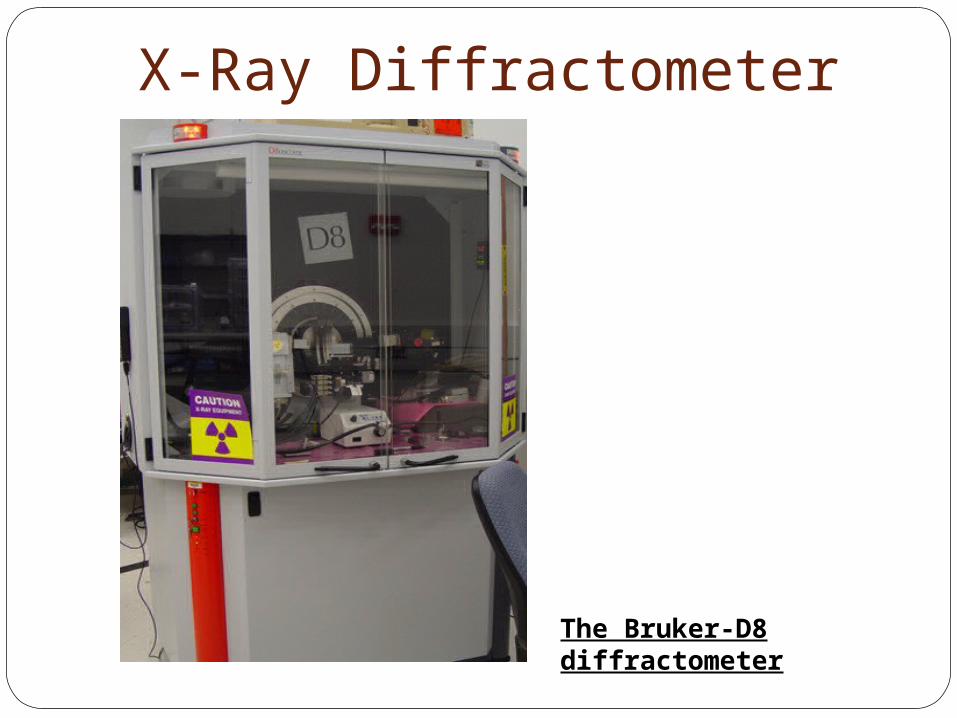
X-Ray Diffractometer
The Bruker-D8 diffractometer
APPLICATIONS
Structure of crystalsPolymer characterizationParticle size determination complexes determination Determination of linkage isomerism
Miscellaneous applicationsSoil classification based on crystallinity.Analysis of industrial dusts.Assessment of weathering & degradation of
minerals & polymers.Study of corrosion products.Examination of tooth enamel & dentine.Examination of bone state & tissue state.Structure of DNA&RNA.X-ray Diffraction on aerosol drugs -
Measurements on small quantities of pharmaceutical samples using the X'Celerator detector.
List of Institutes where XRD analysis facility available
IIT, Ropar, HaryanaIIT BombayOcean & Atmospheric Science and Technology Cell, Mangalore University.Centre for Earth Science Studies,Thiruvananthapuram Kerala STIC, CochinIICT, HyderabadWadia Institute of Himalayan Geology, Dehradun NIPER , MohaliPunjab UniversityCDRI , Luckhnow

About 95% of all solid materials can be described as crystalline. When x-rays interact with a crystallinesubstance (Phase), one gets a diffraction pattern.In 1919 A. W. Hull gave a paper titled, "A New Method of Chemical Analysis." Here he pointed out that "…. every crystalline substance gives a pattern; the same substance always gives the same pattern; and in a mixture of substances each produces its pattern independently of the others."The x-ray diffraction pattern of a pure substance is, therefore, like a fingerprint of the substance. The powder diffraction method is thus ideally suited for characterization and identification of polycrystalline phases.Today about 50,000 inorganic and 25,000 organic single component, crystalline phases, diffraction patterns have been collected and stored on magnetic or optical media as standards. The main use of powder diffraction is to identify components in a sample by a search/match procedure. Furthermore, the areas under the peak are related to the amount of each phase present in the sample.
Why Powder Diffraction?
The purpose of a Soller slit is to take a line source of radiation and slice it into smaller, parallel beams.divergence slit is balance the illuminated light area.Receiving beam; It's purpose is to further define the diffracted beam and eliminate scatter into the detector.