Embed Size (px)
Citation preview
8/2/2019 Year 3 Cardiovascular Examination
http://slidepdf.com/reader/full/year-3-cardiovascular-examination 1/18
Cardiovascular history
& examination
8/2/2019 Year 3 Cardiovascular Examination
http://slidepdf.com/reader/full/year-3-cardiovascular-examination 2/18
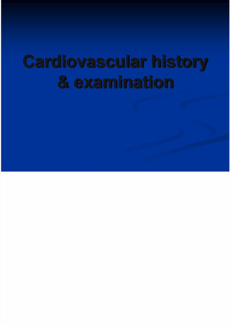
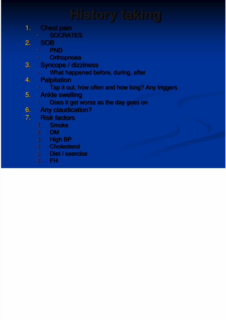
History taking1. Chest pain
• SOCRATES
2. SOB• PND• Orthopnoea
3. Syncope / dizziness• What happened before, during, after
4. Palpitation• Tap it out, how often and how long? Any triggers
5. Ankle swelling• Does it get worse as the day goes on
6. Any claudication?
7. Risk factors1. Smoke2. DM3. High BP4. Cholesterol5. Diet / exercise6. FH
8/2/2019 Year 3 Cardiovascular Examination
http://slidepdf.com/reader/full/year-3-cardiovascular-examination 3/18
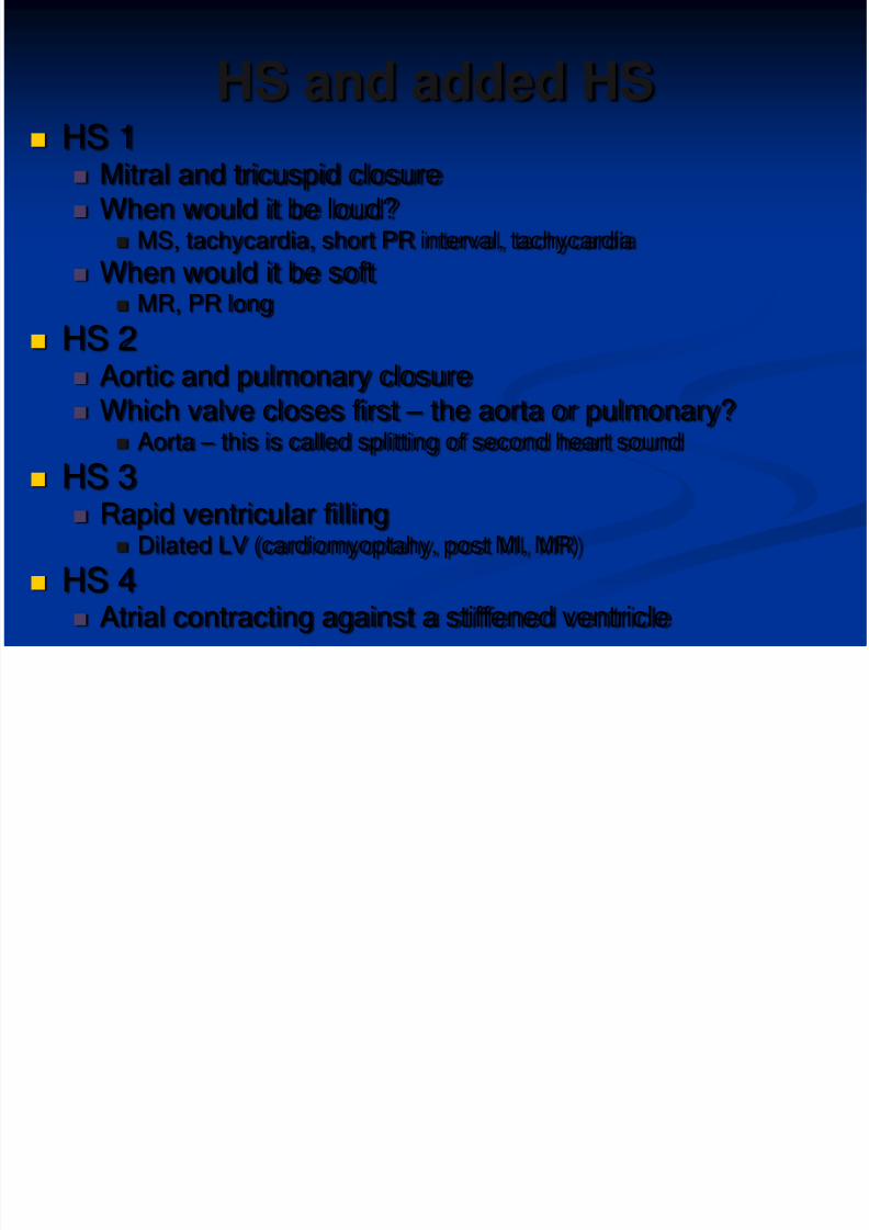
HS and added HS
HS 1 Mitral and tricuspid closure When would it be loud?
MS, tachycardia, short PR interval, tachycardia
When would it be soft MR, PR long
HS 2 Aortic and pulmonary closure Which valve closes first – the aorta or pulmonary?
Aorta –
this is called splitting of second heart sound HS 3
Rapid ventricular filling Dilated LV (cardiomyoptahy, post MI, MR)
HS 4 Atrial contracting against a stiffened ventricle
8/2/2019 Year 3 Cardiovascular Examination
http://slidepdf.com/reader/full/year-3-cardiovascular-examination 4/18
Introduction
WASH HANDS
Introduce
Consent Expose
Offer them a blanket
45 degrees Are you in any pain?
8/2/2019 Year 3 Cardiovascular Examination
http://slidepdf.com/reader/full/year-3-cardiovascular-examination 5/18
Inspection
General inspection
Do they look well or not
Look around the bed: GTN spray
Hands
Clubbing
Capillary refill
Tar stains
Pulse: rate, rhythm, character
8/2/2019 Year 3 Cardiovascular Examination
http://slidepdf.com/reader/full/year-3-cardiovascular-examination 6/18
Inspection 2
Hands ctd
Radio radial delay
Radio femoral delay
Lift arm upcollapsing pulse
Arm
BP
Neck
JVP
Eyes
Anaemia
Xanthalasma
Corneal arcus Fundoscopy
Face
Malar flush
Tongue
Dental hygiene
8/2/2019 Year 3 Cardiovascular Examination
http://slidepdf.com/reader/full/year-3-cardiovascular-examination 7/18
Palpation
Apex beat
5th ICS – MCL
Heaves RVH
Thrills
Palpable murmur

8/2/2019 Year 3 Cardiovascular Examination
http://slidepdf.com/reader/full/year-3-cardiovascular-examination 8/18
Auscultate
www.kumc.edu
8/2/2019 Year 3 Cardiovascular Examination
http://slidepdf.com/reader/full/year-3-cardiovascular-examination 9/18
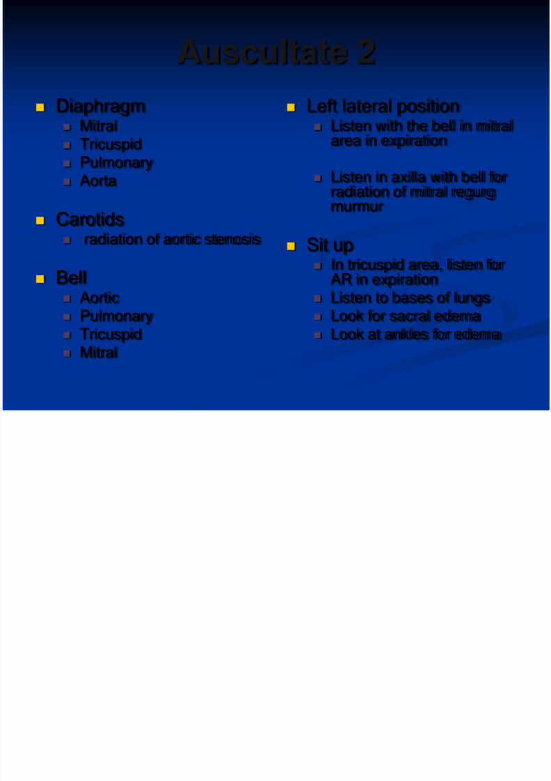
Auscultate 2
Diaphragm Mitral Tricuspid Pulmonary Aorta
Carotids radiation of aortic stenosis
Bell Aortic Pulmonary Tricuspid Mitral
Left lateral position Listen with the bell in mitral
area in expiration
Listen in axilla with bell for
radiation of mitral regurgmurmur
Sit up In tricuspid area, listen for
AR in expiration Listen to bases of lungs Look for sacral edema Look at ankles for edema
8/2/2019 Year 3 Cardiovascular Examination
http://slidepdf.com/reader/full/year-3-cardiovascular-examination 10/18
Finish
Summarise 32 year old male. O/E heart sounds 1 & 2
present with no added sounds AND no
murmurs Also like to
Feel peripheral pulses
Do abdo exam looking for ascites andhepatomegaly in right heart failure
Do respiratory exam – looking for pleuraleffusion etc.
8/2/2019 Year 3 Cardiovascular Examination
http://slidepdf.com/reader/full/year-3-cardiovascular-examination 11/18
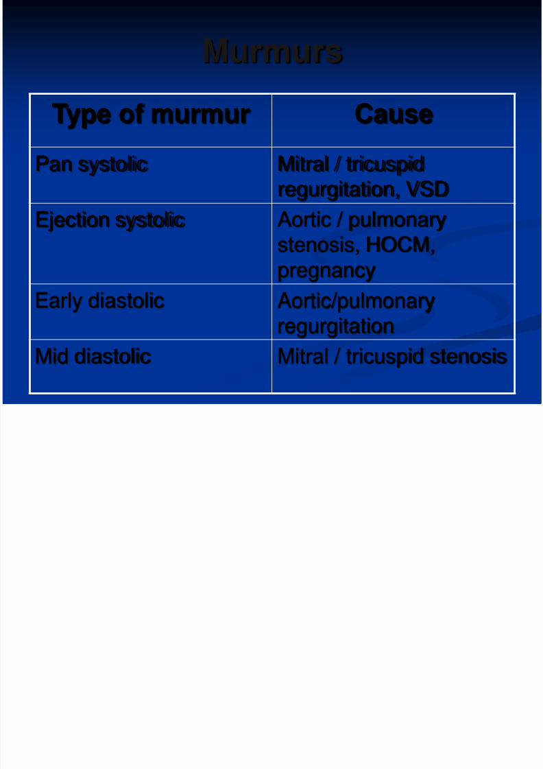
Murmurs
Type of murmur Cause
Pan systolic Mitral / tricuspid
regurgitation, VSDEjection systolic Aortic / pulmonary
stenosis, HOCM,pregnancy
Early diastolic Aortic/pulmonaryregurgitation
Mid diastolic Mitral / tricuspid stenosis
8/2/2019 Year 3 Cardiovascular Examination
http://slidepdf.com/reader/full/year-3-cardiovascular-examination 12/18
Describing a murmur
1. Position heard loudest
2. Pitch
3. Radiation4. Intensity (grade 1-6)
5. Timing
1. Systolic2. Diastolic
8/2/2019 Year 3 Cardiovascular Examination
http://slidepdf.com/reader/full/year-3-cardiovascular-examination 13/18
Jugular venous pressure
Which vein is it
Internal jugular vein
Why are we examining it
Gives us an indication of RA pressure
Normal
<4cm from angle of louis
What happens in inspiration to the JVP? Waveform
ASK ME
8/2/2019 Year 3 Cardiovascular Examination
http://slidepdf.com/reader/full/year-3-cardiovascular-examination 14/18
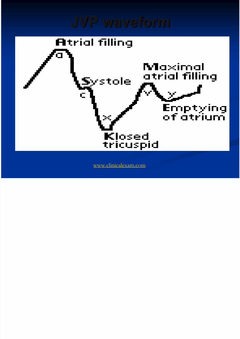
JVP waveform
www.clinicalexam.com
8/2/2019 Year 3 Cardiovascular Examination
http://slidepdf.com/reader/full/year-3-cardiovascular-examination 15/18
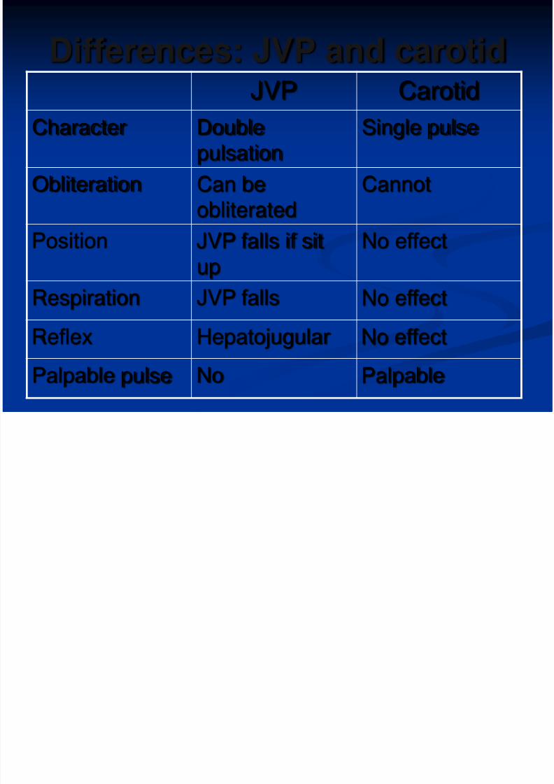
Differences: JVP and carotid
JVP CarotidCharacter Double
pulsationSingle pulse
Obliteration Can beobliterated Cannot
Position JVP falls if situp
No effect
Respiration JVP falls No effect
Reflex Hepatojugular No effect
Palpable pulse No Palpable
8/2/2019 Year 3 Cardiovascular Examination
http://slidepdf.com/reader/full/year-3-cardiovascular-examination 16/18
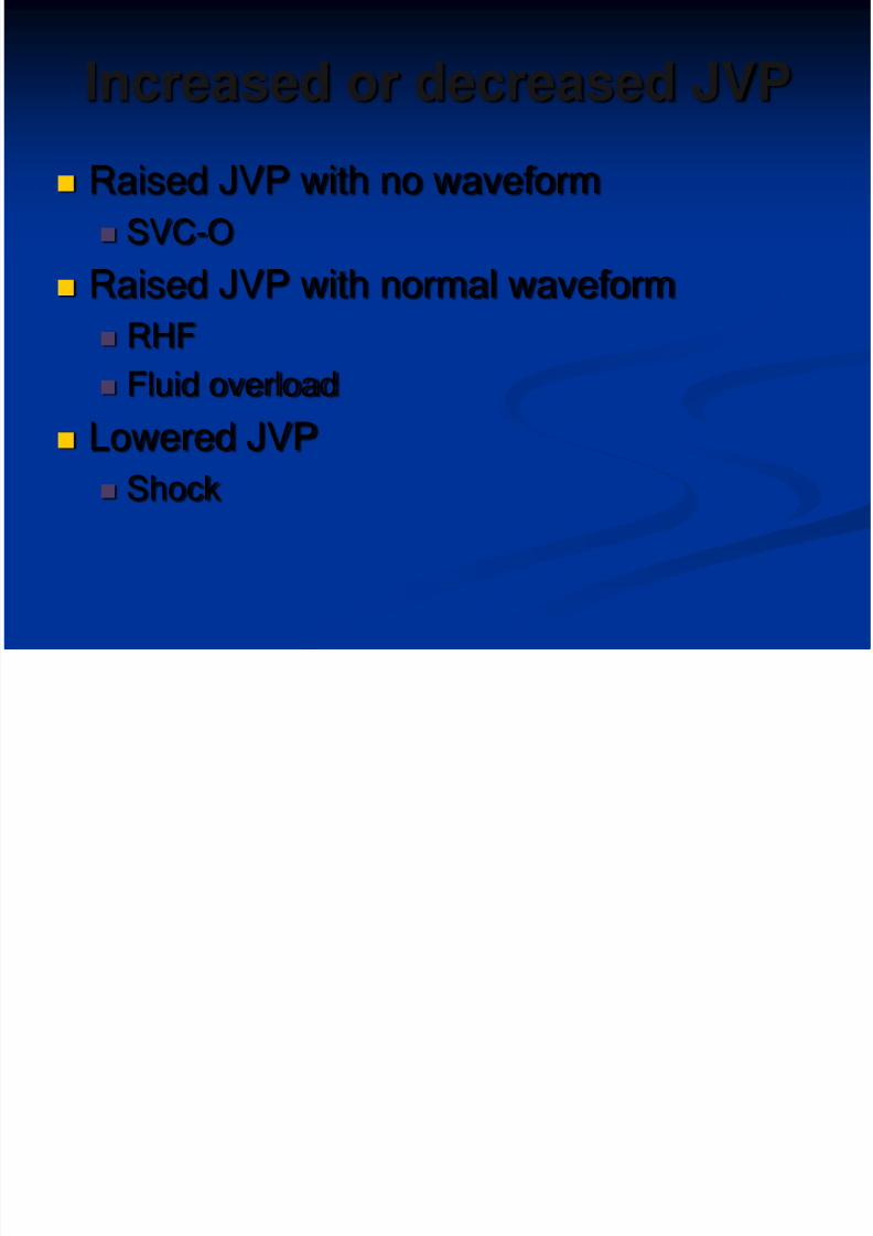
Increased or decreased JVP
Raised JVP with no waveform
SVC-O
Raised JVP with normal waveform
RHF
Fluid overload
Lowered JVP
Shock
8/2/2019 Year 3 Cardiovascular Examination
http://slidepdf.com/reader/full/year-3-cardiovascular-examination 17/18
Thank you for listening
Any questions ??
8/2/2019 Year 3 Cardiovascular Examination
http://slidepdf.com/reader/full/year-3-cardiovascular-examination 18/18
Download PPT for free at……
http://www.medics-mashup.com/