Embed Size (px)
Citation preview
Yeast and Fungal Prions
Reed B. Wickner
Laboratory of Biochemistry and Genetics, National Institute of Diabetes and Digestive and Kidney Diseases,National Institutes of Health, Bethesda, Maryland 20892-0830
Correspondence: [email protected]
Yeast and fungal prions are infectious proteins, most being self-propagating amyloids ofnormally soluble proteins. Their effects range from a very mild detriment to lethal, withspecific effects dependent on the prion protein and the specific prion variant (“prionstrain”). The prion amyloids of Sup35p, Ure2p, and Rnq1p are in-register, parallel, foldedb-sheets, an architecture that naturally suggests a mechanism by which a protein can tem-plate its conformation, just as DNA or RNA templates its sequence. Prion propagation iscriticallyaffected byan arrayof chaperone systems, most notably the Hsp104/Hsp70/Hsp40combination, which is responsible for generating new prion seeds from old filaments. TheBtn2/Cur1 antiprion system cures most [URE3] prions that develop, and the Ssb antiprionsystem blocks [PSIþ] generation.
In 1989, I was writing a review of nonchromo-somal genetic elements of yeast (Wickner
1991). At that time, the infectious proteinconcept had been proposed to explain the trans-missible spongiform encephalopathies (TSEs)(Alper et al. 1967; Griffith 1967; Dickinson etal. 1968; Bolton et al. 1982; Prusiner 1982; Chese-bro et al. 1985; Oesch et al. 1985), but thereremained considerable debate. In reviewing thenonchromosomal genetic element [URE3],I noted that Aigle and Lacroute (1975) had re-ported that ure2 strains were unable to propa-gate the [URE3] element, but that the pheno-types of ure2 and URE2þ [URE3] strains wereessentially identical—namely, de-repression ofnitrogen catabolism genes. As I had been study-ing the mak mutants unable to propagate thekiller virus (Wickner 1978), I recognized thatthe relation of ure2 and [URE3] was not whatwas expected of a chromosomal gene important
for propagation of a nucleic acid replicon, butthat this was the relation expected if [URE3]was an inactive form of Ure2p able to inactivatethe normal form and, thus, act as a prion. Thiswas the beginning of our work showing that[URE3] was a prion of Ure2p, and our inferencethat [PSIþ] was a prion of Sup35p based onthe re-interpretation of published experiments(Wickner 1994).
DISCOVERY OF YEAST PRIONS
The genetic criteria that identified yeast prions(infectious proteins) (Wickner 1994) distin-guish them from nucleic acid–based infectiouselements such as viruses, plasmids, and mito-chondria. Assuming that the prion changewould inactivate the normal function of theprotein, the phenotype of the prion should re-semble that of a recessive mutation in the gene
Editor: Stanley B. Prusiner
Additional Perspectives on Prion Biology available at www.cshperspectives.org
Copyright # 2016 Cold Spring Harbor Laboratory Press; all rights reserved; doi: 10.1101/cshperspect.a023531
Cite this article as Cold Spring Harb Perspect Biol 2016;8:a023531
1
on April 1, 2019 - Published by Cold Spring Harbor Laboratory Press http://cshperspectives.cshlp.org/Downloaded from
for the prion protein, and elimination of theprion protein gene should result in failure ofprion propagation (Wickner 1994). Overpro-duction of the prion protein should result inincreased de novo generation of the prion.Finally, if the prion can be cured, it should bepossible for it to arise again in the cured strain(at low frequency) because the normal form ofthe protein is still being produced (Wickner1994). These genetic criteria are not true forany known mammalian prion, in part becausethe TSEs are not a consequence of PrP defi-ciency and in part because some of the requiredexperiments are not feasible in mammals.
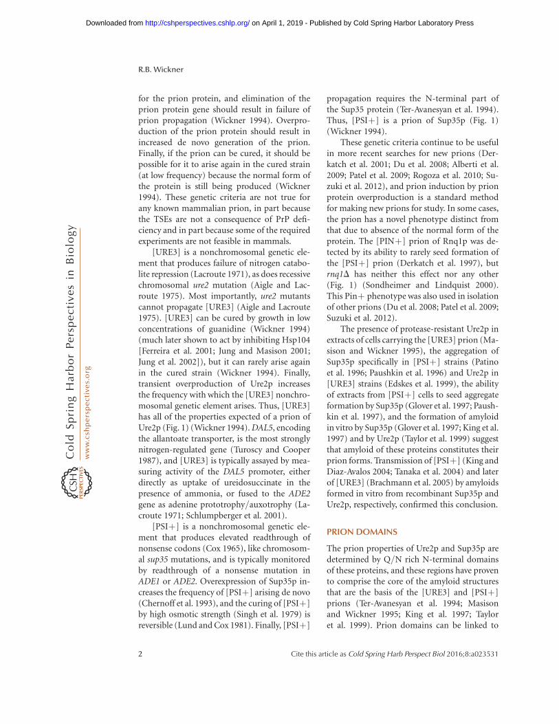
[URE3] is a nonchromosomal genetic ele-ment that produces failure of nitrogen catabo-lite repression (Lacroute 1971), as does recessivechromosomal ure2 mutation (Aigle and Lac-route 1975). Most importantly, ure2 mutantscannot propagate [URE3] (Aigle and Lacroute1975). [URE3] can be cured by growth in lowconcentrations of guanidine (Wickner 1994)(much later shown to act by inhibiting Hsp104[Ferreira et al. 2001; Jung and Masison 2001;Jung et al. 2002]), but it can rarely arise againin the cured strain (Wickner 1994). Finally,transient overproduction of Ure2p increasesthe frequency with which the [URE3] nonchro-mosomal genetic element arises. Thus, [URE3]has all of the properties expected of a prion ofUre2p (Fig. 1) (Wickner 1994). DAL5, encodingthe allantoate transporter, is the most stronglynitrogen-regulated gene (Turoscy and Cooper1987), and [URE3] is typically assayed by mea-suring activity of the DAL5 promoter, eitherdirectly as uptake of ureidosuccinate in thepresence of ammonia, or fused to the ADE2gene as adenine prototrophy/auxotrophy (La-croute 1971; Schlumpberger et al. 2001).
[PSIþ] is a nonchromosomal genetic ele-ment that produces elevated readthrough ofnonsense codons (Cox 1965), like chromosom-al sup35 mutations, and is typically monitoredby readthrough of a nonsense mutation inADE1 or ADE2. Overexpression of Sup35p in-creases the frequency of [PSIþ] arising de novo(Chernoff et al. 1993), and the curing of [PSIþ]by high osmotic strength (Singh et al. 1979) isreversible (Lund and Cox 1981). Finally, [PSIþ]
propagation requires the N-terminal part ofthe Sup35 protein (Ter-Avanesyan et al. 1994).Thus, [PSIþ] is a prion of Sup35p (Fig. 1)(Wickner 1994).
These genetic criteria continue to be usefulin more recent searches for new prions (Der-katch et al. 2001; Du et al. 2008; Alberti et al.2009; Patel et al. 2009; Rogoza et al. 2010; Su-zuki et al. 2012), and prion induction by prionprotein overproduction is a standard methodfor making new prions for study. In some cases,the prion has a novel phenotype distinct fromthat due to absence of the normal form of theprotein. The [PINþ] prion of Rnq1p was de-tected by its ability to rarely seed formation ofthe [PSIþ] prion (Derkatch et al. 1997), butrnq1D has neither this effect nor any other(Fig. 1) (Sondheimer and Lindquist 2000).This Pinþ phenotype was also used in isolationof other prions (Du et al. 2008; Patel et al. 2009;Suzuki et al. 2012).
The presence of protease-resistant Ure2p inextracts of cells carrying the [URE3] prion (Ma-sison and Wickner 1995), the aggregation ofSup35p specifically in [PSIþ] strains (Patinoet al. 1996; Paushkin et al. 1996) and Ure2p in[URE3] strains (Edskes et al. 1999), the abilityof extracts from [PSIþ] cells to seed aggregateformation by Sup35p (Glover et al. 1997; Paush-kin et al. 1997), and the formation of amyloidin vitro by Sup35p (Glover et al. 1997; King et al.1997) and by Ure2p (Taylor et al. 1999) suggestthat amyloid of these proteins constitutes theirprion forms. Transmission of [PSIþ] (King andDiaz-Avalos 2004; Tanaka et al. 2004) and laterof [URE3] (Brachmann et al. 2005) by amyloidsformed in vitro from recombinant Sup35p andUre2p, respectively, confirmed this conclusion.
PRION DOMAINS
The prion properties of Ure2p and Sup35p aredetermined by Q/N rich N-terminal domainsof these proteins, and these regions have provento comprise the core of the amyloid structuresthat are the basis of the [URE3] and [PSIþ]prions (Ter-Avanesyan et al. 1994; Masisonand Wickner 1995; King et al. 1997; Tayloret al. 1999). Prion domains can be linked to
R.B. Wickner
2 Cite this article as Cold Spring Harb Perspect Biol 2016;8:a023531
on April 1, 2019 - Published by Cold Spring Harbor Laboratory Press http://cshperspectives.cshlp.org/Downloaded from
other proteins, with the other protein serving asa reporter of prion formation (Li and Lindquist2000). This approach has also been useful insearching for new prions (Alberti et al. 2009).Of course, it must be confirmed that a segmentacting as a prion domain in a fusion can also doso in its native context.
Surprisingly, the sequence of the prion do-mains of Ure2p and Sup35p are of minimalimportance in their ability to form prions, asrandomly shuffling these segments, fusing theshuffled segments to the remainder of the gene,
and integrating these constructs in place of thenormal gene uniformly (each of five shufflesfor each protein), results in proteins that can stillform prions (Ross et al. 2004, 2005a). This workmade it clear that it is the amino acid com-position, and not the sequence, that determinesprion-forming ability; and within the Q/N-rich context, this method showed that hydro-phobic, aromatic, and hydrophilic residues fa-vor prion formation, whereas charged residuesor prolines are unfavorable (Toombs et al. 2010).In fact, artificial prion domains, designed based
Solubleinactive
Solubleactive
Genes for usingpoor N sources
[URE3] - a prion of Ure2p [PSI+] - a prion of Sup35p
Solubleactive
Translationtermination
UAA
Soluble formactivity unknown
Amyloidprimes [PSI+]generation
Amyloidinactiveand toxic
Prionchange
Prionchange
Prionchange
Prioninactive
[Het-s] - a prion of HETs[PIN+] - a prion of Rnq1p
Amyloid activeheterokaryonincompatibility
Prionchange
Figure 1. The yeast and fungal prions [URE3], [PSIþ], [PINþ], and [Het-s]. Formation of amyloid by Ure2p,Sup35p, Rnq1p, and HET-s results in either the loss of their normal functions (for Ure2p and Sup35p) or theappearance of novel activity (in Rnq1p and HET-s), all of which are heritable because the amyloid catalyzes theconversion of the nonamyloid to the amyloid form. Most variants of [URE3] are also toxic to the cell, and most[PSIþ] prions are lethal or severely impair growth because the essential function of Sup35p is impaired or lost(McGlinchey et al. 2011). The prion domains of Ure2p and Sup35p that form the amyloid cores also have normalnonprion functions (see text).
Yeast and Fungal Prions
Cite this article as Cold Spring Harb Perspect Biol 2016;8:a023531 3
on April 1, 2019 - Published by Cold Spring Harbor Laboratory Press http://cshperspectives.cshlp.org/Downloaded from
on these results, actually worked as a prion(Toombs et al. 2012). The oligopeptide repeatsin the Sup35 prion domain, reminiscent of thePrP octapeptide repeats, were proposed to playa key role in the [PSIþ] prion, but shufflingthe amino acid residues of this region did notaffect prion propagation or generation, show-ing that it was the composition of this domain,not the repeats, that made it a prion (Toombset al. 2011).
THERE ARE MANY YEAST PRIONS
Swi1p, a component of a chromatin-remodel-ing complex, can form the [SWIþ] prion, re-sulting in poor growth on raffinose, galactose,or glycerol, like an swi1 mutation (Du et al.2008). Cyc8p, a subunit of a transcription re-pressor complex, can form the [OCTþ] prion,with phenotypes like those of cyc8 mutants (Pa-tel et al. 2009). A large screen of proteins withdomains similar in composition to known pri-on domains revealed several protein domainscapable of acting as a prion domain, and thetranscription factor Mot3p was shown to actu-ally form a prion called [MOT3þ] (Albertiet al. 2009). Like mot3 mutants, [MOT3þ] cellsshow pseudohyphal growth and biofilm for-mation (Holmes et al. 2013). Sfp1p, a tran-scription factor regulating expression of manytranslation-related proteins, can form a prion,[ISPþ], named for its phenotype, which is thereverse of [PSIþ] (Rogoza et al. 2010). [ISPþ]is a largely nuclear-limited prion and thereforeshows limited infectivity in cytoplasmic mix-ing experiments (Rogoza et al. 2010). Mod5p,a tRNA-isopentenyltransferase, can form the[MODþ] prion with a fluconazole-resistantphenotype similar to mot5 mutants (Suzukiet al. 2012). In contrast to the other known yeastprions, the prion domain of Mod5p is not Q/Nrich (Suzuki et al. 2012). [Het-s] is a prionof the filamentous fungus Podospora anserina(Fig. 1) and is of interest as a clearly functionalprion (for a review, see Saupe 2011).
The definition of the word “prion” as aninfectious protein (Prusiner 1982) does not re-quire amyloid formation as its basis. Indeed,the vacuolar protease B, Prb1p (Jones 1991),
can form a prion called [b] (Roberts and Wick-ner 2003), which is simply the active form ofthe protease. Prb1p is made as an inactive pre-cursor, which is activated by cleavage byprotease A. In the absence of protease A, activePrb1p itself can cleave and activate the precur-sor. This process is self-propagating, and [b]has all of the properties of a prion (Robertsand Wickner 2003). In the absence of proteaseA, this prion is important for survival in sta-tionary phase and required for meiotic sporu-lation (Roberts and Wickner 2003). The [GAR]nonchromosomal genetic element conferringglucosamine-resistance (Ball et al. 1976; Kunzand Ball 1977) is also proposed to be a prionbased on a self-propagating physiological state(Brown and Lindquist 2009), but the nature ofthe state and the mechanism of resistance toglucosamine are still unclear.
PRION VARIANTS/STRAINS AND PRIONCLOUDS
A single protein sequence can be the basis formany different prion variants (called strainsin mammalian prions), with differing biologi-cal and structural properties. Derkatch et al.(1996) first recognized strong (fully Adeþ)and weak (leaky Adeþ) variants of [PSIþ],but there are now variants of [PSIþ] and[URE3] differing in their strength and stability(Schlumpberger et al. 2001; Tanaka et al. 2004;Brachmann et al. 2005), the effects of over-production or deficiency of various chaperones(Kushnirov et al. 2000b; Kryndushkin et al.2002; Borchsenius et al. 2006), prion–prion in-teractions (Bradley and Liebman 2003), theirability to be transmitted to other prion proteinsequences (interspecies or intraspecies barriers)(King 2001; Edskes et al. 2009; Vishveshwaraand Liebman 2009; Chen et al. 2010; Batemanand Wickner 2012), their lethality/toxicity tothe host (McGlinchey et al. 2011), and theirsensitivity to curing by the Btn2/Cur1 anti-prion system (Wickner et al. 2014). Moreover,combinations of these differences multiply thenumber of possible strains. There are bothstrong and weak variants with each of the fourpossible intraspecies transmission variants of
R.B. Wickner
4 Cite this article as Cold Spring Harb Perspect Biol 2016;8:a023531
on April 1, 2019 - Published by Cold Spring Harbor Laboratory Press http://cshperspectives.cshlp.org/Downloaded from
[PSIþ], all propagated with the same Sup35psequence (Bateman and Wickner 2012, 2013).
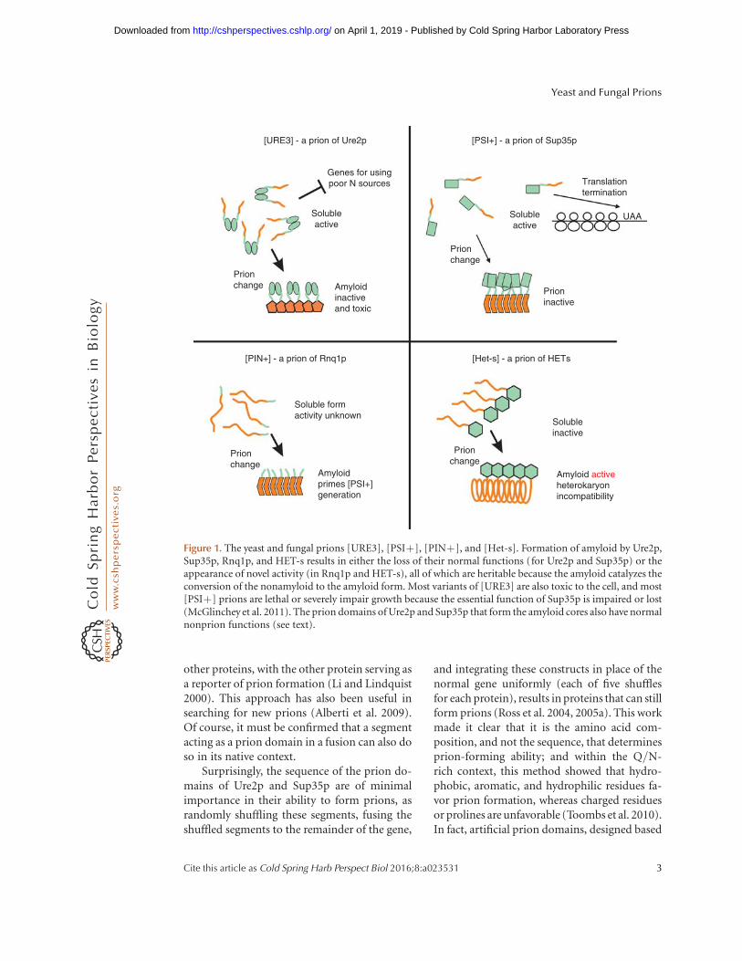
Although prion variants are propagatedwith relative stability, changes in variant prop-erties can occur under selection pressure in bothmammals and yeast, such as in crossing a spe-cies barrier or application of a drug (Kimberlinet al. 1987; Vishveshwara and Liebman 2009; Liet al. 2010). However, even without evident se-lection pressure, variant properties can change,and a mixture of variants can segregate duringgrowth (Bateman and Wickner 2013). Extensiveclonal isolation appears to purify single vari-ants, but on further propagation, all of the othervariants arise again, showing that variant mu-tation also occurs, even under nonselective con-ditions (Fig. 2) (Bateman and Wickner 2013).These results provide strong direct support forthe “prion cloud” model proposed by Collingeto explain the selectability of new variants (Col-linge and Clarke 2007). Previous data were con-sistent with this model but could also have beenexplained by induction of new variants as a re-sult of effects of the selection scheme (new pro-
tein sequence or amyloid-binding drug) on theamyloid structure.
PRION AMYLOID STRUCTURE EXPLAINSINHERITANCE OF PRION VARIANTINFORMATION
Solid-state nuclear magnetic resonance (NMR)and electron microscopic studies of infectiousamyloids of the prion domains of Sup35p,Ure2p, and Rnq1p have shown that each hasan in-register parallel-folded b-sheet architec-ture (Fig. 3) (Shewmaker et al. 2006; Baxa et al.2007; Wickner et al. 2008; Gorkovskiy et al.2014; for a review, see Tycko and Wickner2013). Shuffled versions of the Ure2p andSup35p prion domains also had this architec-ture (Shewmaker et al. 2008). Indeed, the factthat shuffled prion domains remained able toform prions was the basis for an early proposalthat these domains must have an in-register par-allel architecture (Ross et al. 2005b). Mass perunit length measurements show that these fila-ments have one monomer per �4.8 A, the dis-
Variant purificationby random segregation
Raremistemplating
Figure 2. The prion cloud model. A cell with a single predominant prion variant is generally a mixture of variantsas a result of occasional mistemplating, and these variants will gradually be purified from each other by randomsegregation during growth. This model was proven for the yeast prion [PSIþ] (Bateman and Wickner 2013) butwas first hypothesized to explain the properties of mammalian prions (Collinge and Clarke 2007).
Yeast and Fungal Prions
Cite this article as Cold Spring Harb Perspect Biol 2016;8:a023531 5
on April 1, 2019 - Published by Cold Spring Harbor Laboratory Press http://cshperspectives.cshlp.org/Downloaded from
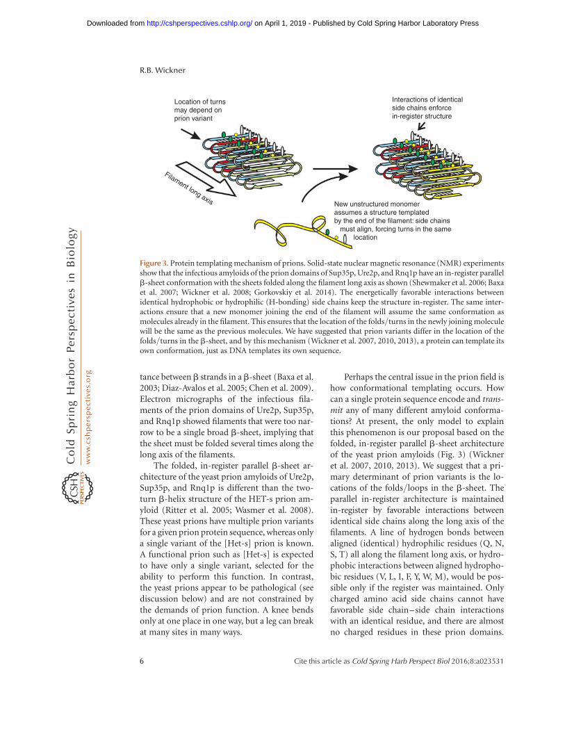
tance between b strands in a b-sheet (Baxa et al.2003; Diaz-Avalos et al. 2005; Chen et al. 2009).Electron micrographs of the infectious fila-ments of the prion domains of Ure2p, Sup35p,and Rnq1p showed filaments that were too nar-row to be a single broad b-sheet, implying thatthe sheet must be folded several times along thelong axis of the filaments.
The folded, in-register parallel b-sheet ar-chitecture of the yeast prion amyloids of Ure2p,Sup35p, and Rnq1p is different than the two-turn b-helix structure of the HET-s prion am-yloid (Ritter et al. 2005; Wasmer et al. 2008).These yeast prions have multiple prion variantsfor a given prion protein sequence, whereas onlya single variant of the [Het-s] prion is known.A functional prion such as [Het-s] is expectedto have only a single variant, selected for theability to perform this function. In contrast,the yeast prions appear to be pathological (seediscussion below) and are not constrained bythe demands of prion function. A knee bendsonly at one place in one way, but a leg can breakat many sites in many ways.
Perhaps the central issue in the prion field ishow conformational templating occurs. Howcan a single protein sequence encode and trans-mit any of many different amyloid conforma-tions? At present, the only model to explainthis phenomenon is our proposal based on thefolded, in-register parallel b-sheet architectureof the yeast prion amyloids (Fig. 3) (Wickneret al. 2007, 2010, 2013). We suggest that a pri-mary determinant of prion variants is the lo-cations of the folds/loops in the b-sheet. Theparallel in-register architecture is maintainedin-register by favorable interactions betweenidentical side chains along the long axis of thefilaments. A line of hydrogen bonds betweenaligned (identical) hydrophilic residues (Q, N,S, T) all along the filament long axis, or hydro-phobic interactions between aligned hydropho-bic residues (V, L, I, F, Y, W, M), would be pos-sible only if the register was maintained. Onlycharged amino acid side chains cannot havefavorable side chain–side chain interactionswith an identical residue, and there are almostno charged residues in these prion domains.
New unstructured monomerassumes a structure templatedby the end of the filament: side chains must align, forcing turns in the same location
Location of turnsmay depend onprion variant
Filament long axis
Interactions of identicalside chains enforcein-register structure
Figure 3. Protein templating mechanism of prions. Solid-state nuclear magnetic resonance (NMR) experimentsshow that the infectious amyloids of the prion domains of Sup35p, Ure2p, and Rnq1p have an in-register parallelb-sheet conformation with the sheets folded along the filament long axis as shown (Shewmaker et al. 2006; Baxaet al. 2007; Wickner et al. 2008; Gorkovskiy et al. 2014). The energetically favorable interactions betweenidentical hydrophobic or hydrophilic (H-bonding) side chains keep the structure in-register. The same inter-actions ensure that a new monomer joining the end of the filament will assume the same conformation asmolecules already in the filament. This ensures that the location of the folds/turns in the newly joining moleculewill be the same as the previous molecules. We have suggested that prion variants differ in the location of thefolds/turns in the b-sheet, and by this mechanism (Wickner et al. 2007, 2010, 2013), a protein can template itsown conformation, just as DNA templates its own sequence.
R.B. Wickner
6 Cite this article as Cold Spring Harb Perspect Biol 2016;8:a023531
on April 1, 2019 - Published by Cold Spring Harbor Laboratory Press http://cshperspectives.cshlp.org/Downloaded from
The unstructured prion domain of the solubleform of the prion protein (e.g., Pierce et al.2005) joins the end of the filament in such away as to maximize these favorable interactionsamong identical residues, and that requirementresults in the new molecule on the end of thefilament adopting the same conformation as themolecules already in the filament, with its turnsin the same places as the molecules already inthe filament (see Fig. 3). Just as DNA templatesits sequence, a prion amyloid can template itsconformation by this mechanism and so can actas a gene with many different heritable alleles(variants/amyloid conformations). Our recentdata identify the locations of some of these foldsin the sheets in support of this model (Gorkov-skiy et al. 2014).
PRION BIOLOGY: PATHOLOGICAL YEASTPRIONS AND A FUNCTIONAL FUNGALPRION
The [Het-s] prion of P. anserina is necessaryfor heterokaryon incompatibility, a process bywhich the fungus limits fusion of hypae to fu-sion partners that are genetically identical at adozen chromosomal loci to limit the spreadof detrimental viruses and plasmids (Saupe2011). This prion plainly serves a function use-ful for the host and is a case study in what toexpect for such “functional prions.” There isonly one known variant of [Het-s], presumablybecause the HETs protein sequence has beenselected during evolution to form a prion withthe desired characteristics. Accordingly, amy-loid of HETs made in vitro has a uniform struc-ture, reflected in sharp peaks in solid-state NMRstudies (Wasmer et al. 2008). This functionalprion is found in .90% of wild isolates withthe het-s allele, as expected for a beneficial prion(Debets et al. 2012).
Lethal or toxic variants of [PSIþ] and[URE3] are more common than the mild vari-ants usually studied (McGlinchey et al. 2011).The yeast prions [PSIþ], [URE3], and [PINþ]form many variants (see above), and even themildest variants are rare in wild populations(Chernoff et al. 2000; Resende et al. 2003;Nakayashiki et al. 2005; Halfmann et al. 2012),
both implying that they are pathological. Re-ports of marginal benefits of carrying the mild-est [PSIþ] prion variants (Eaglestone et al.1999; True and Lindquist 2000; Halfmannet al. 2012) have not been reproduced (Trueand Lindquist 2000; Namy et al. 2008; Wickneret al. 2015).
Sup35p from several different species canform [PSIþ] prions (Chernoff et al. 2000;Kushnirov et al. 2000a; Santoso et al. 2000; Na-kayashiki et al. 2001; Chen et al. 2007; Afana-sieva et al. 2011), but many others cannot(Edskes et al. 2014). Ure2p of most species ofthe genus Saccharomyces can form the [URE3]prion (Edskes and Wickner 2002; Baudin-Bail-lieu et al. 2003; Edskes et al. 2009), but that ofSaccharomyces castellii cannot (Edskes et al.2009). A careful study showed that the Kluyver-omyces lactis Ure2p cannot form [URE3] evenin K. lactis itself (Safadi et al. 2011). Ure2p ofCandida albicans, although closely related tothat of Saccharomyces cerevisiae, cannot form[URE3], whereas that of Candida glabrata,more distant from the S. cerevisiae Ure2p, canform [URE3] with all of its properties (Edskeset al. 2011; Engel et al. 2011; Edskes and Wick-ner 2013). Thus, [PSIþ] and [URE3] prion-forming ability appears to be sporadically dis-tributed. The prion domains of these proteinshave nonprion functions, for example, roles inmRNA turnover of the Sup35 prion domain(Hoshino et al. 1999; Hosoda et al. 2003) andmicrotubule binding (Li et al. 2014) and stabi-lization of the full-length protein in the caseof the Ure2p prion domain (Shewmaker et al.2007). These functions may explain the persis-tence of these domains in evolution despitetheir occasional formation of prions.
PRION ECOLOGY AND EVOLUTION
Yeast and fungal prions are determined by twolevels of inheritance. First, the chromosomalgene encoding the prion protein is under posi-tive or negative selection pressure to maintainits normal function and to maintain or lose itsability to form a prion. Second, having formeda prion, the prion itself is under positive ornegative selection pressure depending on the
Yeast and Fungal Prions
Cite this article as Cold Spring Harb Perspect Biol 2016;8:a023531 7
on April 1, 2019 - Published by Cold Spring Harbor Laboratory Press http://cshperspectives.cshlp.org/Downloaded from
phenotype it confers on the cell and its stability.In considering whether prion formation by aparticular protein is beneficial or detrimental,one must consider the full range of prion vari-ants that can arise, their relative frequencies,and the frequencies with which yeast will en-counter an environmental condition underwhich there is a prion-specific beneficial or det-rimental effect. Likewise, the nonprion func-tions of the protein may be affected by the prionconversion. This is quite unlike most genes,whose encoded protein usually has one basicheritable state, determined by the protein se-quence. The protein activity may be regulatedby the environment, but generally not heri-tably so. The preceding program may be impos-sible to execute, but a beginning effort has beenmade to examine some aspects of the evolu-tion and ecology of prion proteins and prionsthemselves.
It is not difficult to find detrimental virusesand prions in wild populations. For example,the uniformly fatal chronic wasting disease(CWD) prion is found in �10% of wild deerand elk in several areas of the United States. Theinfectivity of CWD can outweigh the fatality ofthe disease. For a beneficial virus or prion, in-fectivity and benefit to the host are working inthe same direction, so the infection shouldspread rapidly in the wild, resulting in a highprevalence of the prion. Mitochondria began asbacterial endosymbionts, infectious elements.Although the mitochondrial genome is ratherunstable, it is so beneficial that nearly all wildisolates carry this nonchromosomal DNA. The2-mm DNA plasmid replicates and, like the yeastprions, is spread by mating. Although threegroups have found that the 2-mm DNA plasmidmildly slows growth of yeast about 1%–3%(Futcher and Cox 1983; Mead et al. 1986;Futcher et al. 1988; Kelly et al. 2012), it wasfound in 38 of 70 wild strains examined (Na-kayashiki et al. 2005). This provides a standardof comparison against which prions may bejudged. [URE3], [PSIþ], and [SWIþ] werenot found in any of the same 70 wild strains,indicating that they must confer a substantiallygreater detriment than does 2-mm DNA (Na-kayashiki et al. 2005; Bateman and Wickner
2012; Kelly et al. 2012). Note that this is thedetriment of the most mild variants of [URE3]or [PSIþ] or other prions. The [PINþ] prionwas found in �15% of these wild strains (Na-kayashiki et al. 2005), and [MOT3þ] was de-tected in �6% of a different set of 96 wild iso-lates (Halfmann et al. 2012), indicating thatboth are also detrimental on the net.
The preceding discussion shows that eventhe mildest variants of the prions examined aredetrimental to their hosts. Nonetheless, they arefound in nature at frequencies above their fre-quency of de novo generation. For [PINþ], twopossible scenarios were considered: (1) theremay be some part of the ecological niche ofS. cerevisiae in which [PINþ], should it arise,would be beneficial to its host, and this has ledto expansion of the line in which it arose becauseof this benefit; and (2) [PINþ] is mildly detri-mental in all niches, but it has spread by matingdespite this detriment. Detailed examination ofthe occasional wild [PINþ] strains has shownthat the presence of [PINþ] is associated withthe recent occurrence of outcross mating, asshown by heterozygosity (Kelly et al. 2014).This result favors the second explanation.
The polymorphs of Sup35 among wildS. cerevisiae that limit the spread of [PSIþ]are also a reflection of the ecology of this prion(Bateman and Wickner 2012). It would be ofinterest to know whether certain ecologicalniches favor certain prion domain sequences.
ANTIPRION SYSTEMS
Ribosome-Associated Hsp70s (Ssbs) Inhibit[PSIþ] Generation
Ssb1 or Ssb2 are members of the Hsp70 familyassociated with the ribosome and involved infolding of nascent proteins (Fig. 4) (Nelsonet al. 1992; Pfund et al. 1998). Hsp104 overpro-duction cures the [PSIþ] prion by an as-yetuncertain mechanism (Chernoff et al. 1995).Overproduction of Ssb1p or Ssb2p facilitatesthe curing of [PSIþ] by overproduced Hsp104(Chernoff et al. 1999). Moreover, in an ssb1Dssb2D strain, the frequency of [PSIþ] formationis elevated, whether spontaneous or induced by
R.B. Wickner
8 Cite this article as Cold Spring Harb Perspect Biol 2016;8:a023531
on April 1, 2019 - Published by Cold Spring Harbor Laboratory Press http://cshperspectives.cshlp.org/Downloaded from

Sup35p overexpression (Chernoff et al. 1999).Restoration of Ssb levels to [PSIþ] strains gen-erated in the ssb1D ssb2D strain did not cure[PSIþ], indicating that Ssb1 and Ssb2, at theirnormal levels, inhibit the generation of [PSIþ]rather than its propagation (Chernoff et al.1999). Since yeast prions are genes and the gen-eration of [PSIþ] constitutes a “mutation”from [psi-], it is proposed that Ssbs are analo-gous to DNA repair systems, or an antimutatorsystem (Chernoff et al. 1999).
Btn2/Cur1 at Normal Levels Cure Most[URE3] Variants
Overproduction of Btn2p, or its paralog Cur1p,efficiently cures [URE3-1], the original [URE3]variant isolated by Lacroute (Kryndushkin et al.2008). Remarkably, in the process of curing,Ure2p aggregates are concentrated at a singlecellular site, co-localized with Btn2p, suggestingthat overproduced Btn2p collects the prion ag-gregates, preventing their distribution to both
Monomeraddition
Unfolded monomerpulled from the middle
New prion seeds
Hsp104Hsp70Hsp40
Amyloidfiber
?Endosome
Nucleus
Vacuole
[URE3]amyloid
Btn2
Btn2
Hsp42
Hsp42
Figure 4. Prion-handling systems. Prion protein monomers are synthesized and are soon incorporated intoamyloid fibers (see Fig. 2). These fibers grow and are split by the ability of the Hsp104–Hsp70–Hsp40 machineto remove a monomer from the middle of the fiber. This ensures sufficient prion seeds to distribute to daughtercells, allowing prion propagation. Some filaments are sequestered by the Btn2p–Hsp42 system. If the seednumber is sufficiently low, this results in curing the prion from progeny cells. The Ssb1/2 Hsp70s act to inhibitconversion of the normal form (of Sup35p) to the prion form, an inhibition of prion generation not ofpropagation (Chernoff et al. 1999).
Yeast and Fungal Prions
Cite this article as Cold Spring Harb Perspect Biol 2016;8:a023531 9
on April 1, 2019 - Published by Cold Spring Harbor Laboratory Press http://cshperspectives.cshlp.org/Downloaded from
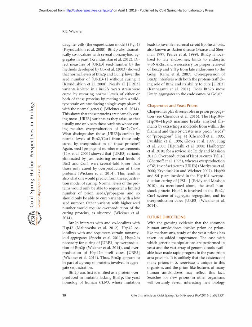
daughter cells (the sequestration model) (Fig. 4)(Kryndushkin et al. 2008). Btn2p also dramat-ically co-localizes with several nonamyloid ag-gregates in yeast (Kryndushkin et al. 2012). Di-rect measures of [URE3] seed-number by themethods developed by Cox et al. (2003) showedthat normal levels of Btn2p and Cur1p lower theseed number of [URE3-1] without curing it(Kryndushkin et al. 2008). Nearly all [URE3]variants isolated in a btn2D cur1D strain werecured by restoring normal levels of either orboth of these proteins by mating with a wild-type strain or introducing a single-copy plasmidwith the normal gene(s) (Wickner et al. 2014).This shows that these proteins are normally cur-ing most [URE3] variants as they arise, so thatusually one only sees those variants whose cur-ing requires overproduction of Btn2/Cur1.What distinguishes those [URE3]s curable bynormal levels of Btn2/Cur1 from those onlycured by overproduction of these proteins?Again, seed (propagon) number measurements(Cox et al. 2003) showed that [URE3] variantseliminated by just restoring normal levels ofBtn2 and Cur1 were several-fold lower thanthose only cured by overproduction of thoseproteins (Wickner et al. 2014). This result isalso what one would predict from the sequestra-tion model of curing. Normal levels of the pro-teins would only be able to sequester a limitednumber of prion seeds/propagons and soshould only be able to cure variants with a lowseed number. Other variants with higher seednumber would require overproduction of thecuring proteins, as observed (Wickner et al.2014).
Btn2p interacts with and co-localizes withHsp42 (Malinovska et al. 2012), Hsp42 co-localizes with and sequesters certain nonamy-loid aggregates (Specht et al. 2011), Hsp42 isnecessary for curing of [URE3] by overproduc-tion of Btn2p (Wickner et al. 2014), and over-production of Hsp42p itself cures [URE3](Wickner et al. 2014). Thus, Btn2p appears tobe part of a group of proteins involved in aggre-gate sequestration.
Btn2p was first identified as a protein over-produced in mutants lacking Btn1p, the yeasthomolog of human CLN3, whose mutation
leads to juvenile neuronal ceroid lipofuscinosis,also known as Batten disease (Pearce and Sher-man 1997; Pearce et al. 1999). Btn2p is loca-lized to late endosomes, binds to endocyticv-SNAREs, and is necessary for proper retrievalof Kex2p and Yif1p from late endosomes to theGolgi (Kama et al. 2007). Overexpression ofBtn3p interferes with both the protein-traffick-ing role of Btn2 and its ability to cure [URE3](Kanneganti et al. 2011). Does Btn2p moveUre2p aggregates to the endosomes or Golgi?
Chaperones and Yeast Prions
Chaperones play diverse roles in prion propaga-tion (see Chernova et al. 2016). The Hsp104–Hsp70–Hsp40 machine breaks amyloid fila-ments by extracting a molecule from within thefilament and thereby creates new prion “seeds”or “propagons” (Fig. 4) (Chernoff et al. 1995;Paushkin et al. 1996; Glover et al. 1997; Junget al. 2000; Higurashi et al. 2008; Haslbergeret al. 2010; for a review, see Reidy and Masison2011). Overproduction of Hsp104 cures [PSIþ](Chernoff et al. 1995), whereas overproductionof Ydj1p or Sse1p cures [URE3] (Moriyama et al.2000; Kryndushkin and Wickner 2007). Hsp90and Sti1p are involved in the Hsp104 overpro-duction curing of [PSIþ] (Reidy and Masison2010). As mentioned above, the small heat-shock protein Hsp42 is involved in the Btn2/Cur1 system of aggregate segregation, and itsoverproduction cures [URE3] (Wickner et al.2014).
FUTURE DIRECTIONS
With the growing evidence that the commonhuman amyloidoses involve prion or prion-like mechanisms, study of the yeast prions hastaken on added importance. The ease withwhich genetic manipulations are performed inyeast and the vast array of genomic tools avail-able have made rapid progress in the yeast prionarea possible. It is unlikely that the existence ofmany prions in S. cerevisiae is unique to thisorganism, and the prion-like features of manyhuman amyloidoses may reflect this fact.Searches for new prions in other organismswill certainly reveal interesting new biology
R.B. Wickner
10 Cite this article as Cold Spring Harb Perspect Biol 2016;8:a023531
on April 1, 2019 - Published by Cold Spring Harbor Laboratory Press http://cshperspectives.cshlp.org/Downloaded from
(e.g., the P. anserina [Het-s] prion) and pathol-ogy. Structural work on yeast prions has pro-duced for the first time a working model thatcan explain how prion variants are inherited,but additional structural detail will require ex-tensive further studies. The mechanisms of theBtn2/Cur1 prion curing will be particularly im-portant because this antiprion system normallycures prions as they arise, and enhancementof homologous or analogous human system(s)may be utilized for treatment of amyloidoses.Continued dissection of the mechanisms bywhich chaperones shape prion formation andpropagation is revealing important aspects ofthese complex systems.
ACKNOWLEDGMENTS
This work is supported by the Intramural Pro-gram of the National Institute of Diabetes andDigestive and Kidney Diseases of the NationalInstitutes of Health.
REFERENCES
Afanasieva EG, Kushnirov VV, Tuite MF, Ter-Avanesyan MD.2011. Molecular basis for transmission barrier and inter-ference between closely related prion proteins in yeast.J Biol Chem 286: 15773–15780.
Aigle M, Lacroute F. 1975. Genetical aspects of [URE3], anon-mitochondrial, cytoplasmically inherited mutationin yeast. Mol Gen Genet 136: 327–335.
Alberti S, Halfmann R, King O, Kapila A, Lindquist S. 2009.A systematic survey identifies prions and illuminatessequence features of prionogenic proteins. Cell 137:146–158.
Alper T, Cramp WA, Haig DA, Clarke MC. 1967. Does theagent of scrapie replicate without nucleic acid? Nature214: 764–766.
Ball AJ, Wong DK, Elliott JJ. 1976. Glucosamine resistancein yeast. I: A preliminary genetic analysis. Genetics 84:311–317.
Bateman DA, Wickner RB. 2012. [PSIþ] prion transmis-sion barriers protect Saccharomyces cerevisiae frominfection: Intraspecies “species barriers.” Genetics 190:569–579.
Bateman D, Wickner RB. 2013. The [PSIþ] prion exists asa dynamic cloud of variants. PLoS Genet 9: e1003257.
Baudin-Baillieu A, Fernandez-Bellot E, Reine F, Coissac E,Cullin C. 2003. Conservation of the prion proper-ties of Ure2p through evolution. Mol Biol Cell 14:3449–3458.
Baxa U, Taylor KL, Wall JS, Simon MN, Cheng N, WicknerRB, Steven A. 2003. Architecture of Ure2p prion fila-
ments: The N-terminal domain forms a central core fiber.J Biol Chem 278: 43717–43727.
Baxa U, Wickner RB, Steven AC, Anderson D, Marekov L,Yau WM, Tycko R. 2007. Characterization of b-sheetstructure in Ure2p1-89 yeast prion fibrils by solid statenuclear magnetic resonance. Biochemistry 46: 13149–13162.
Bolton DC, McKinley MP, Prusiner SB. 1982. Identificationof a protein that purifies with the scrapie prion. Science218: 1309–1311.
Borchsenius AS, Muller S, Newnam GP, Inge-VechtomovSG, Chernoff YO. 2006. Prion variant maintained onlyat high levels of the Hsp104 disaggregase. Curr Genet 49:21–29.
Brachmann A, Baxa U, Wickner RB. 2005. Prion generationin vitro: Amyloid of Ure2p is infectious. EMBO J 24:3082–3092.
Bradley ME, Liebman SW. 2003. Destabilizing interactionsamong [PSIþ] and [PINþ] yeast prion variants. Genetics165: 1675–1685.
Brown JC, Lindquist S. 2009. A heritable switch in carbonsource utilization driven by an unusual yeast prion. GenesDev 23: 2320–2332.
Chen B, Newnam GP, Chernoff YO. 2007. Prion speciesbarrier between the closely related yeast proteins is de-tected despite coaggregation. Proc Natl Acad Sci 104:2791–2796.
Chen B, Thurber KR, Shewmaker F, Wickner RB, Tycko R.2009. Measurement of amyloid fibril mass-per-length bytilted-beam transmission electron microscopy. Proc NatlAcad Sci 106: 14339–14344.
Chen B, Bruce KL, Newnam GP, Gyoneva S, Romanyuk AV,Chernoff YO. 2010. Genetic and epigenetic control of theefficiency and fidelity of cross-species prion transmis-sion. Mol Microbiol 76: 1483–1499.
Chernoff YO, Derkach IL, Inge-Vechtomov SG. 1993. Multi-copy SUP35 gene induces de-novo appearance of psi-likefactors in the yeast Saccharomyces cerevisiae. Curr Genet24: 268–270.
Chernoff YO, Lindquist SL, Ono BI, Inge-Vechtomov SG,Liebman SW. 1995. Role of the chaperone proteinHsp104 in propagation of the yeast prion-like factor[ psiþ]. Science 268: 880–884.
Chernoff YO, Newnam GP, Kumar J, Allen K, Zink AD. 1999.Evidence for a protein mutator in yeast: Role of theHsp70-related chaperone Ssb in formation, stabilityand toxicity of the [PSI] prion. Mol Cell Biol 19: 8103–8112.
Chernoff YO, Galkin AP, Lewitin E, Chernova TA, NewnamGP, Belenkiy SM. 2000. Evolutionary conservation of pri-on-forming abilities of the yeast Sup35 protein. Mol Mi-crobiol 35: 865–876.
Chernova TA, Wilkinson KD, Chernoff YO. 2016. Prions,chaperones, and proteostasis in yeast. Cold Spring HarbPerspect Biol doi: 10.1101/cshperspect.a023663.
Chesebro B, Race R, Wehrly K, Nishio J, Bloom M, LechnerD, Bergstrom S, Robbins K, Mayer L, Keith JM, et al. 1985.Identification of scrapie prion protein-specific mRNA inscrapie-infected brain. Nature 315: 331–333.
Collinge J, Clarke AR. 2007. A general model of prion strainsand their pathogenicity. Science 318: 930–936.
Yeast and Fungal Prions
Cite this article as Cold Spring Harb Perspect Biol 2016;8:a023531 11
on April 1, 2019 - Published by Cold Spring Harbor Laboratory Press http://cshperspectives.cshlp.org/Downloaded from

Cox BS. 1965. PSI, a cytoplasmic suppressor of super-sup-pressor in yeast. Heredity 20: 505–521.
Cox BS, Ness F, Tuite MF. 2003. Analysis of the generationand segregation of propagons: Entities that propagate the[PSIþ] prion in yeast. Genetics 165: 23–33.
Debets AJ, Dalstra HJ, Slakhorst M, Koopmanschap B,Hoekstra RF, Saupe SJ. 2012. High natural prevalence ofa fungal prion. Proc Natl Acad Sci 109: 10432–10437.
Derkatch IL, Chernoff YO, Kushnirov VV, Inge-VechtomovSG, Liebman SW. 1996. Genesis and variability of [PSI]prion factors in Saccharomyces cerevisiae. Genetics 144:1375–1386.
Derkatch IL, Bradley ME, Zhou P, Chernoff YO, LiebmanSW. 1997. Genetic and environmental factors affectingthe de novo appearance of the [PSIþ] prion in Saccharo-myces cerevisiae. Genetics 147: 507–519.
Derkatch IL, Bradley ME, Hong JY, Liebman SW. 2001. Pri-ons affect the appearance of other prions: The story of[PIN]. Cell 106: 171–182.
Diaz-Avalos R, King CY, Wall JS, Simon M, Caspar DLD.2005. Strain-specific morphologies of yeast prion amy-loids. Proc Natl Acad Sci 102: 10165–10170.
Dickinson AG, Meikle VMH, Fraser H. 1968. Identificationof a gene which controls the incubation period of somestrains of scrapie in mice. J Comp Path 78: 293–299.
Du Z, Park KW, Yu H, Fan Q, Li L. 2008. Newly identifiedprion linked to the chromatin-remodeling factor Swi1 inSaccharomyces cerevisiae. Nat Genet 40: 460–465.
Eaglestone SS, Cox BS, Tuite MF. 1999. Translation termi-nation efficiency can be regulated in Saccharomyces cer-evisiae by environmental stress through a prion-mediatedmechanism. EMBO J 18: 1974–1981.
Edskes HK, Wickner RB. 2002. Conservation of a portion ofthe S. cerevisiae Ure2p prion domain that interacts withthe full-length protein. Proc Natl Acad Sci 99: 16384–16391.
Edskes HK, Wickner RB. 2013. The [URE3] prion in Can-dida. Eukaryot Cell 12: 551–558.
Edskes HK, Gray VT, Wickner RB. 1999. The [URE3] prionis an aggregated form of Ure2p that can be cured by over-expression of Ure2p fragments. Proc Natl Acad Sci 96:1498–1503.
Edskes HK, McCann LM, Hebert AM, Wickner RB. 2009.Prion variants and species barriers among SaccharomycesUre2 proteins. Genetics 181: 1159–1167.
Edskes HK, Engel A, McCann LM, Brachmann A, Tsai HF,Wickner RB. 2011. Prion-forming ability of Ure2 of yeastsis not evolutionarily conserved. Genetics 188: 81–90.
Edskes HK, Khamar HJ, Winchester CL, Greenler AJ, ZhouA, McGlinchey RP, Gorkovskiy A, Wickner RB.2014. Sporadic distribution of prion-forming ability ofSup35p from yeasts and fungi. Genetics 198: 605–616.
Engel A, Shewmaker F, Edskes HK, Dyda F, Wickner RB.2011. Amyloid of the Candida albicans Ure2p prion do-main is infectious and has a parallel in-register b-sheetstructure. Biochemistry 50: 5971–5978.
Ferreira PC, Ness F, Edwards SR, Cox BS, Tuite MF. 2001.The elimination of the yeast [PSIþ] prion by guanidinehydrochloride is the result of Hsp104 inactivation. MolMicrobiol 40: 1357–1369.
Futcher AB, Cox BS. 1983. Maintenance of the 2 mm circleplasmid in populations of Saccharomyces cerevisiae.J Bacteriol 154: 612–622.
Futcher B, Reid E, Hickey DA. 1988. Maintenance of the2 mm circle plasmid of Saccharomyces cerevisiae by sexualtransmission: An example of selfish DNA. Genetics 118:411–415.
Glover JR, Kowal AS, Shirmer EC, Patino MM, Liu JJ, Lind-quist S. 1997. Self-seeded fibers formed by Sup35, theprotein determinant of [PSIþ], a heritable prion-likefactor of S. cerevisiae. Cell 89: 811–819.
Gorkovskiy A, Thurber KR, Tycko R, Wickner RB. 2014.Locating the folds of the in-register parallel b-sheet ofthe Sup35p prion domain infectious amyloid. Proc NatlAcad Sci 111: E4615–E4622.
Griffith JS. 1967. Self-replication and scrapie. Nature 215:1043–1044.
Halfmann R, Jarosz DF, Jones SK, Chang A, Lancaster AK,Lindquist S. 2012. Prions are a common mechanism forphenotypic inheritance in wild yeasts. Nature 482: 363–368.
Haslberger T, Bukau B, Mogk A. 2010. Towards a unifyingmechanism for the ClpB/Hsp104-mediated protein dis-aggregation and prion propagation. Biochem Cell Biol 88:63–75.
Higurashi T, Hines JK, Sahi C, Aron R, Craig EA. 2008.Specificity of the J-protein Sis1 in the propagation of 3yeast prions. Proc Natl Acad Sci 105: 16596–16601.
Holmes DL, Lancaster AK, Lindquist S, Halfmann R. 2013.Heritable remodeling of yeast multicellularity by an en-vironmentally responsive prion. Cell 153: 153–165.
Hoshino S, Imai M, Kobayashi T, Uchida N, Katada T. 1999.The eukaryotic polypeptide chain releasing factor (eRF3/GSPT) carrying the translation termination signal to the30-poly(A) tail of mRNA. Direct association of eRF3/GSPT with polyadenylate-binding protein. J Biol Chem274: 16677–16680.
Hosoda N, Kobayashii T, Uchida N, Funakoshi Y, Kikuchi Y,Hoshino S, Katada T. 2003. Translation termination fac-tor eRF3 mediates mRNA decay through the regulation ofdeadenylation. J Biol Chem 278: 38287–38291.
Jones EW. 1991. Three proteolytic systems in the yeast Sac-charomyces cerevisiae. J Biol Chem 266: 7963–7966.
Jung G, Masison DC. 2001. Guanidine hydrochloride inhib-its Hsp104 activity in vivo: A possible explanation for itseffect in curing yeast prions. Curr Microbiol 43: 7–10.
Jung G, Jones G, Wegrzyn RD, Masison DC. 2000. A role forcytosolic Hsp70 in yeast [PSIþ] prion propagation and[PSIþ] as a cellular stress. Genetics 156: 559–570.
Jung G, Jones G, Masison DC. 2002. Amino acid residue 184of yeast Hsp104 chaperone is critical for prion-curing byguanidine, prion propagation, and thermotolerance.Proc Natl Acad Sci 99: 9936–9941.
Kama R, Robinson M, Gerst JE. 2007. Btn2, a Hook1 ortho-log and potential Batten disease-related protein, mediateslate endosome-Golgi protein sorting in yeast. Mol CellBiol 27: 605–621.
Kanneganti V, Kama R, Gerst JE. 2011. Btn3 is a negativeregulator of Btn2-mediated endosomal protein traffick-ing and prion curing in yeast. Mol Biol Cell 22: 1648–1663.
R.B. Wickner
12 Cite this article as Cold Spring Harb Perspect Biol 2016;8:a023531
on April 1, 2019 - Published by Cold Spring Harbor Laboratory Press http://cshperspectives.cshlp.org/Downloaded from
Kelly AC, Shewmaker FP, Kryndushkin D, Wickner RB.2012. Sex, prions and plasmids in yeast. Proc Natl AcadSci 109: E2683–E2690.
Kelly AC, Busby B, Wickner RB. 2014. Effect of domestica-tion on the spread of the [PINþ] prion in Saccharomycescerevisiae. Genetics 197: 1007–1024.
Kimberlin RH, Cole S, Walker CA. 1987. Temporary andpermanent modifications to a single strain of mouse scra-pie on transmission to rats and hamsters. J Gen Virol 68:1875–1881.
King CY. 2001. Supporting the structural basis of prionstrains: Induction and identification of [PSI] variants.J Mol Biol 307: 1247–1260.
King CY, Diaz-Avalos R. 2004. Protein-only transmission ofthree yeast prion strains. Nature 428: 319–323.
King CY, Tittmann P, Gross H, Gebert R, Aebi M, WuthrichK. 1997. Prion-inducing domain 2–114 of yeast Sup35protein transforms in vitro into amyloid-like filaments.Proc Natl Acad Sci 94: 6618–6622.
Kryndushkin D, Wickner RB. 2007. Nucleotide exchangefactors for Hsp70s are required for [URE3] prion prop-agation in Saccharomyces cerevisiae. Mol Biol Cell 18:2149–2154.
Kryndushkin D, Smirnov VN, Ter-Avanesyan MD, Kush-nirov VV. 2002. Increased expression of Hsp40 chaper-ones, transcriptional factors, and ribosomal proteinRpp0 can cure yeast prions. J Biol Chem 277: 23702–23708.
Kryndushkin D, Shewmaker F, Wickner RB. 2008. Curing ofthe [URE3] prion by Btn2p, a Batten disease-related pro-tein. EMBO J 27: 2725–2735.
Kryndushkin D, Ihrke G, Piermartiri TC, Shewmaker F.2012. A yeast model of optineurin proteinopathy revealsa unique aggregation pattern associated with cellular tox-icity. Mol Microbiol 86: 1531–1547.
Kunz BA, Ball AJ. 1977. Glucosamine resistance in yeast.II: Cytoplasmic determinants conferring resistance. MolGen Genet 153: 169–177.
Kushnirov VV, Kochneva-Pervukhova NV, Cechenova MB,Frolova NS, Ter-Avanesyan MD. 2000a. Prion propertiesof the Sup35 protein of yeast Pichia methanolica. EMBO J19: 324–331.
Kushnirov VV, Kryndushkin D, Boguta M, Smirnov VN,Ter-Avanesyan MD. 2000b. Chaperones that cure yeastartificial [PSIþ] and their prion-specific effects. CurrBiol 10: 1443–1446.
Lacroute F. 1971. Non-Mendelian mutation allowing urei-dosuccinic acid uptake in yeast. J Bacteriol 106: 519–522.
Li L, Lindquist S. 2000. Creating a protein-based element ofinheritance. Science 287: 661–664.
Li J, Browning S, Mahal SP, Oelschlegel AM, Weissmann C.2010. Darwinian evolution of prions in cell culture. Sci-ence 327: 869–872.
Li X, Rayman JB, Kandel ER, Derkatch IL. 2014. Functionalrole of Tia1/Pub1 and Sup35 prion domains: Directingprotein synthesis machinery to the tubulin cytoskeleton.Mol Cell 55: 1–14.
Lund PM, Cox BS. 1981. Reversion analysis of [ psi2] mu-tations in Saccharomyces cerevisiae. Genet Res 37: 173–182.
Malinovska L, Kroschwald S, Munder MC, Richter D, Al-berti S. 2012. Molecular chaperones and stress-inducibleprotein-sorting factors coordinate the spaciotemporaldistribution of protein aggregates. Mol Biol Cell 23:3041–3056.
Masison DC, Wickner RB. 1995. Prion-inducing domain ofyeast Ure2p and protease resistance of Ure2p in prion-containing cells. Science 270: 93–95.
McGlinchey R, Kryndushkin D, Wickner RB. 2011. Suicidal[PSIþ] is a lethal yeast prion. Proc Natl Acad Sci 108:5337–5341.
Mead DJ, Gardner DCJ, Oliver SG. 1986. The yeast 2 mmplasmid: Strategies for the survival of a selfish DNA. MolGen Genet 205: 417–421.
Moriyama H, Edskes HK, Wickner RB. 2000. [URE3] prionpropagation in Saccharomyces cerevisiae: Requirementfor chaperone Hsp104 and curing by overexpressed chap-erone Ydj1p. Mol Cell Biol 20: 8916–8922.
Nakayashiki T, Ebihara K, Bannai H, Nakamura Y. 2001.Yeast [PSIþ] “prions” that are crosstransmissible andsusceptible beyond a species barrier through a quasi-pri-on state. Mol Cell 7: 1121–1130.
Nakayashiki T, Kurtzman CP, Edskes HK, Wickner RB. 2005.Yeast prions [URE3] and [PSIþ] are diseases. Proc NatlAcad Sci 102: 10575–10580.
Namy O, Galopier A, Martini C, Matsufuji S, Fabret C,Rousset C. 2008. Epigenetic control of polyamines bythe prion [PSIþ]. Nat Cell Biol 10: 1069–1075.
Nelson RJ, Ziegilhoffer T, Nicolet C, Werner-Washburne M,Craig EA. 1992. The translation machinery and 70 kDalheat shock protein cooperate in protein synthesis. Cell 71:97–105.
Oesch B, Westaway D, Walchli M, McKinley MP, Kent SB,Aebersold R, Barry RA, Tempst P, Templow DB, Hood LE,et al. 1985. A cellular gene encodes scrapie PrP 27–30protein. Cell 40: 735–746.
Patel BK, Gavin-Smyth J, Liebman SW. 2009. The yeast glob-al transcriptional co-repressor protein Cyc8 can propa-gate as a prion. Nat Cell Biol 11: 344–349.
Patino MM, Liu JJ, Glover JR, Lindquist S. 1996. Support forthe prion hypothesis for inheritance of a phenotypic traitin yeast. Science 273: 622–626.
Paushkin SV, Kushnirov VV, Smirnov VN, Ter-AvanesyanMD. 1996. Propagation of the yeast prion-like [ psiþ]determinant is mediated by oligomerization of theSUP35-encoded polypeptide chain release factor.EMBO J 15: 3127–3134.
Paushkin SV, Kushnirov VV, Smirnov VN, Ter-AvanesyanMD. 1997. In vitro propagation of the prion-like stateof yeast Sup35 protein. Science 277: 381–383.
Pearce DA, Sherman F. 1997. BTN1, a yeast gene correspond-ing to the human gene responsible for Batten’s disease, isnot essential for viability, mitochondrial function, ordegradation of mitochondrial ATP synthase. Yeast 13:691–697.
Pearce DA, Ferea T, Nosel SA, Das B, Sherman F. 1999. Ac-tion of BTN1, the yeast ortholog of the gene mutated inBatten disease. Nat Genet 22: 55–58.
Pfund C, Lopez-Hoyo N, Ziegelhoffer T, Schilke BA, Lopez-Buesa P, Walter WA, Wiedmann M, Craig EA. 1998. Themolecular chaperone Ssb from Saccharomyces cerevisiae is
Yeast and Fungal Prions
Cite this article as Cold Spring Harb Perspect Biol 2016;8:a023531 13
on April 1, 2019 - Published by Cold Spring Harbor Laboratory Press http://cshperspectives.cshlp.org/Downloaded from
a component of the ribosome-nascent chain complex.EMBO J 17: 3981–3989.
Pierce MM, Baxa U, Steven AC, Bax A, Wickner RB. 2005. Isthe prion domain of soluble Ure2p unstructured? Bio-chemistry 44: 321–328.
Prusiner SB. 1982. Novel proteinaceous infectious particlescause scrapie. Science 216: 136–144.
Reidy M, Masison DC. 2010. Sti1 regulation of Hsp70 andHsp90 is critical for curing of Saccharomyces cerevisiae[PSIþ] prions by Hsp104. Mol Cell Biol 30: 3542–3552.
Reidy M, Masison DC. 2011. Modulation and elimination ofyeast prions by protein chaperones and co-chaperones.Prion 5: 245–249.
Resende CG, Outeiro TF, Sands L, Lindquist S, Tuite MF.2003. Prion protein gene polymorphisms in Saccharomy-ces cerevisiae. Mol Microbiol 49: 1005–1017.
Ritter C, Maddelein ML, Siemer AB, Luhrs T, Ernst M, MeierBH, Saupe SJ, Riek R. 2005. Correlation of structuralelements and infectivity of the HET-s prion. Nature435: 844–848.
Roberts BT, Wickner RB. 2003. A class of prions that prop-agate via covalent auto-activation. Genes Dev 17: 2083–2087.
Rogoza T, Goginashvili A, Rodionova S, Ivanov M, Viktor-ovskaya O, Rubel A, Volkov K, Mironova L. 2010. Non-Mendelian determinant [ISPþ] in yeast is a nuclear-re-siding prion form of the global transcriptional regulatorSfp1. Proc Natl Acad Sci 107: 10573–10577.
Ross ED, Baxa U, Wickner RB. 2004. Scrambled prion do-mains form prions and amyloid. Mol Cell Biol 24: 7206–7213.
Ross ED, Edskes HK, Terry MJ, Wickner RB. 2005a. Primarysequence independence for prion formation. Proc NatlAcad Sci 102: 12825–12830.
Ross ED, Minton AP, Wickner RB. 2005b. Prion domains:Sequences, structures and interactions. Nat Cell Biol 7:1039–1044.
Safadi RA, Talarek N, Jacques N, Aigle M. 2011. Yeast prions:Could they be exaptations? The URE2/[URE3] system inKluyveromyces lactis. FEMS Yeast Res 11: 151–153.
Santoso A, Chien P, Osherovich LZ, Weissman JS. 2000.Molecular basis of a yeast prion species barrier. Cell100: 277–288.
Saupe SJ. 2011. The [Het-s] prion of Podospora anserina andits role in heterokaryon incompatibility. Sem Cell DevBiol 22: 460–468.
Schlumpberger M, Prusiner SB, Herskowitz I. 2001. Induc-tion of distinct [URE3] yeast prion strains. Mol Cell Biol21: 7035–7046.
Shewmaker F, Wickner RB, Tycko R. 2006. Amyloid of theprion domain of Sup35p has an in-register parallel b-sheet structure. Proc Natl Acad Sci 103: 19754–19759.
Shewmaker F, Mull L, Nakayashiki T, Masison DC, WicknerRB. 2007. Ure2p function is enhanced by its prion do-main in Saccharomyces cerevisiae. Genetics 176: 1557–1565.
Shewmaker F, Ross ED, Tycko R, Wickner RB. 2008. Amy-loids of shuffled prion domains that form prions have aparallel in-register b-sheet structure. Biochemistry 47:4000–4007.
Singh AC, Helms C, Sherman F. 1979. Mutation of the non-Mendelian suppressor c in yeast by hypertonic media.Proc Natl Acad Sci 76: 1952–1956.
Sondheimer N, Lindquist S. 2000. Rnq1: An epigeneticmodifier of protein function in yeast. Mol Cell 5: 163–172.
Specht S, Miller SBM, Mogk A, Bukau B. 2011. Hsp42 isrequired for sequestration of protein aggregates into dep-osition sites in Saccharomyces cerevisiae. J Cell Biol 195:617–629.
Suzuki G, Shimazu N, Tanaka M. 2012. A yeast prion, Mod5,promotes acquired drug resistance and cell survival un-der environmental stress. Science 336: 355–359.
Tanaka M, Chien P, Naber N, Cooke R, Weissman JS.2004. Conformational variations in an infectious pro-tein determine prion strain differences. Nature 428:323–328.
Taylor KL, Cheng N, Williams RW, Steven AC, WicknerRB. 1999. Prion domain initiation of amyloid for-mation in vitro from native Ure2p. Science 283: 1339–1343.
Ter-Avanesyan A, Dagkesamanskaya AR, Kushnirov VV,Smirnov VN. 1994. The SUP35 omnipotent suppressorgene is involved in the maintenance of the non-Mende-lian determinant [ psiþ] in the yeast Saccharomyces cer-evisiae. Genetics 137: 671–676.
Toombs JA, McCarty BR, Ross ED. 2010. Compositionaldeterminants of prion formation in yeast. Mol Cell Biol30: 319–332.
Toombs JA, Liss NM, Cobble KR, Ben-Musa Z, Ross ED.2011. [PSIþ] maintenance is dependent on the compo-sition, not the primary sequence, of the oligopeptiderepeat domain. PLoS ONE 6: e21953.
Toombs JA, Petri M, Paul KR, Kan GY, Ross ED. 2012.De novo design of synthetic prion domains. Proc NatlAcad Sci 109: 6519–6524.
True HL, Lindquist SL. 2000. A yeast prion provides a mech-anism for genetic variation and phenotypic diversity. Na-ture 407: 477–483.
Turoscy V, Cooper TG. 1987. Ureidosuccinate is transportedby the allantoate transport system in Saccharomyces cer-evisiae. J Bacteriol 169: 2598–2600.
Tycko R, Wickner RB. 2013. Molecular structures of amyloidand prion fibrils: Consensus versus controversy. AccChem Res 46: 1487–1496.
Vishveshwara N, Liebman SW. 2009. Heterologous cross-seeding mimics cross-species prion conversion in a yeastmodel. BMC Biol 7: 26.
Wasmer C, Lange A, Van Melckebeke H, Siemer AB, Riek R,Meier BH. 2008. Amyloid fibrils of the HET-s(218–279)prion form a b solenoid with a triangular hydrophobiccore. Science 319: 1523–1526.
Wickner RB. 1978. Twenty-six chromosomal genesneeded to maintain the killer double-stranded RNAplasmid of Saccharomyces cerevisiae. Genetics 88: 419–425.
Wickner RB. 1991. Methods in classical genetics. In Saccha-romyces (ed. Tuite MF and Oliver SG), pp. 101–147.Plenum, New York.
R.B. Wickner
14 Cite this article as Cold Spring Harb Perspect Biol 2016;8:a023531
on April 1, 2019 - Published by Cold Spring Harbor Laboratory Press http://cshperspectives.cshlp.org/Downloaded from
Wickner RB. 1994. [URE3] as an altered URE2 protein:Evidence for a prion analog in S. cerevisiae. Science 264:566–569.
Wickner RB, Edskes HK, Shewmaker F, Nakayashiki T. 2007.Prions of fungi: Inherited structures and biological roles.Nat Rev Microbiol 5: 611–618.
Wickner RB, Dyda F, Tycko R. 2008. Amyloid of Rnq1p,the basis of the [PINþ] prion, has a parallel in-regi-ster b-sheet structure. Proc Natl Acad Sci 105: 2403–2408.
Wickner RB, Shewmaker F, Edskes H, Kryndushkin D,Nemecek J, McGlinchey R, Bateman D, WinchesterCL. 2010. Prion amyloid structure explains templat-
ing: How proteins can be genes. FEMS Yeast Res 10:980–991.
Wickner RB, Edskes HK, Bateman DA, Kelly AC, GorkovskiyA, Dayani Y, Zhou A. 2013. Amyloids and yeast prionbiology. Biochemistry 52: 1514–1527.
Wickner RB, Beszonov E, Bateman DA. 2014. Normal levelsof the antiprion proteins Btn2 and Cur1 cure most newlyformed [URE3] prion variants. Proc Natl Acad Sci 111:E2711–E2720.
Wickner RB, Shewmaker FP, Bateman DA, Edskes HE, Gor-kovskiy A, Dayani Y, Beszonov EE. 2015. Yeast prions:Structure, biology and prion-handling systems. MicrobiolMol Biol Rev 79: 1–17.
Yeast and Fungal Prions
Cite this article as Cold Spring Harb Perspect Biol 2016;8:a023531 15
on April 1, 2019 - Published by Cold Spring Harbor Laboratory Press http://cshperspectives.cshlp.org/Downloaded from
August 1, 20162016; doi: 10.1101/cshperspect.a023531 originally published onlineCold Spring Harb Perspect Biol
Reed B. Wickner Yeast and Fungal Prions
Subject Collection Prion Biology
Genetic PrP Prion Diseases
et al.Mee-Ohk Kim, Leonel T. Takada, Katherine Wong, Neurodegenerative Diseases
Clinical Neurology and Epidemiology of the Major
GeschwindMichael G. Erkkinen, Mee-Ohk Kim and Michael D.
Transgenesis in MiceNeurodegenerative Disease Transmission and
CarlsonBrittany N. Dugger, Daniel P. Perl and George A.
Sclerosis and Potential TherapyPrion Properties of SOD1 in Amyotrophic Lateral
Caroline Sibilla and Anne Bertolotti
ScToward the Atomic Structure of PrP
EisenbergJose A. Rodriguez, Lin Jiang and David S. Progression
Mapping Neurodegenerative Disease Onset and
William W. SeeleyBioassays and Inactivation of Prions
al.Kurt Giles, Amanda L. Woerman, David B. Berry, et
Erratum: Functional Prions in the BrainJoseph B. Rayman and Eric R. Kandel
Functional Prions in the BrainJoseph B. Rayman and Eric R. Kandel
Pathology of Neurodegenerative DiseasesBrittany N. Dugger and Dennis W. Dickson
Human DiseaseThe Amyloid Phenomenon and Its Links with
Christopher M. Dobson
TIA-1 Is a Functional Prion-Like ProteinJoseph B. Rayman and Eric R. Kandel
Tau Positron Emission Tomography ImagingHartmuth C. Kolb and José Ignacio Andrés Dementias
Molecular Genetics of Neurodegenerative
Flora I. Hinz and Daniel H. Geschwind
InflammationPrion-Like Polymerization in Immunity and
Xin Cai, Hui Xu and Zhijian J. ChenSequence Domains
Polymerization of Low ComplexityβCross-
Masato Kato and Steven L. McKnight
http://cshperspectives.cshlp.org/cgi/collection/ For additional articles in this collection, see
Copyright © 2016 Cold Spring Harbor Laboratory Press; all rights reserved
on April 1, 2019 - Published by Cold Spring Harbor Laboratory Press http://cshperspectives.cshlp.org/Downloaded from