Embed Size (px)
Citation preview
87:1215-1284, 2007. doi:10.1152/physrev.00017.2006 Physiol RevYehezkel Ben-Ari, Jean-Luc Gaiarsa, Roman Tyzio and Rustem Khazipov
You might find this additional information useful...
670 articles, 281 of which you can access free at: This article cites http://physrev.physiology.org/cgi/content/full/87/4/1215#BIBL
on the following topics: http://highwire.stanford.edu/lists/artbytopic.dtlcan be found at Medline items on this article's topics
Oncology .. Glutamate Signaling Oncology .. Gaba Signaling Neuroscience .. Gaba Neuroscience .. Synapse Formation Neuroscience .. Glutamate Physiology .. Calcium Channel
including high-resolution figures, can be found at: Updated information and services http://physrev.physiology.org/cgi/content/full/87/4/1215
can be found at: Physiological Reviewsabout Additional material and information http://www.the-aps.org/publications/prv
This information is current as of October 12, 2007 .
http://www.the-aps.org/.website at MD 20814-3991. Copyright © 2005 by the American Physiological Society. ISSN: 0031-9333, ESSN: 1522-1210. Visit ourpublished quarterly in January, April, July, and October by the American Physiological Society, 9650 Rockville Pike, Bethesda
provides state of the art coverage of timely issues in the physiological and biomedical sciences. It isPhysiological Reviews
on October 12, 2007
physrev.physiology.orgD
ownloaded from
GABA: A Pioneer Transmitter That Excites Immature Neuronsand Generates Primitive Oscillations
YEHEZKEL BEN-ARI, JEAN-LUC GAIARSA, ROMAN TYZIO, AND RUSTEM KHAZIPOV
Institut de Neurobiologie de la Mediterranee, Institut National de la Sante
et de la Recherche Medicale U. 29, Marseille, France
I. Introduction and Historical Perspectives 1216II. Basic Properties of GABA Signaling 1217
A. GABAA receptors 1217B. GABAB receptors 1217
III. Depolarizing/Excitatory Actions of GABA During Development 1218A. Early studies on actions of GABA on immature neurons 1218B. Multiple facets of depolarizing and excitatory GABA during development 1218C. Developmental changes in chloride homeostasis 1228D. Bicarbonate-mediated GABAA excitation 1230E. Voltage-gated chloride channels 1230F. Time course of the excitatory to inhibitory developmental switch 1231G. Intrinsic and extrinsic factors modulate the developmental switch 1231H. A dramatic shift of EGABA during delivery 1233I. Depolarizing actions of GABA in other brain structures and animal species 1235
IV. Early Operation of GABAA Signaling Prior to Synapse Formation 1235A. GABA signaling is present at a very early stage 1235B. A nonvesicular release of GABA in developing cortical networks 1236C. GABA influences DNA synthesis in precursor neocortical cells 1236D. GABA modulates neuronal migration 1237E. Early trophic actions of GABA 1239F. Conclusion: GABA is an ancillary communicating signal with multiple actions at early
developmental stages 1239V. GABAergic Synapses Are Established Before Glutamate Synapses 1240
A. GABA receptor antagonists block early ongoing activity in the hippocampus 1240B. GABAergic synapses are the first functional synapses on principal cells of the
hippocampal formation 1240C. GABAergic interneurons mature before principal neurons and follow the same sequence 1243D. GABAergic interneurons innervate first the dendrites of principal neurons 1244E. Use of knockout strategies to determine the role of GABA in development 1244F. GABA transporters are functional after glutamate transporters 1245G. The GABA-glutamate sequence in primate hippocampal neurons in utero 1246H. Glutamatergic mossy fiber synapses have an early mixed GABA/glutamate phenotype in the
developing hippocampus 1246I. The GABA-glutamate sequence in proliferating neurons in adult networks 1248J. The GABA-glutamate sequence in other brain regions 1249K. A developmental switch from GABA to glycine 1251L. Conclusion: a model that integrates these findings 1251
VI. Primitive Patterns in the Developing Brain Are Largely Based On Excitatory GABA 1252A. GDPs in the hippocampus: a prototype of early network patterns 1252B. A problem of terminology 1254C. How are GDPs generated? How do they propagate? 1254D. GDPs in subhuman primates 1255E. Other early patterns in the developing hippocampus 1256F. Oscillations in other developing structures 1256G. Conclusions 1260
VII. Plasticity of Developing GABAergic and Glycinergic Synaptic Transmission 1260A. Induction of long-term alterations of synaptic efficacy in developing GABAergic and
glycinergic synapses 1260B. Long-term changes in the efficacy of GABAergic and glycinergic synapses are mediated by
presynaptic mechanisms 1261
Physiol Rev 87: 1215–1284, 2007;doi:10.1152/physrev.00017.2006.
www.prv.org 12150031-9333/07 $18.00 Copyright © 2007 the American Physiological Society
on October 12, 2007
physrev.physiology.orgD
ownloaded from
C. Long-term plasticity: contribution to the establishment of GABAergic and glycinergic synapsesin the developing brain 1262
D. Conclusion 1263VIII. Pathogenic Aspects of Depolarizing GABA 1263
A. GABA and the high incidence of seizures of the immature brain 1263B. Seizures beget seizures: GABA and high-frequency oscillations 1264C. Excitatory GABA in migration disorders 1266
IX. General Conclusions: A Sequence That Equilibrates GABA and Glutamate During Development 1267
Ben-Ari Y, Gaiarsa JL, Tyzio R, Khazipov R. GABA: A Pioneer Transmitter That Excites Immature Neurons andGenerates Primitive Oscillations. Physiol Rev 87: 1215–1284, 2007; doi:10.1152/physrev.00017.2006.—Developingnetworks follow common rules to shift from silent cells to coactive networks that operate via thousands of synapses.This review deals with some of these rules and in particular those concerning the crucial role of the neurotransmitter�-aminobuytric acid (GABA), which operates primarily via chloride-permeable GABAA receptor channels. In alldeveloping animal species and brain structures investigated, neurons have a higher intracellular chloride concen-tration at an early stage leading to an efflux of chloride and excitatory actions of GABA in immature neurons. Thistriggers sodium spikes, activates voltage-gated calcium channels, and acts in synergy with NMDA channels byremoving the voltage-dependent magnesium block. GABA signaling is also established before glutamatergic trans-mission, suggesting that GABA is the principal excitatory transmitter during early development. In fact, even beforesynapse formation, GABA signaling can modulate the cell cycle and migration. The consequence of these rules is thatdeveloping networks generate primitive patterns of network activity, notably the giant depolarizing potentials(GDPs), largely through the excitatory actions of GABA and its synergistic interactions with glutamate signaling.These early types of network activity are likely required for neurons to fire together and thus to “wire together” sothat functional units within cortical networks are formed. In addition, depolarizing GABA has a strong impact onsynaptic plasticity and pathological insults, notably seizures of the immature brain. In conclusion, it is suggested thatan evolutionary preserved role for excitatory GABA in immature cells provides an important mechanism in theformation of synapses and activity in neuronal networks.
I. INTRODUCTION AND
HISTORICAL PERSPECTIVES
�-Aminobutyric acid (GABA) is an inhibitory trans-mitter, acting on a receptor channel complex permeablemainly to chloride anions that act to reduce neuronalexcitability. As such, GABAergic signaling plays a majorrole in brain physiology, and dysfunction of GABAergicsignaling can result in pathological conditions such asepilepsies that are generated when the balance betweenexcitation and inhibition is impaired (16, 178, 179, 181,284, 525–527). Recent studies suggest a more complexscope of functions for GABAergic signaling than justglobal inhibition. For example, the heterogeneity in typesof GABAergic synapses and interneurons unraveled in thelast decade suggests that an array of GABAergic signalingfunctions may exist (139, 165, 201, 497, 497). GABAergicneurons also control the generation of behaviorallyrelevant patterns and oscillations that may turn out tobe far more important than inhibition per se. In addi-tion, GABA depolarizes neurons because of a “re-versed” chloride gradient in a wide range of neurontypes and animal species, notably invertebrates (82,163, 177, 178, 182, 229, 285, 355, 356, 674). Even in adultmammalian cortical neurons, dendritic GABAergic ac-tion is depolarizing because of a locally reversed Cl�
gradient and not a different ionic mechanism, as was
thought for some years (14; also see Refs. 111, 244, 418,587; for reviews, see Refs. 420, 589). However, the vastmajority of central actions of GABA are inhibitory.
In this review, we discuss these issues from thestandpoint of brain maturation. Studies on brain devel-opment have greatly increased our understanding ofhow the brain operates and how cortical networksintegrate neuronal activity. The observation that hasrenewed interest in studying GABA in developmentis the discovery of a higher [Cl�]i in immature neuronsthat leads to excitatory actions of GABA in immatureneurons (50). The progressive reduction of [Cl�]i hasnow been confirmed in every animal species and brainstructure investigated, suggesting that the depolarizingto hyperpolarizing (D-H) switch associated with an ex-citatory to inhibitory (E-I) shift has been preservedduring evolution and provides a solution to a majordevelopmental problem. Hence, the central issues areas follows: Why is the chloride gradient reduced duringbrain maturation? What are the underlying mechanismsand functional significance? What are the implicationsof these rules in the construction of cortical networks?It is has been suggested (49) that this sequence enablesdeveloping neurons and networks to equilibrate gluta-matergic and GABAergic drives and avoid transientoverexcitation or overinhibition if the former or thelatter predominate.
1216 BEN-ARI, GAIARSA, TYZIO, AND KHAZIPOV
Physiol Rev • VOL 87 • OCTOBER 2007 • www.prv.org
on October 12, 2007
physrev.physiology.orgD
ownloaded from
Here, we first review the main features of GABAreceptors and GABAergic synapses. Bearing in mind thatGABA exerts a multitude of actions on developmentalprocesses well before synapses are functional, we shallthen review the early actions of GABA on migration, cellgrowth, and synapse formation. The earlier formation ofGABAergic synapses, the initial excitatory actions ofGABA, and the generation of primitive activity patternsare then analyzed. The GABAB metabotropic receptor Gprotein-activated channels during development will bebriefly reviewed. We then review the mechanisms ofGABAergic synapse plasticity. Finally, we discuss the roleof depolarizing GABA in relation to the high prevalence ofseizures during early development and the pathologicalplasticity of GABA signaling in epileptogenesis. Sincestudies using the hippocampus have provided many of theinitial observations and the concepts derived from them,we shall review these first before discussing other brainstructures. We shall review only in brief the organizationof GABA receptor subunits as this has been extensivelyreviewed recently (490, 492).
II. BASIC PROPERTIES OF GABA SIGNALING
The amino acid GABA prevails in the adult centralnervous system (CNS) as an inhibitory neurotransmitterthat mediates most of its effects through two classes ofreceptors: GABAA and GABAB receptors.
A. GABAA Receptors
GABAA receptors consist of pentameric assembly ofdistinct subunits that forms a central ion channel perme-able to chloride, and to a lesser extent, bicarbonate an-ions. To date, 19 GABAA receptors subunits have beencloned in the mammalian CNS. This diversity offers agreat potential heterogeneity of GABAA receptor subunitcomposition, which is further increased by alternativesplicing. The molecular composition of the GABAA recep-tors has important functional consequences as it deter-mines the properties, pharmacological modulation, andtargeting of the native receptors.
GABAA receptors are ligand-gated ion channels per-meable to chloride and bicarbonate with a net effect thatdepends on the electrochemical gradient of these anions(297). Under physiological conditions, GABAA receptoractivation generates a membrane hyperpolarization and areduction of action potential firing. However, this classi-cal view has been challenged by recent studies showingthat GABAA receptor-mediated responses reversal poten-tial (EGABA) is close to, or even at a more depolarizedpotential than, the resting membrane potential (Em), thusleading to a membrane depolarization (244, 418). Shunt-ing inhibition is an alternative mechanism of inhibition, in
which hyperpolarizing and depolarizing GABAA receptor-mediated responses reduce dendritic excitatory glutama-tergic responses via a local increase in conductanceacross the plasma membrane. GABAA receptor-mediatedshunting occurs in a narrow window near the peak ofGABAA receptor-induced synaptic responses and requiresa close temporal overlap between glutamatergic andGABAergic synaptic responses (244, 586). Hyperpolariz-ing and depolarizing GABAA receptor-mediated synapticresponses can enhance cell excitability; thus hyperpolar-izing responses trigger rebound spikes that can pace pop-ulation activity (202). Dendritic GABAergic depolarizingresponses combined with subthreshold membrane depo-larization can elicit action potentials in adult corticalpyramidal neurons (244). In some cerebellar interneu-rons, GABAA receptor-mediated responses reversedat �58 mV, and activation of presynaptic GABAergic af-ferents leads to postsynaptic firing (111). The polarity ofGABAA receptor-mediated responses can also changeduring physiological cycles or pathological conditions. Inthe suprachiasmatic nucleus, GABA triggers excitationduring the day and inhibition during the night (645). Fol-lowing repeated activation, GABAA receptor-mediated re-sponses can switch from a hyperpolarizing to depolariz-ing direction and can enhance cell firing (499). This ac-tivity-dependent switch also occurs during epileptiformactivity where it may contribute to generate epileptiformactivity (206, 325, 339).
The activation of GABAA receptors by the release ofGABA leads to both phasic inhibitory postsynaptic cur-rents (IPSCs) and tonic currents as revealed by the out-ward holding current and decrease in background noiseinduced by GABAA receptors antagonists (303, 477, 554).Tonic GABAA receptor-mediated currents were observedearly in pre- and postnatal life (158) but not in adultpyramidal cells, unless the concentration of GABA wasincreased (96, 554, 665). The tonic current results fromGABA spillover acting on extrasynaptic receptors withdifferent subunit composition and pharmacological pro-file compared with the synaptic receptors (250, 477, 554,590). The functional role of the tonic current remains tobe determined. The net effect of the tonic current is anincrease in input conductance, thus decreasing the input-output relationship of the neurons (107). Moreover, thetotal charge carried by the tonic current in granule cellsand interneurons is larger than the averaged charge car-ried by the spontaneous phasic current, thus pointing toan important role in regulating the network excitability.
B. GABAB Receptors
GABA also acts on GABAB receptors that operatethrough Gi and Go proteins (68, 140, 449) localized onboth pre- and postsynaptic membranes. Activation of
EXCITATORY GABA IN DEVELOPMENT 1217
Physiol Rev • VOL 87 • OCTOBER 2007 • www.prv.org
on October 12, 2007
physrev.physiology.orgD
ownloaded from
postsynaptic receptors generally causes activation of in-wardly rectifying potassium channels (GIRK or Kir3) thatunderlie the late phase of inhibitory postsynaptic poten-tials (170, 407). Activation of presynaptic GABAB recep-tors decreases neurotransmitter release by inhibiting volt-age-activated Ca2� channels of the N or P/Q types (13,435, 448, 508, 545), although mechanisms independent ofchanges in membrane conductance have also been pro-posed (449). Activation of GABAB receptors also modulatescAMP production (256, 564), leading to a wide range ofactions on ion channels and proteins that are targets of thecAMP-dependent kinase (protein kinase A or PKA), and thusmodulate neuronal and synaptic functions (228, 538).
To date, genes encoding two different subunits,GABAB1 and GABAB2, have been identified (81, 283, 310,503, 551). Fully functional GABAB receptors require thecoassembly of the two different subunits, since neitherthe GABAB1 nor the GABAB2 is active when expressedindependently (194, 294, 311, 361, 524, 662). However,when coexpressed, recombinant GABAB1,2 receptors me-diate all predominant effects of native receptors, i.e., mod-ulation of cAMP production, activation of GIRK channels,and inhibition of P/Q- and N-type Ca2� channels (175, 194,416). Moreover, in GABAB1 or GABAB2 knockout mice, allGABAB receptor-mediated functions were absent (226, 511,547). However, the general assumption that heterodimeriza-tion of GABAB1 and GABAB2 subunits is required has beenrecently challenged by the observation of responses withreceptor subunit expressed in isolation (226, 417).
III. DEPOLARIZING/EXCITATORY ACTIONS OF
GABA DURING DEVELOPMENT
A. Early Studies on Actions of GABA
on Immature Neurons
Contradictory observations were made in early stud-ies concerning the maturation of GABAergic inhibition(174, 254, 512, 549). Thus in vivo studies of kitten hip-pocampus suggested that inhibition is the predominantform of early synaptic activity (512). In contrast, studiesin hippocampal slices suggested that excitatory synapticevents are more common in young animals and that in-hibitory synaptic activity appears fairly late in the kitten(549), rabbit (548), and rat (166, 254) hippocampus. Prob-ably the first suggestion of a developmentally regulatedshift of GABA actions was made by Obata et al. (481) inspinal cord neurons. Applications of GABA or glycinedepolarized 6-day-old chick spinal neurons in culture andhyperpolarized 10-day-old embryos (481). Using intracel-lular recordings, Schwartzkroin and colleagues found de-polarizing responses to somatic GABA application and adepolarizing GABAergic component of synaptic re-sponses in neonatal (P6–10) rabbit hippocampal CA1 py-
ramidal neurons. The Em was of �53 mV, and the reversalpotentials of the somatic responses to GABA andGABAergic postsynaptic potentials were of �36 and �46mV, respectively (467), although a more negative valueof �54 mV of the somatic EGABA was reported in a pre-vious study (466). The authors suggested that depolariz-ing GABA inhibits via shunting mechanisms. In maturepyramidal cells, the Em was of �59 mV, and the reversalpotentials of the somatic responses to GABA andGABAergic IPSPs were of �71 and �67 mV, respectively.The authors suggested that these developmental changesare due to two types of GABA receptors/channels: ahyperpolarizing type permeable to chloride and a de-polarizing type permeable to sodium and/or calcium inaddition to chloride. Although subsequent studies sug-gested different actions of GABA in dendrites and so-mata of adult neurons (9, 10, 14, 612), the developmentalchanges in GABAergic signaling are clearly due to alter-ations of [Cl�]i.
In a study performed in 1989, the developmentalchanges of GABAergic signaling in neonatal hippocampalslices were investigated using intracellular recordingsfrom CA3 pyramidal cells (56). The principal findings ofthis study can be summarized as follows: 1) GABA actingvia GABAA receptors depolarizes and excites the imma-ture neurons, due to an elevated concentration of [Cl�]i inimmature cells that is reduced progressively with devel-opment; 2) neuronal activity at an early developmentalstage is provided by a network primarily driven by syn-chronized GABAA-mediated giant depolarizing potentials(GDPs); 3) GABAergic activity is expressed first and pre-cedes glutamatergic (AMPA receptor-mediated) synaptictransmission during development; and 4) early glutama-tergic synapses are predominantly based on postsynapticNMDA receptors. The developmental excitatory to inhib-itory (E-I) switch in the action of GABA and reversalpotential of GDPs and GABAergic responses occurred atpostnatal day P5–P7. Although various details of theseobservations have been recently revised (see below), theprincipal conclusions of this study have been confirmedin a wide range of preparations suggesting that the pro-gressive reduction of [Cl�]i is a general developmentalrule that has been conserved throughout evolution.
B. Multiple Facets of Depolarizing and Excitatory
GABA During Development
1. GABA depolarizes immature neurons
A) INTRACELLULAR RECORDINGS. Early demonstrations ofdepolarizing actions of GABA on immature neurons wereobtained mainly using intracellular recordings (56, 405,467, 481). However, intracellular recordings introduceseveral sources of errors including alterations in the in-tracellular ionic composition which affects EGABA and
1218 BEN-ARI, GAIARSA, TYZIO, AND KHAZIPOV
Physiol Rev • VOL 87 • OCTOBER 2007 • www.prv.org
on October 12, 2007
physrev.physiology.orgD
ownloaded from
neuronal depolarization, both errors being particularlyimportant in immature neurons. Indeed, sharp electrodesused for intracellular recordings are filled with electrolytein the molar range that exceeds severalfold the ioniccomposition in intact cells. Dialysis of the cell duringrecordings with such electrodes will alter intracellularionic composition. For example, using intracellular record-ings, EGABA in the neonatal CA3 pyramidal cells at P2–5 wasestimated at �25 mV with KCl-filled electrodes and at �51mV with potassium methylsulfate-filled electrodes (56). Inaddition to direct dialysis of anions, [Cl�]i may change as aresult of the alteration in the activity of the cation-chloridecotransporters. For example, potassium-filled sharp elec-trodes could elevate [K�]i and increase KCC2 driving force,whereas cesium-filled electrodes could block KCC2. Besidesthe dialysis problem, intracellular recordings using sharpelectrodes introduce leak conductance in the range of 500M� that could also affect [Cl�]i because of exchange of Cl�
between the cell and external solution via leak conductance.Leak conductance also introduces an important error in theestimation of Em causing neuronal depolarization, the arti-fact being maximal in small neurons with high membraneresistance (37, 626). Nevertheless, the depolarizing effects ofGABA have now been confirmed with less invasive record-ing techniques.
B) GRAMICIDIN PERFORATED PATCH RECORDINGS. To over-come the problem of intracellular dialysis, Marty andcolleagues have developed a technique of perforatedpatch recordings. This is based on ionophores (polyeneantibiotics) that are inserted in plasma membrane in cell-attached configuration to obtain electrical access to cell(278, 419, 514). Polypeptide antibiotic gramicidin formschannels in membranes that are selectively permeable tosmall cations but not anions (469) and therefore are suit-able for noninvasive recordings of GABAA and glycineresponses (1, 176, 364, 517).
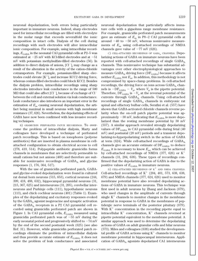
With the use of gramicidin perforated patch, GABA-and glycine-evoked depolarization were found in culturedrat dorsal horn neurons (515, 653), cortical neurons (244,399, 418, 488, 632), hippocampal pyramidal neurons (31,215, 367, 625) and interneurons (31, 205), cerebellar inter-neurons and Purkinje cells (111), hypothalamic neurons(113), and chick cochlear neurons (401) (Table 1). Exam-ples of the depolarizing and excitatory responses evokedby the GABAA agonist isoguvacine and synaptic activationof the GABAA receptors in a P2 CA3 pyramidal cell re-corded using gramicidin perforated patch are shown inFigure 1. In CA3 pyramidal cells, EGABA measured usinggramicidin perforated patch was of �55 mV during theearly neonatal period and progressively shifted to �74 mVby the end of the second postnatal week (625) (see alsoRef. 31). However, while gramicidin perforated patch re-cordings eliminate the problem of intracellular dialysisand thus provide accurate estimate of EGABA, it does notsolve the problem of leak conductance and associated
neuronal depolarization that particularly affects imma-ture cells with a gigaohms range membrane resistance.For example, gramicidin perforated patch measurementsgave an estimate of Em in P0–2 CA3 pyramidal cells ataround �40 to �50 mV, whereas noninvasive measure-ments of Em using cell-attached recordings of NMDAchannels gave value of �77 mV (626).
C) CELL-ATTACHED RECORDINGS OF GABAA CHANNELS. Depo-larizing actions of GABA on immature neurons have beenreported with cell-attached recordings of single GABAA
channels. This noninvasive technique has substantial ad-vantages over other electrophysiological approaches tomeasure GABAA driving force (DFGABA) because it affectsneither EGABA nor Em. In addition, this methodology is notcompromised by space-clamp problems. In cell-attachedrecordings, the driving force on ions across GABAA chan-nels is �DFGABA � Vp, where Vp is the pipette potential.Therefore, DFGABA is �Vp at the reversal potential of thecurrents through GABAA channels. Using cell-attachedrecordings of single GABAA channels in embryonic ratspinal and olfactory bulbar cells, Serafini et al. (557) haveshown that GABA-activated chloride channels reverse po-larity when the on-cell patch pipette potential was ap-proximately �30 mV, indicating that EGABA is more depo-larized than the resting membrane potential by 30 mV(557). A similar approach revealed strongly depolarizingvalues of DFGABA in CA3 pyramidal cells during fetal (40mV) and postnatal (20 mV) periods and a transient depo-larizing-to-hyperpolarizing switch in the action of GABAat term (624). While cell-attached recordings of GABAA
channels give an accurate estimate of DFGABA, to deduceEGABA it is necessary to know Em, which can be achievedby cell-attached recordings of NMDA (377, 626) or K�
channels (31, 204, 638). These types of recordings con-firmed that the depolarizing action of GABA is due to thepositive values of EGABA in immature neurons.
D) CELL-ATTACHED RECORDINGS OF K�
AND NMDA CHANNELS.Cell-attached recordings of K� (204, 401, 573, 638, 638,679) and NMDA channels (377, 624, 626) used to monitormembrane potential have also revealed depolarizing ac-tions of GABA in immature neurons. This technique wasfirst used in adult neurons by Zhang and Jackson (679),who used changes in the amplitude of currents throughsingle K� channels to monitor changes in the membranepotential in response to GABA in the membranes of pep-tidergic nerve terminals of the posterior pituitary (679).With K� concentration in the recording pipette equal tointracellular K� concentration, K� channels reversed atpipette potential equivalent to the membrane potential. Asimilar approach was used to determine the depolarizingactions of GABA on adult granular cells and hilar neurons(573). Miles and colleagues (638) studied the developmen-tal profile of GABA actions using K� channels to monitormembrane potential in hippocampal interneurons. Appli-cation of GABAA agonists depolarized CA1 interneurons
EXCITATORY GABA IN DEVELOPMENT 1219
Physiol Rev • VOL 87 • OCTOBER 2007 • www.prv.org
on October 12, 2007
physrev.physiology.orgD
ownloaded from

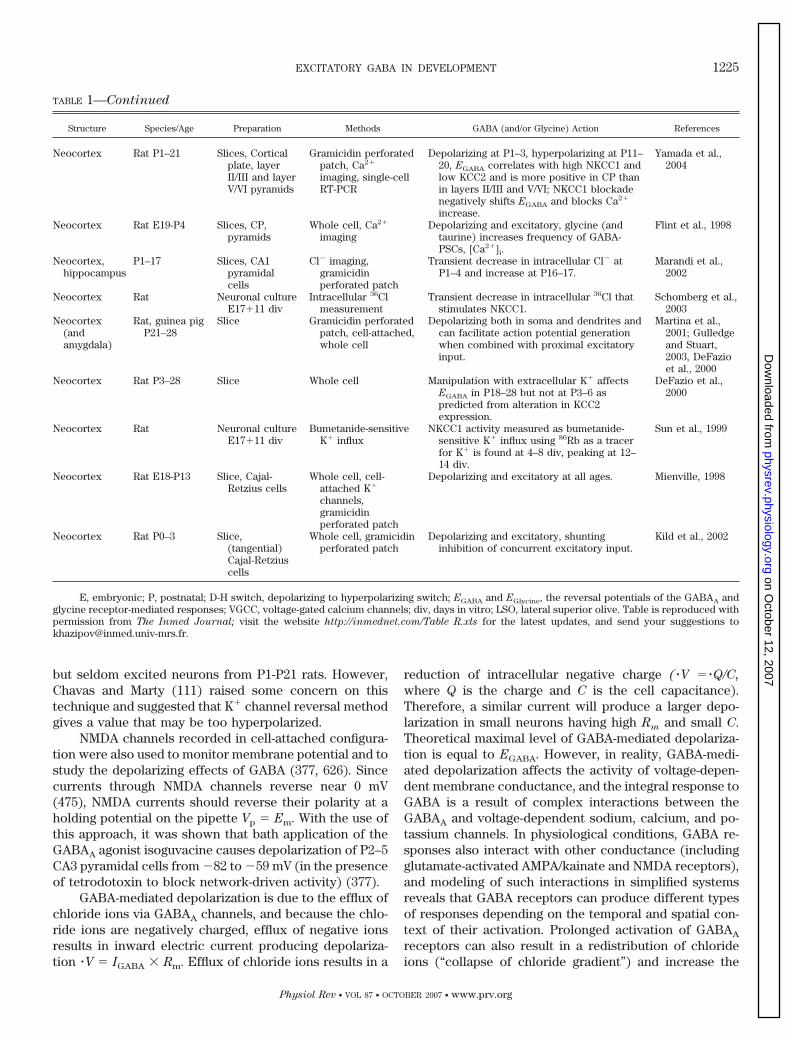
TABLE 1. Depolarizing and excitatory effects of GABA and glycine in developing brain structures
Structure Species/Age Preparation Methods GABA (and/or Glycine) Action References
Retina Chick E3 Retina Ca2� imaging,intracellularrecordings
GABA and muscimol depolarize and increaseintracellular Ca2� via activation of VGCCs.
Yamashita andFukuda, 1993
Retina Ferret P0–25 Intact retina,retinal slices
Ca2� imaging, wholecell
Both GABA and glycine increase Ca2� inretinal ganglion cells at P0–10 anddecrease Ca2� after P15; GABAA
antagonists suppress spontaneous burstingactivity of ganglion cells at P0–10; diverseeffects at P15 and increase in burstfrequency at �P21. ON and OFF cellsbursting difference emerges as GABAbecomes inhibitory.
Fischer et al.,1998
Retina Turtle S22-PH3 Retina Ca2� imaging GABA is excitatory at S25 and switches toinhibitory around hatching, coincidingwith a transformation of retinal waves tostationary patches.
Sernagor et al.,2003
Retina Rabbit E29-P26 Retina starburstcells
Ca2� imaging Muscimol increases Ca2� in starburst cells atE29 and has no detectable effect at P5,concomitant with an emergence of stronginhibitory GABA action on waves.
Zheng et al., 2004
Spinal cord Chick E6–10 Spinal cordexplants
Intracellularrecordings
Deplarizing and excitatory at E6–8 andhyperpolarizing at E10.
Obata et al., 1978
Spinal cord Chick E11–16 Isolated spinalcord
Intracellular recording Hyperpolarization and inhibition at E11–16. Velumian 1984
Spinal cord Chick E10–11 Isolated spinalcord
MEQ intracellular Cl�
measurementsDepolarizing; Cl� reduces during network
burst and recovers partly via NKCC1.Chub et al., 2006
Spinal cord Rat E14–18 Dissociated cells Voltage-sensitive dye No detectable response before E14;depolarizing at E14–18.
Mandler et al.,1990
Spinal cord Rat E16–P2 Hemisectedspinal cordpreparation,motoneurons
Intracellularrecordings
Depolarization at E16–21 and almostisoelectric at P1–2; glycine and GABAA
receptor antagonists block response todorsal root stimulation at E16–18 andincreases it at E19-P2.
Wu et al., 1992
Spinal cord Rat E15–16�3–28 div
Neuronalculture, dorsalhorn
Gramicidin perforatedpatch, whole cell,Ca2� imaging
GABA and glycine increase intracellularCa2� via activation of VGCCs and causeneuronal depolarization during the firstweek in culture; number of respondingcells decreases with age, and none of cellsresponds at �30 div.
Wang et al., 1994;Reichling et al.,1994
Spinal cordandolfactorybulb
Rat E15 Dissociated cells Cell-attached rec. ofGABAA channels
Depolarization by 30 mV; GABAA channelopenings occasionally trigger actionpotentials.
Serafini et al.,1995
Spinal cord Xenopus laevis
larvae 3–8days old
Spinal cord invivo; primarysensoryRohon-Beardneurons anddorsolateralinterneurons
Intracellular, wholecell, amphotericin Band gramicidinperforated patchrecordings
Depolarizing, action of GABAA via GABAA
receptors on Rohon-Beard neurons andhyperpolarizing effects in dorsolateralinterneurons; bumetanide negatively shiftsEGABA in Rohon-Beard neurons.
Rohrbough andSpitzer, 1996
Spinal cord Rat E13–18 Spinal cordpreparation
Ventral root potentials Depolarizing and excitatory at E13–15;GABA and glycine antagonists suppressnetwork bursts at E14–5.
Nishimaru et al.,1996
Spinal cord Rat E15–19 Slices, lumbarmotoneurons
Perforated patchclamp; Ca2�
imaging
Depolarization; entry of Ca2� via Ca2�
channels.Kulik et al., 2000
Spinal cord Mouse KCC2knockoutE18.5
Slice, ventralpart,motoneurons
Perforated patchclamp, fieldpotentials
KCC2�/� mice die after birth due to motordeficits that also abolish respiration.GABA and glycine are more depolarizingin KCC2�/� mice than in wild type.
Hubner et al.,2001
Spinal cord Rat P0–5 Brain stem-spinal cordpreparation
Intracellular recordingfrom L2–5motoneurons;extracellular rootrecordings
Isoelectric inhibitory in motoneurons;depolarizing and excitatory in primaryafferents.
Vinay and Clarac1999; Fellippa-Marques et al.,2000
Spinal cord Rat P0–7 Slices, dorsalhorn (mainlyL11 neurons)
Gramicidin perforatedpatch
Depolarizing in 40% of neurons at P0–2;hyperpolarizing in all cells by P6–7.
Baccei &Fitzgerald,2004
1220 BEN-ARI, GAIARSA, TYZIO, AND KHAZIPOV
Physiol Rev • VOL 87 • OCTOBER 2007 • www.prv.org
on October 12, 2007
physrev.physiology.orgD
ownloaded from
TABLE 1—Continued
Structure Species/Age Preparation Methods GABA (and/or Glycine) Action References
Spinal cord Rat P0–60 Slices, dorsalhorn L1neurons
Gramicidin perforatedpatch, whole cell,Ca2� imaging
Depolarizing during the first week, D-Hswitch is complete by P7; GABA causesCa2� increase until P21 coincides withbiphasic H-D response to GABA; chlorideextrusion does not reach maturity byP10–11.
Cordero-Erausquinet al., 2005
Brain stem Chick E17-P10 Slices, cochlearmucleus
Cell-attachedpotassium channels,gramicidinperforated patch
Depolarizing, dual excitatory and inhibitorydepending on context of their activation.
Lu and Trussel,2001,Monsivais andRubel, 2001
Brain stem Rat E18-P17 Slices, LSOneurons
Intracellular recording Depolarizing at E18-P4 and hyperpolarizingafter P8.
Kandler andFriauf, 1995
Brain stem Rat P2–13 Slices, LSOneurons
Gramicidin perforatedpatch
Depolarizing in 59%, hyperpolarizing in 5%,and biphasic in 34% of cells; EGlycine shiftspositively during the response to glycine,independently of HCO3.
Backus et al.,1998
Brain stem Rat P2–11 Slices, LSOneurons
Gramicidin perforatedpatch
Depolarizing and excitatory (30% of cells) atP1–4 and hyperpolarizing at P9–11; D-Hswitch around P5–8.
Ehrlich et al.,1999
Brain stem Rat, miceP3–12
Slices, LSOneurons
Gramicidin perforatedpatch, molecularbiology
Depolarizing during the first week, D-Hswitch occurs at P8 in wild-type but not inKCC2 KO; KCC2 is present early but is notactive; NKCC1 mRNA not detected duringthe early D phase.
Balakrishnanet al., 2003
Brain stem Rat, miceP1–15
Slices, LSOneurons
Ca2� imaging Both GABA and glycine (exogenous andevoked by MNTB stimulation) increaseCa2� during the first postnatal week;slightly decrease Ca2� in 2-wk-old animals.
Kullman et al.,2002
Brain stem Rat, E18-P10 Slices, LSO,MSO, MSN,MTNB
Voltage-sensitivefluorescent dye,gramicidinperforated patch
D-H switch was determined in four superiorolivary complex nuclei: MSO P5–9, SPNE18-P1, no D-H switch in MTBN neurons,LSO P4–5 but delayed in low-frequencyregions; mere expression of KCC2 did notcorrelate with depolarizing GABA.
Lohrke et al.,2005
Brain stem Rat P0–18 Slices,hypoglossalmotoneurons
Gramicidin perforatedpatch
Depolarizing in P0–3 and hyperpolarizing inP10–18 motorneurons.
Singer et al., 1998
Brain stem Mouse P0–15 Slices, Pre-Botzingercomplexneurons
Gramicidin perforatedpatch from PBCneurons, fieldpotentials from XIIrootlets
Depolarizing at P0–2, hyperpolarizing at P4(D-H switch at around P3), depolarizingaction persists in bicarbonate-free saline.Bicuculline does not affect the frequencyof rhythmic XII motor output at P0–3 andincreases it after P3.
Ritter and Zhang,2000
Basal ganglia Rat P4–26 Slices,substantianigra parsreticulata
Gramicidin perforatedpatch
Depolarizing in neonatal cells; D-H switchoccurred in males at around P17 and infemales at around P10.
Kyrosis et al.,2006
Hypothalamus Rat Neuronal cultureE15–18 � 2–25 div
Ca2� imaging GABA elevates, and GABAA antagonistdepresses intracellular Ca2� via VGCCs inthe immature neurons; both effects onintracellular Ca2� switch between 8–13div.
Obrietan and vanden Pol, 1995
Hypothalamus Rat Neuronal cultureE15 �1–7 div
Ca2� imaging GABA increases and bicuculline reducesCa2� levels in the growth cones.
Obrietan and vanden Pol, 1996
Hypothalamus Rat Neuronal culture Gramicidin perforatedpatch clamp
GABA depolarizes and often excites neuronsin young cultures (1–7 div) andhyperpolarizes and inhibits at 20–33 div.EGABA shifts from �40 to �70 mV. EGABA
is more negative in neurons dissociatedfrom P5 compared the E15. EndogenousGABA excites as bicuculline reducesongoing firing. GABA-evokeddepolarization could facilitate or shuntother depolarizing input depending ontemporal relationship between GABA-evoked depolarization and other excitatoryevents.
Chen et al., 1996
EXCITATORY GABA IN DEVELOPMENT 1221
Physiol Rev • VOL 87 • OCTOBER 2007 • www.prv.org
on October 12, 2007
physrev.physiology.orgD
ownloaded from
TABLE 1—Continued
Structure Species/Age Preparation Methods GABA (and/or Glycine) Action References
Hypothalamus Rat Neuronal cultureE15–18�2–5div
Gramicidin perforatedpatch, whole cell
Depolarizing; shunts glutamate-mediateddepolarization at the peak and facilitatesexcitatory action of glutamate during thedecay phase of GABA response.
Chen et al., 1998
Hypothalamus(and spinalcord)
Mice Slice, P1–10neuronalcultures E15–18�4–�20 div
Gramicidin perforatedpatch, extracellular
Depolarizing and excitatory in the immatureneurons (slices: P1–4; cultures: 4–7 div)and inhibitory in mature (slices: P8–10;cultures: �20 div). GABAA antagonistdecreases firing in immature neurons andincreases in mature neurons. GABAA
excitatory drive is more important thanglutamatergic in the immature neurons.
Gao et al., 2001
Hypothalamus Mice Slice andneuronalculture
Gramicidin perforatedpatch, whole cell
Depolarizing and excitatory at P2–9(exogenous and synaptic GABA) and 2–23div cultures; patterns of GABA-dependentaction potentials (single or multiplespikes) depend on intrinsic membraneproperties of hypothalamic neurons.
Wang et al., 2001
Inferiorcolliculus
Gerbil Slices Ca2� imaging,intracellular
Ca2� increase, biphasic hyperpolarizing-depolarizing response.
Lo et al., 1998
Cerebellum Rat P3�5�4–30 div
Explant culture,granule cells
Ca2� imaging Ca2� increase that outlasts the exposure toGABA by several minutes.
Connor et al.,1987
Cerebellum Rat P2–22 Slice, Purkinjeneurons
Ca2� imaging,gramicidinperforated patch
Depolarizes and increases Ca2� during thefirst postnatal week; D-H switch and lossof Ca2� increase to GABA occurs at theend of the 1st postnatal week.
Eilers et al., 2001
Cerebellum Rat P4–28 Slice, granulecells
Gramicidin perforatedpatch, whole cell
Depolarizing and occasionally excitatory atP7; isoelectric and shunting at P18–21.
Brickley et al.,1996
Hippocampus Kitten P1–18 In vivo,responses tofornixstimulation
Intracellularrecordings
Inhibitory (hyperpolarizing) IPSPs areevoked in hippocampal neurons by fornixstimulation at P1–3; spontaneous IPSPsare similar in duration, EPSPs are notprominent in young kittens.
Purpura et al.,1968
Hippocampus Kitten P2–28 Slice, CA1neurons
Intracellularrecordings
IPSPs and EPSPs are seen at all ages. IPSPsblock cell discharge even in the youngestpreparations.
Schwartzkroinand Altschuler,1977
Hippocampus Rabbit P6-1mo
Slice, CA1 pyr.cells
Intracellularrecordings
Depolarizing at P6–10 in soma and dendrites;depolarizing in dendrites andhyperpolarizing in soma in adult.
Mueller et al.,1993, 1984;Janigro andSchwartzkroin,1988
Hippocampus Rat P1–64 Slice, CA1 Field Potential Paired-pulse simulation reveals facilitation atP1–5 and inhibition at P6–64;“spontaneous unison firing” pattern(GDPs?) blossoms at P4–5 and disappearsat P6.
Harris andTeyler, 1983
Hippocampus Rat P0–P18 Slice, CA3 pyr.cells
Intracellularrecordings
Depolarizing and excitatory at P2–5;hyperpolarizing and inhibitory from P6onwards. Giant depolarizing potentials(GDPs) are generated by excitatory GABA.Blockade of GABAA receptors suppressesGDPs until P5 and induces epileptoformactivity from P6 onwards. Glycine actiondisplays similar D-H developmental profile.
Ben-Ari et al.,1989;Cherubiniet al., 1990; Itoand Cherubini1991; Chesnutand Swann1989
Hippocampus Rat P10–P15 Slice Field Potential,intracellularrecordings
Muscimol produces epileptiform activity anddisinhibition at P10–15.
Chesnut andSwann, 1989
Hippocampus Rat P3-adult Slice, CA3 andCA1
Intracellular and fieldpotential recording
Hyperpolarizing IPSPs are first observed byP5–6 in CA3 and by P9 in CA1 neurons. InCA3, bicuculline causes epileptiformevents in slices from immature but notmonth-old rats. Equilibrium potential ofIPSPs in CA3 neurons was similar whenmade during the first postnatal week andat 1 mo of age.
Swann et al.,1989
Hippocampus Rat E17–21and P5–7
Dissociated cells Fluorescence-activated cell sorter
Depolarizing at E17–21; depolarizing andhyperpolarizing in cell subpopulations atP5–7.
Fiszman et al.,1990
1222 BEN-ARI, GAIARSA, TYZIO, AND KHAZIPOV
Physiol Rev • VOL 87 • OCTOBER 2007 • www.prv.org
on October 12, 2007
physrev.physiology.orgD
ownloaded from
TABLE 1—Continued
Structure Species/Age Preparation Methods GABA (and/or Glycine) Action References
Hippocampus Rat P2–20 Slice, CA1pyramidalcells
Whole cell recordings Isoelectric response at P2–5; EGABA at P2–5is predicted by GHK, hyperpolarizing andmore negative than GHK value at P8–20.
Zhang et al., 1991
Hippocampus Rat P2–5 Slice, CA3 pyr.cells
Cell-attached actionpotentials; cell-attached NMDAchannels; Ca2�
imaging
Depolarizing and excitatory; decrease inMg2� blockade of NMDA receptors viadepolarization; influx of Ca 2� via VGCCsand NMDA channels. Synergistic action ofGABAA and NMDA receptors is involved inCa2� oscillations associated with theGDPs.
Leinekuge et al.,1995; 1997
Hippocampus Rat E17�5–8div
Neuronal culture MQAE Cl�
measurements indendrites and soma
Pericarionic [Cl�]i is lower than dendritic[Cl�]i; furosemide and bumetanidestronger decrease in [Cl�]i in dendritesthan in soma.
Hara et al., 1992
Hippocampus Rat P0–16 Slice, CA1 Ca2� imaging, wholecell
Early network oscillations (ENOs �equivalent of GDPs) in CA1 are blocked bybicuculline; muscimol increasesintracellular Ca2� in all cells at P1–12involved in ENOs. Ca2� increase graduallydecreases over the first 2 postnatal weeks.
Garaschuk et al.,1998
Hippocampus Rat P0–2 Slice, CA3 andCA1 pyramidalcells
Field potentials,gramicidinperforated patch,whole cell
Depolarizing and excitatory at cellular levelbut inhibitory at network level; Muscimolblocks GDPs; bicuculline induces interictalactivity.
Lamsa et al.,2000
Hippocampus Rat P2–5 Slice, CA3interneurons
Cell-attached actionpotentials
Depolarizing and excitatory; synergisticaction of GABA and glutamate (AMPAand NMDA) receptors in interneuronexcitation during GDPs; blockade ofGABAA receptors induces epileptiformactivity from P2.
Khazipov et al.,1997
Hippocampus Rat (Wistar)P0–5
Slice, intacthippocampusin vitro, CA3pyramidalcells
Cell-attached; wholecell
Excitatory at P2–5 and inhibitory at P10–15(synaptic-GABA on single unit); GABAA
agonist and diazepam transiently increaseGDPs frequency; GABAA antagonist blocksGDPs and induces interictal events fromP0 and ictal events from P2.
Khalilov et al.,1999
Hippocampus Rat P1–34(Sprague-Dawley)
Slice, CA3pyramidalcells
Field potentials,whole cell, cell-attached
Excitatory from P1 to P10, excitation-to-inhibition switch from P10 to P14;inhibitory after P15 (GABAA agonist teston MUA); dual role in seizure control.
Khazipov et al.,2004
Hippocampus Rat P1–30 Slice, CA3pyramidalcells
Field potential Bicuculline action switches from depressingto increasing CA3 MUA at P12 (3.5 mMexternal K�) and at P23 (8.5 mM externalK�). Ictal events caused by 8.5 mM K�
are blocked by gabazine and bicucullineand are enhanced by isoguvacine andmuscimol.
Dzhala et al.,2003
Hippocampus Rat Slice, CA3pyramidalcells
Field potential, wholecell
Depolarizing and excitatory, NKCC1blockade shifts EGABA negatively andalleviates high-potassium seizures in vitroand kainate seizures in vivo.
Dzhala et al.,2005
Hippocampus(andneocortex)
Mouse P0–7,rat P1–5
Slice Field potentials Anticonvulsive; starting from P2–3, GABAA
blockade induces paroxysmal activity bothin the hippocampus and neocortex.
Wells et al., 2000
Hippocampus Rat P1–21 Slice, CA1interneurons
Cell-attachedpotassium channels
Depolarizing response to GABAA agonist atP1–4 and at P7–21 to similar level (�55mV).
Verhuegen et al.,1999
Hippocampus Mice P3–32 Neuronalculture; slice,CA1 pyr. cells
Whole-cell, localGABA photolysis
Chloride extrusion progressively developsduring the two first postnatal weeks.
Khirug et al.,
Hippocampus Rat E18�div4–18
Neuronal culture Ca2� imaging,gramicidinperforated patch
Induces influx of Ca2� via voltage-gatedCa2� channels and depolarizes during 2wk after plating; D-H switch correlateswith KCC2 expression and is promotedby GABA.
Ganguly er et al.,2001
Hippocampus Rat P1–9 In vivo P7–9 andslices P1–8
Field potentials,gramicidinperforated patch
Depolarizing, blockade of NKCC1 rendersGABA hyperpolarizing and blocks sharpwaves in vivo and GDPs in vitro.
Sipila et al., 2006
EXCITATORY GABA IN DEVELOPMENT 1223
Physiol Rev • VOL 87 • OCTOBER 2007 • www.prv.org
on October 12, 2007
physrev.physiology.orgD
ownloaded from
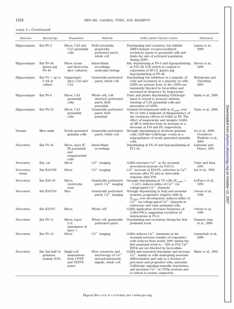
TABLE 1—Continued
Structure Species/Age Preparation Methods GABA (and/or Glycine) Action References
Hippocampus Rat P0–2 Slices, CA3 andCA1 pyramidalcells
Field potentials,gramicidinperforated patch,whole cell
Depolarizing and excitatory but inhibitsAMPA-kainate receptor-mediatedexcitatory inputs to pyramidal cells andlimits the size of activated populationduring GDPs.
Lamsa et al.,2000
Hippocampus Rat P0–30,guinea pigP0–40
Slices (acuteand short-termslice cultures)
Intracellularrecordings,molecular biology
Rat: depolarizing at P0–4 and hyperpolarizingat P12–30; D-H switch is coupled toexpression of KCC2; guniea pig:hyperpolarizing at P0–40.
Rivera et al.,1999
Hippocampus Rat P4 � up to6 wk inculture
Organotypicslice; CA3 andCA1
Gramicidin perforatedpatch, whole cell
Depolarizing but inhibitory in a majority ofcells and excitatory in a minority of cells;GDPs are present from 12 div; GDPs aretransiently blocked by bicuculline andincreased in frequency by isoguvacine.
Mohajerani andCherubini,2005
Hippocampus Rat P0–6 Slices, CA3pyramidalcells
Whole cell, cell-attached, perforatedpatch, fieldpotentials
Tonic and phasic depolarizing GABAergicinput is crucial to promote intrinsicbursting of CA3 pyramidal cells andgeneration of GDPs.
Sipila et al., 2005
Hippocampus Rat P0–15 Slices, CA3pyramidalcells
Gramicidin perforatedpatch, fieldpotential
Gradual developmental shift in EGABA overP0–15 with a midpoint of disappearance ofthe excitatory effects of GABA at P8. Theeffect of isoguvacine and synaptic GABAon MUA switches from an increase to adecrease at P10 and P8, respectively.
Tyzio et al., 2006
Dentate Mice adult Newly generatedgranular cells
Gramicidin perforatedpatch, whole cell
Strongly depolarizing in newborn granularcells; GDP-like GABAergic events in asubpopulation of newly generated granularcells.
Ge et al., 2006;Overstreet-Wadiche et al.,2006
Neocortex Rat P4–41 Slices, layer II/III pyramidalandnonpyramidalcells
Intracellularrecordings
Depolarizing at P4–10 and hyperpolarizing atP11–41.
Luhmann andPrince, 1991
Neocortex Rat, cat Slices Ca2� imaging GABA increases Ca2� in the neonatalneocortical neurons via VGCCs.
Yuste and Katz,1991
Neocortex,visual
Rat E18-P30 Slices Ca2� imaging Ca2� increase at E18-P5, reduction in Ca2�
increase after P5 and no detectableresponse after P20.
Lin et al., 1994
Neocortex Rat E16–18 Slices,ventricularzone
Gramicidin perforatedpatch; Ca2� imaging
Strongly depolarizing in VZ cells (EGABA ��5 mV); induces influx of Ca2� viavoltage-gated Ca2� channels.
LoTurco et al.,1995
Neocortex Rat E16-P16 Slice Gramicidin perforatedpatch, Ca2� imaging
Strongly depolarizing in fetal and neonatalneurons; progressive negative shift inEGABA over development; induces influx ofCa2� via voltage-gated Ca2� channels inembryonic and early postnatal cells.
Owens et al.,1996
Neocortex Rat E19-P5 Slices Whole cell GABA application increases frequency ofGABA-PSCs, suggesting excitation ofinterneurons at P3–4.
Owens et al.,1999
Neocortex Rat P0–11 Slices, Layer2–6,stimulation oflayer 1
Whole cell, gramicidinperforated patch
Depolarizing and excitatory during the firstpostnatal week.
Dammer manet al., 2000
Neocortex Rat P0–12 Slices Ca2� imaging GABA induces Ca2� transients in theneonatal neurons; number of responsivecells reduces from nearly 100% during thefirst postnatal week to �20% at P12; Ca2�
ENOs are not blocked by bicuculline.
Garaschuk et al.,2000
Neocortex Rat 2nd half ofgestation(mainly E19)
Single-cellsuspensionsfrom CP/SPand VZ/SVZzones
Flow cytometry andmicroscopy of Ca2�
and potentiometricsignals, whole cell
GABA and muscimol depolarize and increaseCa2� mainly in cells undergoing neuronaldifferentiation and only in a fraction ofprecursor and progenitor cells; autocrineGABAergic signaling tonically depolarizesand increases Ca2� in CP/Sp neurons andis critical to neurite outgrowth.
Marie et al., 2001
1224 BEN-ARI, GAIARSA, TYZIO, AND KHAZIPOV
Physiol Rev • VOL 87 • OCTOBER 2007 • www.prv.org
on October 12, 2007
physrev.physiology.orgD
ownloaded from
but seldom excited neurons from P1-P21 rats. However,Chavas and Marty (111) raised some concern on thistechnique and suggested that K� channel reversal methodgives a value that may be too hyperpolarized.
NMDA channels recorded in cell-attached configura-tion were also used to monitor membrane potential and tostudy the depolarizing effects of GABA (377, 626). Sincecurrents through NMDA channels reverse near 0 mV(475), NMDA currents should reverse their polarity at aholding potential on the pipette Vp � Em. With the use ofthis approach, it was shown that bath application of theGABAA agonist isoguvacine causes depolarization of P2–5CA3 pyramidal cells from �82 to �59 mV (in the presenceof tetrodotoxin to block network-driven activity) (377).
GABA-mediated depolarization is due to the efflux ofchloride ions via GABAA channels, and because the chlo-ride ions are negatively charged, efflux of negative ionsresults in inward electric current producing depolariza-tion �V � IGABA � Rm. Efflux of chloride ions results in a
reduction of intracellular negative charge ( �V � �Q/C,
where Q is the charge and C is the cell capacitance).Therefore, a similar current will produce a larger depo-larization in small neurons having high Rm and small C.Theoretical maximal level of GABA-mediated depolariza-tion is equal to EGABA. However, in reality, GABA-medi-ated depolarization affects the activity of voltage-depen-dent membrane conductance, and the integral response toGABA is a result of complex interactions between theGABAA and voltage-dependent sodium, calcium, and po-tassium channels. In physiological conditions, GABA re-sponses also interact with other conductance (includingglutamate-activated AMPA/kainate and NMDA receptors),and modeling of such interactions in simplified systemsreveals that GABA receptors can produce different typesof responses depending on the temporal and spatial con-text of their activation. Prolonged activation of GABAA
receptors can also result in a redistribution of chlorideions (“collapse of chloride gradient”) and increase the
TABLE 1—Continued
Structure Species/Age Preparation Methods GABA (and/or Glycine) Action References
Neocortex Rat P1–21 Slices, Corticalplate, layerII/III and layerV/VI pyramids
Gramicidin perforatedpatch, Ca2�
imaging, single-cellRT-PCR
Depolarizing at P1–3, hyperpolarizing at P11–20, EGABA correlates with high NKCC1 andlow KCC2 and is more positive in CP thanin layers II/III and V/VI; NKCC1 blockadenegatively shifts EGABA and blocks Ca2�
increase.
Yamada et al.,2004
Neocortex Rat E19-P4 Slices, CP,pyramids
Whole cell, Ca2�
imagingDepolarizing and excitatory, glycine (and
taurine) increases frequency of GABA-PSCs, [Ca2�]i.
Flint et al., 1998
Neocortex,hippocampus
P1–17 Slices, CA1pyramidalcells
Cl� imaging,gramicidinperforated patch
Transient decrease in intracellular Cl� atP1–4 and increase at P16–17.
Marandi et al.,2002
Neocortex Rat Neuronal cultureE17�11 div
Intracellular 36Clmeasurement
Transient decrease in intracellular 36Cl thatstimulates NKCC1.
Schomberg et al.,2003
Neocortex(andamygdala)
Rat, guinea pigP21–28
Slice Gramicidin perforatedpatch, cell-attached,whole cell
Depolarizing both in soma and dendrites andcan facilitate action potential generationwhen combined with proximal excitatoryinput.
Martina et al.,2001; Gulledgeand Stuart,2003, DeFazioet al., 2000
Neocortex Rat P3–28 Slice Whole cell Manipulation with extracellular K� affectsEGABA in P18–28 but not at P3–6 aspredicted from alteration in KCC2expression.
DeFazio et al.,2000
Neocortex Rat Neuronal cultureE17�11 div
Bumetanide-sensitiveK� influx
NKCC1 activity measured as bumetanide-sensitive K� influx using 86Rb as a tracerfor K� is found at 4–8 div, peaking at 12–14 div.
Sun et al., 1999
Neocortex Rat E18-P13 Slice, Cajal-Retzius cells
Whole cell, cell-attached K�
channels,gramicidinperforated patch
Depolarizing and excitatory at all ages. Mienville, 1998
Neocortex Rat P0–3 Slice,(tangential)Cajal-Retziuscells
Whole cell, gramicidinperforated patch
Depolarizing and excitatory, shuntinginhibition of concurrent excitatory input.
Kild et al., 2002
E, embryonic; P, postnatal; D-H switch, depolarizing to hyperpolarizing switch; EGABA and EGlycine, the reversal potentials of the GABAA andglycine receptor-mediated responses; VGCC, voltage-gated calcium channels; div, days in vitro; LSO, lateral superior olive. Table is reproduced withpermission from The Inmed Journal; visit the website http://inmednet.com/Table R.xls for the latest updates, and send your suggestions [email protected].
EXCITATORY GABA IN DEVELOPMENT 1225
Physiol Rev • VOL 87 • OCTOBER 2007 • www.prv.org
on October 12, 2007
physrev.physiology.orgD
ownloaded from
relative contribution of bicarbonate permeability ofGABAA channels producing a delayed depolarization(587). This mechanism does not, however, operate inimmature cells because of delayed expression of intracel-lular carbonic anhydrase (520, 533).
2. GABA triggers sodium action potentials
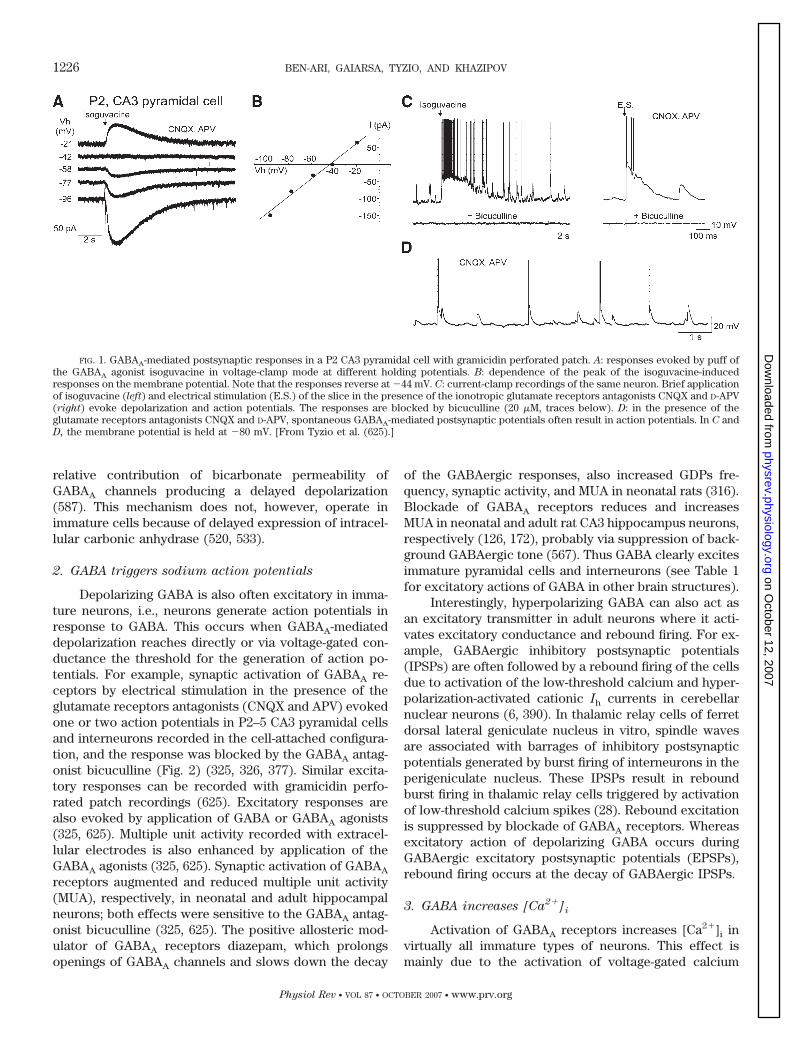
Depolarizing GABA is also often excitatory in imma-ture neurons, i.e., neurons generate action potentials inresponse to GABA. This occurs when GABAA-mediateddepolarization reaches directly or via voltage-gated con-ductance the threshold for the generation of action po-tentials. For example, synaptic activation of GABAA re-ceptors by electrical stimulation in the presence of theglutamate receptors antagonists (CNQX and APV) evokedone or two action potentials in P2–5 CA3 pyramidal cellsand interneurons recorded in the cell-attached configura-tion, and the response was blocked by the GABAA antag-onist bicuculline (Fig. 2) (325, 326, 377). Similar excita-tory responses can be recorded with gramicidin perfo-rated patch recordings (625). Excitatory responses arealso evoked by application of GABA or GABAA agonists(325, 625). Multiple unit activity recorded with extracel-lular electrodes is also enhanced by application of theGABAA agonists (325, 625). Synaptic activation of GABAA
receptors augmented and reduced multiple unit activity(MUA), respectively, in neonatal and adult hippocampalneurons; both effects were sensitive to the GABAA antag-onist bicuculline (325, 625). The positive allosteric mod-ulator of GABAA receptors diazepam, which prolongsopenings of GABAA channels and slows down the decay
of the GABAergic responses, also increased GDPs fre-quency, synaptic activity, and MUA in neonatal rats (316).Blockade of GABAA receptors reduces and increasesMUA in neonatal and adult rat CA3 hippocampus neurons,respectively (126, 172), probably via suppression of back-ground GABAergic tone (567). Thus GABA clearly excitesimmature pyramidal cells and interneurons (see Table 1for excitatory actions of GABA in other brain structures).
Interestingly, hyperpolarizing GABA can also act asan excitatory transmitter in adult neurons where it acti-vates excitatory conductance and rebound firing. For ex-ample, GABAergic inhibitory postsynaptic potentials(IPSPs) are often followed by a rebound firing of the cellsdue to activation of the low-threshold calcium and hyper-polarization-activated cationic Ih currents in cerebellarnuclear neurons (6, 390). In thalamic relay cells of ferretdorsal lateral geniculate nucleus in vitro, spindle wavesare associated with barrages of inhibitory postsynapticpotentials generated by burst firing of interneurons in theperigeniculate nucleus. These IPSPs result in reboundburst firing in thalamic relay cells triggered by activationof low-threshold calcium spikes (28). Rebound excitationis suppressed by blockade of GABAA receptors. Whereasexcitatory action of depolarizing GABA occurs duringGABAergic excitatory postsynaptic potentials (EPSPs),rebound firing occurs at the decay of GABAergic IPSPs.
3. GABA increases [Ca2�]i
Activation of GABAA receptors increases [Ca2�]i invirtually all immature types of neurons. This effect ismainly due to the activation of voltage-gated calcium
FIG. 1. GABAA-mediated postsynaptic responses in a P2 CA3 pyramidal cell with gramicidin perforated patch. A: responses evoked by puff ofthe GABAA agonist isoguvacine in voltage-clamp mode at different holding potentials. B: dependence of the peak of the isoguvacine-inducedresponses on the membrane potential. Note that the responses reverse at �44 mV. C: current-clamp recordings of the same neuron. Brief applicationof isoguvacine (left) and electrical stimulation (E.S.) of the slice in the presence of the ionotropic glutamate receptors antagonists CNQX and D-APV(right) evoke depolarization and action potentials. The responses are blocked by bicuculline (20 �M, traces below). D: in the presence of theglutamate receptors antagonists CNQX and D-APV, spontaneous GABAA-mediated postsynaptic potentials often result in action potentials. In C andD, the membrane potential is held at �80 mV. [From Tyzio et al. (625).]
1226 BEN-ARI, GAIARSA, TYZIO, AND KHAZIPOV
Physiol Rev • VOL 87 • OCTOBER 2007 • www.prv.org
on October 12, 2007
physrev.physiology.orgD
ownloaded from
channels as it persists when sodium channels are blockedand is suppressed by calcium channel blockers. Interest-ingly, blockade of GABAA receptors induces a significantdecrease in [Ca2�]i, indicating that tonic release of GABAis sufficient to increase Ca2� in immature neurons (484).
Connor et al. (132) were among the first to use digitalimaging of the Ca2� to study the depolarizing responses ofdeveloping granule cells in culture to GABA. Applicationof GABA induced a transient increase in membrane con-ductance and caused [Ca2�]i increase that outlasted theexposure to GABA by several minutes. Glutamate or kai-nate also elevated [Ca2�]i, but unlike GABA, this [Ca2�]i
response reversed rapidly upon removal of the transmit-ter. In keeping with these results, GABA and glycineincreased [Ca2�]i in a number of embryonic and neonatalneurons including neocortex (223, 385, 399, 675), hip-pocampus (222, 377, 378), spinal cord (359), dorsal hornneurons (515, 653), hypothalamus, olfactory bulb, cortex,medulla, striatum, thalamus, hippocampus, and colliculus(484) (see Table 1).
Synaptically released GABA also increases intracel-lular calcium in immature neurons. Obrietan and van den
Pol (484) have shown that addition of bicuculline tomonosynaptically connected hypothalamic neurons de-creased [Ca2�]i, indicating that hypothalamic neuronswere secreting GABA at an early age of development, andthat sufficient GABA was released to elicit an increase in[Ca2�]i. This effect was seen even after blocking all glu-tamatergic activity with glutamate receptor antagonists(484). Leinekugel et al. (378) have demonstrated thatelectrical stimulation of afferent fibers induces a transientincrease in [Ca2�]i in neonatal pyramidal cells and inter-neurons (P5). This elevation of [Ca2�]i was reversiblyblocked by bicuculline but not by glutamate receptorantagonists. During simultaneous electrophysiological re-cording in current-clamp mode and [Ca2�]i monitoringfrom P5 pyramidal cells, electrical stimulation of afferentfibers, in the presence the glutamate receptors antago-nists, caused synaptic depolarization accompanied by afew action potentials and a transient increase in [Ca2�]i.In voltage-clamp mode, however, there was no increase in[Ca2�]i following synaptic stimulation, showing that it isdepolarization dependent (378).
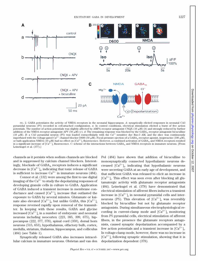
FIG. 2. GABA potentiates the activity of NMDA receptors in the neonatal hippocampus. A: synaptically elicited responses in neonatal CA3pyramidal neurons (P5) recorded in cell-attached configuration. a: In control conditions, electrical stimulation elicited a burst of five actionpotentials. The number of action potentials was slightly affected by AMPA receptor antagonist CNQX (10 �M) (b) and strongly reduced by furtheraddition of the NMDA receptor antagonist APV (50 �M) (c). d: The remaining response was blocked by the GABAA receptor antagonist bicuculline(10 �M). B: a CA3 pyramidal neuron (P5) was loaded extracellularly with the Ca2�-sensitive dye fluo-3 AM, and the slice was continuouslysuperfused with the voltage-gated Ca2� channel blocker D600 (50 �M). Focal pressure ejection of a GABAA-receptor agonist, isoguvacine (100 �M),or bath application NMDA (10 �M) had no effect on [Ca2�]i fluorescence. However, a combined activation of GABAA and NMDA receptors resultedin a significant increase of [Ca2�]i fluorescence. C: scheme of the interactions between GABAA and NMDA receptors in immature neurons. [FromLeinekugel et al. (377).]
EXCITATORY GABA IN DEVELOPMENT 1227
Physiol Rev • VOL 87 • OCTOBER 2007 • www.prv.org
on October 12, 2007
physrev.physiology.orgD
ownloaded from
An additional factor to consider is that voltage de-pendence of calcium conductance is developmentally reg-ulated. Ganguly et al. (215) have shown that mild depo-larization produced by application of 6 mM extracellularpotassium evokes robust increase in [Ca2�]i in P7 neuronsbut no response at P13 in organotypic slices (215). Only8–10 mM [K�]o produced calcium signals in P13 neurons.Whole cell study of the voltage dependence of calciumcurrents revealed a developmental shift in the activationprofile of calcium currents toward more hyperpolarizedpotentials (215). Calcium fluorescence measurements canbe also affected by the developmental changes in theCa2�-buffering properties of neurons. Several develop-mental studies indicate that calcium-binding proteins areprogressively expressed during development in varioustypes of neurons including calretinin (543), parvalbumin(130), and calbindin (79). Recently, Chavas and Marty(111) raised concern on using [Ca2�]i to monitor depolar-izing actions of GABA showing that, in cerebellar inter-neurons, GABAA agonists induce a somatodendritic[Ca2�]i rise that persists at least until postnatal day 20 andis not mediated by depolarization-induced Ca2� entry. Alocal [Ca2�]i elevation could likewise be elicited by repet-itive stimulation of presynaptic GABAergic afferent fibers.Following GABAA receptor activation, bicarbonate-in-duced Cl� entry led to cell depolarization, Cl� accumula-tion, and osmotic tension. The authors proposed that thistension induces the [Ca2�]i rise as part of a regulatoryvolume decrease reaction (110).
4. GABA reduces the voltage-dependent magnesium
block of NMDA channels
The depolarization produced by GABA also attenu-ates the voltage-dependent magnesium block of NMDAchannels (Fig. 2). Using cell-attached recordings of singleNMDA channels from P2–5 CA3 pyramidal cells, Leineku-gel et al. (377) have shown that activation of GABAA
receptors strongly reduces the magnesium block ofNMDA channels by reducing the affinity of magnesiumions to NMDA channels from 16 to 118 �M. This effectwas entirely due to neuronal depolarization from �82to �59 mV. Confocal microscopy with the permanent dyefluo 3-AM revealed that in the presence of the calciumchannels blocker D600, applications of isoguvacine andNMDA increase [Ca2�]i when applied together but notseparately. In the presence of an AMPA receptor antago-nist (CNQX), electrical stimulation evoked on average 3.6action potentials in immature pyramidal cells and inter-neurons (326); adding an NMDA receptor antagonist(APV) further reduced the response to 1.4 action poten-tials, and the remaining spikes were fully blocked bybicuculline (Fig. 2). Therefore, synaptic activation ofGABAA receptors attenuates the magnesium voltage-de-pendent block of NMDA receptors. This “synergistic” in-
teraction between GABAA and NMDA receptors contrib-utes to the generation of the physiological pattern ofGDPs (59, 326, 377) (and see below).
5. GABA interferes with ionotropic
glutamatergic transmission
In developing hypothalamic neurons in culture,GABA acting via GABAA receptors exerts depolarizingactions that will exert different effects on AMPA-medi-ated responses depending on the delay between the acti-vation of GABAA and AMPA receptors (219). GABAergicdepolarization reduced and augmented glutamatergicpostsynaptic responses at short and longer latencies, re-spectively. The reduction is due to the shunting effect ofGABAA-mediated conductance. In contrast, subthresholdglutamatergic responses summated with GABAA-medi-ated depolarization generated spikes if they occurred atthe end of GABAA depolarization, when the shuntingGABAA conductance ceased. These observations suggestthat under certain temporal conditions GABAA andAMPA/kainate receptors may work in synergy to excitethe immature neurons. Similar synergistic excitatory ac-tions of GABA-mediated depolarization and glutamatergicEPSPs may also occur in mature cortical neurons (244).
C. Developmental Changes
in Chloride Homeostasis
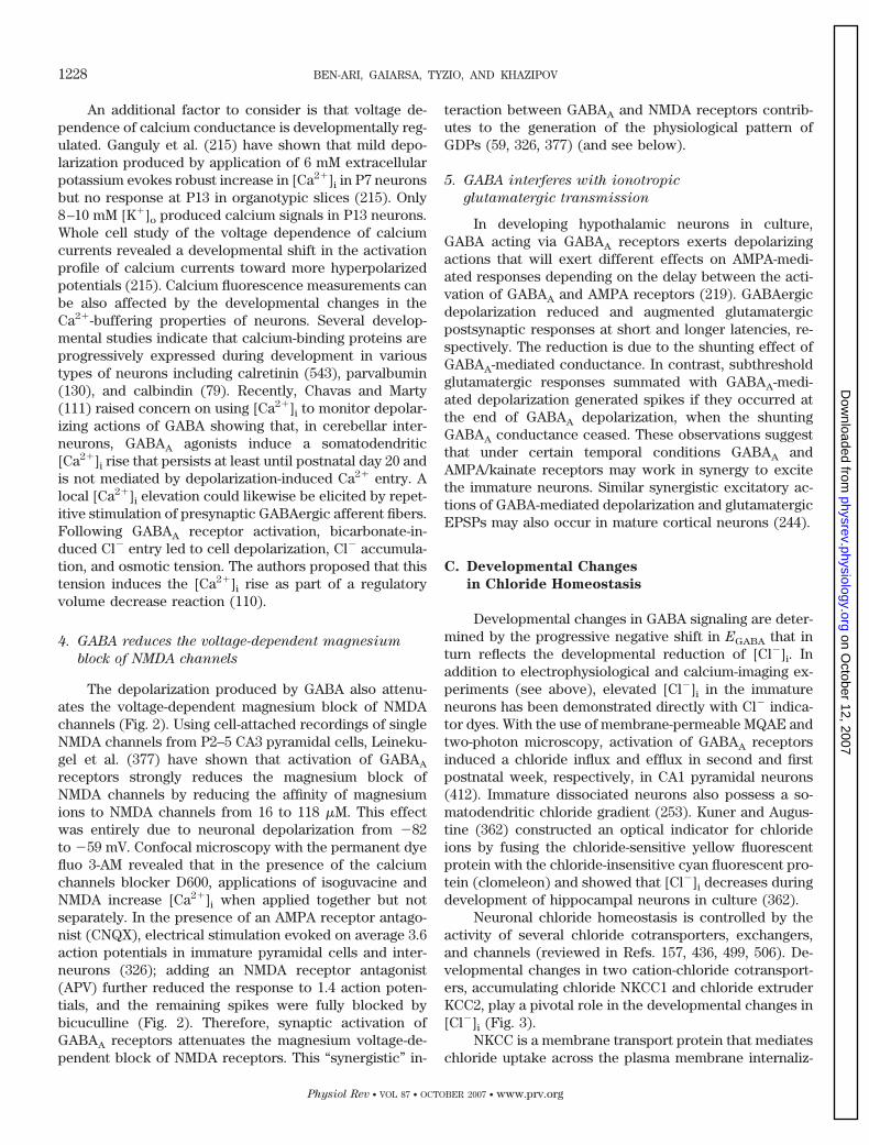
Developmental changes in GABA signaling are deter-mined by the progressive negative shift in EGABA that inturn reflects the developmental reduction of [Cl�]i. Inaddition to electrophysiological and calcium-imaging ex-periments (see above), elevated [Cl�]i in the immatureneurons has been demonstrated directly with Cl� indica-tor dyes. With the use of membrane-permeable MQAE andtwo-photon microscopy, activation of GABAA receptorsinduced a chloride influx and efflux in second and firstpostnatal week, respectively, in CA1 pyramidal neurons(412). Immature dissociated neurons also possess a so-matodendritic chloride gradient (253). Kuner and Augus-tine (362) constructed an optical indicator for chlorideions by fusing the chloride-sensitive yellow fluorescentprotein with the chloride-insensitive cyan fluorescent pro-tein (clomeleon) and showed that [Cl�]i decreases duringdevelopment of hippocampal neurons in culture (362).
Neuronal chloride homeostasis is controlled by theactivity of several chloride cotransporters, exchangers,and channels (reviewed in Refs. 157, 436, 499, 506). De-velopmental changes in two cation-chloride cotransport-ers, accumulating chloride NKCC1 and chloride extruderKCC2, play a pivotal role in the developmental changes in[Cl�]i (Fig. 3).
NKCC is a membrane transport protein that mediateschloride uptake across the plasma membrane internaliz-
1228 BEN-ARI, GAIARSA, TYZIO, AND KHAZIPOV
Physiol Rev • VOL 87 • OCTOBER 2007 • www.prv.org
on October 12, 2007
physrev.physiology.orgD
ownloaded from
ing one Na�, one K�, and two Cl� in electroneutral cou-pled fashion (499). Cloning and functional expressionstudies have identified two major isoforms of NKCC co-transporters that are products of distinct genes. TheNKCC1 isoform is the so-called secretory isoform bestcharacterized in secretory epithelial cells. NKCC1 is ex-pressed in virtually all mammalian cells and is thought toplay a housekeeping role in cell volume homeostasis andthe common control of cytosolic ion content. NKCC1does not use ATP but operates using electrochemicalgradient for Na� and K� produced by Na�-K�-ATPase.There is considerable evidence that uptake of Cl� inimmature neurons is mediated by NKCC1. High expres-sion of NKCC1 in immature neurons plays an importantrole in maintaining high intracellular Cl� (157, 173, 207,383, 440, 499, 506, 528, 596, 651, 671).
KCC2 is the principal transporter for Cl� extrusionfrom neurons. KCC2 extrudes K� and Cl� using the elec-trochemical gradient for K�. Cl� extrusion is weak inimmature neurons and increases with neuronal matura-tion (330, 405, 678). The KCC2 isoform of KCC cotrans-porters is expressed in mature neurons, thus underlyingthe developmental changes in Cl� extrusion (400, 521,562, 651, 671). K�-Cl� cotransport also contributes to thelow [Cl�]i in mature neurons (156, 286, 450, 613–615).Additionally, the KCC1, KCC3, and KCC4 isoforms havebeen also found in the central nervous system but with alimited expression in neurons (499).
Using ribonuclease protection analysis and in situhybridization, Clayton et al. (121) determined the devel-
opmental expression of the members of the cation-Cl�
cotransporter gene family in rat brain (121). Of the in-wardly directed cotransporters, NKCC-1, NKCC-2, andNCC-1, only NKCC-1 was detected at significant levels inbrain. NKCC-1 was expressed in neurons, appearing firstin the cortical plate but not in the ventricular or subven-tricular zone. Expression levels peaked by the third post-natal week and were maintained in adults. Outwardlydirected cotransporters demonstrated a different timecourse of expression: KCC-1 was expressed prenatally atlow levels that increased slightly over the course of de-velopment; KCC-2 expression appeared around birth andincreased dramatically after the first week of postnatallife.
Using single-cell PCR, Rivera et al. (521) showedthat at birth, KCC2 mRNA was barely detectable; asteep increase in the expression was evident at P5,reaching adult level by P15. Interestingly, mature dorsalroot ganglion neurons that have depolarizing GABA(160) did not express KCC2. In contrast, KCC2 mRNAwas present in abundant amounts at embryonic day E42in the hippocampus of guinea pig, a species with earlymaturation. Spatiotemporal expression pattern ofKCC2 mRNA expression follows a caudorostral gradi-ent reflecting functional maturation of various brainareas. Electrophysiological recordings using Cl�-freesharp electrodes revealed strong correlation betweenthe hyperpolarizing GABAA-mediated responses andKCC2 mRNA expression in the pyramidal hippocampalneurons of the rat, guinea pig, and dorsal root ganglion
FIG. 3. Developmental changes in chloride ho-meostasis during development. A: during develop-ment, the intracellular chloride concentration de-creases. In the immature neurons, efflux of the nega-tively charged chloride ions produces inward electriccurrent and depolarization. In the mature neurons,chloride enters the cell and produces outward elec-tric current and hyperpolarization. B: developmentalchange in the intracellular chloride is due to the changesin the expression of the two major chloride cotransport-ers, KCC2 and NKCC1. Chloride extruder KCC2 isexpressed late in development, whereas NKCC1,which accumulates chloride in the cell, is more ex-pressed in the immature neurons.
EXCITATORY GABA IN DEVELOPMENT 1229
Physiol Rev • VOL 87 • OCTOBER 2007 • www.prv.org
on October 12, 2007
physrev.physiology.orgD
ownloaded from
neurons. Antisense oligodeoxynucleotides againstKCC2 mRNA produced a fivefold reduction in KCC2protein in P11–13 rat hippocampal slices associatedwith a strong reduction of DFGABA (from �10.9 to �2.8mV). Similarly, early overexpression of KCC2 in imma-ture cortical neurons, before the upregulation of KCC2,produced a negative shift in GABA reversal potentialand reduced GABA-elicited calcium responses in cul-tured neurons (118, 374, 588). Taken together, theseresults indicated that developmental expression ofKCC2 is pivotal for development of hyperpolarizingGABAA-mediated inhibition.
Using perforated patch-clamp method and single-cell multiplex RT-PCR to measure cation-Cl� cotrans-porter mRNAs in postnatal rat neocortical neurons,Yamada et al. (671) reported that the mRNA expressionlevels of NKCC1 and KCC2 were positively and nega-tively correlated, respectively, with EGABA (671). [Cl�]i
and NKCC1 mRNA were higher in cortical plate (CP)neurons than in the presumably older layer V/VI pyra-midal neurons in a given slice. The pharmacologicaleffects of selective NKCC1 blocker bumetanide onEGABA were consistent with the different expressionlevels of NKCC1 mRNA. The expression of NKCC1 andKCC2 was also analyzed by Western blot and immuno-fluorescence and double labeling in the rat and humancortex (173). In the rat cortex, NKCC1 expressionreached 14-fold higher levels between P3–14 than atP21 and adulthood. In contrast, KCC2 levels were sig-nificantly lower during the first two postnatal weeksthan at P21 and adulthood. These ontogenic findingswere confirmed by immunofluorescence double label-ing using the neuronal marker NeuN and either NKCC1or KCC2 specific antibodies. In human cortex, NKCC1expression was significantly higher at 31– 41 postcon-ceptional weeks than at 1 year and older. During thefirst year of life, NKCC1 expression rapidly decreasedto levels of the adult. Between 31 and 41 postconcep-tional weeks, when NKCC1 levels were peaking, KCC2expression was 2–25% of adult levels, rising over thefirst year of life. Immunofluorescence double labelingwith neuronal marker NeuN and antibodies againstNKCC1 and KCC2 was consistent with Western blotresults. Whole cell recordings from P4 – 6 rat CA1 pyra-midal cells demonstrated that blockade of NKCC1 ac-tivity with bumetanide shifted EGABA from �37 � 2.7 to�40.4 � 2.7 mV. Modest effect of bumetanide on EGABA
in these experiments could be due to the error of EGABA
measurements introduced by whole cell recordings.Taken together, these observations suggest that neo-cortical neurons, like hippocampal neurons, express aD-H change mediated by a developmental shift fromNKCC1 to KCC2.
D. Bicarbonate-Mediated GABAA Excitation
Strong activation of GABA receptors produces depo-larizing shift in EGABA and excitatory action of GABA inmature neurons (9, 10, 14, 298, 533, 570, 587, 612). Thiseffect is maximal in small cell compartments with highreceptor-to-volume ratio such as dendrites (587). The ac-tivity-dependent GABAA-mediated depolarization/excita-tion is contingent on HCO3
�, which is permeable viaGABAA receptor channels (74, 297). HCO3
� equilibriumpotential is set at approximately �10 mV, and therefore,bicarbonate currents via GABAA receptors are depolariz-ing. Efflux of HCO3
� is compensated by rapid synthesis ofHCO3
� from CO2 by carbonic anhydrase (498). Depolar-ization caused by the HCO3
� current leads to accumula-tion of [Cl�]i, a positive shift in EGABA (299, 300, 644), andan increase of extracellular K� that depolarizes neu-rons to more positive levels than EGABA (300, 570).Furthermore, inhibitors of carbonic anhydrase sup-press GABAergic excitation (533).
However, HCO3�-dependent excitation is expressed
relatively late in development and does not contribute todepolarizing and excitatory actions of GABA in immatureneurons. Also, GABA-mediated excitation of immatureneurons persists after removal of bicarbonate/CO2 fromthe external medium and its substitution for HEPES-based buffer (378). Also, carbonic anhydrase VII, akey molecule in the generation of HCO3
�-dependentGABAergic excitation, is not expressed at an early stage,and high-frequency stimulation of GABAergic inputs gen-erates excitatory actions of GABA at P10–12 but notbefore (520, 533). HCO3
� efflux however contributes to thedepolarizations mediated by GABA and glycine in fetalspinal cord motoneurons (359).
E. Voltage-Gated Chloride Channels
Several members of the voltage-gated chloride chan-nel family are expressed in the CNS including ClC-2,ClC-3, ClC-4, ClC-5, ClC-6, and ClC-7 (121, 122). ClC-2channels are inwardly rectifying, with significant conduc-tance only at membrane potentials more negative thanCl� equilibrium potential. They play a role in regulation ofcell volume regulation and the maintenance of a balancebetween electrolytes and intracellular ions (241; see alsoRef. 534). Acute hyposmotic challenges increase cell vol-ume and activate ion fluxes (K� and Cl�), and conversely,hyperosmotic challenge causes shrinkage consequently tothe activation of K� and Cl� conductances. These chan-nels are intracellular, membrane bound, or both (119). Inmature hippocampal neurons, hyperpolarization activatedCl� conductance mediated by ClC-2 channels is of suffi-cient magnitude and duration to stabilize the relationshipbetween ECl and resting membrane potential indepen-
1230 BEN-ARI, GAIARSA, TYZIO, AND KHAZIPOV
Physiol Rev • VOL 87 • OCTOBER 2007 • www.prv.org
on October 12, 2007
physrev.physiology.orgD
ownloaded from
dently of electroneutral Cl� transport (that is KCC2) (571,584). Transgenic expression of ClC-2 in neurons that ac-cumulate Cl� is sufficient to reduce intracellular Cl� lev-els to concentrations that approach the passive Cl� equi-librium potential (585). ClC-2 is expressed early in devel-opment and may play a role in the developmental declinein [Cl�]i (122). The relatively low expression of ClC-2 atan early stage may contribute to the depolarizing actionsof GABA and glycine in the hippocampus (451). Morerecent studies suggest that CLC-3 may play an importantrole in maturation. Thus Strobawa et al. (592) have re-ported an almost complete degeneration of the hippocam-pus and retina in CLC-3 knockouts and have shown thatthis effect is mediated by an intracellular action of theprotein since the volume changes evoked identical re-sponses in controls and CLC-3 knockouts. More recentlyhowever, Nelson and co-workers (655) showed a chlorideconductance activated by calmodulin kinase II that inhippocampal cultures increases the duration and ampli-tude of exogenous and synaptic currents generated byNMDA receptors in immature neurons at a time when[Cl�]i is elevated. This effect is reduced with maturation,when [Cl�]i is lower, and thus acts to augment excitabilityand NMDA receptor-mediated events in immature neu-rons. The channels are postsynaptically located close toNMDA receptors and may play an important role in par-allel to the synergistic actions of GABA with glutamate toaugment the contribution of NMDA receptor-mediatedevents. This important observation points to an alterna-tive mechanism for increasing excitability when [Cl�]i iselevated.
F. Time Course of the Excitatory to Inhibitory
Developmental Switch
The general conclusion derived from studies on thetime course of the developmental E-I switch in GABAer-gic actions is that it parallels the negative shift in EGABA.The timing of the shift also depends on the species, sex,brain structures, and neuronal type. Studies using viralinfections to label proliferating neurons in adult networksstrongly suggest that the shift of GABAA signaling reflectsthe “age” of the recorded neuron (227, 487; and see be-low). Timing of the E-I switch in the GABA actions hasbeen most thoroughly investigated in the rat hippocam-pus. Originally it was reported in rat CA3 pyramidal cellsthat E-I switch occurs at around P5 (56). In agreementwith these results, intracellularly recorded inhibitoryGABAergic postsynaptic potentials were observed in themajority of CA3 pyramidal cell by P5–6 and by P9 in CA1pyramidal cells (600). Different estimates were made withwhole cell recordings from CA1 pyramidal cells (678).However, these techniques are invasive and will affectEGABA, reflecting the importance of combining noninva-sive imaging with electrophysiological techniques.
With measurement of [Ca2�]i using fluorescentcalcium-sensitive dyes, the activation of GABA receptorsinduced an influx of calcium ions via voltage-gated cal-cium channels at P2–5 CA3 pyramidal neurons and inter-neurons but not at P12–13, suggesting that the switchoccurs during the second postnatal week (378). With theuse of a similar approach, the switch in CA1 pyramidalcells was estimated to be around P5–6; at P7–10, �20% ofcells were still excited by GABA, and none from thebeginning of the third postnatal week onwards (222). Incultures of hippocampal neurons, the midpoint of disap-pearance of the GABAA-mediated increase in [Ca2�]i oc-curred at �11 days in vitro (215).
Noninvasive extracellular and cell-attached record-ings revealed a developmental switch in the effect ofGABAA agonist isoguvacine on MUA in CA3 Sprague-Dawley rat hippocampus at around P13 (325) (Fig. 4).Similar experiments performed in Wistar rat hippocam-pus revealed that the effect of isoguvacine switched froman increase to a decrease MUA at around P10 (625), thatis 3 days earlier than in Sprague-Dawley rats, suggesting astrain difference in the timing of the E-I switch. The effectof synaptic GABAA-mediated responses evoked by elec-trical stimulation in the presence of the glutamate iono-tropic and GABAB receptors antagonists on MUA in theWistar rat CA3 pyramidal cell layer switched from anincrease to a decrease at around P8 (625). With gramici-din perforated patch recordings, the E-I switch was alsocentered at P8 (625). The GABAA antagonist bicucullineswitched from decreasing to increasing MUA in WistarCA3 pyramidal cell layer at around P12 (172). The timecourse of the E-I switch of GABA also depends on the sexof the animal: with the use of the gramicidin perforatedpatch-clamp recordings from the rat substantia nigra parsreticularis, it has been shown that the switch occurs inmales around P17 and in females around P10 (363). Sincethe postnatal development is associated with dramaticchanges in physiological activity patterns and seizure sus-ceptibility, these minor differences may have an impor-tant physiological and clinical relevance (see sects. VI
and IX).
G. Intrinsic and Extrinsic Factors Modulate the
Developmental Switch
What are the genetic and environmental mechanismsunderlying the developmental switch? So far, our knowl-edge on this topic is fragmentary. Information is nowavailable on the developmental expression of moleculesthat control chloride concentrations (see sect. III), but thecontrol of the expression of these molecules remains tobe fully determined.
EXCITATORY GABA IN DEVELOPMENT 1231
Physiol Rev • VOL 87 • OCTOBER 2007 • www.prv.org
on October 12, 2007
physrev.physiology.orgD
ownloaded from
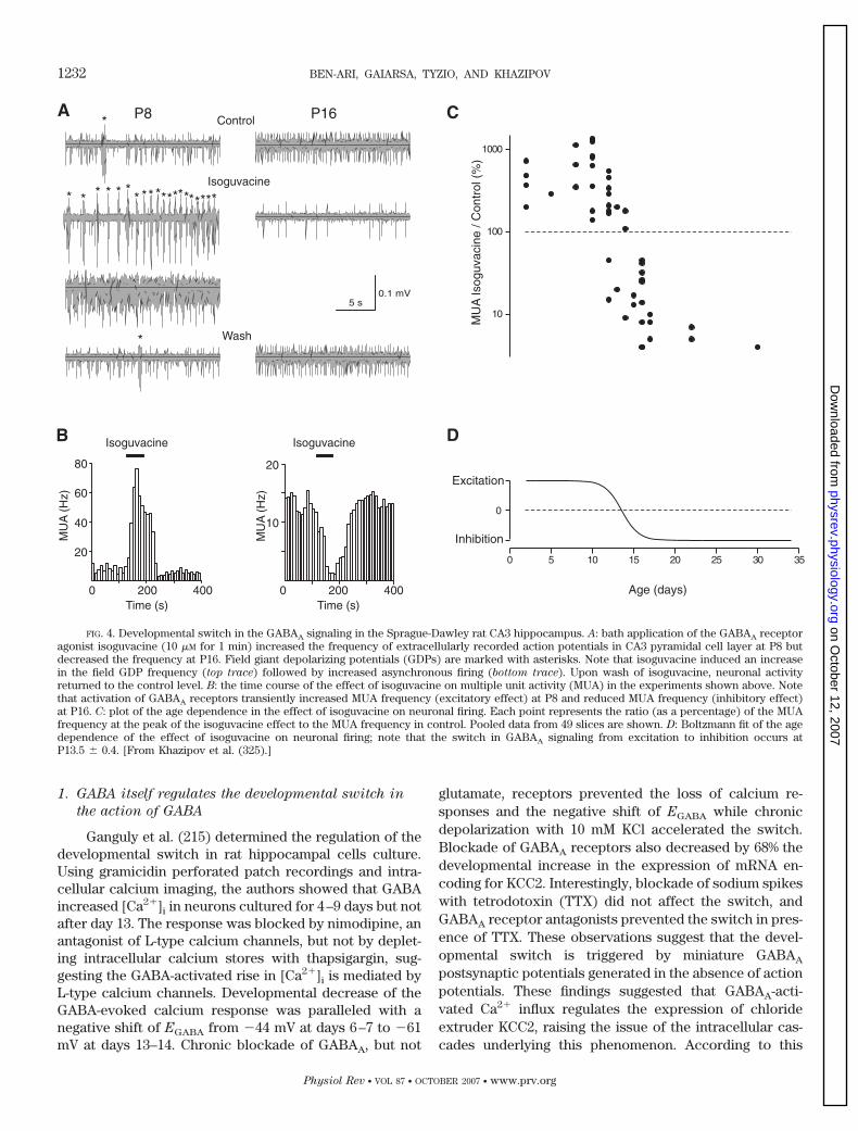
1. GABA itself regulates the developmental switch in
the action of GABA
Ganguly et al. (215) determined the regulation of thedevelopmental switch in rat hippocampal cells culture.Using gramicidin perforated patch recordings and intra-cellular calcium imaging, the authors showed that GABAincreased [Ca2�]i in neurons cultured for 4–9 days but notafter day 13. The response was blocked by nimodipine, anantagonist of L-type calcium channels, but not by deplet-ing intracellular calcium stores with thapsigargin, sug-gesting the GABA-activated rise in [Ca2�]i is mediated byL-type calcium channels. Developmental decrease of theGABA-evoked calcium response was paralleled with anegative shift of EGABA from �44 mV at days 6–7 to �61mV at days 13–14. Chronic blockade of GABAA, but not
glutamate, receptors prevented the loss of calcium re-sponses and the negative shift of EGABA while chronicdepolarization with 10 mM KCl accelerated the switch.Blockade of GABAA receptors also decreased by 68% thedevelopmental increase in the expression of mRNA en-coding for KCC2. Interestingly, blockade of sodium spikeswith tetrodotoxin (TTX) did not affect the switch, andGABAA receptor antagonists prevented the switch in pres-ence of TTX. These observations suggest that the devel-opmental switch is triggered by miniature GABAA
postsynaptic potentials generated in the absence of actionpotentials. These findings suggested that GABAA-acti-vated Ca2� influx regulates the expression of chlorideextruder KCC2, raising the issue of the intracellular cas-cades underlying this phenomenon. According to this
FIG. 4. Developmental switch in the GABAA signaling in the Sprague-Dawley rat CA3 hippocampus. A: bath application of the GABAA receptoragonist isoguvacine (10 �M for 1 min) increased the frequency of extracellularly recorded action potentials in CA3 pyramidal cell layer at P8 butdecreased the frequency at P16. Field giant depolarizing potentials (GDPs) are marked with asterisks. Note that isoguvacine induced an increasein the field GDP frequency (top trace) followed by increased asynchronous firing (bottom trace). Upon wash of isoguvacine, neuronal activityreturned to the control level. B: the time course of the effect of isoguvacine on multiple unit activity (MUA) in the experiments shown above. Notethat activation of GABAA receptors transiently increased MUA frequency (excitatory effect) at P8 and reduced MUA frequency (inhibitory effect)at P16. C: plot of the age dependence in the effect of isoguvacine on neuronal firing. Each point represents the ratio (as a percentage) of the MUAfrequency at the peak of the isoguvacine effect to the MUA frequency in control. Pooled data from 49 slices are shown. D: Boltzmann fit of the agedependence of the effect of isoguvacine on neuronal firing; note that the switch in GABAA signaling from excitation to inhibition occurs atP13.5 � 0.4. [From Khazipov et al. (325).]
1232 BEN-ARI, GAIARSA, TYZIO, AND KHAZIPOV
Physiol Rev • VOL 87 • OCTOBER 2007 • www.prv.org
on October 12, 2007
physrev.physiology.orgD
ownloaded from
work, GABA may play a self-regulatory role in the kineticsof the developmental switch in the GABA actions. Pooand co-workers (196) have recently also reported thatprolonged postsynaptic spiking of hippocampal neuronsled to a shift in the reversal potential of GABA-inducedCl� currents toward positive levels in a duration- andfrequency-dependent manner. This shift requires an ele-vation of [Ca2�]i and is occluded by inhibition of KCC2.Interestingly, these changes are larger in mature neuronsthat express the cotransporter than in immature neurons.These observations further reinforce the link betweenactivity and the actions of GABA (196).
Using a turtle model and calcium imaging, Sernagorand colleagues (379) investigated the effects of bicucul-line on the GABAergic polarity switch (from 1 wk beforehatching until 4 wk after hatching) and the presence ofpatterns of spontaneously generated retinal waves in theretinal ganglion cell layer. During that period, spontane-ous activity normally switches from propagating waves tostationary patches of coactive cells, until correlated ac-tivity completely disappears. The authors reported that inthe presence of bicuculline, GABAA responses remainexcitatory and spontaneous waves were generated andpropagated across the retinal ganglionic layer. This actionwas associated with a reduction of the developmentallyregulated expression of the KCC2 transporter, thus inkeeping with the observations of Ganguly et al. (215).
However, other studies suggest that GABAergic ac-tivity is not required for the switch to take place. Inhippocampal cell cultures, the developmental increase inthe KCC2 expression and hyperpolarizing shift of EGABA
can take place in spite of a chronic blockade of GABAA
receptors (404, 616). A similar conclusion was reached ina recent study in which the vesicular amino acid trans-porter was mutated leading to a cleft palate, but did notimpair the development of inhibitory synapses or of theexpression of KCC2 (666).
An additional mechanism has been proposed byOuardouz and Sastry (486). The authors showed that indeveloping deep cerebellar nuclei, a train of high-fre-quency stimulations triggered the D-H shift, an effectmediated by an apparent upregulation of the KCC cotrans-porter. This action was prevented by protein synthesisinhibitors and by protein phosphatase inhibitors, suggest-ing that a phosphorylation of the protein and the synthesisof new molecules may be involved in the transformation.
2. Brain-derived neurotrophic factor and tyrosine
kinase receptors regulate the developmental switch
Other factors have been implicated in the D-H devel-opmental switch of GABA. Thus cultured hippocampalneurons initially contain an inactive form of the KCC2protein, which becomes activated with neuronal matura-tion (314). This process is accelerated by transient stim-
ulation of insulin-like growth factor I (IGF-I) receptors.Because the transporter could be rapidly activated bycoapplication of IGF-I and Src kinase and deactivated bymembrane-permeable protein tyrosine kinase inhibitors,it was suggested that activation of K�/Cl cotransporterfunction by endogenous protein tyrosine kinases medi-ates the developmental D-H switch of GABAergic re-sponses (314). Brain-derived neurotrophic factor (BDNF)acting via tyrosine kinase receptor also increased expres-sion of KCC2 during the early development (5). Thisupregulation of KCC2 expression may be mediated byShc pathway (522). Interestingly, at older developmentalstages, BDNF affected EGABA in the hippocampal inter-neurons in an opposite direction (656). BDNF induced ashift in EGABA to more positive levels for GABAergic butnot glutamatergic postsynaptic neurons. It was concludedthat BDNF may decrease the efficacy of inhibitory trans-mission by acute postsynaptic downregulation of Cl�
transport, in addition to its well-known presynaptic ef-fect. TrkB-mediated downregulation of KCC2 in adultneurons requires activation of the two major cascades:phospholipase C (PLC)-� and Shc (444). Thus the polarityof the effect of BDNF on KCC2 expression is age depen-dent.
Khirug et al. (330) compared the developmental ex-pression and function of KCC2 in acute slices and cul-tures. Imposing a fixed Cl� load via patch pipette, theymeasured the resultant somatodendritic gradients in re-versal potential of GABAergic currents to determine thetime course of functional maturation of KCC2-mediatedCl� extrusion. In both preparations, the gradient wasinitially small or not detectable in immature neurons. Anabrupt increase occurs at around days 13–14 in culture,while a more gradual increase occurs between postnataldays 5–14 in slices. Consistent with the presence of anonfunctional form of KCC2 in immature hippocampalneurons grown in culture, application of the broad-spec-trum kinase inhibitor staurosporine produced a rapid andpotent upregulation of KCC2 function in these culturedneurons, but not in neonatal slices. These results indi-cated that the functional activity of KCC2 in vivo parallelsthe developmental expression of the protein, whereascultured neurons require an additional activation step(mimicked by staurosporine) for KCC2 to become func-tional.
H. A Dramatic Shift of EGABA During Delivery
A dramatic illustration of the importance of the shiftof the actions of GABA was shown recently in a study inwhich using single GABAA channel recordings in slices,Tyzio et al. (624) investigated the properties of GABAaround delivery time (Fig. 5). GABA was expected todepolarize neurons in utero and a few days after delivery,
EXCITATORY GABA IN DEVELOPMENT 1233
Physiol Rev • VOL 87 • OCTOBER 2007 • www.prv.org
on October 12, 2007
physrev.physiology.orgD
ownloaded from
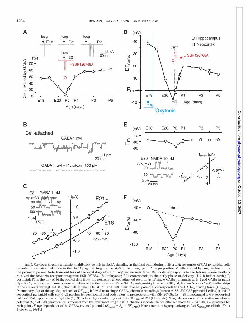
FIG. 5. Oxytocin triggers a transient inhibitory switch in GABA signaling in the fetal brain during delivery. A: responses of CA3 pyramidal cellsrecorded in cell-attached mode to the GABAA agonist isoguvacine. Bottom: summary plot of the proportion of cells excited by isoguvacine duringthe perinatal period. Note transient loss of the excitatory effect of isoguvacine near term. Red code corresponds to the fetuses whose mothersreceived the oxytocin receptor antagonist SSR126768A. [E, embryonic; E21 corresponds to the early phase of delivery (1–2 h before birth); P,postnatal; P0 is the day of birth; pooled data from 146 neurons]. B: cell-attached recordings of single GABAA channels with 1 �M GABA in patchpipette (top trace); the channels were not observed in the presence of the GABAA antagonist picrotoxin (100 �M; bottom trace). C: I-V relationshipsof the currents through GABAA channels in two cells, at E21 and E18; their reversal potential corresponds to the GABAA driving force (DFGABA).D: summary plot of the age dependence of DFGABA inferred from single GABAA channels recordings [means � SE; 209 CA3 pyramidal cells (E) and 17neocortical pyramidal cells (�); 6–24 patches for each point]. Red code refers to pretreatment with SSR126768A (n � 25 hippocampal and 9 neocorticalpatches). Bath application of oxytocin (1 �M) induced hyperpolarizing switch in DFGABA at E18 (blue code). E: age dependence of the resting membranepotential (Em) of CA3 pyramidal cells inferred from the reversal of single NMDA channels recorded in cell-attached mode (n � 84 cells; 4–12 patches foreach point). F: age dependence of the GABAA reversal potential (EGABA � Em � DFGABA). Note a transient hyperpolarizing shift of EGABA near birth. [FromTyzio et al. (624).]
1234 BEN-ARI, GAIARSA, TYZIO, AND KHAZIPOV
on October 12, 2007
physrev.physiology.orgD
ownloaded from
but shortly before, during, and after delivery, there was adramatic transient fall of the concentration of [Cl�]i (from�25 to 4 mM) and a D-H shift in DFGABA. Single NMDAchannel recordings made in parallel showed that Em wasslightly hyperpolarized by 7 mV during delivery, indicatinga genuine shift of EGABA. Cell-attached recordings con-firmed that GABA transiently lost its excitatory actionduring this narrow window. The mechanisms underlyingthe perinatal D-H shift were due to the maternal hormoneoxytocin that is released by the mother to trigger labor asoxytocin and antagonists of oxytocin receptors, respec-tively, produced and blocked this shift. Most significantly,administration of the antagonist to the mother preventedperinatal DFGABA shift in neocortical and hippocampalfetal neurons. Administration of bumetanide alleviatedthe actions of oxytocin receptor antagonists, suggestingthat the actions of the hormone are mediated by a rapidreduction of NKCC1 activity leading to a reduction of[Cl�]i. The authors suggested that the fetus is informed ofthe imminence of delivery by maternal release of a hor-mone that both triggers labor and exerts a massive inhib-itory action on fetal neurons. These effects are neuropro-tective. Indeed, blocking oxytocin receptors in pregnantrats, using oxytocin receptor antagonists also used toavoid preterm labor, aggravated the effects of hypoxicepisodes in fetal neurons. This study illustrates the phys-iological relevance of the developmental shift of GABAactions (624).
I. Depolarizing Actions of GABA in Other Brain
Structures and Animal Species
Studies performed in a wide range of developingbrain structures confirmed a developmental switch in theactions of GABA from depolarizing (and often excitatory)to hyperpolarizing (and often inhibitory). These includethe neocortex, the cerebellum, the spinal cord, the olfac-tory bulb, sensory structures, and several subcortical andperipheral structures. Table 1 lists available informationwith references.
IV. EARLY OPERATION OF GABAA SIGNALING
PRIOR TO SYNAPSE FORMATION
The starting point is that like at all central or periph-eral synapses, postsynaptic receptors are functional wellbefore synapses are formed and prior to the arrival ofpresynaptic axons. The earlier formation of receptorssuggests that the postsynaptic neuron or muscle receivessome information from the extracellular environment andcan communicate with arriving terminals to engage thesequence of events that will ultimately lead to the forma-tion of a synapse. This has been extensively investigatedin the neuromuscular junction that has provided most of
the concepts concerning the role of presynaptic axons onthe construction of the postsynaptic complex (360, 465).
GABA and GABAergic markers are expressed wellbefore synapses are formed, and GABA is released at anearly developmental stage and acts as a trophic factor tomodulate several essential developmental processes in-cluding neuronal proliferation, migration, differentiation,synapse formation, neuronal growth, or network con-struction. This early intercellular communication is basedon diffusion and distal paracrinic actions that contrastswith the local fast communication provided by synapticcurrents. These early effects are briefly summarized hereas they provide an excellent illustration of the multiplefacets of the actions of GABA and the unique role ofGABA as an early communicating signal. Excellent re-views of these effects have been published (36, 352, 492,579).
GABA, like other neurotransmitters, is released bygrowth cones (220) and can act on distal sites to generatereceptor-mediated currents. Neurotransmitters have nowbeen shown to influence directly or indirectly severalaspects of neuronal maturation in various experimentalsystems (36, 94, 183, 368, 370, 492). The depolarizingaction of GABA on developing systems is particularlysuited and constitutes a common denominator to its ac-tions on a wide range of processes.
A. GABA Signaling Is Present at a Very
Early Stage
In the neocortex, all the components of GABA sig-naling are present at an early stage. GABA immunoreac-tive neurons are found in all fetal zones of the cerebralanlage starting from embryonic day 15 (123, 633), and asecond population of GABAergic cells is observed in theintermediate zone. Beginning on day 16 of gestation andcontinuing throughout gestation, GABAergic neurons areobserved in the marginal zone, the zone, the cortical plate,and the ventricular and subventricular zones (30). As withNMDA-generated currents, GABA-mediated currents arepresent in proliferative zones well before synapses havebeen established (398, 399, 493). Other physiological stud-ies that failed to detect synaptic currents at an early stagereported tonic outward currents in response to applica-tions of GABA receptor antagonists (492, 493). Therefore,a tonic release of GABA acts can generate long-lastingcurrents in neurons that bear no functional synapse (alsosee below).
In the rodent hypothalamus, immunogold and perox-idase studies showed that GABA immunoreactivity ispresent in fibers and weakly stained perikarya at thebeginning of hypothalamic neurogenesis (E15) (631).Studies of neurons in culture suggest an earlier presenceof GABAA receptors and currents than other transmitters
EXCITATORY GABA IN DEVELOPMENT 1235
Physiol Rev • VOL 87 • OCTOBER 2007 • www.prv.org
on October 12, 2007
physrev.physiology.orgD
ownloaded from
including glutamate and glycine. Thus, in cultured hypo-thalamic embryonic neurons, GABA evoked currents al-ready a few hours after plating (E15), and all neuronsrecorded expressed GABAA receptors, whereas onlysome expressed glutamate or glycine receptors (112). Thesame group (483) showed also that applications of GABAelevate calcium in growth cones, suggesting that GABAreceptors may play a significant role in modulating theformation of early connections in the hypothalamus.
Similar conclusions were reached in studies of thematuration of the cerebellum. GABA immunoreactivity ispresent throughout the dendrites, cell bodies, and growthcones (607). In contrast, the vesicular GABA transporter(VGAT) was confined to the growth cones and axon var-icosities where GABA receptors are not present, suggest-ing that GABA may be extrasynaptically released fromaxon varicosities and growth cones by vesicular secretion“exocytosis” at a time when synapses have not formed. Inaddition, GABA release can occur from all part of thedeveloping neuron by nonvesicular secretion that the au-thors suggest may be due to a paracrine type of releasethrough a reversal of the transporters (also see Ref. 673and below). GABA is present in forebrain neurons at birthand can be released from growth cones without beingassociated with synaptic vesicles (610). This is in keepingwith the Ca2�-independent release of endogenous and3H-labeled GABA from growth cones (see below).
In rat embryonic dorsal spinal cord, GABAergicmarkers are present already at embryonic day 13 (E13),while neurons are still proliferating. GABA-evoked re-sponses were inhibited by bicuculline and picrotoxin andpotentiated by benzodiazepines (97, 556). Similarly, em-bryonic rat striatal neurons respond selectively to theGABAA agonist muscimol (195).
The developmental regulation of neurotransmittersynthesis has been extensively studied in the central ner-vous system of the Xenopus embryo (579). GABA immu-noreactivity appeared at an early stage, also suggestingthat GABA may play additional roles early in development(580, 657). This system is an attractive model for investi-gation, since action potentials are elicited from spinalneurons at the time of closure of the neural tube and canbe used to study the role of activity in the phenotypicexpression of transmitters. Spitzer and co-workers (52,75, 576, 577) have shown that the GABA phenotype isregulated by calcium signaling providing a direct linkbetween activity and network operation.
B. A Nonvesicular Release of GABA in Developing
Cortical Networks
Barker and co-workers (629, 630) also reported aseries of observations that suggest a tonic release ofGABA on cultured hippocampal neurons. Washing the
surface of neurons with a steam of saline rapidly andreversibly reduced the baseline current and fluctuations,both of which were completely eliminated by bicuculline(629, 630). Using hippocampal embryonic and early post-natal slices, Demarque et al. (158) confirmed that bicucul-line generates tonic currents during maturation. A briefelectrical pulse applied to the vicinity of the recordedneuron generated a GABAergic “nonsynaptic” current insilent neurons that have no spontaneous or evokedpostsynaptic currents. Procedures that release GABA alsofailed to generate a response indicating that vesicularrelease mechanisms are not operant at that stage. Thisearly slow current (ESC) is much longer than synapticcurrents, several seconds duration, and the electriccharges are several orders of magnitude larger than thatof conventional synaptic responses. ESCs are observedshortly after birth but disappear subsequently, around 2wk postnatal. Thus, during a transient postnatal period,neurons can have a slow GABAergic ESC generated withor without the faster more conventional synapse-drivenGABAergic currents. In contrast to EPSCs, ESCs are notblocked by a wide range of agents that block calciumchannels and vesicular release, including cobalt, TTX,zero calcium media, or botulinum toxin, and are observedin knockout mice in which the snare family proteins havebeen knocked out and vesicular release completely oblit-erated (637). Therefore, a nonvesicular release of GABAprecedes the conventional vesicular mechanisms and op-erates to flood in postsynaptic target neurons prior tosynapse formation. The source of this release has notbeen identified. However, growth cones do release GABAboth in a calcium-dependent (220) or -independent man-ner (611). The presence of this “noncanonical release”(158) of transmitter suggests that procedures that blockvesicular release may be inadequate to infer on the role oftransmission on brain construction. Blocking sodium/cal-cium spikes or even vesicular release by knockout strat-egies does not prevent the action of nonvesicular releaseof transmitter. The nonvesicular release of GABA may besufficient to modulate functions that will develop nor-mally in the absence of ongoing network dependent ac-tivity (516).
C. GABA Influences DNA Synthesis in Precursor
Neocortical Cells
Activation of GABA in the neocortical proliferativezone leads to significant decrease in DNA synthesis and areduction in the number of bromodeoxyuridine (BrdU)-labeled cells (399). Depolarization by high K� producedthe same effect (148, 399), and blocking GABA receptorsprevented the actions of GABA and increased DNA syn-thesis, suggesting that ongoing GABA release may reduceprotein synthesis (399). More recent experiments re-
1236 BEN-ARI, GAIARSA, TYZIO, AND KHAZIPOV
Physiol Rev • VOL 87 • OCTOBER 2007 • www.prv.org
on October 12, 2007
physrev.physiology.orgD
ownloaded from
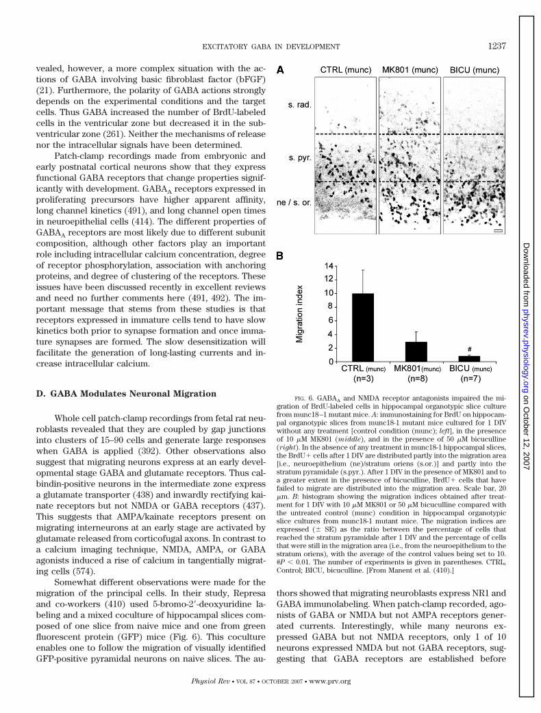
vealed, however, a more complex situation with the ac-tions of GABA involving basic fibroblast factor (bFGF)(21). Furthermore, the polarity of GABA actions stronglydepends on the experimental conditions and the targetcells. Thus GABA increased the number of BrdU-labeledcells in the ventricular zone but decreased it in the sub-ventricular zone (261). Neither the mechanisms of releasenor the intracellular signals have been determined.
Patch-clamp recordings made from embryonic andearly postnatal cortical neurons show that they expressfunctional GABA receptors that change properties signif-icantly with development. GABAA receptors expressed inproliferating precursors have higher apparent affinity,long channel kinetics (491), and long channel open timesin neuroepithelial cells (414). The different properties ofGABAA receptors are most likely due to different subunitcomposition, although other factors play an importantrole including intracellular calcium concentration, degreeof receptor phosphorylation, association with anchoringproteins, and degree of clustering of the receptors. Theseissues have been discussed recently in excellent reviewsand need no further comments here (491, 492). The im-portant message that stems from these studies is thatreceptors expressed in immature cells tend to have slowkinetics both prior to synapse formation and once imma-ture synapses are formed. The slow desensitization willfacilitate the generation of long-lasting currents and in-crease intracellular calcium.
D. GABA Modulates Neuronal Migration
Whole cell patch-clamp recordings from fetal rat neu-roblasts revealed that they are coupled by gap junctionsinto clusters of 15–90 cells and generate large responseswhen GABA is applied (392). Other observations alsosuggest that migrating neurons express at an early devel-opmental stage GABA and glutamate receptors. Thus cal-bindin-positive neurons in the intermediate zone expressa glutamate transporter (438) and inwardly rectifying kai-nate receptors but not NMDA or GABA receptors (437).This suggests that AMPA/kainate receptors present onmigrating interneurons at an early stage are activated byglutamate released from corticofugal axons. In contrast toa calcium imaging technique, NMDA, AMPA, or GABAagonists induced a rise of calcium in tangentially migrat-ing cells (574).
Somewhat different observations were made for themigration of the principal cells. In their study, Represaand co-workers (410) used 5-bromo-2-deoxyuridine la-beling and a mixed coculture of hippocampal slices com-posed of one slice from naive mice and one from greenfluorescent protein (GFP) mice (Fig. 6). This cocultureenables one to follow the migration of visually identifiedGFP-positive pyramidal neurons on naive slices. The au-
thors showed that migrating neuroblasts express NR1 andGABA immunolabeling. When patch-clamp recorded, ago-nists of GABA or NMDA but not AMPA receptors gener-ated currents. Interestingly, while many neurons ex-pressed GABA but not NMDA receptors, only 1 of 10neurons expressed NMDA but not GABA receptors, sug-gesting that GABA receptors are established before
FIG. 6. GABAA and NMDA receptor antagonists impaired the mi-gration of BrdU-labeled cells in hippocampal organotypic slice culturefrom munc18–1 mutant mice. A: immunostaining for BrdU on hippocam-pal organotypic slices from munc18-1 mutant mice cultured for 1 DIVwithout any treatment [control condition (munc); left], in the presenceof 10 �M MK801 (middle), and in the presence of 50 �M bicuculline(right). In the absence of any treatment in munc18-1 hippocampal slices,the BrdU� cells after 1 DIV are distributed partly into the migration area[i.e., neuroepithelium (ne)/stratum oriens (s.or.)] and partly into thestratum pyramidale (s.pyr.). After 1 DIV in the presence of MK801 and toa greater extent in the presence of bicuculline, BrdU� cells that havefailed to migrate are distributed into the migration area. Scale bar, 20�m. B: histogram showing the migration indices obtained after treat-ment for 1 DIV with 10 �M MK801 or 50 �M bicuculline compared withthe untreated control (munc) condition in hippocampal organotypicslice cultures from munc18-1 mutant mice. The migration indices areexpressed (� SE) as the ratio between the percentage of cells thatreached the stratum pyramidale after 1 DIV and the percentage of cellsthat were still in the migration area (i.e., from the neuroepithelium to thestratum oriens), with the average of the control values being set to 10.#P 0.01. The number of experiments is given in parentheses. CTRL,Control; BICU, bicuculline. [From Manent et al. (410).]
EXCITATORY GABA IN DEVELOPMENT 1237
Physiol Rev • VOL 87 • OCTOBER 2007 • www.prv.org
on October 12, 2007
physrev.physiology.orgD
ownloaded from
NMDA receptors. This sequential expression of receptorsfollows the general rules shown in acute slices for theformation of synapses (see sect. VI). Also, GABA but notglutamate receptor antagonists generated tonic currents.The generation of currents in migrating neurons couldcontrol neuronal excitability and motility (555). The pres-ence of functional AMPA currents in neocortical neurons(438) but not hippocampal (410) principal neurons couldreflect genuine differences between different neuronalpopulations. In keeping with this, Represa and co-work-ers (411) confirmed that tangentially migrating interneu-rons express AMPA but not GABA or NMDA receptors.Therefore, migrating principal cells and interneurons maybe modulated by different receptors during their journey.
Are these receptors functional and involved in mod-ulating neuronal migration? Using mouse cerebellar slicepreparations and a laser scanning confocal microscope,Komuro and Rakic (345) showed that selective blockadeof N-type functional calcium channels in granule cellsretarded cell movement. In contrast, blocking T- or L-typecalcium channels or sodium and potassium channels hadno effect on granule cell migration. In another study, thesame group (346) showed that blockade or activation ofNMDA receptors, respectively, retarded and increasedcell migration. Blocking uptake of glutamate acceleratedthe rate of cell migration reflecting a contribution ofendogenous extracellular glutamate to the process. Theamplitude and frequency components of Ca2� fluctua-tions correlated positively with the rate of granule cellmovement, suggesting a modulation of migration byspikes and waves of calcium oscillations (347, see alsoRef. 348). These observations collectively suggest thatmigration speed and directions are modulated by extrin-sic calcium signals to which transmitters participate priorto synapse formation. Molnar and co-workers (396) stud-ied the role of GABAB receptors by combining preembed-ding immunocytochemistry with cell tracking in embry-onic brain slice cultures. The authors found that a GABAB
receptor antagonist produced a dose-dependent accumu-lation of tangentially migrating neurons in the ventricular/subventricular zones of the cortex as well as a shorterleading process. The effects of the antagonist are mostlikely presynaptic as agonists did not generate currents inthese neurons in keeping with the observations thatGABAB agonists mature first presynaptically and are ex-pressed only subsequently in the postsynaptic target neu-rons (see sect. IX). Other studies suggest developmentallyregulated expression of different types of glutamate andGABA receptor subunits; some have prominent expres-sion in the embryonic and/or postnatal brain, whereasothers are mainly present in the adult brain (406).Changes of subunit composition may constitute essentialfactors in the development of particular brain regions andspecific neuronal populations within these regions.
The role of GABAA receptors in the neuronal migra-tion has also been investigated in the newborn rat parietalcortex in vivo and in vitro. Local in vivo application ofbicuculline methiodide via cortical surface Elvax im-plants revealed heterotopic cell clusters in the upper lay-ers (264). In organotypic neocortical slices from embry-onic day 18–19 embryos, bicuculline increased the migra-tion speed, suggesting an inhibitory action of GABA onmigration (264). Similarly, in embryonic rat cortical slices,bicuculline enhanced migration of neurons at E18 (44).
Bolteus and Bordey (71) studied the role of GABA inproliferation and migration of neuronal precursor migra-tion in the anterior subventricular zone and the rostralmigratory stream. In acute brain slices, application of lowconcentrations of GABA reduced the rate of cell migra-tion, whereas bicuculline enhanced the migration ratesignificantly, suggesting that endogenous GABA tonicallyreduces the speed of cell migration via GABAA receptoractivation. Interestingly, inhibition of the uptake of GABAor enhancement of the release of GABA by high K� fromneuronal precursors reduced migration, the later effectbeing blocked by bicuculline. The authors suggested thatastrocytes generate a microenvironment that controls thedegree of GABAA receptor activation and the migration ofneuronal precursors. In an earlier study, the same groupshowed that GABA is depolarizing at resting membranepotential, suggesting that the depolarization by means ofa rise of calcium constitutes the basis for a paracrinesignal to dynamically regulate their proliferation and/ormigration (43, 652).
Different results were obtained in the hippocampus.Using two-slice cocultures obtained from GFP and naivemice (see above), Represa and co-workers (410) mea-sured the effects of various agents on the migration of theGFP neurons that can be readily visualized when theymigrate on the naive slice. The authors showed that bothbicuculline and a NMDA receptor antagonist retard con-siderably the migration of GFP neurons. Most interest-ingly, when the GFP slice was apposed to a slice obtainedfrom a Munc18 knockout mouse, in which there is novesicular release, the effects of bicuculline and NMDAreceptor antagonist were preserved, suggesting that anendogenous nonvesicular release of GABA and glutamatemodulates the migration of neurons. Therefore, the stim-ulation of electrically silent neurons induces the releaseof GABA and glutamate in a “noncanonical” mechanism(410) that modulates the migration of adjacent neurons byan action on receptors in the absence of functional syn-apses (see Ref. 516 for review). The reasons for thedifferent actions of bicuculline in the hippocampus andneocortex are not known; one possibility is the generationby bicuculline of seizures in adjacent more developedneocortical neurons or a different involvement of astro-cytes as suggested by Bolteus and Bordey (see below).
1238 BEN-ARI, GAIARSA, TYZIO, AND KHAZIPOV
Physiol Rev • VOL 87 • OCTOBER 2007 • www.prv.org
on October 12, 2007
physrev.physiology.orgD
ownloaded from
Collectively, these observations suggest that recep-tors are expressed well before synapses and are activatedduring migration, providing a possibility for extrinsic fac-tors to modulate migration. This is in keeping with theneed for intracellular calcium alterations for cytoskeletalchanges that are indispensable for motility and migration(348). Other major functions are also regulated by cal-cium oscillations including expression of the GABA phe-notype (see below). These observations have importantclinical implications considering the importance of migra-tion disorders and the effects of widely used transmitteracting drugs during pregnancy (see sect. IX).
E. Early Trophic Actions of GABA
Early studies showed that applications of GABA tocultures of mouse neuroblastoma cells triggered a seriesof changes including proliferation of coated vesicles andthe appearance of electron-dense material aggregated atthe inner aspect of the plasma membrane (136, 186). Thesame group showed that GABA treatments affect thegrowth and development of embryonic chick cortical andretinal neurons in culture, including the length andbranching of the neurites (582). GABA treatment alsoincreased the density of neurotubules, rough endoplasmicreticulum (RER), and Golgi apparatus. Similar observa-tions were made by Schousboe and colleagues (46, 252,428). Treatment of cultures with the GABA agonist 7-tetrahydroxyazolo[5,4-c]pyridin-3-ol (THIP) of 7-day-oldcultures but not older ones increased the cytoplasmicdensity of RER and Golgi apparatus.
GABAergic embryonic hippocampal neurons expressa high potential for growth and sprouting in vitro andin vivo. Thus Robain et al. (523) compared the maturationof neurons taken from E18-E19 embryos, dissociated insuspension, and either grafted to an adult hippocampus orcultivated. The authors reported a substantial proportionof GABAergic cells in vivo and in vitro that exceeds thepercentage of GABA positive neurons in situ. The samegroup then tested directly the effects of GABA or GABAreceptor antagonists on the maturation and neuronalgrowth in vitro (35). Whereas neuritic outgrowth of cul-tured hippocampal neurons was not affected by musci-mol, a GABAA agonist, addition of bicuculline to the cul-ture medium severely reduced the number of primaryneurites and branching points (35). The lack of effects ofthe GABAA agonist may be due to a degradation of theagonist or a desensitization of receptors (or the fact thatendogenous GABA saturates the receptors, thus addingmore GABA will have no effect). The powerful effects ofGABAA antagonists suggest that an endogenous release ofGABA stimulates growth of hippocampal neurons proba-bly via the excitatory actions of GABA at an early stage.
Groc et al. (239, 240) investigated in vivo the earlyroles for GABA- and glutamate-driven hippocampal neu-
ronal network activity in the morphological differentia-tion of CA1 pyramidal cells. Injection of tetanus toxin intohippocampus of P1 rats induced strong reduction in thefrequency of spontaneous GABA and glutamatergic syn-aptic currents and led to a complete blockade of GDPsduring the first postnatal week. Morphology of neurobi-otin-filled CA1 pyramidal cells was analyzed at the end ofthe first postnatal week (P6–10). In activity-reduced neu-rons, the total length of basal dendritic tree was threetimes less than control. The number, but not the length, ofbasal dendritic branches was affected. The growth impair-ment was restricted to the basal dendrites. The apicaldendrite, the axons, or the soma grew normally duringactivity deprivation. Thus the in vivo neural activity in theneonate hippocampus seems to promote neuronal growthby initiating novel branches (239). However, quantitativeanalysis of dendritic parameters and spine density fromreconstructed CA1 pyramidal cells showed that youngadult CA1 pyramidal (postnatal day 31–34) cells that wereexposed to activity deprivation in the neonatal periodwere almost indistinguishable from control cells of thesame age. These results suggest that the early hippocam-pal activity driven by depolarizing GABA and glutamatecontrols the growth rate but is not necessary for thegeneration of an adult normal basal dendritic tree (240).
F. Conclusion: GABA Is an Ancillary
Communicating Signal With Multiple
Actions at Early Developmental Stages
GABA is not a novel signaling molecule. GABA andits receptors appeared very early during evolution (531).Glutamic acid decarboxylase (GAD) is ubiquitous in pro-karyotes and eukaryotes. In hydra, neurosteroids andGABA antagonists like picrotoxin act on GABAA receptorswith a modulation of the feeding response (131, 153). GABAexerts important roles in plants, and these actions are notonly metabolic (77, 78). The synthesizing enzyme of GABA,GAD, has been identified in several plants including arabi-dopsis where it is involved in several important functions.Severe morphological abnormalities, such as short stems,are induced in transgenic tobacco plants expressing a mu-tant GAD lacking the calmodulin-binding domain (38). Inplants, disruption of enzymes for GABA degradation resultsin abnormal development (76), and a GABA gradient isinvolved in guidance of the pollen tube (494). Interestingly,stress leads to extremely high GABA and low glutamate.Thus the synthesis of GABA is rapidly stimulated inwounded plant tissues consequent to the activation of GAD,and GABA is activated by infection of plants (117).
Fibers showing GABA-like immunoreactivity arepresent in abundance in the longitudinal nerve cords,lateral nerves, and fibers forming commissures in thebrain of a flatworm Dugesia tigrina (188). GABA medi-
EXCITATORY GABA IN DEVELOPMENT 1239
Physiol Rev • VOL 87 • OCTOBER 2007 • www.prv.org
on October 12, 2007
physrev.physiology.orgD
ownloaded from
ates enteric muscle contraction in the nematode Caeno-
rhabditis elegans via a receptor coupled to a cation-selective ligand-gated ion channel (EXP-1) (42). TheEXP-1 protein resembles ionotropic GABA receptor sub-units, but the pore-forming domain has residues that con-fer cation selectivity. Hence, GABA exerts a cation-medi-ated excitatory action. GABAergic neurons that are alsoGAD positive are present in the first brain nerves andcerebral and caudal ganglia of the urochordate Oiko-
pleura dioica (575). Therefore, although GABA has amajor metabolic role at a very early stage of evolution, itsmultiple facets have served many functions includingearly signaling actions notably as a transmitter.
These observations raise the possibility that at leastsome of the fundamental properties associated with theactions of GABA have been exploited early on in evolu-tion. The anionic permeability of its channels and thechloride fluxes associated with its operation appear to beuseful at an early developmental stage well before syn-apses are established. The most parsimonious hypothesisis that GABA as an informative molecule is a pioneersignal that contributes to organize a heterogeneous pop-ulation of immature neurons. The paracrine actions ofGABA at that stage and the use of a nonvesicular releaseof GABA to flood the environment and the delayed devel-opment of GABA transporters (see below) will facilitatedistal actions of GABA. The stimulation of neuronalgrowth appears to be mediated by a positive loopwhereby the release of GABA excites postsynaptic neu-rons that release BDNF leading to an enhancement of theactions of GABA (see sect. VIII). Although important shiftsin the action of other transmitters during developmenthave been reported, those involving GABA are of partic-ular importance as GABA receptors are present in everyneuron and GABAergic inhibition in every circuit. In ad-dition, the reversal of GABA actions has no parallel situ-ation in glutamatergic synapses.
Since glutamatergic signaling operates somewhatlater that GABAergic synapses (see sect. V), GABA will bede facto the principal excitatory transmitter early on. Anadditional factor to consider is the earlier maturation offunctional NMDA receptors than AMPA receptor-medi-ated EPSCs, the so-called “silent” synapses (169; but seealso Refs. 225, 238). There are some indications that thevoltage-dependent Mg2� blockade is less efficient in neo-natal neurons than adults (57, 80, 279, 338, 606; but seeRef. 328) consequent to important changes in subunitcomposition that occur during maturation (458, 519). In-terestingly, GABA is an important source to remove theblockade and enable the activation of NMDA receptorchannels (59). This “synergistic” action of GABA withNMDA receptors (377) contrasts with adult neurons andconstitutes a major aspect of early GABA signaling inaddition to the generation by GABA of action potentials(see sect. III). Although glutamate is likely also released at
an early stage, in physiological conditions, circulatingconcentrations of GABA are more elevated than those ofglutamate because of the earlier maturation of glutamatetransporters than GABA transporters (see sect. VII). Thisexplains why GABA but not glutamate tonic currents havebeen recorded at an early stage. Therefore, the system istuned to favor the actions of GABA early on.
V. GABAERGIC SYNAPSES ARE ESTABLISHED
BEFORE GLUTAMATE SYNAPSES
A. GABA Receptor Antagonists Block Early
Ongoing Activity in the Hippocampus
Since the actions of GABA and glutamate receptorantagonists are selective on immature neurons (56, 467),they can be used to determine the contribution of GABAand glutamate PSCs to ongoing activity. In their initialobservations, Ben-Ari and colleagues (56, 115) reportedthat during the first postnatal week, bicuculline blockedongoing activity and often fails to generate seizures as inadults. At an early stage, ongoing activity is dominated bythe so-called GDP, a transient network-driven patternmediated primarily by GABA that disappears progres-sively with the D-H shift (see sect. VI). GDPs reversed at apotential close to that of currents generated by exogenousapplications of GABA, suggesting that immature PSCs aremediated primarily by GABAA receptors. Both occurred atmore depolarized potentials in immature neurons thanadult ones, confirming the early depolarizing actions ofGABA (see sect. III). GDPs were, however, also blocked byglutamate receptor antagonists, notably by the NMDAreceptor antagonist APV (135, 210, 212), suggesting asynergistic contribution of depolarizing GABA and NMDAreceptors (see also below).
B. GABAergic Synapses Are the First Functional
Synapses on Principal Cells of the
Hippocampal Formation
The use of GABA receptor antagonists to determinethe sequential maturation of GABA and glutamate syn-apses has however several limitations in immature net-works as reflected by the observation that in P0-P1 slices:bicuculline generates seizures in some studies but not inothers (see sects. VI and IX). Antagonists can affect differ-ently various neuronal populations reducing the excita-tory drive and disinhibiting principal neurons (see, forinstance, Refs. 600, 602). Bicuculline also blocks potas-sium channels (155). GABA receptor antagonists generateseizures more readily in intact hippocampal preparationsthan in slices, most likely because of the loss of a signif-icant number of axonal inputs and synapses that at that
1240 BEN-ARI, GAIARSA, TYZIO, AND KHAZIPOV
Physiol Rev • VOL 87 • OCTOBER 2007 • www.prv.org
on October 12, 2007
physrev.physiology.orgD
ownloaded from
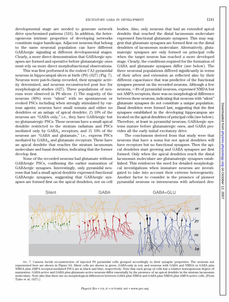
developmental stage are needed to generate networkdrive synchronized patterns (316). In addition, the heter-ogeneous intrinsic properties of developing networksconstitute major handicaps. Adjacent neurons that belongto the same neuronal population can have differentGABAergic signaling at different developmental stages.Clearly, a more direct demonstration that GABAergic syn-apses are formed and operative before glutamatergic onesmust rely on more direct morphofunctional observations.
This was first performed in the rodent CA1 pyramidalneurons in hippocampal slices at birth (P0) (627) (Fig. 7).Neurons were patch-clamp recorded, their synaptic activ-ity determined, and neurons reconstructed post hoc formorphological studies (627). Three populations of neu-rons were observed in P0 slices: 1) The majority of theneurons (80%) were “silent” with no spontaneous orevoked PSCs including when strongly stimulated by var-ious agents; neurons have small somata and either nodendrites or an anlage of apical dendrite; 2) 10% of theneurons are “GABA only,” i.e., they have GABAergic butno glutamatergic PSCs. These neurons have a small apicaldendrite restricted to the stratum radiatum and PSCsmediated only by GABAA receptors; and 3) 10% of theneurons are “GABA and glutamate,” i.e., express PSCsmediated by GABAA and glutamate receptors. These havean apical dendrite that reaches the stratum lacunosummoleculare and basal dendrites, indicating that the formerdevelop first.
None of the recorded neurons had glutamate withoutGABAergic PSCs, confirming the earlier maturation ofGABAergic synapses. Interestingly, only pyramidal neu-rons that had a small apical dendrite expressed functionalGABAergic synapses, suggesting that GABAergic syn-apses are formed first on the apical dendrites, not on cell
bodies. Also, only neurons that had an extended apicaldendrite that reached the distal lacunosum moleculareexpressed functional glutamate synapses. This may sug-gest that glutamate synapses are formed first on the distaldendrites of lacunosum moleculare. Alternatively, gluta-matergic synapses are only formed on principal cellswhen the target neuron has reached a more developedstage. Clearly, the conditions required for the formation ofGABA and glutamate synapses differ (see below). Thethree neuronal populations differed significantly in termsof their arbor and extension as reflected also by theirdifferent capacitance that was predictive of the functionalsynapses present on the recorded neurons. Although a fewneurons, 4% of pyramidal neurons, expressed NMDA butnot AMPA receptors, there was no morphological differencebetween these neurons, indicating that neurons with “silent”glutamate synapses do not constitute a unique population.Basal dendrites were formed last, suggesting that the firstsynapses established in the developing hippocampus arelocated on the apical dendrites of principal cells (see below).Therefore, at least in pyramidal neurons, GABAergic sys-tems mature before glutamatergic ones, and GABA pro-vides all the early initial excitatory drive.
The conclusions derived from that study were thatneurons that have a soma but not apical dendrites willhave receptors but no functional synapses. Then the api-cal dendrites start growing and GABA synapses are firstformed. Only when the apical dendrites reach the distallacunosum moleculare are glutamatergic synapses estab-lished. This reinforces the need for detailed morphologi-cal investigations when immature neurons are investi-gated to take into account their extreme heterogeneity.Another factor to consider is the presence of pioneerpyramidal neurons or interneurons with arborized den-
FIG. 7. Camera lucida reconstruction of injected P0 pyramidal cells grouped accordingly to their synaptic properties. The neurons notrepresented here are shown in Figure 9A. Silent cells are shown in green, GABA-only in red, and neurons with GABA and NMDA or GABA plusNMDA plus AMPA receptor-mediated PSCs are in black and blue, respectively. Note that each group of cells has a relative homogeneous degree ofmaturation. GABA-active and GABA plus glutamate-active neurons differ essentially by the presence of an apical dendrite in the stratum lacunosummoleculare. Note also that there are no morphological differences between GABA plus NMDA and GABA plus NMDA plus AMPA-active cells. [FromTyzio et al. (627).]
EXCITATORY GABA IN DEVELOPMENT 1241
Physiol Rev • VOL 87 • OCTOBER 2007 • www.prv.org
on October 12, 2007
physrev.physiology.orgD
ownloaded from
drites at an early stage when most neurons are quiescentwith little or no functional synapses; this may explain theobservations of AMPA and NMDA receptor-mediated pop-ulation field EPSPs already at birth in CA1 (161).
One important implication of this study (627) is thatthe formation of synapses is strongly dependent on infor-mation provided by the target neuron. Indeed, as gluta-matergic axonal afferents are present at an early devel-opmental stage in the appropriate layer (see below), theGABA-glutamate sequence is not due to a later arrival ofthe inputs. Therefore, glutamatergic axons that meet im-mature apical dendrites will not establish synapses,whereas GABAergic axons will: different clues are re-quired for the formation of glutamatergic and GABAergicsynapses. Studies in culture offer several convenient toolsto examine these issues. GABAergic and glutamatergicneurons develop in culture and express similar morpho-logical and biological features as in vivo (62). With the useof this preparation, a detailed description of the formationof GABA and glutamate has been reported and the role ofneuronal activity analyzed (87, 141, 224, 456, 684). Thusthe axons of cultured hippocampal neurons form presyn-aptic specializations soon after they emerge and do notrequire conditions other than encountering appropriatetargets (197). However, cell bodies and dendrites cannotinduce the formation of presynaptic specializations untilthey have reached a critical stage of “maturation” (197).These and other observations are in keeping with thenotion that axons develop early and reach their targetsand then wait for appropriate cues from the targets toestablish functional synapses. One possibility is a retro-grade messenger that could be targeted to glutamate butnot GABA synapses and would be required for the forma-tion of synapses. In several systems, specific cues re-quired for synapse formation between pre- and postsyn-aptic neurons have been investigated (151).
Poo and colleagues (544) have shown recently that atrophic factor, BDNF, induced rapid and persistent poten-tiation of evoked glutamate release when the postsynap-tic neuron was glutamatergic but not when it wasGABAergic. In a single presynaptic neuron innervatingglutamatergic and GABAergic neurons, only the formertarget was modulated by BDNF, suggesting a target-spe-cific action of BDNF on individual terminals. Corticalcocultures containing neurons that were obtained earlyduring development (E16) readily evoked GABA PSCs,whereas glutamate PSCs were mainly evoked when thecocultures were obtained from more adult neurons (E19)(454). This was due to a reduction of the number ofrelease sites, the quantal amplitude, but not the meanrelease probability. This suggests that the formation ofglutamatergic but not GABAergic synapses is influencedby a postsynaptic factor. In another study (641), the roleof axonal guidance in the choice of the correct targetneuron was investigated using an in vitro system in which
neuritic outgrowth of rat cortical neurons is accuratelyguided along the narrow pathways of a surface micropat-tern. The authors found that precise axonal guidance isinsufficient for a target-specific synapse formation; addi-tional cues are required to enable recognition betweenindividual cells (641). Molecular matches between pre-and postsynapse play a central role in the establishmentof the correct connections. The role of extrinsic andintrinsic cues in brain development and the establishmentof neuronal phenotypes has been extensively documentedat earlier developmental stages (183, 368). However, morestudies are required to examine in parallel the maturationof GABA and glutamate synapses and compare their de-velopment and cross-relations. This information will inturn facilitate the definition of cues that govern the for-mation of an equilibrated network with the appropriatedensity of GABA and glutamate synapses. We shall notreview here homeostatic mechanisms in the formation ofsynapses as this has been almost exclusively concernedwith glutamatergic synapses and reviewed extensivelyrecently (623).
The earlier maturation of GABAergic synapses hasalso been validated in another major component of thehippocampal formation: the granule cells of the fasciadentate. As developing granule cells have a developmen-tal period that is primarily postnatal, they are useful can-didates to examine this rule in postnatal slices. In thisregion, inhibitory interneurons are generated in utero inrodents, and their circuitry is well developed even as theirtarget excitatory connections are still being formed.GABAergic neurotransmission was found to prevail inimmature cells and the contribution of glutamatergic neu-rotransmission increased with neuronal maturation (387).In another study (12), two GABA currents evoked inimmature granule cells by medial perforant path stimula-tion showed only depolarizing GABAergic synaptic inputat first and then also glutamatergic synaptic inputs. Neu-rons were identified on the basis of their decreasing inputresistance and resting membrane potential getting morehyperpolarized with maturation. Miniature GABA PSCshave a slower kinetics in immature granule cells (P0-P15)resulting in a larger decrease in synaptic charge transferthan in adults (270). Other unique features of immatureGABAergic currents have been reported (125, 387, 388).Studies by Soltez and co-workers (269, 270) using pairrecordings from neonatal granule cells show that thespontaneous activation of GABAA receptors displaysunique properties that are distinct from the temporalpatterns and biophysical features observed followingGABAA receptor activation in the adult dentate gyrus.Therefore, the earlier formation of GABA-operated syn-apses is a general rule that applies to the various sub-populations of glutamatergic neurons in the hippocam-pus.
1242 BEN-ARI, GAIARSA, TYZIO, AND KHAZIPOV
Physiol Rev • VOL 87 • OCTOBER 2007 • www.prv.org
on October 12, 2007
physrev.physiology.orgD
ownloaded from
Parallel studies have suggested that GABA neuronsplay a pioneer role in the formation of glutamatergicsynapses. Thus Soriano and colleagues (598) have shownthat GABA interneurons are the first targets of commis-sural projections; over 60% of the targets of commis-sural axons according to quantitative electron microsco-pic (EM) studies. The authors suggested that earlyGABAergic neurons may constitute pioneer cells thatguide the formation of commissural axons and die subse-quently (291). The targets of commissural connectionswere identified in this study as being on interneurons, andsigns of cell death were observed with EM techniques. Inthis model, early maturing neurons will help guiding glu-tamatergic axons to their targets. In contrast, anotherstudy suggests that when genes required for the tangentialmigration of interneurons are deleted and interneuronsfail to reach the hippocampus, the organization of hip-pocampal connections remains quite normal (505; seealso below).
C. GABAergic Interneurons Mature Before
Principal Neurons and Follow the
Same Sequence
If GABAergic synapses are operative before gluta-mate synapses, then GABAergic interneurons and theiraxons must mature before glutamatergic pyramidal neu-rons. Morphological studies suggest an earlier maturationof GABAergic neuronal elements. GABAergic neurons inthe rat hippocampus appeared at the 18th day of gestationdeeply in the intermediate zone near the ventricular zone,and superficially in the marginal zone near the hippocam-pal fissure (39, 529). GABA immunolabeling is enriched inthe neuropil initially rather than in the principal layers inkeeping with the early formation of dendrite targetedsynapses. At E19, most pyramidal neurons have just fin-ished their formation (11) and have not developed theapical dendrites (445, 446) required for the formation ofsynaptic contacts in the marginal zone (197, 627). Theearly formation of GABAergic neurons is also reflected bya variety of markers including GAD (168), GABA (529),and calbindin (598). These and other synaptic markersincluding synapsin-1 and synaptophysin labeling arepresent in dendritic layers but not in the stratum pyrami-dale, suggesting that the first synapses formed in thehippocampus are GABAergic peridendritic synapses thatprecede perisomatic GABAergic synapses (see below).The presence of GAD containing terminals in utero and atbirth in the neuropil of stratum radiatum (529) also sug-gests that afferent fibers are present prior to synapseformation. Golgi impregnation was used to study the den-dritic differentiation of granule cells in the rat fasciadentate (403). This was followed by gold-toned analysis toidentify the fine structural study of the same identified
neurons and of the input synapses onto their cell bodiesand dendrites. Authors reported that the plexus ofGABAergic axons is already well developed at an earlystage when granule cells are still forming.
A direct validation of the proposed model came frommorphofunctional studies in which a large number ofinterneurons recorded in the CA1 region of slices of newlyborn rat pups (P0) and reconstructed after physiologicalcharacterization (51, 266; also see Refs. 211, 213). At birth,three types of interneurons were found: 1) noninnervated“silent” interneurons (5%) with no spontaneous or evokedsynaptic currents, 2) GABA only interneurons (17%) withGABAA but no glutamate synapses, and 3) GABA andglutamate interneurons with GABA and glutamatergicsynapses (78%).
Silent neurons remained silent when ongoing activitywas artificially augmented and latrotoxin applied to de-plete vesicular pools confirming the lack of functionalsynapses. Also, high-frequency electrical stimuli in thevicinity of visually patched clamped neurons did notevoke PSCs or action potentials/currents when recordedwith cell-attached recordings. In contrast, functionalGABA and glutamate receptors were present on silentinterneurons since applications of agonists evoked theexpected currents. Reconstruction of these neurons re-vealed that the three types of interneurons correspond tothree developmental stages: noninnervated neurons thathave little or no dendrites, interneurons with only GABAA
synapses having a more extended dendritic arbor, andinterneurons with both GABA and glutamate synapsesthat are extensively developed. In fact, the total surface ofthe dendrites, axons, and somata were significantly lowerin silent neurons than in GABA only, and the latter werealso significantly smaller than neurons endowed with op-erative GABA and glutamate synapses. Neuronal capaci-tance enabled the prediction of the recorded electricalresponse: the curve relating surface and capacitance re-vealed three groups of neurons that corresponded to thethree neuronal populations.
These studies confirmed the early maturation of in-terneurons at a time when pyramidal neurons are primar-ily silent. Thus, at birth, 80% of CA1 pyramidal neuronsbut only 5% of interneurons are silent, although 10% ofpyramidal neurons and �60% of interneurons have bothGABA and glutamate synapses at that stage. In fetuses(E18–20), the majority of interneurons (65%) had opera-tive synapses, whereas nearly 90% of principal neuronswere silent. Similarly, the ratio of GABA only versusGABA and glutamate interneurons was more than six-fold higher at E18 –20 than at birth, confirming the lateformation of glutamate synapses on interneurons that areonly established once target neurons have functionalGABAergic synapses. Putative pioneer interneurons werealso reconstructed; these had an extensive dendritic andaxonal arbor that innervated the CA3 region, the distal
EXCITATORY GABA IN DEVELOPMENT 1243
Physiol Rev • VOL 87 • OCTOBER 2007 • www.prv.org
on October 12, 2007
physrev.physiology.orgD
ownloaded from
lacunosum moleculare, or the subiculum. These neuronsare similar to the back-projecting interneurons describedin adults (201). Therefore, interneurons follow the samesequence as principal cells but earlier: at all developmen-tal stages, there is a GABAergic source of synaptic activityto stimulate neuronal growth.
D. GABAergic Interneurons Innervate First the
Dendrites of Principal Neurons
As stressed above, GABAergic interneurons consti-tute an extremely heterogeneous population of neurons,and there has been enormous difficulties in classifying thevarious neuronal types relying on morphological and elec-trophysiological or biochemical markers (201, 497). Thisis even more difficult in developing networks becauseimmature neurons lack many of the chemical markers andthe extended dendritic or axonal arbor required to iden-tify them (266, 369). Determining the sequence of matu-ration of various interneurons is important to determinewhether indeed GABAergic synapses are formed on thedendrites of principal neurons before their somata. This isalso important since the first patterns recorded in thedeveloping hippocampus are generated by interneurons(51, 236).
In spite of these difficulties, morphofunctional stud-ies suggest that interneurons that innervate other inter-neurons and/or the dendrites of principal cells maturebefore those that innervate the somata of principal cells.Thus, in the adult hippocampus, calretinin immunoreac-tive interneurons innervate exclusively GABAergic den-drites and other calretinin or calbindin D28k or vasoactiveintestinal polypeptide (VIP)-containing basket cells,whereas parvalbumin-containing basket and axo-axoniccells are selectively avoided (245). The unique connectiv-ity of these neurons confers important contribution tocontrol the generation of synchronous, rhythmic hip-pocampal activity. Its target specificity is also an impor-tant asset to determine the sequential maturation ofGABAergic and glutamatergic terminals. Calretinin neu-rons are detected as early as embryonic day 15 in theprimordial hippocampus in the primitive plexiform layer(292, 397).
In a more recent study (670), neurons from the pro-liferative zone from GFP mouse embryos were trans-planted onto neonatal cortical feeder cells and assessedfor their ability to generate specific interneuron subtypes.Parvalbumin- and somatostatin-expressing interneuronsoriginate primarily within the medial ganglionic emi-nence, whereas the calretinin-expressing interneurons de-rive mainly from the caudal ganglionic eminence (see alsoRef. 88). Therefore, GABAergic interneurons that inner-vate dendrites and somata of principal cells may have adifferent origin and migration trajectory. Since the fate of
these neurons and their discharge properties appear to bedetermined by their origin (88, 353), it will be of interestto examine which factors control their phenotype duringmigration and their integration within the networks. Oneprediction is that interneurons that innervate other inter-neurons or the dendrites of principal cells are functionalbefore neurons that innervate cell bodies.
An interesting example is also provided by the mat-uration of cholecystokinin (CCK)-immunoreactive inter-neurons. These neurons innervate the dendrites of prin-cipal cells in all hippocampal subfields at birth, and theaxon arbors begin to concentrate around pyramidal cellbodies only at P8 (463). Synapses formed on CCK-con-taining interneurons are primarily symmetric initially andsubsequently shift to asymmetric synapses, suggesting anearlier formation of GABAergic terminals (463). Theseobservations also suggest that the interconnections be-tween interneurons mature first. The authors suggestedthat the shift from dendritic to somatic targets occurswhen the D-H shift occurs. In sum, these observations arein accord with the suggestion that the earliest functionalsynapses are between interneurons, then on dendrites ofprincipal neurons, and only lastly on the somata of prin-cipal neurons.
The maturation of endocannabinoid systems has alsobeen investigated since type 1 cannabinoid receptors thatare selectively located on axon terminals of GABAergicinterneurons mediate a retrograde synaptic signaling thatcontrols the release of GABA (308, 663). Type 1 cannabi-noid receptors are expressed at an early stage, and itscellular and subcellular patterns of expression duringearly postnatal life are similar to that of CCK-containingaxons (462). This distribution suggests that endocannabi-noid receptors can modulate at an early stage, well beforethe D-H shift has occurred, the dominant ongoingGABAergic activity (461, 462). This hypothesis was testedrecently in vitro and in vivo (66). The authors reportedthat ongoing activity during early maturation is modu-lated by endocannabinoid receptors and GDPs, and otherpolysynaptic events activate these presynaptic receptorsleading to a retrograde negative-feedback loop acting onthe terminals of CCK interneurons to reduce the releaseof GABA (see sects. VI and VII). In summary, interneuronsinnervate other interneurons before innervating principalcells, with functional GABAergic synapses operating at atime when glutamatergic activity is sparse or nonexistent.Furthermore, dendritic targets are favored by axons andby connections established in neurons that have little orno operative synapses.
E. Use of Knockout Strategies to Determine the
Role of GABA in Development
Obata and co-workers (22) have generated GAD 67and/or GAD 65 knockouts to investigate the conse-
1244 BEN-ARI, GAIARSA, TYZIO, AND KHAZIPOV
Physiol Rev • VOL 87 • OCTOBER 2007 • www.prv.org
on October 12, 2007
physrev.physiology.orgD
ownloaded from
quences on brain development. Deleting the 65 isoform ofGAD did not produce gross behavioral alterations, and thebrain was histologically quite normal, although with alower threshold for seizure generation (22). In contrast,mice lacking the 67 isoform of GAD died of severe cleftpalate during the first morning after birth. As expected,these mice have much reduced GAD activities and GABAcontents in the cerebral cortex (to 20% for the 65 and 7%for the 67, respectively). Their brain, however, did notshow any discernible defects in its construction (23).Mice with both knockouts (290) also do not survive afterbirth (cleft palate) and have quite normal brains on grosshistological examination in spite of an almost completeabsence of GABA. However, the compensatory develop-ment of other transmitters cannot be discarded as GABAand glycine are released from the same terminals (seebelow), and reorganizations during brain developmentcould explain some of these relatively minor conse-quences of knocking out GAD 65 or 67. A detailed micro-scopic investigation of these connections and distributionof various cell types in the knockouts remains to beperformed. A detailed study of this phenotype is alsoneeded as studies in this laboratory (R. Tyzio, A. Represa,and Y. Ben-Ari, unpublished data) have revealed a highdensity of GABAergic terminals and a quite normal fre-quency of GABAergic synaptic currents in GAD 65 knock-outs.
Other studies have performed more selective dele-tions of GABA signaling. Thus the deletion of genes re-quired for the migration of GABAergic interneurons to thehippocampus and cortex has been investigated by Ruben-stein and colleagues (124). Mice lacking two homeodo-main proteins needed for migration, DLX-1 and DLX-2,show no detectable cell migration from the subcorticaltelencephalon to the neocortex and a massive loss ofGABA-expressing cells in the neocortex (17, 415). Lossof Dlx1/2 homeobox function also prevents migration ofmost GABAergic interneurons to the hippocampus (505).It is possible to differentially disrupt the arrival to variouspopulations of neurons to the hippocampus. Thus delet-ing Nkx2.1 homeobox function depletes the hippocampusof a distinct subset of hippocampal interneurons. In con-trast to the conclusions of Soriano et al. (598), loss ofhippocampal interneurons had neither major effects onthe early development of hippocampal projection neuronsnor on the path-finding of afferent tracts: the commis-sural, entorhinal, and Schaeffer collaterals were intact.These mutations led to a reduction of GABAergic IPSCs inneocortex and hippocampus and cortical dysrhythmiain vivo. Dlx1 mutant mice show generalized electro-graphic seizures and histological evidence of seizure-in-duced reorganization. Whether this mutation leads to ab-normal migration of principal cells and organization in thepyramidal layer of the hippocampus has not been deter-mined.
F. GABA Transporters are Functional After
Glutamate Transporters
In adult networks, the extracellular concentration ofglutamate in the synaptic cleft is controlled by high-affin-ity Na�-dependent transporters required both for physio-logical processes and for prevention of receptor overac-tivation and possible excitotoxic actions (152). Glutamatetransporters also contribute to the synthesis of GABA viathe supply of glutamate to GABAergic synaptic terminals(423). A different situation prevails in the developingbrain where glutamate transporters are functional wellbefore synapses have been established (410, 490), sug-gesting that the tight control of glutamate is essential toprevent possible toxic actions of glutamate. In keepingwith this, blockade of glutamate transport in immaturebut not adult neurons by DL-threo-�-benzyloxyaspartate(TBOA) that blocks the three main neuronal transporters(561) generates in hippocampal slices, a pattern of recur-rent synchronous oscillations that is readily blocked by aNMDA receptor antagonist (159). Intracerebroventricularinjections of the blocker to pups generate NMDA recep-tor-dependent seizures. These oscillations are networkdriven and associated with an activity in the beta fre-quency range (15–20 Hz). Applications of NMDA alsogenerated similar oscillations that are blocked by gluta-mate but not GABA receptor antagonists, reflecting theimportant role of NMDA receptors in their generation.Similar oscillations are readily generated in the intacthippocampus in vitro where their propagation can beinvestigated. In more recent studies, the same group hasshown that the electroencephalogram (EEG) signature ofthese oscillations in pups is reminiscent of the seizureswith suppression bursts recorded in various encephalop-athies in children (442). Interestingly, a familial form ofOtahara encephalopathy is associated with a mutation ofa mitochondrial glutamate transporter (455). Therefore,deficiencies of glutamate transport can be deleteriousduring development in keeping with extensive data (431).
At birth, glutamate but not GABA transporters arefunctional. Thus the current generated by local applica-tions of GABA were not modified by bath applications ofSKF899756A, a selective powerful and irreversible block-ade of GAT1 (72, 158). A similar experiment in P14 neu-rons revealed a consistent increase in currents generatedby exogenous GABA (158). In contrast, TBOA augmentedthe currents produced by pressure ejections of glutamateand generated a large and persistent tonic current, sug-gesting that glutamate transporters operate in physiolog-ical conditions to prevent an accumulation of glutamate inthe extracellular space. These effects do not require ve-sicular release of transmitters as they are observed also inthe presence of botulinum toxin (158). The retarded func-tional expression of a GABA transporter is also reflectedby the observation that in adult but not neonatal neurons,
EXCITATORY GABA IN DEVELOPMENT 1245
Physiol Rev • VOL 87 • OCTOBER 2007 • www.prv.org
on October 12, 2007
physrev.physiology.orgD
ownloaded from
an inhibitor of GABA transporters unravels a GABAB
receptor-dependent paired pulse depression (92). Kailaand co-workers (566) showed that blockers of GABAtransporters increase the frequency of GDPs at the agesstudied (P4-P5), suggesting, in agreement with observa-tions made by the authors of this review (92, 158), thatblockers of GABA transport are functional when tested onpolysynaptic PSCs, like GDPs, but not on single PSCs orcurrents evoked by local applications of GABA. In con-trast, the efficient glutamate transport mechanisms leadto a rapid removal of glutamate. The generation at birthof tonic currents by glutamate but not GABA receptorantagonists is also in keeping with an early maturation ofglutamate transporters. The lack of efficient GABA trans-porters will facilitate the diffusion of GABA to distal siteswell before synapses are functional.
G. The GABA-Glutamate Sequence in Primate
Hippocampal Neurons in Utero
Are these trends restricted to rodents and rabbits, orare they general to other species including human andsubhuman primates? GABA receptor sites are present incortical samples in autopsies of normal infants during thethird trimester of gestation. In monkeys, GABA markersare present in utero, and labeling is increased duringpostnatal life (672). The activity of GAD increases twofoldduring postnatal development (672).
The basket cell subclass of GABAergic interneuronsreaches their cellular target cells several weeks beforebirth (64). Huntley et al. (280) reported that in the monkeysensory motor cortex, GABA receptor immunoreactivityis enriched in the neuropil, often outlining the nonlabeledpyramidal cells and interneurons. The laminar distribu-tion of GABA immunolabeling and GABA receptors sug-gests that GABAergic circuits may contribute to the es-tablishment of afferent connections (559). In a systematicstudy of the emergence and distribution of GABA-contain-ing cells in both the association (prefrontal) and primarysensory (visual) cortices using light and electron micro-scopic immunohistochemistry (430), the authors repor-ted that GABA-immunolabeled neurons were presentthroughout the full thickness of the cerebral wall, as earlyas E41. In addition, migrating bipolar neurons expressedthe markers, suggesting that a subset of cortical neuronsmay be committed to a given phenotype prior to reachingtheir adult position. In the occipital lobe of the embryonicmonkey (429), subplate neurons contain the GABA ma-chinery. The establishment of GABAergic neuronal cir-cuits may be involved in the development of long tractconnections that are present in the region prior to theirtransfer to the overlying cortical plate. This putative roleof subplate neurons has received strong support recentlyfrom various studies in rodents (167, 306, 460). Therefore,
in spite of their long journey in monkeys, GABAergicinterneurons are present at an early stage and may mod-ulate major functions.
The maturation of GABA and glutamatergic PSCswas investigated in a study in which primate neuronswere recorded during the fetal stage of development (323)(Fig. 8). The authors made patch-clamp recordings of CA1hippocampal pyramidal neurons in slices of macaquecynomolgus fetuses delivered by cesarean sections frommidgestation to a few weeks before delivery. After func-tional identification of GABA and glutamate PSCs, neu-rons were biocytin-filled and reconstructed, and the infor-mation was used to provide a quantitative estimate of thetime frame of maturation. Several major factors werequantified including GABA and glutamate PSCs, axonaland dendritic arbors, and spine density as a measure ofsynapse formation. As in the rodent, the reconstructedneurons consisted of three neuronal populations that cor-related with the level of morphological differentiation ofpyramidal cells: 1) silent neurons with no apical (or basal)dendrites and no synaptic currents (these constitute themajority of CA1 pyramidal neurons around mid-gesta-tion); 2) “GABA only neurons” with no operative gluta-mate synapses appeared somewhat later, at a time whenneurons expressed an apical dendrite that penetrated intostratum radiatum; and 3) “GABA and glutamate” neuronsthat were first observed around E100 and that expressedfunctional GABAergic and glutamatergic PSCs. These py-ramidal neurons had a well-developed axodendritic treeand dendritic spines.
The estimation made on the basis of this analysis wasthat hundreds of spines and functional glutamatergic syn-apses are established every day during the last trimesterof gestation, leading to a shift from almost no synapses atmidgestation in pyramidal neurons of CA1 to over 7,000synapses before delivery. Also, axons are formed beforedendrites and grow at a fast speed with a peak reached atmid gestation. The Boltzmann equation of these functions(Fig. 9) provides a good template of maturation of corticalnetworks in primates and is in excellent parallelism withpostnatal rodent data. Results suggest that this templatehas been conserved across the mammalian evolution butshifted toward fetal life in primates. These observationshave also important clinical implications that are dis-cussed in section IX.
H. Glutamatergic Mossy Fiber Synapses Have an
Early Mixed GABA/Glutamate Phenotype in the
Developing Hippocampus
In adults, the granule cells mossy fiber synapses(MFs) form excitatory glutamatergic synapses with CA3principal cells and on inhibitory interneurons. Gutierrezand co-workers (246–248) showed that in contrast devel-
1246 BEN-ARI, GAIARSA, TYZIO, AND KHAZIPOV
Physiol Rev • VOL 87 • OCTOBER 2007 • www.prv.org
on October 12, 2007
physrev.physiology.orgD
ownloaded from
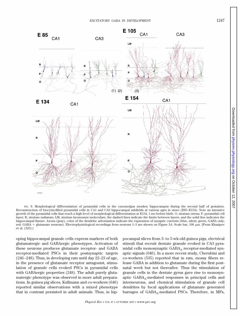
oping hippocampal granule cells express markers of bothglutamatergic and GABAergic phenotypes. Activation ofthese neurons produces glutamate receptor- and GABAreceptor-mediated PSCs in their postsynaptic targets(246–248). Thus, in developing rats until day 22–23 of age,in the presence of glutamate receptor antagonist, stimu-lation of granule cells evoked PSCs in pyramidal cellswith GABAergic properties (248). The adult purely gluta-matergic phenotype was observed in more adult prepara-tions. In guinea pig slices, Kullmann and co-workers (646)reported similar observations with a mixed phenotypethat in contrast persisted in adult animals. Thus, in hip-
pocampal slices from 3- to 5-wk-old guinea pigs, electricalstimuli that recruit dentate granule evoked in CA3 pyra-midal cells monosynaptic GABAA receptor-mediated syn-aptic signals (646). In a more recent study, Cherubini andco-workers (535) reported that in rats, mossy fibers re-lease GABA in addition to glutamate during the first post-natal week but not thereafter. Thus the stimulation ofgranule cells in the dentate gyrus gave rise to monosyn-aptic GABAA-mediated responses in principal cells andinterneurons, and chemical stimulation of granule celldendrites by focal applications of glutamate generatedbarrages of GABAA-mediated PSCs. Therefore, in MFs,
FIG. 8. Morphological differentiation of pyramidal cells in the cynomolgus monkey hippocampus during the second half of gestation.Reconstruction of biocytin-filled pyramidal cells in CA1 and CA3 hippocampal subfields at various ages in utero (E85–E154). Note an intensivegrowth of the pyramidal cells that reach a high level of morphological differentiation at E134, 1 mo before birth. O, stratum oriens; P, pyramidal celllayer; R, stratum radiatum; LM, stratum lacunosum moleculare; the dashed lines indicate the limits between layers, and the solid line indicates thehippocampal fissure. Axons (gray), color of the dendritic arborization indicate the expression of synaptic currents (blue, silent; green, GABA only;red, GABA � glutamate neurons). Electrophysiological recordings from neurons 1–3 are shown on Figure 3A. Scale bar, 100 �m. [From Khazipovet al. (323).]
EXCITATORY GABA IN DEVELOPMENT 1247
Physiol Rev • VOL 87 • OCTOBER 2007 • www.prv.org
on October 12, 2007
physrev.physiology.orgD
ownloaded from
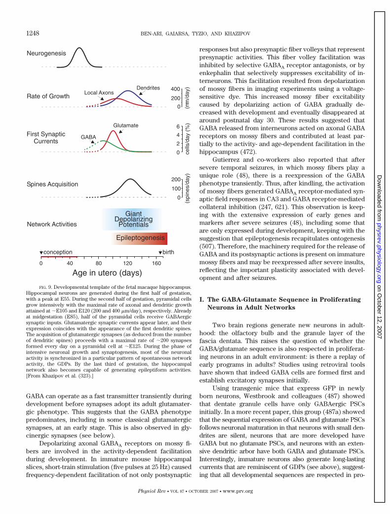
GABA can operate as a fast transmitter transiently duringdevelopment before synapses adopt its adult glutamater-gic phenotype. This suggests that the GABA phenotypepredominates, including in some classical glutamatergicsynapses, at an early stage. This is also observed in gly-cinergic synapses (see below).
Depolarizing axonal GABAA receptors on mossy fi-bers are involved in the activity-dependent facilitationduring development. In immature mouse hippocampalslices, short-train stimulation (five pulses at 25 Hz) causedfrequency-dependent facilitation of not only postsynaptic
responses but also presynaptic fiber volleys that representpresynaptic activities. This fiber volley facilitation wasinhibited by selective GABAA receptor antagonists, or byenkephalin that selectively suppresses excitability of in-terneurons. This facilitation resulted from depolarizationof mossy fibers in imaging experiments using a voltage-sensitive dye. This increased mossy fiber excitabilitycaused by depolarizing action of GABA gradually de-creased with development and eventually disappeared ataround postnatal day 30. These results suggested thatGABA released from interneurons acted on axonal GABAreceptors on mossy fibers and contributed at least par-tially to the activity- and age-dependent facilitation in thehippocampus (472).
Gutierrez and co-workers also reported that aftersevere temporal seizures, in which mossy fibers play aunique role (48), there is a reexpression of the GABAphenotype transiently. Thus, after kindling, the activationof mossy fibers generated GABAA receptor-mediated syn-aptic field responses in CA3 and GABA receptor-mediatedcollateral inhibition (247, 621). This observation is keep-ing with the extensive expression of early genes andmarkers after severe seizures (48), including some thatare only expressed during development, keeping with thesuggestion that epileptogenesis recapitulates ontogenesis(507). Therefore, the machinery required for the release ofGABA and its postsynaptic actions is present on immaturemossy fibers and may be reexpressed after severe insults,reflecting the important plasticity associated with devel-opment and after seizures.
I. The GABA-Glutamate Sequence in Proliferating
Neurons in Adult Networks
Two brain regions generate new neurons in adult-hood: the olfactory bulb and the granule layer of thefascia dentata. This raises the question of whether theGABA/glutamate sequence is also respected in proliferat-ing neurons in an adult environment: is there a replay ofearly programs in adults? Studies using retroviral toolshave shown that indeed GABA cells are formed first andestablish excitatory synapses initially.
Using transgenic mice that express GFP in newlyborn neurons, Westbrook and colleagues (487) showedthat dentate granule cells have only GABAergic PSCsinitially. In a more recent paper, this group (487a) showedthat the sequential expression of GABA and glutamate PSCsfollows neuronal maturation in that neurons with small den-drites are silent, neurons that are more developed haveGABA but no glutamate PSCs, and neurons with an exten-sive dendritic arbor have both GABA and glutamate PSCs.Interestingly, immature neurons also generate long-lastingcurrents that are reminiscent of GDPs (see above), suggest-ing that all developmental sequences are respected in pro-
FIG. 9. Developmental template of the fetal macaque hippocampus.Hippocampal neurons are generated during the first half of gestation,with a peak at E55. During the second half of gestation, pyramidal cellsgrow intensively with the maximal rate of axonal and dendritic growthattained at �E105 and E120 (200 and 400 �m/day), respectively. Alreadyat midgestation (E85), half of the pyramidal cells receive GABAergicsynaptic inputs. Glutamatergic synaptic currents appear later, and theirexpression coincides with the appearance of the first dendritic spines.The acquisition of glutamatergic synapses (as deduced from the numberof dendritic spines) proceeds with a maximal rate of �200 synapsesformed every day on a pyramidal cell at �E125. During the phase ofintensive neuronal growth and synaptogenesis, most of the neuronalactivity is synchronized in a particular pattern of spontaneous networkactivity, the GDPs. By the last third of gestation, the hippocampalnetwork also becomes capable of generating epileptiform activities.[From Khazipov et al. (323).]
1248 BEN-ARI, GAIARSA, TYZIO, AND KHAZIPOV
Physiol Rev • VOL 87 • OCTOBER 2007 • www.prv.org
on October 12, 2007
physrev.physiology.orgD
ownloaded from
liferating neurons. Similar conclusions were reached by Geet al. (227) using a retroviral strategy to express GFP spe-cifically in proliferating cells and their progeny. They re-ported that newly born granule cells of the dentate gyrusexperience first a GABA tonic current, followed by GABA-ergic synapse formation, and then glutamate synapses.Perforated patch recordings showed that the concentra-tion of [Cl�]i is reduced progressively with an initial de-polarization produced by GABA. However, Em alsochanged during maturation, the effect most likely due tothe inherent problems related to the use of patch-clamprecording in immature neurons (37, 626). Interestingly,the authors also showed that the polarity of the actions ofGABA is dependent on the expression of the NKCC1 andKCC2 transporters as in neonates, suggesting that the D-Hshift is respected during the formation of neurons inadults. The density of glutamatergic PSCs is reducedwhen these transporters are knocked out and/or whenGABA synapses are blocked at an early stage, suggestingthat the formation of glutamatergic synapses requires anearlier D-H shift and the formation of GABAergic syn-apses (227). This also confirms the important actions ofGABA as a trophic agent as the growth of dendrites in theprincipal cells is retarded when GABA signaling isblocked. The early expression and importance of GABAsignaling has also been demonstrated in the maturation ofolfactory bulb interneurons where GABAergic responsesdevelop before glutamatergic responses in adult-borngranule and periglomerular neurons (47, 249, 389). Bordeyand colleagues (71, 386, 652) have shown that neuronalprogenitors of the subventricular zone contain GABA andare depolarized by GABA release, suggesting a paracrinesignal among neuronal progenitors that regulates prolif-eration and/or migration.
Therefore, the integration of newly formed neuronsfollows the same rules as during brain maturation. Thisimplies that even in an adult environment, the activationof GABA receptors and synapses exerts excitatory actionson neurons, and only once they are fully integrated dothey progressively acquire the adult properties. This is inkeeping with a developmental program endowed in theseneurons that includes the sequential establishment of re-ceptors, transporters, chloride regulation, and synapseformation.
J. The GABA-Glutamate Sequence in Other
Brain Regions
Morphofunctional studies comparing both the physi-ological and morphological maturation of identified neu-rons have, to the best of our knowledge, not been per-formed in structures other than the hippocampus. How-ever, there are several indications in many brainstructures that GABAergic signaling matures before other
transmitters signals including glutamate but also glycinein the spinal cord. We shall briefly review these studiesbefore discussing the transient GABA phenotype and thedevelopment of mixed synapses that use GABA and gly-cine or GABA and glutamate.
1. The GABA-Glutamate sequence in the neocortex
The migration of GABAergic interneuron of the neo-cortex has been described in detail recently. In contrast toprincipal cells, interneurons are generated in the medialganglionic eminence and migrate tangentially to their finalcortical destinations (352, 415). However, there are alsospecies-specific programs for the generation of neocorti-cal local circuit neurons, since many primate corticalinterneurons are generated in the ventricular and subven-tricular zone (381). Neurons that are just born and mi-grate to the rodent cortical plate (493) have a spontane-ous synaptic activity, the majority of which is GABAergic.First detected as early as embryonic day 18 (E18), spon-taneous GABAergic PSCs are present at birth in �50% ofrecordings. Kriegstein and collaborators (150) have alsoshown that electrical stimulation of layer I gener-ates a GABAergic excitatory PSC in pyramidal neurons,whereas activation of intrinsic layer I neurons with aglutamate agonist fails to produce PSCs in pyramidalcells. This pathway provides spatially restricted excita-tory GABAergic innervations of the distal apical dendritesof pyramidal neurons during the peak period of corticalsynaptogenesis. This group has also reported majorchanges in GABA receptor subunits that result in impor-tant alterations of GABAergic currents during the transi-tion from precursor to fully active neurons. GABA recep-tors expressed in proliferating cells have a higher appar-ent affinity to GABA, long channel open times, and apoorly desensitizing response to activation of GABA re-ceptors (reviewed in Ref. 492). Kriegstein and Prince(354) reported longer and slower synaptic currents inimmature neurons than in adult ones as well as a lack ofinhibitory IPSPs associated with less prominent actions ofGABA receptor antagonists (354). In contrast, GABAergicresponses were readily evoked at all developmentalstages in pyramidal and nonpyramidal neurons from lay-ers II and III of the neocortex (P4 to P10) (405). A differ-ential role of somatic and axon hillock targeted GABAergicaxons in the critical periods has been reported recently,pointing to the important differences of various interneu-ronal populations on the formation of cortical circuits(267). In cortical neurons in culture, operative GABAreceptors are present as early as 1 day after plating,whereas the sensitivity to glutamate was retarded (340).Therefore, a parsimonious conclusion of these observa-tions is that the neocortex follows a similar sequenceduring development even as far as interneurons are con-cerned. This is also reinforced by the observations that
EXCITATORY GABA IN DEVELOPMENT 1249
Physiol Rev • VOL 87 • OCTOBER 2007 • www.prv.org
on October 12, 2007
physrev.physiology.orgD
ownloaded from
initial actions of GABA are excitatory in the neocortex(Table 1). Platel et al. (504) showed recently that appli-cation of glycine to embryonic E12 slices generated adepolarization and an increase of [Ca2�]i via activation ofNa� channels and Na�-Ca2� exchanger. This in turn trig-gers the release of glutamate that will activate AMPA andNMDA receptors, suggesting that tonic release of glycinemay activate chloride-permeable glycine channels provid-ing in the neocortex the excitatory drive needed to acti-vate glutamate ionotropic signaling.
2. The GABA-glutamate sequence in the spinal cord
Early formation of GABA and/or glycine synapseshas been reported in spinal cord neurons in cultures andin situ in several species. In the frog Xenopus laevis,GABA-immunoreactive spinal cord neurons (Kolmer-Agduhr cells) began to appear by 1.2 days shortly afterneural tube closure (69). The pattern of GABA-immuno-reactive cells emerged during embryogenesis, as the den-sity of GABAergic neurons increased. Early maturation ofGABA signaling and the early depolarizing actions ofGABA in Rohon-Beard spinal neurons of Xenopus larvaehave also been extensively studied by Spitzer and co-workers (528, 657). Interestingly, this early maturation ofGABA signaling is also found in cultures with a similartime course as in vivo, and the process requires calciuminflux mediated by spontaneous voltage-dependent chan-nels as well as transcriptional mechanisms (579, 580).Specific GABA immunoreactivity developed in spinal neu-rons in dissociated cell culture with the same time coursepreviously defined in vivo (579, 580).
In the isolated spinal cord preparation from rat fe-tuses (E13.5-E18.5), spontaneous bursts are mediated byglycine receptors at E14.5 and GABA receptors at E15.5;blockers of glutamate synapses have no effects at thatstage (473). Serial sagittal sections of embryos processedfor GAD-65 showed positive axons that extended rostrallyout of the spinal cord into the midbrain at an early devel-opmental stage (E12–15) (618). Clarac et al. (120) devel-oped an isolated brain stem/spinal cord from newbornrats. An early maturation of GABAergic control at variouslevels of the spinal locomotor network was reported in-cluding a locomotor-related GABAergic input onto pri-mary afferent terminals (120, 192). O’Donnovan and co-workers (20, 65) extensively investigated the maturationof GABA signaling in various preparations including theembryonic and posthatched chick lumbosacral spinalcord. GABA immunoreactivity was expressed very early(E4), well before glycine labeling that appeared relativelylate (E8) in embryonic development (20, 65). The authorsconcluded that glycine and GABA provide the earliestfunctional synapses and function transiently as excitatorysynapses at a time when glutamate synapses have notbeen established.
An early expression of GABAergic markers was alsoextensively studies by Barker and co-workers. Thus, inembryonic and postnatal rat spinal cord, GABA is presentas early as embryonic day 13 (E13) (97, 542). Using afluorescence technique to unravel the maturation of var-ious receptors in spinal cord embryos, Barker and co-workers (649) observed, starting from E13, a sequentialexpression of sodium channels followed by GABA andthen only kainate activated glutamate channels.
3. The GABA-glutamate sequence in the
auditory system
Peripheral auditory development proceeds rapidly inkittens between the 7th and 20th postnatal days, andinput/output functions achieve maturity only at that stage(647). Yet, inhibition evaluated by the effects of iono-phoresis of GABA and other amino acids operates at anearly stage (647). In a subsequent study (648), the samegroup showed that capacity of GABA to reduce sponta-neous or acoustically evoked discharge rates was directlyrelated to control discharge rates in both immature andmature animals. The authors suggested that the GABAsignaling operates prior to the arrival of sensory informa-tion. Kandler and collaborators (231) have shown recentlythat in the lateral superior olive (LSO), a nucleus in themammalian sound localization system that receives inhib-itory input from the medial nucleus of the trapezoid body(MNTB), there is a specific elimination/strengthening ofGABAergic and glycinergic synapses. This step is essen-tial for the formation of a precise tonotopic map. Theimmature GABA/glycinergic synapses in the rat LSO alsorelease glutamate, which activates postsynaptic NMDAreceptors. Vesicular glutamate and GABA transporter arecolocalized in single MNTB terminals. At an early stage,glutamatergic transmission relying on synapse-specificactivation of NMDARs may play an important role insynapse elimination and in activity-dependent refinementof inhibitory circuits.
4. Other structures
An electronic microscopy analysis of the develop-ment of the inferior olivary complex of the rat also re-vealed a delayed maturation of GAD-containing terminalswith the classical morphological organization beingreached only after P10-P15 (234). Before that stage,GABAergic projecting elements appear to remain in awaiting compartment. In cultures of septum and hip-pocampus, Koller et al. (340) determined the maturationof functional receptors by applications of selective ago-nists. The authors reported that the GABAA receptor de-veloped prior to the glutamate receptors in both regionsand appeared as early as a few days after plating, whereasover 9 days were required for the appearance of glutamateNMDA and non-NMDA receptor subtypes. Interestingly,
1250 BEN-ARI, GAIARSA, TYZIO, AND KHAZIPOV
Physiol Rev • VOL 87 • OCTOBER 2007 • www.prv.org
on October 12, 2007
physrev.physiology.orgD
ownloaded from
the receptors appeared earlier in the hippocampus than inthe septum (340).
In a pioneer study on hypothalamic neurons in cul-tures and slices, Van Den Pol and co-workers (221)showed that the synaptic release of GABA is not onlyexcitatory but plays a more robust role than glutamate ingenerating spike activity initially. The authors suggested“GABA not glutamate provides the initial excitatorydrive.” Applications of the GABAA receptor antagonistbicuculline induced a dramatic decrease in spike fre-quency (83% decrease) in developing neurons, three timesgreater than that generated by glutamate receptor antag-onists APV and CNQX that block NMDA and AMPA sub-type of receptors, respectively. The membrane propertiesof immature neurons are compatible with robust excita-tion, since GABA generates bursts of action potentials(221, 482).
In the developing retina (365), GABAergic neuronsare immature at birth and become functionally matureby �9 days after birth. Receptive-field properties of manyneurons are determined by GABAergic inhibitory pro-cesses that operate around P11–P28 in the visual cortex ofkitten (667).
K. A Developmental Switch From GABA to Glycine
Since GABA and glycine receptors are permeable tochloride and other anions, they should follow a similarD-H shift during maturation. This indeed has been shownto be the case in several systems (see sect. III). An addi-tional interest in studying the maturation of glycine re-ceptors during development stems from the fact that inadult neurons, single terminals can release GABA andglycine (293). Thus, in the spinal cord, unitary IPSCscomposed of a strychnine-sensitive, glycine receptor-me-diated component and a bicuculline-sensitive, GABA re-ceptor-mediated component have been recorded (293).
In spinal cord slices of 17- to 18-day-old embryos(E17–18) and 1- to 3-day-old postnatal rats (P1–3), Zis-kind-Conhaim and colleagues (217) have shown an earliermaturation of GABAergic signaling in motoneurons. Re-lying on the different time courses and the effects ofselective antagonists, the authors suggested developmen-tal switch from predominantly long-duration GABAergicsynaptic currents to short-duration glycinergic. Duringembryonic development, the conductance increase gener-ated by GABA was sevenfold larger than that generated byglycine in utero but not after birth (216–218). At birth,whole cell patch-clamp recordings from rat superficialdorsal horn neurons suggest that GABAergic but not gly-cinergic PSCs are present (26). At all ages, between P0and P13–P14, GABAergic mechanisms are initially domi-nant over glycinergic events (26; also see Refs. 313, 515).Large, long-lasting spontaneous IPSPs with mini IPSCs
were also recorded in cultures of rat spinal cord neurons(382). These appear to reflect an increased intracellularcalcium concentration in the presynaptic terminals.
In the auditory system, GABA and glycine are presentin several neuronal types. Thus, in the cochlear nucleus ofthe guinea pig, the vast majority of putative inhibitoryendings contain glycine, GABA, or both, suggesting that inat least a proportion of terminals GABA may be releasedwith glycine (296). In the medial nucleus of the trapezoidbody, Trussell and co-workers (25) showed that in younganimals (P5–P7), 80% of the responses evoked by electri-cal stimuli are blocked by GABA receptor antagonists and20% by glycine receptor antagonists. This contrasts withthe situation at the older age group (P13–P15) whereglycine but not GABAA receptor antagonists blocked theresponses. Since GABAergic PSCs have slower kineticsthan glycinergic PSCs, this progressive maturation of gly-cine PSCs has important functional consequences con-cerning the development of hearing. Thus, at P25, IPSCsare 10 times briefer than at birth (25; see also Ref. 622). Asimilar switch from GABA to glycine is also observed inthe lateral superior olive during the first two postnatalweeks, with GABAergic IPSCs declining from almost 80%at postnatal days 3–5 to close to 10% at P12–P16. Therewas an equal and opposite increase in the glycinergiccomponent during this same period (349). In “mixed”mIPSCs, resulting from corelease of glycine and GABAfrom the same vesicles, there is a similar developmentalswitch in this structure (470). In the gerbil lateral superiorolive (LSO), there is a transition from GABAergic to gly-cinergic transmission during the first two postnatal weeks(349). Several other studies have examined this transitionand the role of activity in refinement and maturation ofchloride-dependent glycine and GABA channels (41, 98,231, 237, 281, 333, 560). Collectively, these studies possi-bly reflect the pioneer role of GABA that is expressed atan early stage prior to synapse refinement and expressionof the mature phenotype, as if GABA signaling is “auto-matically” expressed first in developing systems.
L. Conclusion: A Model That Integrates
These Findings
The observations summarized above lead to a hypo-thetical model that can be tested for its functional signif-icance. The main features of the models are 1) a sequen-tial formation of GABA then glutamate synapses; 2) axonsdevelop before dendrites; 3) dendrites are innervated be-fore somata and interneurons that innervate dendrites ofprincipal cells mature before those that are targeted toinnervate the soma; and 4) interneurons follow a similarGABA/glutamate sequence of synapse formation.
At an early stage, GABAergic interneurons and pref-erentially interneurons that innervate the dendrites of
EXCITATORY GABA IN DEVELOPMENT 1251
Physiol Rev • VOL 87 • OCTOBER 2007 • www.prv.org
on October 12, 2007
physrev.physiology.orgD
ownloaded from
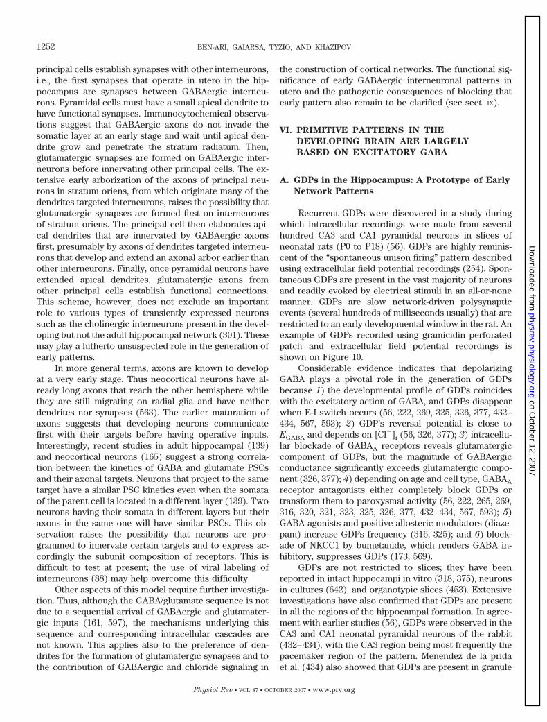
principal cells establish synapses with other interneurons,i.e., the first synapses that operate in utero in the hip-pocampus are synapses between GABAergic interneu-rons. Pyramidal cells must have a small apical dendrite tohave functional synapses. Immunocytochemical observa-tions suggest that GABAergic axons do not invade thesomatic layer at an early stage and wait until apical den-drite grow and penetrate the stratum radiatum. Then,glutamatergic synapses are formed on GABAergic inter-neurons before innervating other principal cells. The ex-tensive early arborization of the axons of principal neu-rons in stratum oriens, from which originate many of thedendrites targeted interneurons, raises the possibility thatglutamatergic synapses are formed first on interneuronsof stratum oriens. The principal cell then elaborates api-cal dendrites that are innervated by GABAergic axonsfirst, presumably by axons of dendrites targeted interneu-rons that develop and extend an axonal arbor earlier thanother interneurons. Finally, once pyramidal neurons haveextended apical dendrites, glutamatergic axons fromother principal cells establish functional connections.This scheme, however, does not exclude an importantrole to various types of transiently expressed neuronssuch as the cholinergic interneurons present in the devel-oping but not the adult hippocampal network (301). Thesemay play a hitherto unsuspected role in the generation ofearly patterns.
In more general terms, axons are known to developat a very early stage. Thus neocortical neurons have al-ready long axons that reach the other hemisphere whilethey are still migrating on radial glia and have neitherdendrites nor synapses (563). The earlier maturation ofaxons suggests that developing neurons communicatefirst with their targets before having operative inputs.Interestingly, recent studies in adult hippocampal (139)and neocortical neurons (165) suggest a strong correla-tion between the kinetics of GABA and glutamate PSCsand their axonal targets. Neurons that project to the sametarget have a similar PSC kinetics even when the somataof the parent cell is located in a different layer (139). Twoneurons having their somata in different layers but theiraxons in the same one will have similar PSCs. This ob-servation raises the possibility that neurons are pro-grammed to innervate certain targets and to express ac-cordingly the subunit composition of receptors. This isdifficult to test at present; the use of viral labeling ofinterneurons (88) may help overcome this difficulty.
Other aspects of this model require further investiga-tion. Thus, although the GABA/glutamate sequence is notdue to a sequential arrival of GABAergic and glutamater-gic inputs (161, 597), the mechanisms underlying thissequence and corresponding intracellular cascades arenot known. This applies also to the preference of den-drites for the formation of glutamatergic synapses and tothe contribution of GABAergic and chloride signaling in
the construction of cortical networks. The functional sig-nificance of early GABAergic interneuronal patterns inutero and the pathogenic consequences of blocking thatearly pattern also remain to be clarified (see sect. IX).
VI. PRIMITIVE PATTERNS IN THE
DEVELOPING BRAIN ARE LARGELY
BASED ON EXCITATORY GABA
A. GDPs in the Hippocampus: A Prototype of Early
Network Patterns
Recurrent GDPs were discovered in a study duringwhich intracellular recordings were made from severalhundred CA3 and CA1 pyramidal neurons in slices ofneonatal rats (P0 to P18) (56). GDPs are highly reminis-cent of the “spontaneous unison firing” pattern describedusing extracellular field potential recordings (254). Spon-taneous GDPs are present in the vast majority of neuronsand readily evoked by electrical stimuli in an all-or-nonemanner. GDPs are slow network-driven polysynapticevents (several hundreds of milliseconds usually) that arerestricted to an early developmental window in the rat. Anexample of GDPs recorded using gramicidin perforatedpatch and extracellular field potential recordings isshown on Figure 10.
Considerable evidence indicates that depolarizingGABA plays a pivotal role in the generation of GDPsbecause 1) the developmental profile of GDPs coincideswith the excitatory action of GABA, and GDPs disappearwhen E-I switch occurs (56, 222, 269, 325, 326, 377, 432–434, 567, 593); 2) GDP’s reversal potential is close toEGABA and depends on [Cl�]i (56, 326, 377); 3) intracellu-lar blockade of GABAA receptors reveals glutamatergiccomponent of GDPs, but the magnitude of GABAergicconductance significantly exceeds glutamatergic compo-nent (326, 377); 4) depending on age and cell type, GABAA
receptor antagonists either completely block GDPs ortransform them to paroxysmal activity (56, 222, 265, 269,316, 320, 321, 323, 325, 326, 377, 432–434, 567, 593); 5)GABA agonists and positive allosteric modulators (diaze-pam) increase GDPs frequency (316, 325); and 6) block-ade of NKCC1 by bumetanide, which renders GABA in-hibitory, suppresses GDPs (173, 569).
GDPs are not restricted to slices; they have beenreported in intact hippocampi in vitro (318, 375), neuronsin cultures (642), and organotypic slices (453). Extensiveinvestigations have also confirmed that GDPs are presentin all the regions of the hippocampal formation. In agree-ment with earlier studies (56), GDPs were observed in theCA3 and CA1 neonatal pyramidal neurons of the rabbit(432–434), with the CA3 region being most frequently thepacemaker region of the pattern. Menendez de la pridaet al. (434) also showed that GDPs are present in granule
1252 BEN-ARI, GAIARSA, TYZIO, AND KHAZIPOV
Physiol Rev • VOL 87 • OCTOBER 2007 • www.prv.org
on October 12, 2007
physrev.physiology.orgD
ownloaded from
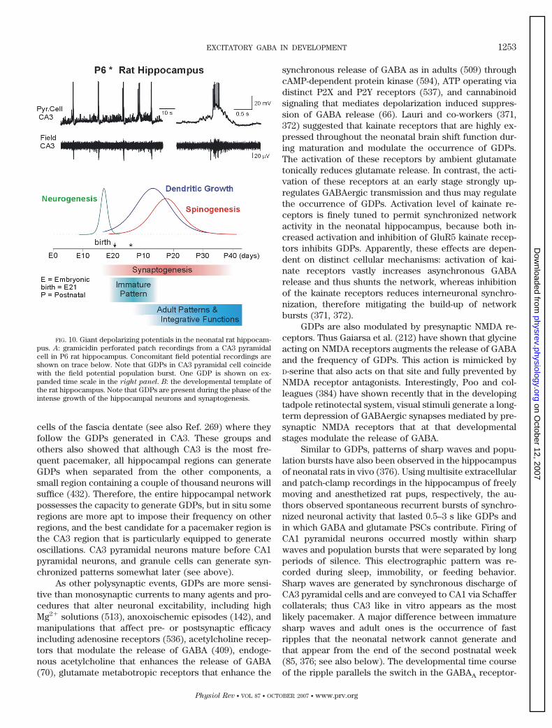
cells of the fascia dentate (see also Ref. 269) where theyfollow the GDPs generated in CA3. These groups andothers also showed that although CA3 is the most fre-quent pacemaker, all hippocampal regions can generateGDPs when separated from the other components, asmall region containing a couple of thousand neurons willsuffice (432). Therefore, the entire hippocampal networkpossesses the capacity to generate GDPs, but in situ someregions are more apt to impose their frequency on otherregions, and the best candidate for a pacemaker region isthe CA3 region that is particularly equipped to generateoscillations. CA3 pyramidal neurons mature before CA1pyramidal neurons, and granule cells can generate syn-chronized patterns somewhat later (see above).
As other polysynaptic events, GDPs are more sensi-tive than monosynaptic currents to many agents and pro-cedures that alter neuronal excitability, including highMg2� solutions (513), anoxoischemic episodes (142), andmanipulations that affect pre- or postsynaptic efficacyincluding adenosine receptors (536), acetylcholine recep-tors that modulate the release of GABA (409), endoge-nous acetylcholine that enhances the release of GABA(70), glutamate metabotropic receptors that enhance the
synchronous release of GABA as in adults (509) throughcAMP-dependent protein kinase (594), ATP operating viadistinct P2X and P2Y receptors (537), and cannabinoidsignaling that mediates depolarization induced suppres-sion of GABA release (66). Lauri and co-workers (371,372) suggested that kainate receptors that are highly ex-pressed throughout the neonatal brain shift function dur-ing maturation and modulate the occurrence of GDPs.The activation of these receptors by ambient glutamatetonically reduces glutamate release. In contrast, the acti-vation of these receptors at an early stage strongly up-regulates GABAergic transmission and thus may regulatethe occurrence of GDPs. Activation level of kainate re-ceptors is finely tuned to permit synchronized networkactivity in the neonatal hippocampus, because both in-creased activation and inhibition of GluR5 kainate recep-tors inhibits GDPs. Apparently, these effects are depen-dent on distinct cellular mechanisms: activation of kai-nate receptors vastly increases asynchronous GABArelease and thus shunts the network, whereas inhibitionof the kainate receptors reduces interneuronal synchro-nization, therefore mitigating the build-up of networkbursts (371, 372).
GDPs are also modulated by presynaptic NMDA re-ceptors. Thus Gaiarsa et al. (212) have shown that glycineacting on NMDA receptors augments the release of GABAand the frequency of GDPs. This action is mimicked byD-serine that also acts on that site and fully prevented byNMDA receptor antagonists. Interestingly, Poo and col-leagues (384) have shown recently that in the developingtadpole retinotectal system, visual stimuli generate a long-term depression of GABAergic synapses mediated by pre-synaptic NMDA receptors that at that developmentalstages modulate the release of GABA.
Similar to GDPs, patterns of sharp waves and popu-lation bursts have also been observed in the hippocampusof neonatal rats in vivo (376). Using multisite extracellularand patch-clamp recordings in the hippocampus of freelymoving and anesthetized rat pups, respectively, the au-thors observed spontaneous recurrent bursts of synchro-nized neuronal activity that lasted 0.5–3 s like GDPs andin which GABA and glutamate PSCs contribute. Firing ofCA1 pyramidal neurons occurred mostly within sharpwaves and population bursts that were separated by longperiods of silence. This electrographic pattern was re-corded during sleep, immobility, or feeding behavior.Sharp waves are generated by synchronous discharge ofCA3 pyramidal cells and are conveyed to CA1 via Schaffercollaterals; thus CA3 like in vitro appears as the mostlikely pacemaker. A major difference between immaturesharp waves and adult ones is the occurrence of fastripples that the neonatal network cannot generate andthat appear from the end of the second postnatal week(85, 376; see also below). The developmental time courseof the ripple parallels the switch in the GABAA receptor-
FIG. 10. Giant depolarizing potentials in the neonatal rat hippocam-pus. A: gramicidin perforated patch recordings from a CA3 pyramidalcell in P6 rat hippocampus. Concomitant field potential recordings areshown on trace below. Note that GDPs in CA3 pyramidal cell coincidewith the field potential population burst. One GDP is shown on ex-panded time scale in the right panel. B: the developmental template ofthe rat hippocampus. Note that GDPs are present during the phase of theintense growth of the hippocampal neurons and synaptogenesis.
EXCITATORY GABA IN DEVELOPMENT 1253
Physiol Rev • VOL 87 • OCTOBER 2007 • www.prv.org
on October 12, 2007
physrev.physiology.orgD
ownloaded from
mediated signaling from excitation to inhibition. Popula-tion bursts were characterized by increase of unit activitywithout prominent field potential typically followed bysharp waves but could also occur independently. Wholecell recordings in situ revealed again in agreement within vitro recordings a contribution of both GABA andglutamate receptor-mediated synaptic signals in their gen-eration (376). Importantly, the NKCC1 blocker bumet-anide that renders GABA inhibitory efficiently suppressedboth sharp waves in vivo and GDPs in vitro during thepostnatal period, suggesting that depolarizing GABA isinstrumental in the generation of the early hippocampalpatterns both in vivo and in vitro (173, 569).
Although depolarizing and excitatory GABA plays apivotal role in the generation of GDPs in the neonatalhippocampus, GABA also exerts inhibitory action in im-mature networks. Indeed, blockade of GABA receptorsinduces interictal-like epileptiform activity in the neonatalhippocampal slices (316, 325, 326, 367, 659) and ictal-liketonic-clonic seizures in the intact hippocampus in vitro(320, 321) and in vivo (282). Although GABAA agonistsand positive allosteric modulators transiently increaseGDPs frequency, the effect is followed by depression dueto shunting inhibition (316, 325). Also, activation of GluR5kainate receptors increases asynchronous GABA releaseand inhibits GDPs (372). Thus GABA exerts dual excita-tory and inhibitory actions in the neonatal hippocampalnetwork. Depolarizing GABA inhibits via a shuntingmechanism by increasing membrane conductance andlimiting depolarization above EGABA. In addition, depolar-izing GABA can also activate voltage-gated K� channels(401, 457) and attenuate the amplitude of the presynapticspike through a combination of shunting and Na� channelinactivation, a phenomenon observed in invertebrate sen-sory fibers, dorsal root ganglion cell terminals, and secre-tory nerve endings (100, 180, 184, 185, 315, 530, 679).
B. A Problem of Terminology
GDPs have been identified initially with intracellularcurrent-clamp recordings, hence the name. The terminol-ogy therefore does not correspond to events recordedwith other techniques. Relying on field recordings andimaging techniques, bursting activity, unison firing pat-tern, and early networks oscillations (ENOs) have, re-spectively, been proposed to term the same phenomenon(222, 254, 496). Clearly none of the names proposed fullyrepresents all the features of the events, and we shall usethe original terminology and restrict it to define patternspresent transiently in immature networks consequent tothe presence of a large number of excitatory GABAergiccurrents.
C. How Are GDPs Generated? How do
They Propagate?
1. The GABA D-H population model
In this model (56, 325, 326, 377), hippocampal localnetworks composed of a few hundred GABAergic inter-neurons and glutamatergic pyramidal neurons are en-dowed with autorhytmicity largely based on synergisticinteractions between recurrent excitatory GABAergic andglutamatergic PSCs. In this model, both glutamatergicand GABAergic neurons and synapses are the key gen-erators. GDPs are considered as a signature of the stateof maturation of the network at a given developmentalstage: GDPs are present as long as a substantial percentof GABAergic synapses are excitatory. In addition tothe E-I shift in the actions of GABA, the late establish-ment of functional perisomatic interneurons-basketcells and axoaxonic neurons may provide an additionalimportant signal for the cessation of GDPs. A centralaspect of the model proposed is that in spite of therequirement of glutamatergic synapses, the level ofneuronal depolarization and excitation during GDPs ismainly determined by GABAergic synapses and inter-neurons. This model is compatible with the main fea-tures of GDPs, its developmental occurrence that par-allels excitatory actions of GABA, and its modulationby various conditions and agents that control networkexcitability.
2. The gap junction pacemaker hilar
interneurons model
Strata et al. (593) have suggested that GDPs aregenerated by an endogenous pacemaker current presentin hilar interneurons “paced” by a hyperpolarization-acti-vated current, with properties of Ih and synchronized viagap junctions. The excitatory actions of GABA are notrequired for that model to operate. The Ih current medi-ated by HCN1 channels has also been suggested to con-tribute to GDPs recently (61). The selective Ih blockerZD7288 disrupted GDP generation and abolished sponta-neous bursting of the CA3 pyramidal cells at frequenciestypical of GDPs without major influence on interneuronalfiring (61). In addition, the “hilar interneurons” modelcannot explain the generation by all components of thehippocampal circuit of GDPs including subslices of vari-ous hippocampal regions that do not contain the hilarzone, and they are blocked by the glutamate receptorsantagonists (56, 60, 432, 434). It is also difficult to recon-cile with all other studies that point to crucial roles of theGABA E-I shift in the generation of GDPs by local net-works.
1254 BEN-ARI, GAIARSA, TYZIO, AND KHAZIPOV
Physiol Rev • VOL 87 • OCTOBER 2007 • www.prv.org
on October 12, 2007
physrev.physiology.orgD
ownloaded from
3. A combined intrinsic current and recurrent
glutamatergic excitation model
Another model (567, 568) incorporates the depolar-izing actions of GABA and intrinsic bursting activity ofCA3 pyramidal neurons generated by voltage-dependentcurrents. In this model, pacemaker intrinsic pyramidalbursting neurons are the generators and GABA only actsas a tonic source of depolarization that enables theseneurons to reach the threshold for burst firing. Phasicexcitation by GABA plays little role in the generation ofGDPs as GDPs in interneurons do not precede GDPsrecorded in field recordings, suggesting that the drive isendogenous to pyramidal cells. The triggering and termi-nation of intrinsic bursts and of GDPs are determined,respectively, by a sodium persistent slow current for theregenerative depolarization that triggers intrinsic burstsand a slow Ca2�-activated K� current for the postburstafterhyperpolarization. The frequency of GDPs is deter-mined by the kinetics of these currents and not by syn-apse-driven events. These intrinsic events are translatedto network-driven GDPs by means of the recurrent gluta-matergic excitatory collaterals present in CA3 pyramidalneurons (567). In that respect, GDPs are similar to inter-ictal patterns generated when inhibition is blocked in CA3neurons (567) except that they require tonic GABA fortheir generation. This model is of obvious interest as itincorporates basic properties of the CA3 pacemaker re-gion in developing networks (see above and Ref. 70).
However, other observations cannot be reconciledwith this model. First, in an earlier intracellular recordingstudy, systematic bursting in CA3 (or CA1) pyramidalneurons was seldom observed (56), in keeping with Yaariand co-workers (374) who reported spontaneous bursts inCA3 after but not before P11 reaching a peak around P18.Also, GDPs are recorded in granule cells where they areclearly not mediated by intrinsic bursting properties butby GABAergic interneurons (269, 434). GDPs can be gen-erated in the isolated CA1 region where neurons are notendowed with intrinsic burst patterns and poorly devel-oped glutamatergic collaterals (222; L. Anikstejn, unpub-lished data). Interneurons clearly generate GDPs, even atan embryonic stage when most pyramidal neurons arequiescent, suggesting that interneurons set the pace ofhippocampal patterns (265). Also, antagonists of GABAB
receptors augment significantly the duration (by several-fold) of GDPs, suggesting that the release of GABA andother presynaptic mechanisms are responsible for termi-nating the event (427). Patterns equivalent to GDPs arepresent in a wide range of structures (see below) that donot seem to possess a pacemaker region equivalent toCA3 pyramidal neurons. Also worth stressing, the mem-brane potential threshold required for the generation of apersistent sodium channel is closer to the threshold ofsodium spikes than to the resting membrane potential
(200). However, clearly several aspects of this model areimportant, and more detailed investigations are needed toclarify this issue.
The propagation of GDPs was studied in detail in theintact hippocampal formation in vitro (320, 375), in whichit is possible to investigate with more detail the propaga-tion of GDPs across the entire hippocampal formation,including the septum and entorhinal cortex. Patch-clamprecordings from a large sample of neurons revealed GDPs(375) that are identical to those recorded in slices. Withthe use of multiple field recordings and dual patch record-ings, GDPs were found to propagate along the septotem-poral axis of the hippocampus with short delays betweenadjacent neurons and long ones between distal sites(375). The spontaneous activity of the septal pole washigher than that of the temporal pole, and separating theseptal and temporal poles did not affect the frequency ofGDPs in the former (11/min) but reduced by half that ofthe temporal pole (to 5/min). Also, GDPs generated in theseptal pole had a much higher probability for propagationto the temporal pole than the converse, suggesting thatthe hippocampus operates like a syncytium in which allcomponents can generate rhythms but some are moredeveloped, have a higher GDP frequency, and imposetheir patterns. The septal pole paces the rest of the hip-pocampus (375), as it has a higher density of active glu-tamatergic synapses and a higher spontaneous activity.The differential maturation of interneurons in the septaland temporal poles most likely also plays a role in thisgradient (89). Recordings from the septum indicate thatGDPs originate in the hippocampus as cutting the con-nections between the septum and hippocampus blockedthe GDPs in the former but not the latter. GDPs are alsosynchronized between both hippocampi by the commis-sural axons, and both septal poles generate synchronizedGDPs, suggesting that commissural axons are operativeat birth already (see also Ref. 319). GDPs propagate toother limbic structures in vitro, including the entorhi-nal cortex (39).
D. GDPs in Subhuman Primates
In primates, the prenatal differentiation of pyramidalneurons, granular neurons (164), and interneurons (63,64) and the early establishment of synaptic connectionsprovide a basis for the in utero emergence of synchro-nized activities. Patch-clamp recordings of hippocampalneurons were performed in slices of macaque cynomol-gus fetuses delivered by cesarean section during the sec-ond half of gestation (323) (see Figs. 8 and 9). No spon-taneous network-driven activity was observed at midges-tation (E85) and seldom close to term (E154; term is 165days). In contrast, between E105 and E134, the majorityof pyramidal neurons displayed periodic network-driven
EXCITATORY GABA IN DEVELOPMENT 1255
Physiol Rev • VOL 87 • OCTOBER 2007 • www.prv.org
on October 12, 2007
physrev.physiology.orgD
ownloaded from
events occurring at a frequency of 0.13 � 0.04 Hz. Thisactivity was highly reminiscent of GDPs. Fetal monkeyGDPs displayed the typical features of polysynaptic net-work-driven events: 1) they were synchronized in simul-taneously recorded neurons; 2) they could be evoked inan all-or-none manner by electrical stimulation; 3) theirfrequency was independent of the membrane potential;and 4) they were completely blocked by TTX. GDPs weresynchronously generated in paired recordings from py-ramidal cells and interneurons and were mediated bysynaptically activated GABAA and glutamate receptors.As in the rat hippocampus, the GDPs were blocked byglutamate receptor antagonists CNQX and APV and weretransformed to paroxysmal discharges by the GABAA re-ceptor antagonist bicuculline. The total count of synapticevents revealed that nearly half of GABAergic and gluta-matergic synaptic activity is synchronized in GDPs. Thussimilar to the neonatal rat hippocampus, GDPs in the fetalmacaque hippocampal slices are generated by GABAergicinputs from interneurons and glutamatergic recurrent col-lateral synapses, and the synaptic activity is highly syn-chronized during the phase of intensive neuronal growthand synaptogenesis.
E. Other Early Patterns in the
Developing Hippocampus
1. Periodic inward currents
Periodic inward currents (PICs) (19) are readily gen-erated in neonatal slices by several agents in the presenceof TTX, TEA, and CsCl to block most sodium and potas-sium channels. Thus recurrent applications of themetabotropic agonist ACPD generated PICs that persistfor several hours. PICs are generated by nonspecific cat-ionic currents and mediated by a pulsatile release ofglutamate that activates ionotropic glutamate receptors.PICs are blocked by removing calcium from the externalmedium as well as the addition of caffeine and thapsigar-gin but not by ryanodine. PICs are triggered by elevationof [Ca2�]i, after mobilization of Ca2� from inositol 1,4,5-trisphosphate (InsP3)-sensitive stores that in turn willlead to a pulsatile release of glutamate from presynapticnerve terminals. The relation of PICs to other uniquefeatures of the developing network remains to be inves-tigated; GABAergic receptors do not appear to play a rolein PICs contrary to GDPs, yet their developmental curvesare quite similar, suggesting that features other than theE-I shift are involved in the generation of early patterns.
2. Gap junction-mediated oscillations
Gap junction-mediated oscillations were recentlyidentified in immature hippocampal networks using fastmultibeam two-photon calcium microscopy to record
hundreds of neurons simultaneously in immature hip-pocampal slices from P0 to P8 (141a). With this technique,it is possible to record the activity of a large number ofneurons simultaneously and identify the pattern presentin every neuron (138). Using this tool, the authors showedthat before birth, most immature neurons in the CA1region do not produce a coherent pattern but intrinsicrecurrent calcium spikes. The first coherent pattern ap-pears around delivery and is characterized by a plateaupotential that synchronizes a small population of gapjunction-coupled neurons. At a subsequent stage, GDPsappear. They are present simultaneously in hundreds ofneurons and propagate to the entire network. Therefore,activity shifts from a voltage-gated localized pattern in-volving selected neuronal assemblies to a synaptic onethat reaches most neurons via excitatory GABA and glu-tamtergic synapses. These observations suggest that inaddition to a sequential maturation of individual ionicchannels (490) there is a sequential maturation of net-work events with gap junction-mediated signals preced-ing synapse-mediated synchronized events (see Ref. 459for review). Interestingly, in the neocortex, gap junction-mediated oscillations are generated by carbachol primar-ily through the action of subplate cortical neurons (167).
F. Oscillations in Other Developing Structures
1. The neocortex
A) IN VITRO PATTERNS. Oscillations and network patternsare frequent in various parts of the developing neocortex,consistent with the intrinsic capacity of developing net-works to generate oscillations (302). These patterns arethought to play important roles in wiring essential con-nections notably cortical maps and in other importantfunctions including migration and proliferation (346, 347,489, 658). Calcium oscillations were described by Krieg-stein and co-workers in the neocortical ventricular zone(489); calcium waves also propagate spontaneouslythrough radial glial cells in the proliferative cortical ven-tricular zone. They require connexin hemichannels, P2Y1ATP receptors, and intracellular InsP3-mediated calciumrelease. Disrupting these waves decreases proliferationduring of embryonic neurogenesis (658). The same groupalso showed that metabotropic glutamate receptors alsogenerate oscillations in the developing neocortex (198).
Using imaging techniques and visual patch record-ings, Yuste and co-workers (676, 677) showed that neo-natal neocortical neurons oscillate when strongly stimu-lated by 0 Mg2� and 5 mM K�, or high K� containingACSF (500, 501). These correlated activities were medi-ated by the depolarizing actions of GABA at that age thatalso increases intracellular calcium. However, TTX orblocking glycine receptors did not affect the activity,
1256 BEN-ARI, GAIARSA, TYZIO, AND KHAZIPOV
Physiol Rev • VOL 87 • OCTOBER 2007 • www.prv.org
on October 12, 2007
physrev.physiology.orgD
ownloaded from
whereas cholinergic and glutamatergic antagonists mod-ulated it, suggesting a complex interaction of severalsignals. The authors favored the hypothesis that a releaseof calcium from intracellular stores plays an importantrole in keeping with other lines of evidence. As in otherbrain structures, gap junctions between developing neo-cortical neurons are frequent and may play an importantrole in oscillations (302, 500). Interestingly, the authorsshowed that these patterns might presage adult functionalarchitecture as the cortex could be partitioned in domainsof spontaneously coactive neuronal populations coupledby gap junctions (302, 677).
Using two photon dynamic imaging, Konnerth andco-workers (2, 222) showed the presence of corticalwaves to which they referred to as cortical ENOs(C-ENOs) in the rat neonatal neocortical slices and in vivo(P0 to P12) (2, 222). ENOs that are expressed only duringthe first postnatal week much like hippocampal GDPs(see above) occur at a very low rate (1–12/min) andpropagate across the entire cortical mantle at a speed of2 mm/min, a rate that is much slower than GDPs andother patterns and that may suggest a nonsynaptic prop-agation mechanism. The duration of single events wastens of seconds. C-ENOs are completely blocked byAMPA or NMDA receptor antagonists but not by GABAreceptor antagonists at least until the end of the firstpostnatal week. In more adult slices, bicuculline gener-ated oscillations most likely of an epileptogenic type inkeeping with the parallel to the E-I shift of the actions ofGABA during the transition from immature to more adultpattern. C-ENOs are quite different from GDPs by theearly contribution of glutamate signaling and limited con-tribution of GABA. In contrast to these observations,Luhmann and co-workers (264) showed that at birth bicu-culline but also muscimol block calcium oscillations in P0neocortical slices. Clearly, a detailed morphofunctionaldescription of the sequential maturation of GABA andglutamatergic synapses is required to gain a better picturefor the development of neocortical patterns.
Luhmann and co-workers (167) have described a pat-tern of carbachol-induced propagating network beta os-cillations in the neonatal mouse neocortex. In the new-born mouse, this activity required an intact subplate andwas strongly synchronized within a cortical column bygap junctions. With the developmental disappearance ofthe subplate at the end of the first postnatal week, acti-vation of NMDA receptors in the immature cortical net-work was needed to generate this columnar activity pat-tern. It was suggested that during a brief developmentalperiod the cortical network switches from a subplate-driven, gap junction-coupled syncytium to a synaptic net-work acting through NMDA receptors to generate syn-chronized oscillatory activity, which may function as anearly functional template for the development of the cor-tical columnar architecture (167). These oscillations were
not affected by blockade of GABAA receptors, suggestinglimited participation of GABA interneurons in their gen-eration (167).
Voigt et al. (642) have performed a detailed study ofthe oscillations present in neocortical cultures. In an ini-tial study, these authors (642) reported the presence ofnetwork oscillations driven by GABA and glutamate PSCsin neuronal cultures that are identical to GDPs. GiantGABAergic neurons are key elements in their generationas they are only present when at least two of theseneurons per square millimeter were present in the culture,with lower density GDPs not present. Changes in thedensity of other neurons did not influence the presence ofoscillations. These neurons form an interconnected net-work with extended axonal arborization enabling them to“collect” information from large regions of the cortex;they were also identified as corresponding to largeGABAergic neurons derived from the primordium plexi-form layer that reside in the subplate at the time of birth.Therefore, a subpopulation of giant GABAergic neuronsthat the authors consider as being a type of basket cellsextends its axonal arbor and triggers network oscilla-tions. Using this preparation, the authors suggested inmore recent studies that the survival of a subpopulationof GABAergic interneurons that the authors considermay correspond to the calretinin/vasoactive intestinalpolypeptide interneurons, critically depends on the pres-ence of functional NMDA receptors, since chronic block-ade of these receptors led to a dramatic loss of theseneurons (154). This observation reinforces the importantlinks between GABA and NMDA signaling. Since GABAand NMDA receptors act in synergy to generate GDPs,this feedback control will provide a mechanism for crosscontrol of the maturation of GABA and NMDA signaling.In keeping with this, chronic blockade of NMDA recep-tors exerts a more severe action than blocking AMPAreceptors. In another study, the same group (642) showedthat GABA receptor antagonists fully block GDPs duringthe first 3–4 days after their emergence but not later,suggesting that GABA acts as an excitatory neurotrans-mitter in these young networks. This observation is incontradiction with the observations of Konnerth and co-workers (222) who reported that in intact cortices, GABAreceptor antagonists do not block GDPs (or ENOs) at anearly stage. In older cultures, oscillations persist in thepresence of bicuculline but with a smaller burst frequencyand a more sustained depolarization and burst-associatedCa2� transients (485). In a more recent study, the samegroup used an interesting preparation to investigate therelation between network oscillations and transformationof silent synapses (with NMDA receptors alone) to activeAMPA receptor containing synapses within a few minutes(643). Since oscillatory activity appears a short delay aftercultures, the authors used a preparation that allows neigh-boring networks and their axons to arrive to the culture
EXCITATORY GABA IN DEVELOPMENT 1257
Physiol Rev • VOL 87 • OCTOBER 2007 • www.prv.org
on October 12, 2007
physrev.physiology.orgD
ownloaded from
either before or after the presence of oscillations. Thenetwork interconnected in the former but not in the lattercase. The authors stress the importance of taking intoaccount the developmental stage, as the actions of antag-onists will differ to an important extent.
Generation and propagation of oscillations werestudied in detail using calcium-imaging techniques inmice organotypic neocortical slices (134). At birth, corti-cal neurons generated widespread, highly synchronous[Ca2�]i transients that propagate to large areas and in-volve the majority of cells. These oscillations are bothTTX and nifedipine sensitive and disappear a few dayslater, suggesting that voltage-gated events precede syn-apse-driven oscillations like in other brain regions. Whenslices were exposed to TTX to block ongoing synapticactivity, the pattern was present until P13, suggesting thatthe development of synaptic activity mediates the devel-opmentally regulated loss of activity (424; see also Ref.459 for review).
Therefore, these observations suggest that in the neo-cortex like in the hippocampus and other brain regions,neuronal populations are entrained initially in widespreadpatterns that are restricted to the developing networkbefore being replaced by a multitude of patterns. Thegeneration of this pattern relies on gap junctions at leastin an initial developmental stage, whereas in contrast tothe hippocampus, GABAergic signaling is not a keyplayer, although the loss of these early patterns occurswhen the immature action of GABA has subsided. How-ever, a full picture of the operation of these oscillationswill only be achieved once the link between the occur-rence of these patterns and the morphofunctional matu-ration of individual neurons has been achieved. Also in-triguing is the need for external excitatory drives, sincemost of these patterns are only observed in vitro in thepresence of high K� or other conditions increasing excit-ability.
B) IN VIVO PATTERNS. The dominant pattern of electricalactivity in the neonatal rat neocortex in vivo is a spindle-burst (251, 329, 447). Spindle-bursts are spatially confinedspindle-shaped oscillations at alpha-beta frequency whichare associated with phase-locked neuronal firing and ac-tivation of the glutamatergic and GABAergic synapses.Spindle-bursts are a self-organizing pattern that persistsafter deafferentation. In the intact animal, compartmen-talized spindle-bursts in somatosensory cortex are trig-gered by sensory feedback resulting from spontaneousmovements in a somatotopic manner (329). In visual cor-tex, spindle-bursts are triggered by spontaneous retinalwaves in an eye-specific manner (251). Spindle-bursts inthe neonatal rats are homologous to the pattern of delta-brushes expressed in human neocortex during the fetalperiod of development (327, 366, 443), also known as slowactivity transients (SATs) (634, 635).
Network mechanisms of spindle-bursts were studiedin neonatal rat barrel cortex using a superfused cortexpreparation (324, 447). Pharmacological analysis revealedthat spindle-bursts are driven by glutamatergic synapseswith a major contribution of AMPA/kainate receptors, butslight participation of NMDA receptors and gap junctions.Whole cell recordings revealed GABAergic synaptic cur-rents phase-locked with the local field potential oscilla-tions, suggesting that interneurons are activated duringspindle-bursts. However, blockade of GABAA receptorsdid not significantly affect the frequency of the rapidoscillations associated with spindle-bursts. NKCC1 antag-onist bumetanide, which shifts the reversal potential ofthe GABAA-mediated responses towards negative values(671), did not affect spindle-bursts. This is in contrast tothe inhibitory effects of bumetanide on hippocampalGDPs in vitro and sharp waves in vivo (173, 569). There-fore, it appears that early hippocampal patterns of sharpwaves/GDPs are more dependent on the depolarizing/excitatory GABA than the neocortical pattern of spindle-burst, which is consistent with the observations madein vitro (223).
Although GABAergic interneurons are not directlyinvolved in setting rhythm of spindle-burst oscillations,they play important role in their horizontal compartmen-talization. Blockade of GABAA receptors significantly in-creased the area of activation during spindle-bursts evi-denced by increase in the amplitude and power of oscil-lations, duration of spindle-bursts, and their horizontalspread. Thus compartmentalization of spindle-bursts isdetermined not only by the vertical segregation of thesensory feedback-driven essentially AMPA/kainate recep-tor-mediated somatotopic excitation (3, 86, 193, 268, 329,331, 502) but also by surround GABAergic inhibitionwhich prevents horizontal spread of the activity via long-range glutamatergic cortical connections, a pattern ob-served in the adult neocortex (108, 199, 595). The inhibi-tory action of GABA at the network level is probably dueto the shunting mechanisms amplified by the activation ofthe voltage-gated potassium channels and inactivation ofsodium channels (73, 219, 244, 401). These results are ingeneral agreement with the findings that administration ofGABAA antagonists induces hypersynchronous seizure-like activity in the neocortex in vivo by P3 (34) andin vitro by P2 (659).
Thus, in contrast to hippocampal GDPs, depolarizingGABA exerts mainly an inhibitory role in the neonatalneocortical network in vivo, providing surround inhibi-tion and compartmentalization of spindle-bursts.
2. The spinal cord
The presence of spontaneous synchronized patternsin the developing spinal cord has long been known andrelated to the ongoing motor activity in embryos of vari-
1258 BEN-ARI, GAIARSA, TYZIO, AND KHAZIPOV
Physiol Rev • VOL 87 • OCTOBER 2007 • www.prv.org
on October 12, 2007
physrev.physiology.orgD
ownloaded from
ous species. In humans, the presence of these patterns isclinically relevant as their absence is thought to reflect abad prognosis (510). In a series of investigations,O’Donovan and collaborators (190, 478, 480) reported thatthe embryonic chick spinal cord has spontaneous activitygenerated by somewhat hyperexcitable networks that canbe recorded already in ovo. They exhibit rhythmic dis-charge both with slow episodes (lasting minutes) and fastintraepisode cycling (�1 Hz frequency). The mechanismsthat initiate these patterns and control their durationswere investigated. The duration of an episode of activitywas found to depend on the network excitability at thebeginning of the episode (604) and a model proposedwhereby a bistable network endowed with a slow activity-dependent depression that switches periodically betweenthe active and inactive states (605). Interestingly, a net-work of interneurons appears to play a central role in thegeneration of this pattern as electrical stimulations inconditions that activate these interneurons triggered epi-sodes prematurely, and this was prevented by pharmaco-logical blockade of cholinergic inputs to interneurons orGABAergic outputs from other interneurons (660). Inkeeping with an early important role for GABA/glycinesynapses, the authors also reported that early rhythmi-cally active interneurons project to motoneurons and aremost likely interconnected by recurrent excitatory synap-tic connections (20, 518 and review in Ref. 479). A com-parison of the effects of GABA and glutamate antagonistsrevealed more persistent and generalized actions of theformer, suggesting that although spontaneous rhythmicactivity is a general property of developing spinal net-works, GABA and glycinergic networks are plastic and actrapidly to compensate for the blockade of excitatorytransmission (190). The isolated lumbosacral spinal cordof the neonatal mouse (P0–7) also generated rhythmicmotor activity that included several patterns includingspontaneous, alternating discharge that occurs two tothree times over a 10-min interval (661). The contributionof GABA signaling to these spontaneous and locomotor-related patterns and the role of the D-H shift was shownby Clarac and coworkers (192). A contribution of presyn-aptic GABAB receptors was also reported (102).
3. Retinal waves
Retinal waves are among the most investigated earlypatterns of activity and will not be reviewed here in detail(see Refs. 144, 191, 617). They are thought to play acentral role in the construction of cortical maps and otheressential features of the visual cortex operation (see Refs.95, 144–146, 309). Probably the first indications that reti-nal ganglion cells discharge in utero well before beingsubject to sensory stimulation came from the elegantstudies by Maffei and Gali-Resta (214, 408). The sponta-neous discharges of neighboring retinal ganglion cells
were recorded simultaneously in situ in embryonic days18 and 21 while still connected to the anesthetized motherrat. They found correlated activity between adjacent neu-rons, and the patterns reported in fact are quite similar tothe GDPs as recorded with extracellular recordings in thehippocampus. Subsequent work studied the mechanismsthat underlie the generation of these patterns and the roleof various transmitter systems and lead to quite contro-versial conclusions (e.g., Ref. 617 for discussion). A cen-tral role for starburst neurons and chlolinergic systemshas been suggested, although GABAA and GABAB, gly-cine, and other receptors appear to play important rolesin addition to endogenous voltage-gated calcium currents.
Studies using the embryonic chick retina showed thepresence of highly patterned large-amplitude, rhythmicwaves of excitation that propagate across the retina.These patterns are most prominent between embryonicdays 13 and 18, coinciding with the developmental periodduring which retinal axons refine their connections intheir targets (668). Antagonists of glutamatergic and gly-cinergic transmission and of gap junctional communica-tion suppress spontaneous activity, whereas antagoniststo GABAergic transmission potentiate it. In another studyof embryonic chick retinas, GABAB receptors were re-ported to play a central role as shown by pharmacologicalstudies and immunocytochemistry (99).
An important role of GABA in the generation andmaintenance of retinal waves was suggested by Sernagorand co-workers in the turtle retina and recording fromganglionic cells (379). The authors reported that the D-Hshift of GABA actions coincides with the shift of sponta-neous activity from propagating waves to stationarypatches of coactive cells. Also, blocking GABAA receptorschronically leads to both maintenance of the D phenotypeof GABA actions, and spontaneous waves keep propagat-ing across the RGC layer. This is mediated by a blockadeof the upregulation of the chloride cotransporter KCC2.Therefore, the activity, largely mediated by GABAergicsignals initially is required for the chloride extrusionmechanism to occur as well as for the changes of theongoing pattern (379).
Studies by Feller and co-workers (565) reported acentral role of L-type Ca2� channels in newborn mice.Applications of agonists that selectively activate thesechannels induce large, frequent, rapidly propagatingwaves. These waves propagate independently of fast syn-aptic transmission, and nicotinic acetylcholine, AMPA,NMDA, glycine, and GABAA receptor antagonists did notaffect their frequency. More recent studies by Zhou andco-workers (682, 683) have clarified these issues using theperinatal rabbit retina. Thus, in a flat-mount perinatalrabbit retinal preparation, a cholinergic and a strychnine-sensitive system in the inner retina plays an obligatoryand developmentally regulated role in the initiation andpropagation of spontaneous retinal waves. In that study,
EXCITATORY GABA IN DEVELOPMENT 1259
Physiol Rev • VOL 87 • OCTOBER 2007 • www.prv.org
on October 12, 2007
physrev.physiology.orgD
ownloaded from

glycine not GABA appeared essential and provided anexcitatory action initially before the D-H shift that oc-curred immediately after birth. Using a dual patch-clamprecording and Ca2� uncaging method, the same groupreported a Ca2�-dependent corelease of ACh and GABAfrom starburst cells that is developmentally regulatedwith a dramatic reduction in the action of nicotinic recep-tors that parallels the D-H shift of GABAergic synapses(681). In a subsequent study, the same authors insisted onthe multiple developmental stages that rely on very dif-ferent transmitters and voltage-dependent calcium signals(603). A central role of endogenous voltage-dependentcalcium channels was suggested more recently in phar-macologically isolated starburst amacrine cells (SACs)that generate spontaneous semiperiodic calcium spikesand long-lasting afterhyperpolarizations (AHPs) (680). Inthe presence of a cocktail of blockers that include GABAreceptor antagonists, SACs generate spontaneously cal-cium spikes and long-lasting AHPs that provide the initialstimulation for retinal waves at an early developmentalstage. The model proposed suggests that retinal waves aregenerated by an interaction between spontaneously ac-tive SAC cells and recurrent synaptic connections medi-ated in part by excitatory GABA. This mechanism is some-what similar to that suggested by Kaila and co-workers toexplain the GDPs (567) with intrinsic neuronal clocks thatrely on voltage-gated signals and synaptic connections toenable their propagation. Conceptually, these models relyon the hypothesis that some neurons are endowed withunique features to play the role of conductors of earlyoscillations. This is an obvious attractive hypothesis thathowever will have to be reconciled with the developmen-tal sequences that appear to be intrinsic to all neurons andnot restricted to one neuronal population. How do theearly patterns conducted by a unique population ofneurons shift to the plethora of patterns generated byvarious neuronal populations? The use of selectiveknockouts combined with dynamic imaging techniqueswill help clarify several issues (95, 230), in particular ifthe genetic ablation is developmental stage and neurondependent.
G. Conclusions
In conclusion, this summary reflects the similaritiesand differences between the hippocampus model andother structures. All these structures tend to oscillatemore readily at an early developmental stage and to ex-press the D-H shift even when GABAergic signals are notinstrumental in their generation. Therefore, the D-H shiftrepresents an important check-point after which the pat-tern changes signature and possibly function. It remainsto be determined if these reflect different developmentalstages or genuine different structure-dependent develop-
mental programs. It is also important to stress the majordifferences between oscillations (GDPs or the like) andseizures generated by blocking GABA receptors: theseare recurrent network-driven patterns but have little incommon otherwise (also see sect. IX). Quantitative im-aging studies will be essential to compare in detail thegenerating neurons and signaling cascades.
We suggest that one common feature appears to beuniversal: the occurrence of a transitional stage duringwhich a single pattern provides all the activity of emerg-ing networks. This may differ in various structures in therole of GABA, voltage-gated channels, or glutamatergicinputs, but the common aspect is that the transition fromsilent neurons to networks that generate a multitude ofbehaviorally relevant patterns is not direct, it includes atransition dominated by the presence of a “primitive”pattern that in many brain structures shares many com-mon mechanisms with hippocampal GDPs. This primitivepattern is poor in information content, not necessarilyassociated with specific information, presence in the ret-ina before eye opening, but is indispensable to turn on themachine and ignite the network. It remains to be inves-tigated whether a single neuronal population coordi-nates this emerging pattern or whether it is organizedwith a developmental organized contribution, accord-ing to their developmental stage, various neurons areendowed with the properties required to conduct theoscillation. Stated differently, it will be important todetermine the coincidence between the sequential mat-uration of neuronal and network signaling.
VII. PLASTICITY OF DEVELOPING GABAergic
AND GLYCINERGIC
SYNAPTIC TRANSMISSION
Persistent activity-dependent changes of synaptic ef-ficacy such as those associated with long-term potentia-tion (LTP) or long-term depression (LTD) contribute tosynapse formation during development. These have beenextensively investigated in adult glutamatergic synapses.However, in the past decade, long-term changes in thestrength of GABAergic and glycinergic synapses havebeen reported in different adult and developing brainregions (209). Here we will mainly focus on the plasticityof GABAergic/glycinergic transmission in the developingbrain.
A. Induction of Long-Term Alterations of Synaptic
Efficacy in Developing GABAergic and
Glycinergic Synapses
Both homosynaptic and heterosynaptic long-termplasticity have been reported in the developing lateralsuperior olive (350), cortex (343), and hippocampus
1260 BEN-ARI, GAIARSA, TYZIO, AND KHAZIPOV
Physiol Rev • VOL 87 • OCTOBER 2007 • www.prv.org
on October 12, 2007
physrev.physiology.orgD
ownloaded from
(307, 426). Long-term plasticity was observed on phar-macologically isolated IPSPs or IPSCs or on unitaryIPSCs evoked by direct stimulation of the interneurons.
Not surprisingly, an increase in [Ca2�]i is required toshape GABAergic/glycinergic synapses, although thesource of the Ca2� rise may differ from one structure toanother (208). In the neonatal rat hippocampus, the in-duction of LTPGABA-A requires a membrane depolariza-tion, provided by the activation of GABAA receptors dur-ing tetanic stimulation (90, 243, 427) that activates volt-age-dependent calcium channels (VDCCs), which triggera cascade of intracellular signals leading to LTPGABA-A. Inthe developing rat visual cortex, LTPGABA-A induction re-quires the activation of postsynaptic GABAB receptors,which facilitate the release of Ca2� from InsP3-sensitivestores triggered by monoamines (342). In the neonatal rathippocampus (91, 426) and cortex (343), tetanic stimula-tion triggers heterosynaptic LTDGABA-A. In both cases,LTDGABA-A is triggered postsynaptically via the activationby glutamate of NMDA receptors on neonatal CA3 pyra-midal cells (90, 91, 426, 427) and cortex (343). LTDGABA-A
requires that GABAA and NMDA receptors work in syn-ergy in the former, whereas in the visual cortex,LTDGABA-A was only observed when GABAA receptorswere blocked by bicuculline. This difference is likely dueto the depolarizing and hyperpolarizing actions of GABAin the neonatal rat hippocampus and the visual cortex. Inthe developing Xenopus retinotectal system, visual stim-uli induced LTDGABA-A (384) mediated by the activation ofpresynaptic NMDA receptors and a coincident high levelof GABAergic activity.
Interestingly, in the developing rat hippocampus(426) and visual cortex (343), the same tetanic stimulationcan lead to both LTPGABA-A and LTDGABA-A depending onwhether or not NMDA receptors are activated during theconditioning protocol. Because of their specific distribu-tion and restricted axonal terminals on target cells (203),GABAergic interneurons can selectively influence the ef-ficacy of afferent inputs and the emergence and mainte-nance of network oscillations. Thus, depending on thetype of interneurons, local changes in the efficacy ofGABA/glycinergic synapses will have different conse-quences on the input-output relationship of the targetneurons. For instance, heterosynaptic depression that re-sults from activation of postsynaptic NMDA receptors willbe only expressed by dendritic synapses where glutama-tergic inputs impinge on target cells. The NMDA-depen-dent LTDGABA-A may locally decrease the shunting effectof glutamatergic inputs mediated by GABAA receptor ac-tivation, thereby increasing the glutamatergic drive re-ceived by the target developing neurons. The LTPGABA-A
may increase the shunting effect of GABAA receptor ac-tivation and decrease the glutamatergic drive, but alsoincrease the excitatory actions of GABA thereby increas-
ing the contribution of GABAA receptors to the ongoingsynaptic activity.
B. Long-Term Changes in the Efficacy of
GABAergic and Glycinergic Synapses are
Mediated by Presynaptic Mechanisms
In the developing rat hippocampus, GABAergic syn-aptic plasticity is associated with modifications in thefrequency, but not amplitude, of quantal IPSCs (90, 91),with changes in the coefficient of variation of evokedIPSCs amplitude (91, 307) and change in failure rate ofminimal stimulations (307, 384). Long-term plasticity ofGABAergic synapses is therefore expressed as modifica-tions in the number of functional synapses or changes inthe probability of transmitter release. In the neonatal rathippocampus, application of a conditioning protocol lead-ing to LTPGABA-A also leads to the appearance of func-tional GABAergic synapses in previously “silent” CA3 py-ramidal neurons (243), supporting the idea that a modifi-cation in the number of functional synapses accounts forthe expression of LTPGABA-A.
If the locus of expression is presynaptic and theinduction postsynaptic, a retrograde messenger shouldmodulate the efficacy of presynaptic GABAergic termi-nals. Such retrograde or trans-synaptic feedback contrib-utes to both short- and long-term modulation of inhibitorysynapses in the adult cerebellum (640) and hippocampus(116, 304, 664). In the developing rat hippocampus, theidentification of messengers involved in LTPGABA-A induc-tion awaits further studies. However, a recent study hasdemonstrated the requirement of BDNF in the inductionof Ca2�-LTPGABA-A (242). Thus activation of postsynapticvoltage-dependent Ca2� channel leading to LTPGABA-A incontrol conditions had no effect on the strength ofGABAergic synaptic transmission in the developing rathippocampus in the presence of TrkB immunoglobulin, a“scavenger” on endogenous BDNF, or in the presence ofk252a, an inhibitor of TrkB receptors coupled-tyrosinekinase LTPGABA-A. Interestingly, this protein is primarilyproduced by excitatory neurons such as pyramidal andgranule cells in the hippocampus (189) and is targeted tothe axonal as well as the somato-dendritic compartmentof the cells (84, 257). Moreover, using time lapse videomicroscopy of BDNF-GFP fluorescence, a postsynapticCa2�-dependent release of BDNF from the dendrites ofhippocampal and cortical neurons in culture has beendemonstrated (84, 255). Exogenously applied BDNF mod-ulates GABAergic synaptic efficacy through both pre- (29,242, 425) and postsynaptic (114, 187, 295) modifications indifferent brain structures. Altogether, these data suggestthat BDNF in the developing postsynaptic target cell mayfunction as an autocrine or retrograde messenger thatacts locally to strengthen GABAergic inputs. However, it
EXCITATORY GABA IN DEVELOPMENT 1261
Physiol Rev • VOL 87 • OCTOBER 2007 • www.prv.org
on October 12, 2007
physrev.physiology.orgD
ownloaded from
remains to be determined whether BDNF has an instruc-tive or a permissive role in the induction of GABAergicsynaptic plasticity.
The stability of GABAA receptors is another impor-tant factor for regulating the efficacy of GABAergic syn-aptic transmission (reviewed in Ref. 337). A direct rela-tionship between the number of GABAA receptors and thestrength of the synapses has been reported in adult tissue(476). Moreover, blocking clathrin-dependent endocytosisof GABAA receptors induced a large increase in quantalsize in cultured hippocampal cells (336), and insulinevoked a translocation of GABAA receptors to the sur-face, thereby increasing the amplitude of miniatureIPSCs in cultured neurons (650). Therefore, insertion orremoval of GABAA receptors can result in changes inthe GABAergic strength.
Activation of protein kinases or phosphatases is an-other mechanism through which the postsynaptic Ca2�
rise can be translated into long-term changes of synapticefficacy (reviewed in Ref. 337). Thus in the adult hip-pocampus, the expression of LTDGABA-A involves a down-regulation of GABAA receptors by the calcium-sensitivephosphatase calcineurin (402, 654). In the cerebellum, theexpression of LTPGABA-A required the activation ofpostsynaptic calcium/calmodulin-dependent kinase II(CaMKII) (305, 312). In the developing auditory brainstem, LTDGABA-A is expressed postsynaptically, as theamplitude but not frequency of miniature synaptic eventswas reduced after LTD induction (109). In this structure,the induction of LTD required the activation of postsyn-aptic kinases (351).
Changes in the strength of inhibitory inputs can alsoresult from modification in the reversal potential ofGABAergic synaptic responses as documented in the hip-pocampus (196, 669). In both culture and acute hippocam-pal, coincident pre- and postsynaptic spiking or postsyn-aptic firing of hippocampal neurons led to a persistentdecrease in GABAergic synaptic strength, associated witha depolarizing shift of the reversal potential of GABAA
receptor-mediated synaptic potentials (EGABA). Similarly,pairing exogenously applied GABA with postsynaptic de-polarization leads to a long-lasting transformation of hy-perpolarizing GABAergic responses into depolarizing re-sponses (129). GABAA receptors are permeable to chlo-ride, and the intracellular concentration is regulated bydifferent chloride cotransporters (499). In their study,Woodin et al. (669) have shown that coincident pre- andpostsynaptic spiking decreases the cation/chloride co-transporter KCC2 activity, resulting in the shift of EGABA
to more positive values. Long-lasting change in EGABA
has been also reported in epileptic tissue (127, 321),supporting the contribution of such phenomenon in theemergence and maintenance of pathological networkactivity.
C. Long-Term Plasticity: Contribution to the
Establishment of GABAergic and Glycinergic
Synapses in the Developing Brain
Extensive investigations primarily centered on exci-tatory glutamatergic synaptic transmission (335, 393, 394)suggest that activity plays a role in shaping the principalsteps of brain maturation including cell migration anddifferentiation, axonal path finding, and the establishmentof functional synaptic connections. The mechanisms un-derlying these effects are not well understood but includelong-term changes in synaptic efficacy (133, 558). Morerecent studies suggest that long-term changes in synapticefficacy may also contribute to the establishment of func-tional inhibitory synapses in the developing brain.
Thus, in all developing structures where GABAergicand glycinergic synaptic plasticity has been described, theinduction is restricted to a limited critical period of de-velopment. This period closely matches the period offunctional maturation of networks in the visual cortex(341), hippocampus (243, 426), and auditory system (350).In the auditory system, the tonotopic organization of gly-cinergic projections is achieved through synapse elimina-tion (539), a process involving activity-dependent mecha-nisms (540). The structural refinement of axonal arborsemerges gradually and is preceded by a functional elimi-nation and strengthening of GABA/glycine connections(333). The period during which LTD is induced in thisstructure coincides with the period of functional elimina-tion of inhibitory synapses (350). In the cortex, LTPGABA-A
and LTDGABA-A occurs in young rats in which develop-ment of selective visual responsiveness is ongoing. Inter-estingly, the maintenance of LTPGABA-A requires firing ofpresynaptic inhibitory terminals and presynaptic calciuminflux through VDCCs in the developing rat cortex(344).When stimulation of the test pathway was stoppedafter LTPGABA-A induction, potentiated responses re-turned to baseline level. This observation suggests that, ifGABAergic synaptic plasticity contributes to experience-dependent refinement of visual inputs early in life, thisrefinement may not persist unless strengthened synapsesare activated by visual stimulation.
The second evidence came from the observation thatthe spontaneous synaptic activity involved in the func-tional maturation of inhibitory synapses also induceslong-term plasticity at theses synapses. Thus the sponta-neous patterned synaptic activity that is a universal hall-mark of developing networks plays a crucial role in therefinement of neuronal circuits (55). In the developing rathippocampus, pyramidal cells and interneurons fire ac-tion potentials during GDPs (56, 326) associated with anincrease in intracellular calcium concentration (377). Ex-perimentally induced postsynaptic firing of CA3 pyrami-dal neurons triggers a Ca2�-dependent LTPGABA-A during anarrow postnatal time window and leads to the appear-
1262 BEN-ARI, GAIARSA, TYZIO, AND KHAZIPOV
Physiol Rev • VOL 87 • OCTOBER 2007 • www.prv.org
on October 12, 2007
physrev.physiology.orgD
ownloaded from
ance of functional GABAergic synapses on previouslysilent cells (243), thus mimicking the functional matura-tion of GABAergic synapses that occurs in vivo (627).Moreover, pharmacological blockades of the GDPs pre-vent the functional maturation of GABAergic synapses(128), and pairing evoked GABAergic synaptic responseswith spontaneous GDPs leads to LTPGABA-A in the neona-tal rat hippocampus (307). Therefore, early in postnatallife, GDPs may represent the physiological pattern of activ-ity leading to the functional maturation of GABAergic syn-apses through LTP/LTD-like mechanisms. More recently,Poo and collaborators (384) have shown that repeatedlight stimuli can trigger LTD of GABAergic synaptic trans-mission in the developing Xenopus retinotectal system,thus showing that sensory experience can modify theefficacy of developing GABAergic synapses.
The third evidence linking activity-dependent matu-ration of inhibitory synapses and long-term plasticitycame from the observation that both phenomena share asimilar mechanism. Thus BDNF also participates in theinduction of LTPGABA-A in the developing rat hippocam-pus via trk receptor-coupled protein tyrosine kinase(242). BDNF also exerts long-term effects on the forma-tion and activation of GABAergic synapses in vivo andin vitro. In cerebellar and hippocampal cultures, activitydeprivation decreases the number of GABAergic syn-apses, an effect reversed by the application of BDNF orneurotrophin-4 (422, 553). Overexpression of BDNF (5) orchronic treatment with different neurotrophins (421, 552,639) accelerates the functional maturation of GABAergicsynapses. Several studies have also shown that neurotro-phins can modulate the efficacy of inhibitory synaptictransmission (29, 114, 187, 242, 295, 425, 608).
Interestingly, GABAA receptor activation switchesfrom enhancing to repressing BDNF mRNA synthesis dur-ing hippocampal maturation (67). Similarly, GABAergicstimulation enhances the expression of BDNF throughthe activation of the mitogen-activated protein kinasesignaling cascade during early development, but not inmature hypothalamic neurons (482). These observationstherefore suggest a feedback loop between GABA andBDNF early in development, where GABA facilitates theexpression, and likely the Ca2�-dependent release, ofBDNF, and BDNF facilitates the functional maturationand plasticity of developing GABAergic synapses. Inagreement with this hypothesis, GABAergic stimulationregulates the phenotype of GABAergic interneurons (421)and the developmental switch from depolarizing to hyper-polarizing GABAergic response (656) through a BDNF-dependent mechanism. It remains now to determinewhether spontaneous synaptic activity and excitatoryGABA early in life are able to trigger a BDNF release fromthe target cells, and to determine the biological effect ofthe released BDNF on the efficacy of GABAergic synapticefficacy.
D. Conclusion
Developing GABAergic and glycinergic synapsesclearly undergo calcium-dependent long-term changes insynaptic efficacy notably following conditioning proto-cols that are relevant to physiological conditions. Theassociation between wiring of neurons and their firingduring maturation provides a rational approach to deter-mine just how activity during development enables refine-ment of the construction of functional units and networksand what role GABA and its developmentally regulatedshifts plays in this process.
VIII. PATHOGENIC ASPECTS OF
DEPOLARIZING GABA
A. GABA and the High Incidence of Seizures of the
Immature Brain
The immature brain is prone to seizure, and there areimportant age-dependent differences in the efficiency ofGABAA acting anticonvulsive drugs. The incidence of sei-zures is highest early in life, particularly during the firstfew postnatal months (258, 259). A large number of patho-logical processes could lead to seizures including inbornerrors of metabolism, genetic factors, hypoxic-ischemicinsults or congenital brain anomalies. Fever which rarelycauses seizures in adults is a frequent cause of seizuresduring development (260, 271, 274, 553).
Critical periods of seizure susceptibility have alsobeen documented in animal models. Thus, in the postnatalrat hippocampus, there is a bell-shaped age dependenceof susceptibility to various epileptogenic agents and con-ditions including kainic acid (8, 317, 620), electrical stim-ulation (464), hypoxia (288), GABAA antagonists (233,323, 599), fever (32, 277, 546), GABAB receptor antago-nists (427), adenosine A1 antagonists (171), and high po-tassium (172, 282, 325). The developmental changes inGABAergic function (50, 53) could explain the higherincidence of seizures of immature neurons in addition toother candidate mechanisms (for reviews, see Refs. 33,272, 274, 601).
In the adult brain, hyperpolarizing and inhibitoryGABA prevents generation and propagation of paroxys-mal activity (203, 441), and many antiepileptic drugs actby enhancing GABAergic transmission. Depolarizing andexcitatory action of GABA can be a potential factor forenhanced excitability in the immature brain. Thus GABA-enhancing drugs have a limited anticonvulsive efficiencyor even a proconvulsive action, and GABAA receptor an-tagonists exert anticonvulsive effects in several models ofictogenesis (172, 173, 319, 321, 325). Anticonvulsive ef-fects of GABA in immature neurons have also been re-
EXCITATORY GABA IN DEVELOPMENT 1263
Physiol Rev • VOL 87 • OCTOBER 2007 • www.prv.org
on October 12, 2007
physrev.physiology.orgD
ownloaded from
ported (316, 326, 439, 659), possibly because of the shunt-ing actions of depolarizing GABA (113, 316, 367, 401, 659).
Studies in hippocampal slices using the high-potas-sium model of ictogenesis that is relevant as elevation of[K�]o occurs during seizures (287, 289, 380, 532, 619, 685)showed that a rise in [K�]o from physiological level (3.5mM) to 8.5 mM induced seizure-like events (SLEs) be-tween P7 and P16 peaking at postnatal day 11 (325). SLEsincluded an initial bursting discharge as well as a tonicand clonic-like after discharge period ending with postic-tal depression. The developmental curve of these eventscorrelates well with hypoxia (288) and fever (32)-inducedseizures in vivo.
Studies performed in the hippocampus in vivo (282)or in vitro (172, 325) showed that blockade of GABAA
receptors caused different effects depending on the back-ground epileptiform activity. Thus GABAA receptor antag-onists either replaced SLEs by recurrent interictal-likeevents (325) or reduced the frequency of SLEs whileincreasing the amplitude of population spikes. In the high-K�/low-Mg2� model, ictal-like activity was reduced by theGABAA agonist isoguvacine and exacerbated by bicucul-line (282). In another study, GABAA-enhancing drugs po-tentiated SLEs, whereas NKCC1 antagonist bumetanidenegatively shifted EGABA and suppressed SLE during thesecond postnatal week, emphasizing the proconvulsiverole of depolarizing GABA (172, 173). Intrahippocampalinjection of epileptogenic agents (high K� and low Mg2�)in vivo induced in P8–P12 rat hippocampal generatedelectrographic seizures that were facilitated by coinfu-sions of the GABAA receptor antagonist and blocked bycoinfusions of isoguvacine or diazepam, suggesting anti-convulsants effects of GABA (282). Although barbituratesand benzodiazepines suppressed seizures in neonatal ratsin various models of ictogenesis (357, 358, 572, 636),hippocampal seizures induced by systemic injection ofkainate were suppressed by bumetanide, phenobarbital,and bicuculline, suggesting that depolarizing GABA facil-itates seizures in the developing brain (173).
Interestingly, the effects of GABAA receptor antago-nists are strongly age dependent as they generate interic-tal-like activity in slices at birth (316, 367, 659), ictal-likeSLEs starting from P2 in the intact hippocampus in vitro(316, 320), and SLEs in slices only around the end of thefirst postnatal week (233, 282, 599). Interictal- and ictal-like activity were also induced by bicuculline in the ma-caque hippocampal slices during the last third of gestation(323). In the mirror-focus model of secondary epilepto-genesis, there is a strong depolarizing shift in EGABA andpowerful excitatory action of GABA and bicuculline sup-pressed interictal-like events in slices prepared from epi-leptic hippocampi, suggesting proconvulsive action ofGABA (319).
Taken together, these observations suggest a com-plex contribution of GABA to the initiation and genera-
tion of paroxysmal activity in the immature hippocampus.On one hand, GABA excites immature neurons, but on theother hand, depolarizing GABA also inhibits because ofshunting mechanisms due to increase in membrane con-ductance as a result of opening GABAA channels andvoltage-gated K� channels and Na� channel inactivation(401, 586). These dual effects of GABA probably explainthe complexity of the phenomena observed during devel-opment.
When comparing the sequence of events that occurduring brain maturation (325), it is manifest that the peaksusceptibility to seizures triggering events or procedurescorresponds to the intersection of three curves: the fall ofthe depolarizing drive of GABA signaling that at P10 or soGABA is clearly less excitatory than at earlier stages buthas not reached the inhibitory efficacy that will be at-tained a couple of days later at a time when the density ofglutamatergic synapses has increased. The peak suscep-tibility therefore does not correspond to the maximalexcitatory drive of GABAergic signaling, this occurs inutero, but to the convergence of a reduced GABAergicexcitation and a significant density of glutamatergic in-puts that are not fully compensated by the shunting ac-tions of GABA.
Depolarizing and excitatory actions of GABA duringseizure have also been reported in adults. Indeed, there isnow considerable evidence that the dynamic switch in theaction of GABA from inhibitory to excitatory as a result ofmassive release of GABA occurs during seizures in adults(24, 395). Moreover, GABAA-mediated excitation contrib-utes substantially to neuronal synchronization duringSLEs generated in a low-magnesium model (339), long-term exposure to the GABAB receptor antagonists (628),the posttetanic afterdischarges (206), and interictal activ-ity in slices of subiculum from TLE patients (127). Afundamental age difference in the GABAA-depolarizingresponses is that in adult neurons, GABA is hyperpolar-izing at resting state in a majority of neurons (but seeRefs. 40, 127, 244, 401, 418, 586, 645) and becomes depo-larizing/excitatory only as a result of intense activation ofGABAA receptors. In contrast, in the immature neurons,because of the delayed expression of chloride extruderKCC2 (286, 450, 499, 521) and early expression of chlorideloader NKCC1 (157, 173, 499, 506, 528, 596, 671), GABAA-mediated responses are depolarizing/excitatory at rest.This is likely to be one of the major factors contributing toenhanced seizure susceptibility.
B. Seizures Beget Seizures: GABA and
High-Frequency Oscillations
The suggestion that “seizures beget seizures” (235)has long been considered as a basic mechanism in epilep-sies, but experimental compelling evidence has been dif-
1264 BEN-ARI, GAIARSA, TYZIO, AND KHAZIPOV
Physiol Rev • VOL 87 • OCTOBER 2007 • www.prv.org
on October 12, 2007
physrev.physiology.orgD
ownloaded from
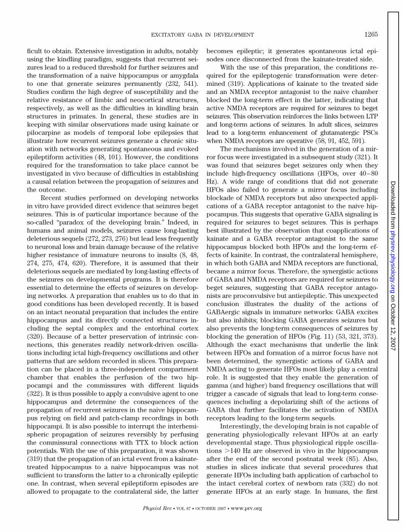
ficult to obtain. Extensive investigation in adults, notablyusing the kindling paradigm, suggests that recurrent sei-zures lead to a reduced threshold for further seizures andthe transformation of a naive hippocampus or amygdalato one that generate seizures permanently (232, 541).Studies confirm the high degree of susceptibility and therelative resistance of limbic and neocortical structures,respectively, as well as the difficulties in kindling brainstructures in primates. In general, these studies are inkeeping with similar observations made using kainate orpilocarpine as models of temporal lobe epilepsies thatillustrate how recurrent seizures generate a chronic situ-ation with networks generating spontaneous and evokedepileptiform activities (48, 101). However, the conditionsrequired for the transformation to take place cannot beinvestigated in vivo because of difficulties in establishinga causal relation between the propagation of seizures andthe outcome.
Recent studies performed on developing networksin vitro have provided direct evidence that seizures begetseizures. This is of particular importance because of theso-called “paradox of the developing brain.” Indeed, inhumans and animal models, seizures cause long-lastingdeleterious sequels (272, 273, 276) but lead less frequentlyto neuronal loss and brain damage because of the relativehigher resistance of immature neurons to insults (8, 48,274, 275, 474, 620). Therefore, it is assumed that theirdeleterious sequels are mediated by long-lasting effects ofthe seizures on developmental programs. It is thereforeessential to determine the effects of seizures on develop-ing networks. A preparation that enables us to do that ingood conditions has been developed recently. It is basedon an intact neonatal preparation that includes the entirehippocampus and its directly connected structures in-cluding the septal complex and the entorhinal cortex(320). Because of a better preservation of intrinsic con-nections, this generates readily network-driven oscilla-tions including ictal high-frequency oscillations and otherpatterns that are seldom recorded in slices. This prepara-tion can be placed in a three-independent compartmentchamber that enables the perfusion of the two hip-pocampi and the commissures with different liquids(322). It is thus possible to apply a convulsive agent to onehippocampus and determine the consequences of thepropagation of recurrent seizures in the naive hippocam-pus relying on field and patch-clamp recordings in bothhippocampi. It is also possible to interrupt the interhemi-spheric propagation of seizures reversibly by perfusingthe commissural connections with TTX to block actionpotentials. With the use of this preparation, it was shown(319) that the propagation of an ictal event from a kainate-treated hippocampus to a naive hippocampus was notsufficient to transform the latter to a chronically epilepticone. In contrast, when several epileptiform episodes areallowed to propagate to the contralateral side, the latter
becomes epileptic; it generates spontaneous ictal epi-sodes once disconnected from the kainate-treated side.
With the use of this preparation, the conditions re-quired for the epileptogenic transformation were deter-mined (319). Applications of kainate to the treated sideand an NMDA receptor antagonist to the naive chamberblocked the long-term effect in the latter, indicating thatactive NMDA receptors are required for seizures to begetseizures. This observation reinforces the links between LTPand long-term actions of seizures. In adult slices, seizureslead to a long-term enhancement of glutamatergic PSCswhen NMDA receptors are operative (58, 91, 452, 591).
The mechanisms involved in the generation of a mir-ror focus were investigated in a subsequent study (321). Itwas found that seizures beget seizures only when theyinclude high-frequency oscillations (HFOs, over 40–80Hz). A wide range of conditions that did not generateHFOs also failed to generate a mirror focus includingblockade of NMDA receptors but also unexpected appli-cations of a GABA receptor antagonist to the naive hip-pocampus. This suggests that operative GABA signaling isrequired for seizures to beget seizures. This is perhapsbest illustrated by the observation that coapplications ofkainate and a GABA receptor antagonist to the samehippocampus blocked both HFOs and the long-term ef-fects of kainite. In contrast, the contralateral hemisphere,in which both GABA and NMDA receptors are functional,became a mirror focus. Therefore, the synergistic actionsof GABA and NMDA receptors are required for seizures tobeget seizures, suggesting that GABA receptor antago-nists are proconvulsive but antiepileptic. This unexpectedconclusion illustrates the duality of the actions ofGABAergic signals in immature networks: GABA excitesbut also inhibits; blocking GABA generates seizures butalso prevents the long-term consequences of seizures byblocking the generation of HFOs (Fig. 11) (53, 321, 373).Although the exact mechanisms that underlie the linkbetween HFOs and formation of a mirror focus have notbeen determined, the synergistic actions of GABA andNMDA acting to generate HFOs most likely play a centralrole. It is suggested that they enable the generation ofgamma (and higher) band frequency oscillations that willtrigger a cascade of signals that lead to long-term conse-quences including a depolarizing shift of the actions ofGABA that further facilitates the activation of NMDAreceptors leading to the long-term sequels.
Interestingly, the developing brain is not capable ofgenerating physiologically relevant HFOs at an earlydevelopmental stage. Thus physiological ripple oscilla-tions �140 Hz are observed in vivo in the hippocampusafter the end of the second postnatal week (85). Also,studies in slices indicate that several procedures thatgenerate HFOs including bath application of carbachol tothe intact cerebral cortex of newborn rats (332) do notgenerate HFOs at an early stage. In humans, the first
EXCITATORY GABA IN DEVELOPMENT 1265
Physiol Rev • VOL 87 • OCTOBER 2007 • www.prv.org
on October 12, 2007
physrev.physiology.orgD
ownloaded from
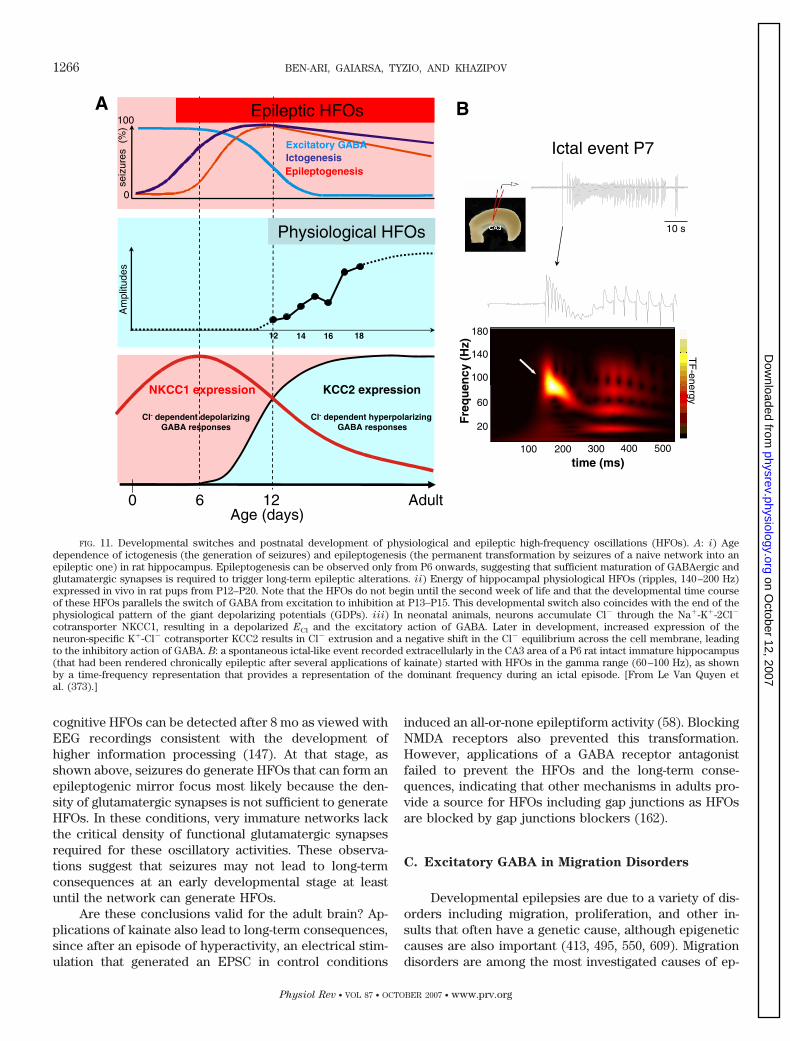
cognitive HFOs can be detected after 8 mo as viewed withEEG recordings consistent with the development ofhigher information processing (147). At that stage, asshown above, seizures do generate HFOs that can form anepileptogenic mirror focus most likely because the den-sity of glutamatergic synapses is not sufficient to generateHFOs. In these conditions, very immature networks lackthe critical density of functional glutamatergic synapsesrequired for these oscillatory activities. These observa-tions suggest that seizures may not lead to long-termconsequences at an early developmental stage at leastuntil the network can generate HFOs.
Are these conclusions valid for the adult brain? Ap-plications of kainate also lead to long-term consequences,since after an episode of hyperactivity, an electrical stim-ulation that generated an EPSC in control conditions
induced an all-or-none epileptiform activity (58). BlockingNMDA receptors also prevented this transformation.However, applications of a GABA receptor antagonistfailed to prevent the HFOs and the long-term conse-quences, indicating that other mechanisms in adults pro-vide a source for HFOs including gap junctions as HFOsare blocked by gap junctions blockers (162).
C. Excitatory GABA in Migration Disorders
Developmental epilepsies are due to a variety of dis-orders including migration, proliferation, and other in-sults that often have a genetic cause, although epigeneticcauses are also important (413, 495, 550, 609). Migrationdisorders are among the most investigated causes of ep-
FIG. 11. Developmental switches and postnatal development of physiological and epileptic high-frequency oscillations (HFOs). A: i) Agedependence of ictogenesis (the generation of seizures) and epileptogenesis (the permanent transformation by seizures of a naive network into anepileptic one) in rat hippocampus. Epileptogenesis can be observed only from P6 onwards, suggesting that sufficient maturation of GABAergic andglutamatergic synapses is required to trigger long-term epileptic alterations. ii) Energy of hippocampal physiological HFOs (ripples, 140–200 Hz)expressed in vivo in rat pups from P12–P20. Note that the HFOs do not begin until the second week of life and that the developmental time courseof these HFOs parallels the switch of GABA from excitation to inhibition at P13–P15. This developmental switch also coincides with the end of thephysiological pattern of the giant depolarizing potentials (GDPs). iii) In neonatal animals, neurons accumulate Cl� through the Na�-K�-2Cl�
cotransporter NKCC1, resulting in a depolarized ECl and the excitatory action of GABA. Later in development, increased expression of theneuron-specific K�-Cl� cotransporter KCC2 results in Cl� extrusion and a negative shift in the Cl� equilibrium across the cell membrane, leadingto the inhibitory action of GABA. B: a spontaneous ictal-like event recorded extracellularly in the CA3 area of a P6 rat intact immature hippocampus(that had been rendered chronically epileptic after several applications of kainate) started with HFOs in the gamma range (60–100 Hz), as shownby a time-frequency representation that provides a representation of the dominant frequency during an ictal episode. [From Le Van Quyen etal. (373).]
1266 BEN-ARI, GAIARSA, TYZIO, AND KHAZIPOV
Physiol Rev • VOL 87 • OCTOBER 2007 • www.prv.org
on October 12, 2007
physrev.physiology.orgD
ownloaded from
ilepsies, and recent studies performed in human and an-imal preparations have investigated those roles ofGABAergic signaling in seizure generation.
Mathern and co-workers (103, 105, 106; for review,see Ref. 104) have extensively investigated the propertiesof neurons in slices obtained after surgical interventionsin migration disorders. In a variety of cortical dysplasia(CD) types, various neuronal populations were identifiedon the basis of post hoc reconstruction including ballooncells, large cytomegalic neurons, and atypical and imma-ture neurons (106). Cytomegalic neurons generatedreadily recurrent calcium currents (103, 105, 106) andexhibited a lower frequency of glutamatergic currents anda higher frequency of GABAergic PSCs (103). The authorssuggest that oscillations produced by GABAergic PSCsmay generate synchronized patterns that would propa-gate to normal neurons located within the focus andgenerate epileptiform activities. Other observations sug-gest a deficit in GABAergic mechanisms. Thus a reductionin IPSC frequency and a potentially compensatory de-crease in transporter-mediated GABA reuptake functionhave been reported (93). Interestingly, GABAergic signal-ing is involved in the relationship between ictal activityonset and the occurrence of slow interictal-like events(149). In focal dysplastic slices, decreasing or increasingGABAA receptor function abolished or potentiated ictaldischarges, respectively. The initiation of ictal events isdue to the occurrence of synchronized GABAergic PSCsleading to [K�]o elevations that promote the generation ofictal events (149; also see Refs. 143, 583). The decrease inNR2B subunit expression in cytomegalic neurons fromCD tissue also can lead to a reduction of the voltage-dependent Mg2� blockade of NMDA receptors and thegeneration of synchronized patterns (18, 471). Furtherstudies are clearly required to determine the role of de-polarizing GABAergic signals in seizure generation in de-velopmental malformations.
IX. GENERAL CONCLUSIONS: A SEQUENCE
THAT EQUILIBRATES GABA AND
GLUTAMATE DURING DEVELOPMENT
The construction of brain networks faces severalissues. First, the process must take into account the ex-treme heterogeneity of immature neurons that will at anypoint in time be in different developmental stages. Thiswill complicate the generation of network-driven patternsthat are common to neurons endowed with many activesynapses and to others that have few if any operativesynapses. If “neurons that fire together wire together,”then slow kinetics and widespread propagation of primi-tive patterns are mandatory to overcome this heterogene-ity and set together neurons in different degrees ofmaturation. Second, neurons have to balance their gluta-
matergic and GABAergic inputs both to generate the en-vironmental support for activity-dependent mechanismsthat regulate many essential developmental processes.This is also needed to prevent deleterious consequencesof a disruption of the balance between GABA and gluta-mate: a glutamatergic excitation that is not compensatedby GABAergic signaling will lead to excitotoxicity, and adominating GABAergic inhibitory drive will slow downdevelopmental processes. Third, neurons and networksmust shift from a silence ensemble of newly differentiat-ing neurons to an ensemble of coactive neurons thatgenerate a plethora of patterns and oscillations. Observa-tions discussed in this review may provide a solution tothese issues (also see Ref. 49). The fact that these se-quences described for the hippocampus have been gener-ally confirmed for many other brain structures and animalspecies suggest that they have been kept throughout evo-lution and reinforce their relevance for development.
At an early stage, GABA receptors are already func-tional when most synapses are not operative. In fact,there is most likely a sequential formation of GABA re-ceptors followed by NMDA and AMPA receptors at leastin principal cells of the hippocampus. In the neocortex,there are some indications that GABA receptors arepresent early (399; also see Refs. 504 and 459 for review),but GABAergic inhibition is delayed probably because ofa delayed synaptic drive to GABAergic interneurons (4,334, 391, 398, 399, 405, 490). In the hippocampus, the lackof an efficient GABA transport system at that early stagewill facilitate the diffusion of GABA and its distal actionson receptors. The long-lasting spontaneous and evokedtonic currents generated by a noncanonical release ofGABA will exert actions on distal neurons. To preventexcitotoxicity, two mechanisms operate: 1) an early mat-uration of functional glutamate transporters that preventexcessive overactivation of NMDA receptors in additionto the voltage-dependent Mg2� blockade and 2) the exci-tatory actions of GABA that can generate sodium andcalcium spikes but with a driving force that is much lowerthan that of glutamate (30–40 mV at best compared with80 mV for glutamate). The shunting actions of GABA willalso reduce the glutamatergic drive. In addition, severalvoltage-gated channels are operative to prevent seizuresand excitotoxicity. The early expression of presynapticGABAB and other G protein-mediated signaling will con-trol transmitter release. Collectively, despite excitatoryGABA, the balance between excitation and inhibition ispreserved thanks to other mechanisms including a pre-synaptic control of transmitter release.
There are only a few exceptions to the rule thatduring maturation there is a reduction of [Cl�]i (401).According to the age of the species, the brain structure,and the neuron, this will take place in utero or after birth.This is by no means incompatible with excitatory actionsof GABA on certain types of neurons or dendrites of
EXCITATORY GABA IN DEVELOPMENT 1267
Physiol Rev • VOL 87 • OCTOBER 2007 • www.prv.org
on October 12, 2007
physrev.physiology.orgD
ownloaded from
neurons. The point here is that the concentrations of[Cl�]i present at an early stage are not reached in adultsexcept in very special conditions (seizures and damage).Perhaps even more importantly, the developmental curve(of a reduction of [Cl�]i) has not been challenged and is infact valid for peripheral neurons and for other organs,indicating that it has served other purposes during devel-opment than converting excitation to inhibition of GABA/glycine. Although speculative, it is possible that the re-moval of chloride from the intracellular milieu was a morerecent selective adaptation for central neurons.
Within this general scheme, there are several pointsto consider. Studies using single-channel cell-attached re-cording show that Em is not modified during maturation(626). Therefore, the driving force that mediates the ac-tions of GABA is strictly dependent on [Cl�]i and not ona shift of Em. However, this developmental gradient in-cludes at least two checkpoints to the excitatory actionsof GABA, the generation of sodium and calcium actionpotentials, and the activation of NMDA receptors by re-moving the voltage-dependent Mg2� blockade. At an earlystage, when [Cl�]i is particularly high, the estimated dif-ference between resting membrane potential and ECl
is �40 mV, the activation of GABA will generate actionpotentials. The high impedance of small cells will com-pensate the smaller density of sodium channels. The de-velopmental time course of the GABA/NMDA synergy isan additional important criterion that has not been stud-ied in detail. This leads to long-term changes in synapticefficacy of GABA, i.e., a long-term potentiation or depres-sion when calcium channels or NMDA receptors are ac-tivated, respectively. This synergistic action of GABA andNMDA is instrumental in the developmental actions ofGABA as it intervenes in neuronal growth, synapse for-mation, and network construction.
The GABA-glutamate sequence is another majorcomponent of the model proposed. The earlier formationof GABAergic synapses provides excitatory drive early onthat will entrain very immature neurons in primitive net-work-driven patterns. In keeping with this, in utero, ex-clusively GABAergic patterns can be recorded in inter-neurons that have matured before principal cells. There-fore, interneurons that modulate many behaviorallyrelevant patterns and oscillations in the adult circuit playa central role in the generation of synchronized patternswell before a significant proportion of principal cells haveestablished a dense network of excitatory synapses. Bydeveloping first and extending an elaborated pattern ofconnections initially with other interneurons, GABAergicneurons generate the first GDPs, setting the pace for themodulation by activity of the formation of glutamatergicsynapses. The longer kinetics of GABAergic PSCs and thesynergistic actions of GABA and NMDA receptors willfacilitate the generation of primitive patterns. Combinedtogether, GABA-NMDA PSCs will have an �300 ms dura-
tion that will readily facilitate the synchronization of long-lasting polysynaptic PSCs or GDPs. Once the excitatorydrive and the density of glutamatergic synapses are sig-nificant and an efficient inhibitory signaling is required,then the mechanisms that shift the chloride gradient areactivated, and neurons and networks resume their adultoperation. This scenario offers a less stringent and pro-grammed way to shift from silent to active neurons. In-deed, the alternative solution, an equilibrated parallelmaturation of excitatory and inhibitory synapses, wouldhave entrained a very strict series of control of the syn-thesis and operation of all the proteins and mechanismsneeded to equilibrate GABA and glutamate PSCs duringdevelopment including transporters, uptake mechanisms,release proteins, vesicular transport and trafficking, po-tentiation, and depression, etc. The earlier formation ofslightly excitatory GABAergic synapses with their inborninhibitory device eliminates the risk for overexcitation yetsupplies the excitatory drive and the calcium signalingneeded for maturation.
The GABA/glutamate sequence is paralleled by asequential maturation of interneurons before principalcells. At this early stage, pyramidal neurons are inessence silent except a few pioneer neurons. Therefore,GABAergic interneurons are organized to operate asthe main pacemaker generators of early pattern. They areendowed with the properties to trigger oscillations and toignite the sequences that will run up the engine before itoperates properly. Extensive investigations by Spitzer andco-workers (577, 581) have shown that calcium waves andspikes enable the establishment of the GABA phenotypeby modulating the synthesis of GAD, procedures thatblock these oscillations also block the expression of thisphenotype.
Several elements of this model remain to be con-firmed. First, do other brain structures indeed follow thesame rules? If in a given network glutamatergic synapsesare formed first, i.e., the neocortex, it will be interesting todetermine how the issue of toxicity is avoided. Second,the role of voltage-gated channels and notably K� chan-nels known to play a crucial role in inhibitory signalingand present early on, has not been discussed at length.Third, the role of GABAergic signaling in the formation ofglutamate synapses and connections remains to be clari-fied. Studies using antagonists or knockout strategies areless than adequate because of the multitude of actionsand heterogeneity of immature neurons. More efficientwill be the use of conditional time- and neuron-specificdeletions of siRNA that have already shown recently to beextremely useful and informative (27). A recent studyshows that GABAergic transmission controls the balanceof excitation and inhibition in the developing retinotectalcircuit, since a premature reduction of [Cl�]i in by theexpression of the Cl� transporter KCC2 blocked the de-velopmental increase in AMPA receptor-mediated trans-
1268 BEN-ARI, GAIARSA, TYZIO, AND KHAZIPOV
Physiol Rev • VOL 87 • OCTOBER 2007 • www.prv.org
on October 12, 2007
physrev.physiology.orgD
ownloaded from
mission (7). Fourth, present understanding of the controlof [Cl�]i suggest that both intrinsic and extrinsic signalsare involved in the developmental reduction of its con-centration. However, this conclusion is based on a staticand often qualitative estimation of [Cl�]i. The regulationof [Cl�]i is a dynamic process, and very few studiesmeasuring in physiological conditions alterations of chlo-ride have been performed. To determine the alteration of[Cl�]i during GDPs or synaptic currents may require dy-namic analysis to fully take into account the speed withwhich [Cl�]i resumes its control concentration. If physi-ological activity indeed regulates rapidly and persistently[Cl�]i, it will be essential to determine the cascade ofsignals that underlies these actions using physiologicallyrelevant preparations. An additional major concern is theidentification of the postmitotic age of the recorded neu-ron. With present available information, it is clearly diffi-cult to rely on data grouped on the basis of the age of theanimal without neuronal reconstruction or neuronal birthdating. The use of lentivirus tranfection techniques torecord neurons after identifying their age will be essen-tial. Transfection studies of the KCC2 cotransporter arealso very useful as they enable the testing of the conse-quences of overexpression at an early stage of the trans-porter. This is particularly useful if an early expression ofKCC2 augments selectively the density of GABAergic syn-apses (7, 118), raising the possibility that [Cl�]i and theactions of GABA influence the formation of synapses onthe postsynaptic targets.
The pathological implications of these observationsand sequences should not be underestimated. With theadvent of imaging tools in humans, it was determined thatmany neurological disorders originate early in life be-cause of the adverse effects of mutations or environmen-tal hazards. There is a concern on the possible deleteriousactions of the widely used GABA-acting drugs duringgestation. Thus GABA-acting antiepileptic agents canhave deleterious actions on neuronal migration and cor-tical construction. The long-lasting actions of seizures on[Cl�]i and the excitatory actions of GABA in human andanimal epileptic material suggest that epileptogenesis re-capitulates ontogenesis. This general scheme appears tobe common to many neurological disorders, suggestingthat [Cl�]i is a nodal point in the posttraumatic responseof neurons and networks. One hypothesis is that theaccumulation of chloride during insults overcomes thecapacity of the neuron to extrude chloride by affectingpersistently cotransporters. This in turn leads to a cas-cade of pathogenic consequences.
In conclusion, the study of how GABAergic signalsare established during brain maturation has illustratedhow studies on development lead to a major break-through in our understanding of how networks operate.This topic is also central to perhaps better understandingthe complex interactions between nature and nurture and
the roles of genetic programs and activity-dependentmechanisms. Perhaps the most exciting is to see how theapparently simple question “what comes first inhibition ofexcitation” leads to the discovery of fundamental devel-opmental rules that appear to have been kept throughoutevolution and that may represent a favorable solution tothese issues.
ACKNOWLEDGMENTS
We are grateful to Profs. K. Krnjevic and E. Cherubini aswell as Dr. J. Ackman for their remarks and suggestions.
Address for reprint requests and other correspondence: Y.Ben-Ari, INMED/INSERM U. 29, 163 Avenue de Luminy, B.P. 13,13273 Marseille, France (e-mail: [email protected]).
GRANTS
This work was supported by Institut National de la Sante etde la Recherche Medicale, Fondation pour la Recherche Medi-cale, L’Agence Nationale de la Recherche, Federation pour laRecherche sur le Cerveau and Rotary International, Conseilregional Provence-Alpes-Cote d’Azur, and European CommunityGrant LSHB-CT-2,004-503467.
REFERENCES
1. Abe Y, Furukawa K, Itoyama Y, Akaike N. Glycine response inacutely dissociated ventromedial hypothalamic neuron of the rat:new approach with gramicidin perforated patch-clamp technique.J Neurophysiol 72: 1530–1537, 1994.
2. Adelsberger H, Garaschuk O, Konnerth A. Cortical calciumwaves in resting newborn mice. Nat Neurosci 8: 988–990, 2005.
3. Agmon A, Hollrigel G, O’Dowd DK. Functional GABAergic syn-aptic connection in neonatal mouse barrel cortex. J Neurosci 16:4684–4695, 1996.
4. Agmon A, O’Dowd DK. NMDA receptor-mediated currents areprominent in the thalamocortical synaptic response before matu-ration of inhibition. J Neurophysiol 68: 345–349, 1992.
5. Aguado F, Carmona MA, Pozas E, Aguilo A, Martinez-Gui-
jarro FJ, Alcantara S, Borrell V, Yuste R, Ibanez CF, Soriano
E. BDNF regulates spontaneous correlated activity at early devel-opmental stages by increasing synaptogenesis and expression ofK�/Cl� co-transport. Development 130: 1287–1280, 2003.
6. Aizenman CD, Linden DJ. Regulation of the rebound depolariza-tion and spontaneous firing patterns of deep nuclear neurons inslices of rat cerebellum. J Neurophysiol 82: 1697–1709, 1999.
7. Akerman CJ, Cline HT. Depolarizing GABAergic conductancesregulate the balance of excitation to inhibition in the developingretinotectal circuit in vivo. J Neurosci 26: 5117–5130, 2006.
8. Albala BJ, Moshe SL, Okada R. Kainic acid-induced seizures: adevelopmental study. Dev Brain Res 13: 139–148, 1984.
9. Alger BE, Nicoll RA. GABA-mediated biphasic inhibitory re-sponses in hippocampus. Nature 281: 315–317, 1979.
10. Alger BE, Nicoll RA. Pharmacological evidence for two kinds ofGABA receptor on rat hippocampal pyramidal cells studied in vitro.J Physiol 328: 125–141, 1982.
11. Altman J, Bayer SA. Prolonged sojourn of developing pyramidalcells in the intermediate zone of the hippocampus and their settlingin the stratum pyramidale. J Comp Neurol 301: 343–364, 1990.
12. Ambrogini P, Lattanzi D, Ciuffoli S, Agostini D, Bertini L,
Stocchi V, Santi S, Cuppini R. Morpho-functional characteriza-tion of neuronal cells at different stages of maturation in granulecell layer of adult rat dentate gyrus. Brain Res 1017: 21–31, 2004.
13. Amico C, Marchetti C, Nobile M, Usai C. Pharmacological typesof calcium channels and their modulation by baclofen in cerebellargranules. J Neurosci 15: 2839–2848, 1995.
EXCITATORY GABA IN DEVELOPMENT 1269
Physiol Rev • VOL 87 • OCTOBER 2007 • www.prv.org
on October 12, 2007
physrev.physiology.orgD
ownloaded from
14. Andersen P, Dingledine R, Gjerstad L, Langmoen A, Laursen
AM. Two different responses of hippocampal pyramidal cells toapplication of �-aminobutyric acid. J Physiol 305: 279–296, 1980.
16. Andersen P, Eccles JC, Loyning Y. Pathway of postsynapticinhibition in the hippocampus. J Neurophysiol 27: 608–619, 1964.
17. Anderson SA, Eisenstat DD, Shi L, Rubenstein JL. Interneuronmigration from basal forebrain to neocortex: dependence on Dlxgenes. Science 278: 474–476, 1997.
18. Andre VM, Flores-Hernandez J, Cepeda C, Starling AJ,
Nguyen S, Lobo MK, Vinters HV, Levine MS, Mathern GW.
NMDA receptor alterations in neurons from pediatric cortical dys-plasia tissue. Cereb Cortex 14: 634–646, 2004.
19. Aniksztejn L, Sciancalepore M, Ben-Ari Y, Cherubini E. Per-sistent current oscillations produced by activation of metabotropicglutamate receptors in immature rat CA3 hippocampal neurons.J Neurophysiol 73: 1422–1429, 1995.
20. Antal M, Berki AC, Horvath L, O’Donovan MJ. Developmentalchanges in the distribution of gamma-aminobutyric acid-immuno-reactive neurons in the embryonic chick lumbosacral spinal cord.J Comp Neurol 343: 228–236, 1994.
21. Antonopoulos J, Pappas IS, Parnavelas JG. Activation of theGABAA receptor inhibits the proliferative effects of bFGF in corti-cal progenitor cells. Eur J Neurosci 9: 291–298, 1997.
22. Asada H, Kawamura Y, Maruyama K, Kume H, Ding R, Ji FY,
Kanbara N, Kuzume H, Sanbo M, Yagi T, Obata K. Mice lackingthe 65 kDa isoform of glutamic acid decarboxylase (GAD65) main-tain normal levels of GAD67 and GABA in their brains but aresusceptible to seizures. BBRC 229: 891–895, 1996.
23. Asada H, Kawamura Y, Maruyama K, Kume H, Ding RG, Kan-
bara N, Kuzume H, Sanbo M, Yagi T, Obata K. Cleft palate anddecreased brain gamma-aminobutyric acid in mice lacking the67-kDa isoform of glutamic acid decarboxylase. Proc Natl Acad Sci
USA 94: 6496–6499, 1997.24. Avoli M, Barbarosie M, Lucke A, Nagao T, Lopantsev V,
Kohling R. Synchronous GABA-mediated potentials and epilepti-form discharges in the rat limbic system in vitro. J Neurosci 16:3912–3924, 1996.
25. Awatramani GB, Turecek R, Trussell LO. Staggered develop-ment of GABAergic and glycinergic transmission in the MNTB.J Neurophysiol 93: 819–828, 2005.
26. Baccei ML, Fitzgerald M. Development of GABAergic and glycin-ergic transmission in the neonatal rat dorsal horn. J Neurosci 24:4749–4757, 2004.
27. Bai J, Ramos RL, Ackman JB, Thomas AM, Lee RV, LoTurco
JJ. RNAi reveals doublecortin is required for radial migration in ratneocortex. Nat Neurosci 6: 1277–1283, 2003.
28. Bal T, von Krosigk M, McCormick DA. Synaptic and membranemechanisms underlying synchronized oscillations in the ferret lat-eral geniculate nucleus in vitro. J Physiol 483: 641–663, 1995.
29. Baldelli P, Hernandez-Guijo JM, Carabelli V, Carbone E.
Brain-derived neurotrophic factor enhances GABA release proba-bility and nonuniform distribution of N- and P/Q-type channels onrelease sites of hippocampal inhibitory synapses. J Neurosci 25:3358–3368, 2005.
30. Balslev Y, Saunders NR, Mollgard K. Synaptogenesis in theneocortical anlage and early developing neocortex of rat embryos.Acta Anat 156: 2–10, 1996.
31. Banke TG, McBain CJ. GABAergic input onto CA3 hippocampalinterneurons remains shunting throughout development. J Neuro-
sci 26: 11720–11725, 2006.32. Baram TZ, Gerth A, Schultz L. Febrile seizures: an appropriate-
aged model suitable for long-term studies. Brain Res 98: 265–270,1997.
33. Baram TZ, Hatalski CG. Neuropeptide-mediated excitability: akey triggering mechanism for seizure generation in the developingbrain. Trends Neurosci 21: 471–476, 1998.
34. Baram TZ, Snead OC. Bicuculline induced seizures in infant rats:ontogeny of behavioral and electrocortical phenomena. Dev Brain
Res 57: 291–295, 1990.35. Barbin G, Pollard H, Gaıarsa JL, Ben-Ari Y. Involvement of
GABAA receptors in the outgrowth of cultured hippocampal neu-rons. Neurosci Lett 152: 150–154, 1993.
36. Barker JL, Behar T, Li YX, Liu QY, Ma W, Maric D, Maric I,
Schaffner AE, Serafini R, Smith SV, Somogyi R, Vautrin JY,
Wen XL, Xian H. GABAergic cells and signals in CNS develop-ment. Perspect Dev Neurobiol 5: 305–322, 1998.
37. Barry PH, Lynch JW. Liquid junction potentials and small celleffects in patch-clamp analysis. J Membr Biol 121: 101–117, 1991.
38. Baum G, Lev-Yadun S, Fridmann Y, Arazi T, Katsnelson H, Zik
M, Fromm H. Calmodulin binding to glutamate decarboxylase isrequired for regulation of glutamate and GABA metabolism andnormal development in plants. EMBO J 15: 2988–2996, 1996.
39. Bayer SA, Altman J. Hippocampal development in the rat: cyto-genesis and morphogenesis examined with autoradiography andlow-level X-irradiation. J Comp Neurol 158: 55–80, 1974.
40. Bazhenov M, Timofeev I, Steriade M, Sejnowski TJ. Self-sustained rhythmic activity in the thalamic reticular nucleus medi-ated by depolarizing GABAA receptor potentials. Nat Neurosci 2:168–174, 1999.
41. Becker M, Nothwang HG, Friauf E. Differential expression pat-tern of chloride transporters NCC, NKCC2, KCC1, KCC3, KCC4,AE3 in the developing rat auditory brainstem. Cell Tissue Res 312:155–165, 2003.
42. Beg AA, Jorgensen EM. EXP-1 is an excitatory GABA-gatedcation channel. Nat Neurosci 6: 1145–1152, 2003.
43. Behar TN, Li YX, Tran HT, Ma W, Dunlap V, Scott C, Barker
JL. GABA stimulates chemotaxis and chemokinesis of embryoniccortical neurons via calcium-dependent mechanisms. J Neurosci
16: 1808–1818, 1996.44. Behar TN, Schaffner AE, Scott CA, Greene CL, Barker JL.
GABA receptor antagonists modulate postmitotic cell migration inslice cultures of embryonic rat cortex. Cereb Cortex 10: 899–909,2000.
45. Behar TN, Schaffner AE, Tran HT, Barker JL. GABA-inducedmotility of spinal neuroblasts develops along a ventrodorsal gradi-ent and can be mimicked by agonists of GABAA and GABAB recep-tors. J Neurosci Res 42: 97–108, 1995.
46. Belhage B, Hansen GH, Elster L, Schousboe A. Effects ofgamma-aminobutyric acid (GABA) on synaptogenesis and synapticfunction. Perspect Dev Neurobiol 5: 235–246, 1998.
47. Belluzzi O, Benedusi M, Ackman J, LoTurco JJ. Electrophysi-ological differentiation of new neurons in the olfactory bulb. J Neu-
rosci 23: 10411–10418, 2003.48. Ben-Ari Y. Limbic seizure and brain damage produced by kainic
acid: mechanisms and relevance to human temporal lobe epilepsy.Neuroscience 14: 375–403, 1985.
49. Ben-Ari Y. Excitatory actions of GABA during development: thenature of the nurture. Nat Rev Neurosci 3: 728–739, 2002.
50. Ben-Ari Y, Holmes GL. The multiple facets of gamma-aminobu-tyric acid dysfunction in epilepsy. Curr Opin Neurol 18: 141–145,2005.
51. Ben-Ari Y, Khalilov I, Represa A, Gozlan H. Interneurons setthe tune of developing networks. Trends Neurosci 27: 422–427,2004.
52. Ben-Ari Y, Spitzer NC. Nature and nurture in brain development.Trends Neurosci 27: 361, 2004.
53. Ben-Ari Y, Holmes GL. Effects of seizures on developmentalprocesses in the immature brain. Lancet Neurol 5: 1055–1063, 2006.
55. Ben-Ari Y. Developing networks play similar melody. Trends Neu-
rosci 24: 354–360, 2001.56. Ben-Ari Y, Cherubini E, Corradetti R, Gaıarsa JL. Giant syn-
aptic potentials in immature rat CA3 hippocampal neurones.J Physiol 416: 303–325, 1989.
57. Ben-Ari Y, Cherubini E, Krnjevic K. Changes in voltage depen-dence of NMDA currents during development. Neurosci Lett 94:88–92, 1988.
58. Ben-Ari Y, Gho M. Long-lasting modification of the synaptic prop-erties of rat CA3 hippocampal neurones induced by kainaic acid.J Physiol 404: 365–384, 1988.
59. Ben-Ari Y, Khazipov R, Leinekugel X, Caillard O, Gaıarsa JL.
GABAA, NMDA and AMPA receptors: a developmentally regulated“menage a trois.” Trends Neurosci 20: 523–529, 1997.
60. Bender RA, Galindo R, Mameli M, Gonzalez-Vega R, Valenzu-
ela CF, Baram TZ. Synchronized network activity in developingrat hippocampus involves regional hyperpolarization-activated cy-
1270 BEN-ARI, GAIARSA, TYZIO, AND KHAZIPOV
Physiol Rev • VOL 87 • OCTOBER 2007 • www.prv.org
on October 12, 2007
physrev.physiology.orgD
ownloaded from
clic nucleotide-gated (HCN) channel function. Eur J Neurosci 22:2669–2674, 2005.
61. Bender RA, Galindo R, Mameli M, Gonzalez-Vega R, Valenzu-
ela CF, Baram TZ. Synchronized network activity in developingrat hippocampus involves regional hyperpolarization-activated cy-clic nucleotide-gated (HCN) channel function. Eur J Neurosci 22:2669–2674, 2005.
62. Benson DL, Watkins FH, Steward O, Banker G. Characteriza-tion of GABAergic neurons in hippocampal cell cultures. J Neuro-
cytol 23: 279–295, 1994.63. Berger B, Alvarez C. Neurochemical development of the hip-
pocampal region in the fetal rhesus monkey, III: calbindin-D28K,calretinin and parvalbumin with special mention of cajal-retziuscells and the retrosplenial cortex. J Comp Neurol 366: 674–699,1996.
64. Berger B, De Grissac N, Alvarez C. Precocious development ofparvalbumin-like immunoreactive interneurons in the hippocampalformation and entorhinal cortex of the fetal cynomolgus monkey.J Comp Neurol 403: 309–331, 1999.
65. Berki AC, O’Donovan MJ, Antal M. Developmental expressionof glycine immunoreactivity and its colocalization with GABA inthe embryonic chick lumbosacral spinal cord. J Comp Neurol 362:583–596, 1995.
66. Bernard C, Milh M, Morozov YM, Ben-Ari Y, Freund TF, Goz-
lan H. Altering cannabinoid signaling during development disruptsneuronal activity. Proc Natl Acad Sci USA 102: 9388–9393, 2005.
67. Berninger B, Marty S, Zafra F, da Penha Berzaghi M, Thoenen
H, Lindholm D. GABAergic stimulation switches from enhancingto repressing BDNF expression in rat hippocampal neurons duringmaturation in vitro. Development 121: 2327–2335, 1995.
68. Bettler B, Kaupmann K, Mosbacher J, Gassmann M. Molecularstructure and physiological functions of GABA(B) receptors.Physiol Rev 84: 835–867, 2004.
69. Binor E, Heathcote RD. Development of GABA-immunoreactiveneuron patterning in the spinal cord. J Comp Neurol 438: 1–11,2001.
70. Bolea S, Avignone E, Berretta N, Sanchez-Andres JV, Cheru-
bini E. Glutamate controls the induction of GABA-mediated giantdepolarizing potentials through AMPA receptors in neonatal rathippocampal slices. J Neurophysiol 81: 2095–2102, 1999.
71. Bolteus AJ, Bordey A. GABA release and uptake regulate neuro-nal precursor migration in the postnatal subventricular zone.J Neurosci 24: 7623–7631, 2004.
72. Borden LA, Murali Dhar TG, Smith KE, Weinshank RL,
Branchek TA, Gluchowski C. Tiagabine, SK&F 89976-A, CI-966,NNC-711 are selective for the cloned GABA transporter GAT-1. Eur
J Pharmacol 269: 219–224, 1994.73. Borg-Graham LJ, Monier C, Fregnac Y. Visual input evokes
transient and strong shunting inhibition in visual cortical neurons.Nature 393: 369–373, 1998.
74. Bormann J, Hamill OP, Sakmann B. Mechanisms of anion per-meation through channels gated by glycine and �-aminobutyricacid in mouse cultured spinal neurones. J Physiol 385: 243–286,1987.
75. Borodinsky LN, Root CM, Cronin JA, Sann SB, Gu X, Spitzer
NC. Activity-dependent homeostatic specification of transmitterexpression in embryonic neurons. Nature 429: 523–530, 2004.
76. Bouche N, Fait A, Bouchez D, Moller SG, Fromm H. Mitochon-drial succinic-semialdehyde dehydrogenase of the gamma-ami-nobutyrate shunt is required to restrict levels of reactive oxygenintermediates in plants. Proc Natl Acad Sci USA 100: 6843–6848,2003.
77. Bouche N, Fromm H. GABA in plants: just a metabolite? Trends
Plant Sci 9: 110–115, 2004.78. Bouche N, Lacombe B, Fromm H. GABA signaling: a conserved
and ubiquitous mechanism. Trends Cell Biol 13: 607–610, 2003.79. Boukhaddaoui H, Sieso V, Scamps F, Vigues S, Roig A,
Valmier J. Q- and L-type calcium channels control the develop-ment of calbindin phenotype in hippocampal pyramidal neuronsin vitro. Eur J Neurosci 12: 2068–2078, 2000.
80. Bowe MA, Nadler JV. Developmental increase in the sensitivity tomagnesium of NMDA receptors on CA1 hippocampal pyramidalcells. Dev Brain Res 56: 55–61, 1990.
81. Bowery NG, Brown DA. Inhibitory synapses: the cloning ofGABAB receptors. Nature 386: 223–224, 1997.
82. Bowery NG, Brown DA, Marsh S. Depolarization of sympatheticganglion cells mediated through the release of gamma-aminobu-tyric acid (GABA) from adjacent glial cells. J Physiol 246: 57P–58P,1975.
83. Brenowitz S, David J, Trussell L. Enhancement of synapticefficacy by presynaptic GABAB receptors. Neuron 20: 135–141,1998.
84. Brigadski T, Hartmann M, Lessmann V. Differential vesiculartargeting and time course of synaptic secretion of the mammalianneurotrophins. J Neurosci 25: 7601–7614, 2005.
85. Buhl DL, Buzsaki G. Developmental emergence of hippocampalfast-field “ripple” oscillations in the behaving rat pups. Neuro-
science 134: 1423–1430, 2005.86. Bureau I, Shepherd GM, Svoboda K. Precise development of
functional and anatomical columns in the neocortex. Neuron 42:789–801, 2004.
87. Burrone J, O’Byrne M, Murthy VN. Multiple forms of synapticplasticity triggered by selective suppression of activity in individualneurons. Nature 420: 414–418, 2002.
88. Butt SJ, Fuccillo M, Nery S, Noctor S, Kriegstein A, Corbin
JG, Fishell G. The temporal and spatial origins of cortical inter-neurons predict their physiological subtype. Neuron 48: 591–604,2005.
89. Buzsaki G, Chen LS, Gage FH. Spatial organization of physiolog-ical activity in the hippocampal region: relevance to memory for-mation. Prog Brain Res 83: 257–268, 1990.
90. Caillard O, Ben-Ari Y, Gaıarsa JL. Long-term potentiation ofGABAergic synaptic transmission in neonatal rat hippocampus.J Physiol 518.1: 109–119, 1999.
91. Caillard O, Ben-Ari Y, Gaıarsa JL. Mechanisms of induction andexpression of long-term depression at GABAergic synapses in neo-natal rat hippocampus. J Neurosci 19: 7568–7577, 1999.
92. Caillard O, McLean HA, Ben-Ari Y, Gaıarsa JL. Ontogenesis ofpresynaptic GABAB receptor-mediated inhibition in the CA3 regionof the rat hippocampus. J Neurophysiol 79: 1341–1348, 1998.
93. Calcagnotto ME, Paredes MF, Tihan T, Barbaro NM, Baraban
SC. Dysfunction of synaptic inhibition in epilepsy associated withfocal cortical dysplasia. J Neurosci 25: 9649–9657, 2005.
94. Cameron HA, Hazel TG, McKay RD. Regulation of neurogenesisby growth factors and neurotransmitters. J Neurobiol 36: 287–306,1998.
95. Cang J, Renteria RC, Kaneko M, Liu X, Copenhagen DR,
Stryker MP. Development of precise maps in visual cortex re-quires patterned spontaneous activity in the retina. Neuron 48:797–809, 2005.
96. Caraiscos VB, Elliott EM, You T, Cheng VY, Belelli D, Newell
JG, Jackson MF, Lambert JJ, Rosahl TW, Wafford KA, Mac-
Donald JF, Orser BA. Tonic inhibition in mouse hippocampalCA1 pyramidal neurons is mediated by alpha5 subunit-containinggamma-aminobutyric acid type A receptors. Proc Natl Acad Sci
USA 101: 3662–3667, 2004.97. Caserta MT, Barker JL. Development of the GABAergic pheno-
type in murine spinal cord-dorsal root ganglion cultures. Int J Dev
Neurosci 12: 753–765, 1994.98. Caspary DM, Milbrandt JC, Helfert RH. Central auditory aging:
GABA changes in the inferior colliculus. Exp Gerontol 30: 349–360,1995.
99. Catsicas S, Mobbs P. GABAB receptors regulate chick retinalcalcium waves. J Neurosci 21: 897–910, 2001.
100. Cattaert D, El Manira A. Shunting versus inactivation: analysis ofpresynaptic inhibitory mechanisms in primary afferents of the cray-fish. J Neurosci 19: 6079–6089, 1999.
101. Cavalheiro EA, Santos NF, Priel MR. The pilocarpine model ofepilepsy in mice. Epilepsia 37: 1015–1019, 1996.
102. Cazalets JR, Bertrand S, Sqalli-Houssaini Y, Clarac F.
GABAergic control of spinal locomotor networks in the neonatalrat. Ann NY Acad Sci 860: 168–180, 1998.
103. Cepeda C, Andre VM, Flores-Hernandez J, Nguyen OK, Wu N,
Klapstein GJ, Nguyen S, Koh S, Vinters HV, Levine MS, Math-
ern GW. Pediatric cortical dysplasia: correlations between neuro-imaging, electrophysiology and location of cytomegalic neurons
EXCITATORY GABA IN DEVELOPMENT 1271
Physiol Rev • VOL 87 • OCTOBER 2007 • www.prv.org
on October 12, 2007
physrev.physiology.orgD
ownloaded from
and balloon cells and glutamate/GABA synaptic circuits. Dev Neu-
rosci 27: 59–76, 2005.104. Cepeda C, Andre VM, Levine MS, Salamon N, Miyata H, Vin-
ters HV, Mathern GW. Epileptogenesis in pediatric cortical dys-plasia: the dysmature cerebral developmental hypothesis. Epilepsy
Behav 9: 219–235, 2006.105. Cepeda C, Andre VM, Vinters HV, Levine MS, Mathern GW.
Are cytomegalic neurons and balloon cells generators of epilepticactivity in pediatric cortical dysplasia? Epilepsia 46 Suppl 5: 82–88,2005.
106. Cepeda C, Hurst RS, Flores-Hernandez J, Hernandez-
Echeagaray E, Klapstein GJ, Boylan MK, Calvert CR, Jocoy
EL, Nguyen OK, Andre VM, Vinters HV, Ariano MA, Levine
MS, Mathern GW. Morphological and electrophysiological char-acterization of abnormal cell types in pediatric cortical dysplasia.J Neurosci Res 72: 472–486, 2003.
107. Chadderton P, Margrie TW, Hausser M. Integration of quanta incerebellar granule cells during sensory processing. Nature 428:856–860, 2004.
108. Chagnac-Amitai Y, Connors BW. Horizontal spread of synchro-nized activity in neocortex and its control by GABA-mediatedinhibition. J Neurophysiol 61: 747–758, 1989.
109. Chang EH, Kotak VC, Sanes DH. Long-term depression of syn-aptic inhibition is expressed postsynaptically in the developingauditory system. J Neurophysiol 90: 1479–1488, 2003.
110. Chavas J, Forero ME, Collin T, Llano I, Marty A. Osmotictension as a possible link between GABA(A) receptor activationand intracellular calcium elevation. Neuron 44: 701–713, 2004.
111. Chavas J, Marty A. Coexistence of excitatory and inhibitoryGABA synapses in the cerebellar interneuron network. J Neurosci
23: 2019–2031, 2003.112. Chen G, Trombley PQ, van den Pol AN. GABA receptors pre-
cede glutamate receptors in hypothalamic development: differen-tial regulation by astrocytes. J Neurophysiol 74: 1473–1484, 1995.
113. Chen G, Trombley PQ, van den Pol AN. Excitatory actions ofGABA in developing rat hypothalamic neurones. J Physiol 494:451–464, 1996.
114. Cheng Q, Yeh HH. PLCgamma signaling underlies BDNF potenti-ation of Purkinje cell responses to GABA. J Neurosci Res 79:616–627, 2005.
115. Cherubini E, Gaıarsa JL, Ben-Ari Y. GABA: an excitatory trans-mitter in early postnatal life. Trends Neurosci 14: 515–519, 1991.
116. Chevaleyre V, Castillo PE. Heterosynaptic LTD of hippocampalGABAergic synapses: a novel role of endocannabinoids in regulat-ing excitability. Neuron 38: 461–472, 2003.
117. Chevrot R, Rosen R, Haudecoeur E, Cirou A, Shelp BJ, Ron E,
Faure D. GABA controls the level of quorum-sensing signal inAgrobacterium tumefaciens. Proc Natl Acad Sci USA 103: 7460–7464, 2006.
118. Chudotvorova I, Ivanov A, Rama S, Hubner CA, Pellegrino C,
Ben-Ari Y, Medina I. Early expression of KCC2 in rat hippocam-pal cultures augments expression of functional GABA synapses.J Physiol 566: 671–679, 2005.
119. Clapham D. How to lose your hippocampus by working on chlo-ride channels. Neuron 29: 1–3, 2001.
120. Clarac F, Brocard F, Vinay L. The maturation of locomotornetworks. Prog Brain Res 143: 57–66, 2004.
121. Clayton GH, Owens GC, Wolf JS, Smith RL. Ontogeny of cation-Cl-cotransporter expression in at neocortex. Dev Brain Res 109:281–292, 1998.
122. Clayton GH, Staley KJ, Wilcox CL, Owens GC, Smith RL.
Developmental expression of C1C-2 in the rat nervous system.Brain Res 108: 307–318, 1998.
123. Cobas A, Fairen A, Alvarez-Bolado G, Sanchez MP. Prenataldevelopment of the intrinsic neurons of the rat neocortex: a com-parative study of the distribution of GABA-immunoreactive cellsand the GABAA receptor. Neuroscience 40: 375–397, 1991.
124. Cobos I, Calcagnotto ME, Vilaythong AJ, Thwin MT, Noebels
JL, Baraban SC, Rubenstein JL. Mice lacking Dlx1 show sub-type-specific loss of interneurons, reduced inhibition and epilepsy.Nat Neurosci 8: 1059–1068, 2005.
125. Cohen AS, Lin DD, Coulter DA. Protracted postnatal develop-ment of inhibitory synaptic transmission in rat hippocampal areaCA1 neurons. J Neurophysiol 84: 2465–2476, 2000.
126. Cohen I, Miles R. Contributions of intrinsic and synaptic activitiesto the generation of neuronal discharges in in vitro hippocampus.J Physiol 524: 485–502, 2000.
127. Cohen I, Navarro V, Clemenceau S, Baulac M, Miles R. On theorigin of interictal activity in human temporal lobe epilepsy in vitro.Science 298: 1418–1421, 2002.
128. Colin-Le Brun I, Ferrand N, Caillard O, Tosetti P, Ben-Ari Y,
Gaıarsa JL. Spontaneous synaptic activity is required for theformation of functional GABAergic synapses in the developing rathippocampus. J Physiol 559: 129–139, 2004.
129. Collin C, Devane WA, Dahl D, Lee CJ, Axelrod J, Alkon DL.
Long-term synaptic transformation of hippocampal CA1 gamma-aminobutyric acid synapses and the effect of anandamide. Proc
Natl Acad Sci USA 92: 10167–10171, 1995.130. Collin T, Chat M, Lucas MG, Moreno H, Racay P, Schwaller B,
Marty A, Llano I. Developmental changes in parvalbumin regulatepresynaptic Ca2� signaling. J Neurosci 25: 96–107, 2005.
131. Concas A, Pierobon P, Mostallino MC, Porcu P, Marino G,
Minei R, Biggio G. Modulation of gamma-aminobutyric acid(GABA) receptors and the feeding response by neurosteroids inHydra vulgaris. Neuroscience 85: 979–988, 1998.
132. Connor JA, Tseng HY, Hockberger PE. Depolarization- andtransmitter-induced changes in intracellular Ca2� of rat cerebellargranular cells in explant cultures. J Neurosci 7: 1384–1400, 1987.
133. Constantine-Paton M, Cline HT. LTP and activity-dependentsynaptogenesis: the more alike they are, the more different theybecome. Curr Opin Neurobiol 8: 139–148, 1998.
134. Corlew R, Bosma MM, Moody WJ. Spontaneous, synchronouselectrical activity in neonatal mouse cortical neurones. J Physiol
560: 377–390, 2004.135. Corradetti R, Gaıarsa JL, Ben-Ari Y. D-Aminophosphonovaleric
acid-sensitive spontaneous giant EPSPs in immature rat hippocam-pal neurones. Eur J Pharmacol 154: 221–222, 1988.
136. Cosi C, Spoerri PE, Comelli MC, Guidolin D, Skaper SD.
Glucocorticoids depress activity-dependent expression of BDNFmRNA in hippocampal neurones. Neuroreport 4: 527–530, 1993.
138. Cossart R, Aronov D, Yuste R. Attractor dynamics of networkUP states in the neocortex. Nature 423: 283–288, 2003.
139. Cossart R, Petanjek Z, Dumitriu D, Hirsch JC, Ben-Ari Y,
Esclapez M, Bernard C. Interneurons targeting similar layersreceive synaptic inputs with similar kinetics. Hippocampus 16:408–420, 2006.
140. Couve A, Moss SJ, Pangalos MN. GABAB receptors: a newparadigm in G protein signaling. Mol Cell Neurosci 16: 296–312,2000.
141. Craig AM, Graf ER, Linhoff MW. How to build a central synapse:clues from cell culture. Trends Neurosci 29: 8–20, 2006.
141a.Crepel V, Aronov D, Jorquera I, Represa A, Ben-Ari Y, Cos-
sart R. A parturition-associated nonsynaptic coherent activity pat-tern in the developing hippocampus. Neuron 54: 105–120, 2007.
142. Crepel V, Krnjevic K, Ben-Ari Y. Developmental and regionaldifferences in the vulnerability of rat hippocampal slices to lack ofglucose. Neuroscience 47: 579–587, 1992.
143. Crino PB, Duhaime AC, Baltuch G, White R. Differential ex-pression of glutamate and GABA-A receptor subunit mRNA incortical dysplasia. Neurology 56: 906–913, 2001.
144. Crowley JC, Katz LC. Development of ocular dominance col-umns in the absence of retinal input. Nat Neurosci 2: 1125–1130,1999.
145. Crowley JC, Katz LC. Early development of ocular dominancecolumns. Science 290: 1321–1324, 2000.
146. Crowley JC, Katz LC. Ocular dominance development revisited.Curr Opin Neurobiol 12: 104–109, 2002.
147. Csibra G, Davis G, Spratling MW, Johnson MH. Gamma oscil-lations and object processing in the infant brain. Science 290:1582–1585, 2000.
148. Cui H, Bulleit RF. Potassium chloride inhibits proliferation ofcerebellar granule neuron progenitors. Brain Res 106: 129–135,1998.
1272 BEN-ARI, GAIARSA, TYZIO, AND KHAZIPOV
Physiol Rev • VOL 87 • OCTOBER 2007 • www.prv.org
on October 12, 2007
physrev.physiology.orgD
ownloaded from
149. D’Antuono M, Louvel J, Kohling R, Mattia D, Bernasconi A,
Olivier A, Turak B, Devaux A, Pumain R, Avoli M. GABAA
receptor-dependent synchronization leads to ictogenesis in thehuman dysplastic cortex. Brain 127: 1626–1640, 2004.
150. Dammerman RS, Flint AC, Noctor S, Kriegstein AR. An exci-tatory GABAergic plexus in developing neocortical layer 1. J Neu-
rophysiol 84: 428–434, 2000.151. Dan Y, Poo MM. Retrograde interactions during formation and
elimination of neuromuscular synapses. Curr Opin Neurobiol 4:95–100, 1994.
152. Danbolt NC. Glutamate uptake. Prog Neurobiol 65: 1–105, 2001.153. Darlison MG, Pahal I, Thode C. Consequences of the evolution
of the GABA(A) receptor gene family. Cell Mol Neurobiol 25: 607–624, 2005.
154. De Lima AD, Opitz T, Voigt T. Irreversible loss of a subpopula-tion of cortical interneurons in the absence of glutamatergic net-work activity. Eur J Neurosci 19: 2931–2943, 2004.
155. Debarbieux F, Brunton J, Charpak S. Effect of bicuculline onthalamic activity: a direct blockade of IAHP in reticularis neurons.J Neurophysiol 79: 2911–2918, 1998.
156. Deisz RA, Lux HD. The role of intracellular chloride in hyperpo-larizing post-synaptic inhibition of crayfish stretch receptor neu-rones. J Physiol 326: 123–138, 1982.
157. Delpire E. Cation-chloride cotransporters in neuronal communi-cation. News Physiol Sci 15: 309–312, 2000.
158. Demarque M, Represa A, Becq H, Khalilov I, Ben-Ari Y,
Aniksztejn L. Paracrine intercellular communication by a Ca2�-and SNARE-independent release of GABA and glutamate prior tosynapse formation. Neuron 36: 1051–1061, 2002.
159. Demarque M, Villeneuve N, Manent JB, Becq H, Represa A,
Ben-Ari Y, Aniksztejn L. Glutamate transporters prevent thegeneration of seizures in the developing rat neocortex. J Neurosci
24: 3289–3294, 2004.160. Deschenes M, Feltz P, Lamour Y. A model for an estimate in vivo
of the ionic basis of presynaptic inhibition: an intracellular analysisof the GABA-induced depolarization in rat dorsal root ganglia.Brain Res 118: 486–493, 1976.
161. Diabira D, Chevassus-Au-Louis N, Ben-Ari Y, Gozlan H. Lateembryonic expression of AMPA receptor function in the CA1 re-gion of the intact hippocampus in vitro. Eur J Neurosci 11: 4015–4023, 1999.
162. Draguhn A, Traub RD, Schmitz D, Jefferys JG. Electrical cou-pling underlies high-frequency oscillations in the hippocampusin vitro. Nature 394: 189–192, 1998.
163. Dudel J, Kuffler SW. Presynaptic inhibition at the crayfish neu-romuscular junction. J Physiol 155: 543–562, 1961.
164. Duffy CJ, Rakic P. Differentiation of granule cell dendrites in thedentate gyrus of the rhesus monkey: a quantitative Golgi study.J Comp Neurol 214: 224–237, 1983.
165. Dumitriu D, Cossart R, Huang J, Yuste R. Correlation betweenaxonal morphologies and synaptic input kinetics of interneuronsfrom mouse visual cortex. Cereb Cortex 17: 81–91, 2007.
166. Dunwiddie T. Age-related differences in the in vitro rat hippocam-pus: development of inhibition and the effects of hypoxia. Dev
Neurosci 4: 165–175, 1981.167. Dupont E, Hanganu IL, Kilb W, Hirsch S, Luhmann HJ. Rapid
developmental switch in the mechanisms driving early corticalcolumnar networks. Nature 439: 79–83, 2006.
168. Dupuy ST, Houser CR. Prominent expression of two forms ofglutamate decarboxylase in the embryonic and early postnatal rathippocampal formation. J Neurosci 16: 6919–6932, 1996.
169. Durand GM, Kovalchuk Y, Konnerth A. Long-term potentiationand functional synapse induction in developing hippocampus. Na-
ture 381: 71–75, 1996.170. Dutar P, Nicoll RA. A physiological role for GABAB receptors in
the central nervous system. Nature 332: 156–158, 1988.171. Dzhala V, Desfreres L, Melyan Z, Ben-Ari Y, Khazipov R.
Epileptogenic action of caffeine during anoxia in the neonatal rathippocampus. Ann Neurol 46: 95–102, 1999.
172. Dzhala VI, Staley KJ. Excitatory actions of endogenously re-leased GABA contribute to initiation of ictal epileptiform activity inthe developing hippocampus. J Neurosci 23: 1840–1846, 2003.
173. Dzhala VI, Talos DM, Sdrulla DA, Brumback AC, Mathews
GC, Benke TA, Delpire E, Jensen FE, Staley KJ. NKCC1 trans-porter facilitates seizures in the developing brain. Nat Med 11:1205–1213, 2005.
174. Dzidzishvili NN, Kvirkvelia LR. Electrophysiological signs ofhippocampal development in ontogenesis. Prog Brain Res 22: 414–426, 1968.
175. Easter A, Spruce AE. Recombinant GABA(B) receptors formedfrom GABA(B1) and GABA(B2) subunits selectively inhibit N-typeCa(2�) channels in NG108-15 cells. Eur J Pharmacol 440: 17–25,2002.
176. Ebihara S, Shirato K, Harata N, Akaike N. Gramicidin-perfo-rated patch recording: GABA response in mammalian neuroneswith intact intracellular chloride. J Physiol 484: 77–86, 1995.
177. Eccles JC. Presynaptic inhibition in the spinal cord. Prog Brain
Res 12: 65–91, 1964.178. Eccles JC. The ionic mechanisms of excitatory and inhibitory
synaptic action. Ann NY Acad Sci 137: 473–494, 1966.179. Eccles JC. The development of the cerebellum of vertebrates in
relation to the control of movement. Naturwissenschaften 56:525–534, 1969.
180. Eccles JC, Eccles RM, Magni F. Central inhibitory action attrib-utable to presynaptic depolarization produced by muscle afferentvolleys. J Physiol 159: 147–166, 1961.
181. Eccles JC, Llinas R, Sasaki K. The inhibitory interneuroneswithin the cerebellar cortex. Exp Brain Res 1: 1–16, 1966.
182. Eccles JC, Schmidt R, Willis WD. Pharmacological studies onpresynaptic inhibition. J Physiol 168: 500–530, 1963.
183. Edlund T, Jessell TM. Progression from extrinsic to intrinsicsignaling in cell fate specification: a view from the nervous system.Cell 96: 211–224, 1999.
184. Edwards DH. Mechanisms of depolarizing inhibition at the cray-fish giant motor synapse. 1. Electrophysiology. J Neurophysiol 64:532–540, 1990.
185. Edwards DH. Mechanisms of depolarizing inhibition at the cray-fish giant motor synapse. 2. Quantitative reconstruction. J Neuro-
physiol 64: 541–550, 1990.186. Eins S, Spoerri PE, Heyder E. GABA or sodium-bromide-induced
plasticity of neurites of mouse neuroblastoma cells in culture. Aquantitative study. Cell Tissue Res 229: 457–460, 1983.
187. Elmariah SB, Crumling MA, Parsons TD, Balice-Gordon RJ.
Postsynaptic TrkB-mediated signaling modulates excitatory andinhibitory neurotransmitter receptor clustering at hippocampalsynapses. J Neurosci 24: 2380–2393, 2004.
188. Eriksson KS, Panula P. gamma-Aminobutyric acid in the nervoussystem of a planarian. J Comp Neurol 345: 528–536, 1994.
189. Ernfors P, Wetmore C, Olson L, Persson H. Identification ofcells in rat brain and peripheral tissues expressing mRNA formembers of the nerve growth factor family. Neuron 5: 511–526,1990.
190. Fedirchuk B, Wenner P, Whelan PJ, Ho S, Tabak J,
O’Donovan MJ. Spontaneous network activity transiently de-presses synaptic transmission in the embryonic chick spinal cord.J Neurosci 19: 202–2112, 1999.
191. Feller MB, Wellis DP, Stellwagen D, Werblin FS, Shatz CJ.
Requirement for cholinergic synaptic transmission in the propaga-tion of spontaneous retinal waves. Science 272: 1182–1187, 1996.
192. Fellipa-Marques S, Vinay L, Clarac F. Spontaneous and loco-motor-related GABAergic input onto primary afferents in the neo-natal rat. Eur J Neurosci 12: 155–164, 2000.
193. Ferezou I, Bolea S, Petersen CCH. Visualizing the cortical rep-resentation of whisker touch: voltage-sensitive dye imaging infreely moving mice. Neuron 50: 617–629, 2006.
194. Filippov AK, Couve A, Pangalos MN, Walsh FS, Brown DA,
Moss SJ. Heteromeric assembly of GABA(B)R1 and GABA(B)R2receptor subunits inhibits Ca2� current in sympathetic neurons.J Neurosci 20: 2867–2874, 2000.
195. Fiszman ML, Behar T, Lange GD, Smith SV, Novotny EA,
Barker JL. GABAergic cells and signals appear together in theearly post-mitotic period of telencephalic and striatal development.Brain Res 73: 243–251, 1993.
EXCITATORY GABA IN DEVELOPMENT 1273
Physiol Rev • VOL 87 • OCTOBER 2007 • www.prv.org
on October 12, 2007
physrev.physiology.orgD
ownloaded from
196. Fiumelli H, Cancedda L, Poo MM. Modulation of GABAergictransmission by activity via postsynaptic Ca2�-dependent regula-tion of KCC2 function. Neuron 48: 773–786, 2005.
197. Fletcher TL, De Camilli P, Banker G. Synaptogenesis in hip-pocampal cultures: evidence indicating that axons and dendritesbecome competent to form synapses at different stages of neuronaldevelopment. J Neurosci 14: 6695–6706, 1994.
198. Flint AC, Dammerman RS, Kriegstein AR. Endogenous activa-tion of metabotropic glutamate receptors in neocortical develop-ment causes neuronal calcium oscillations. Proc Natl Acad Sci USA
96: 12144–12149, 1999.199. Fox K, Wright N, Wallace H, Glazewski S. The origin of cortical
surround receptive fields studied in the barrel cortex. J Neurosci
23: 8380–8391, 2003.200. French CR, Sah P, Buckett KJ, Gage PW. A voltage-dependent
persistent sodium current in mammalian hippocampal neurons.J Gen Physiol 95: 1139–1157, 1990.
201. Freund T, Buzsaki G. Interneurons of the hippocampus. Hip-
pocampus 6: 345–470, 1996.202. Freund TF. Interneuron diversity series: rhythm and mood in
perisomatic inhibition. Trends Neurosci 26: 489–495, 2003.203. Freund TF, Buzsaki G. Interneurons of the hippocampus. Hip-
pocampus 6: 347–470, 1996.204. Fricker D, Verheugen JA, Miles R. Cell-attached measurements
of the firing threshold of rat hippocampal neurones. J Physiol 517:791–804, 1999.
205. Fu LY, van den Pol AN. GABA excitation in hilar neuropeptide Yneurons. J Physiol. In press.
206. Fujiwara-Tsukamoto Y, Isomura Y, Nambu A, Takada M. Ex-citatory GABA input directly drives seizure-like rhythmic synchro-nization in mature hippocampal CA1 pyramidal cells. Neuroscience
119: 265–275, 2003.207. Fukuda A, Muramatsu K, Okabe A, Shimano Y, Hida H, Fuji-
moto I, Nishino H. Changes in intracellular Ca2� induced byGABAA receptor activation and reduction in Cl� gradient in neo-natal rat neocortex. J Neurophysiol 79: 439–446, 1998.
208. Gaiarsa JL. Plasticity of GABAergic synapses in the neonatal rathippocampus. J Cell Mol Med 8: 31–37, 2004.
209. Gaiarsa JL, Caillard O, Ben-Ari Y. Long-term plasticity atGABAergic and glycinergic synapses: mechanisms and functionalsignificance. Trends Neurosci 25: 564–570, 2002.
210. Gaiarsa JL, Corradetti R, Cherubini E, Ben-Ari Y. Modulationof GABA-mediated synaptic potentials by glutamatergic agonists inneonatal CA3 rat hippocampal neurons. Eur J Neurosc 3: 301–309,1990.
211. Gaiarsa JL, Beaudoin M, Ben-Ari Y. Effect of neonatal degran-ulation on the morphological development of rat CA3 pyramidalneurons: inductive role of mossy fibers on the formation of thornyexcrescences. J Comp Neurol 321: 612–625, 1992.
212. Gaiarsa JL, Corradetti R, Cherubini E, Ben-Ari Y. The alloste-ric glycine site of the N-methyl-D-aspartate receptor modulatesGABAergic-mediated synaptic events in neonatal rat CA3 hip-pocampal neurons. Proc Natl Acad Sci USA 87: 343–346, 1990.
213. Gaiarsa JL, Khalilov I, Gozlan H, Ben-Ari Y. Morphology of CA3non-pyramidal cells in the developing rat hippocampus. Dev Brain
Res 127: 157–164, 2001.214. Galli L, Maffei L. Spontaneous impulse activity of rat retinal
ganglion cells in prenatal life. Science 242: 90–91, 1988.215. Ganguly K, Schinder AF, Wong ST, Poo MM. GABA itself pro-
motes the developmental switch of neuronal GABAergic responsesfrom excitation to inhibition. Neuron 105: 521–532, 2001.
216. Gao BX, Cheng G, Ziskind-Conhaim L. Development of sponta-neous synaptic transmission in the rat spinal cord. J Neurophysiol
79: 2277–2287, 1998.217. Gao BX, Stricker C, Ziskind-Conhaim L. Transition from
GABAergic to glycinergic synaptic transmission in newly formedspinal networks. J Neurophysiol 86: 492–502, 2001.
218. Gao BX, Ziskind-Conhaim L. Development of glycine-and GABA-gated currents in rat spinal motoneurons. J Neurophysiol 74: 113–121, 1995.
219. Gao XB, Chen G, van den Pol AN. GABA-dependent firing ofglutamate-evoked action potentials at AMPA/kainate receptors in
developing hypothalamic neurons. J Neurophysiol 79: 716–726,1998.
220. Gao XB, van den Pol AN. GABA release from mouse axonalgrowth cones. J Physiol 523: 629–637, 2000.
221. Gao XB, van den Pol AN. GABA, not glutamate, a primary trans-mitter driving action potentials in developing hypothalamic neu-rons. J Neurophysiol 85: 425–434, 2001.
222. Garaschuk O, Hanse E, Konnerth A. Developmental profile andsynaptic origin of early network oscillations in the CA1 region ofrat neonatal hippocampus. J Physiol 507: 219–236, 1998.
223. Garaschuk O, Linn J, Eilers J, Konnerth A. Large-scale oscil-latory calcium waves in the immature cortex. Nature 3: 452–459,2000.
224. Garner CC, Zhai RG, Gundelfinger ED, Ziv NE. Molecularmechanisms of CNS synaptogenesis. Trends Neurosci 25: 243–251,2002.
225. Gasparini S, Saviane C, Voronin LL, Cherubini E. Silent syn-apses in the developing hippocampus: lack of functional AMPAreceptors or low probability of glutamate release? Proc Natl Acad
Sci USA 97: 9741–9746, 2000.226. Gassmann M, Shaban H, Vigot R, Sansig G, Haller C, Barbieri
S, Humeau Y, Schuler V, Muller M, Kinzel B, Klebs K, Schmutz
M, Froestl W, Heid J, Kelly PH, Gentry C, Jaton AL, van der
PH, Mombereau C, Lecourtier L, Mosbacher J, Cryan JF,
Fritschy JM, Luthi A, Kaupmann K, Bettler B. Redistribution ofGABAB(1) protein and atypical GABAB responses in GABAB(2)-deficient mice. J Neurosci 24: 6086–6097, 2004.
227. Ge S, Goh EL, Sailor KA, Kitabatake Y, Ming GL, Song H.
GABA regulates synaptic integration of newly generated neurons inthe adult brain. Nature 439: 589–593, 2006.
228. Gerber U, Gahwiler BH. GABAB and adenosine receptors medi-ate enhancement of the K� current, IAHP, by reducing adenylylcyclase activity in rat CA3 hippocampal neurons. J Neurophysiol
72: 2360–2367, 1994.229. Gerschenfeld HM. Chemical transmission in invertebrate central
nervous systems and neuromuscular junctions. Physiol Rev 53:1–119, 1973.
230. Gianfranceschi L, Siciliano R, Walls J, Morales B, Kirkwood
A, Huang ZJ, Tonegawa S, Maffei L. Visual cortex is rescuedfrom the effects of dark rearing by overexpression of BDNF. Proc
Natl Acad Sci USA 100: 12486–12491, 2003.231. Gillespie DC, Kim G, Kandler K. Inhibitory synapses in the
developing auditory system are glutamatergic. Nat Neurosci 8:332–338, 2005.
232. Goddard GV, Douglas RM. Does the engram of kindling modelthe engram of normal long term memory? Can J Neurol Sci 2:385–394, 1975.
233. Gomez-Di CC, Smith KL, Rice FL, Swann JW. Axonal remod-eling during postnatal maturation of CA3 hippocampal pyramidalneurons. J Comp Neurol 384: 165–180, 1997.
234. Gotow T, Sotelo C. Postnatal development of the inferior olivarycomplex in the rat. IV. Synaptogenesis of GABAergic afferents,analyzed by glutamic acid decarboxylase immunocytochemistry.J Comp Neurol 263: 526–552, 1987.
235. Gower WR. Epilepsy and Other Chronic Convulsive Diseases.London: Churchill Livingstone, 1881.
236. Gozlan H, Ben-Ari Y. Interneurons are the source and the targetsof the first synapses formed in the rat developing hippocampalcircuit. Cereb Cortex 13: 684–692, 2003.
237. Gras C, Vinatier J, Amilhon B, Guerci A, Christov C, Ravas-
sard P, Giros B, El Mestikawy S. Developmentally regulatedexpression of VGLUT3 during early post-natal life. Neuropharma-
cology 49: 901–911, 2005.238. Groc L, Gustafsson B, Hanse E. Spontaneous unitary synaptic
activity in CA1 pyramidal neurons during early postnatal develop-ment: constant contribution of AMPA and NMDA receptors. J Neu-
rosci 22: 5552–5562, 2002.239. Groc L, Petanjek Z, Gustafsson B, Ben-Ari Y, Hanse E, Khazi-
pov R. In vivo blockade of neural activity alters dendritic develop-ment of neonatal CA1 pyramidal cells. Eur J Neurosci 16: 1931–1938, 2002.
240. Groc L, Petanjek Z, Gustafsson B, Ben-Ari Y, Khazipov R,
Hanse E. Compensatory dendritic growth of CA1 pyramidal cells
1274 BEN-ARI, GAIARSA, TYZIO, AND KHAZIPOV
Physiol Rev • VOL 87 • OCTOBER 2007 • www.prv.org
on October 12, 2007
physrev.physiology.orgD
ownloaded from
following growth impairment in the neonatal period. Eur J Neuro-
sci 18: 1332–1336, 2003.241. Guan YY, Wang GL, Zhou JG. The ClC-3 Cl� channel in cell
volume regulation, proliferation and apoptosis in vascular smoothmuscle cells. Trends Pharmacol Sci 27: 290–296, 2006.
242. Gubellini P, Ben-Ari Y, Gaiarsa JL. Endogenous neurotrophinsare required for the induction of GABAergic long-term potentiationin the neonatal rat hippocampus. J Neurosci 25: 5796–5802, 2005.
243. Gubellini P, Ben-Ari Y, Gaiarsa JL. Activity- and age-dependentGABAergic synaptic plasticity in the developing rat hippocampus.Eur J Neurosci 14: 1937–1946, 2001.
244. Gulledge AT, Stuart GJ. Excitatory actions of GABA in thecortex. Neuron 37: 299–309, 2003.
245. Gulyas A, Hajos N, Freund TF. Interneurons containing calreti-nin are specialized to control other interneurons in the rat hip-pocampus. J Neurosci 16: 3397–3411, 1996.
246. Gutierrez R. The GABAergic phenotype of the “glutamatergic”granule cells of the dentate gyrus. Prog Neurobiol 71: 337–358,2003.
247. Gutierrez R. The dual glutamatergic-GABAergic phenotype ofhippocampal granule cells. Trends Neurosci 28: 297–303, 2005.
248. Gutierrez R, Romo-Parra H, Maqueda J, Vivar C, Ramirez M,
Morales MA, Lamas M. Plasticity of the GABAergic phenotype ofthe “glutamatergic” granule cells of the rat dentate gyrus. J Neu-
rosci 23: 5594–5598, 2003.249. Hack MA, Saghatelyan A, de Chevigny A, Pfeifer A, Ashery-
Padan R, Lledo PM, Gotz M. Neuronal fate determinants of adultolfactory bulb neurogenesis. Nat Neurosci 8: 865–872, 2005.
250. Hamann M, Rossi DJ, Attwell D. Tonic and spillover inhibition ofgranule cells control information flow through cerebellar cortex.Neuron 33: 625–633, 2002.
251. Hanganu IL, Ben-Ari Y, Khazipov R. Retinal waves trigger spin-dle bursts in the neonatal rat visual cortex. J Neurosci 26: 6728–6736, 2006.
252. Hansen GH, Belhage B, Schousboe A, Meier E. �-Aminobutyricacid agonist-induced alteration in the ultrastructure of culturedcerebellar cells is restricted to early development. J Neurochem 51:243–245, 1988.
253. Hara M, Inoue M, Yasukura T, Ohnishi S, Mikami Y, Inagaki C.
Uneven distribution of intracellular Cl� in rat hippocampal neu-rons. Neurosci Lett 143: 135–138, 1992.
254. Harris K, Teyler TJ. Evidence for a late development of inhibitionin area CA1 of the rat hippocampus. Brain Res 268: 339–343, 1983.
255. Hartmann M, Heumann R, Lessmann V. Synaptic secretion ofBDNF after high-frequency stimulation of glutamatergic synapses.EMBO J 20: 5887–5897, 2001.
256. Hashimoto T, Kuriyama K. In vivo evidence that GABA(B) re-ceptors are negatively coupled to adenylate cyclase in rat striatum.J Neurochem 69: 365–370, 1997.
257. Haubensak W, Narz F, Heumann R, Lessmann V. BDNF-GFPcontaining secretory granules are localized in the vicinity of syn-aptic junctions of cultured cortical neurons. J Cell Sci 111: 1483–1493, 1998.
258. Hauser WA. Seizure disorders: the changes with age. Epilepsia 33Suppl 4: S6–14, 1992.
259. Hauser WA. The natural history of drug resistant epilepsy: epide-miologic considerations. Epilepsy Res Suppl 5: 25–28, 1992.
260. Hauser WA. The prevalence and incidence of convulsive disordersin children. Epilepsia 35 Suppl 2: S1–S6, 1994.
261. Haydar TF, Wang F, Schwartz ML, Rakic P. Differential modu-lation of proliferation in the neocortical ventricular and subven-tricular zones. J Neurosci 20: 5764–5774, 2000.
264. Heck N, Kilb W, Reiprich P, Kubota H, Furukawa T, Fukuda A,
Luhmann HJ. GABA-A receptors regulate neocortical neuronalmigration in vitro and in vivo. Cereb Cortex 17: 138–148, 2007.
265. Hennou S, Khalilov I, Diabira D, Ben-Ari Y, Gozlan H. Earlysequential formation of functional GABA(A) and glutamatergicsynapses on CA1 interneurons of the rat foetal hippocampus. Eur
J Neurosci 16: 197–208, 2002.266. Hennou S, Khalilov I, Diabira D, Ben-Ari Y, Gozlan H. Early
sequential formation of functional GABAA and glutamatergic syn-apses on CA1 interneurons of the rat foetal hippocampus. Eur
J Neurosci 16: 197–208, 2002.
267. Hensch TK. Critical period plasticity in local cortical circuits. Nat
Rev Neurosci 6: 877–888, 2005.268. Higashi S, Molnar Z, Kurotani T, Toyama K. Prenatal develop-
ment of neural excitation in rat thalamocortical projections studiedby optical recording. Neuroscience 115: 1231–1246, 2002.
269. Hollrigel GS, Ross ST, Soltesz I. Temporal patterns and depo-larizing actions of spontaneous GABAA receptor activation in gran-ule cells of the early postnatal dentate gyrus. J Neurophysiol 80:2340–2351, 1998.
270. Hollrigel GS, Soltesz I. Slow kinetics of miniature IPSCs duringearly postnatal development in granule cells of the dentate gyrus.J Neurosci 17: 5119–5128, 1997.
271. Holmes GL. Neonatal seizures. Semin Pediatr Neurol 1: 72–82,1994.
272. Holmes GL, Ben-Ari Y. Seizures in the developing brain: Perhapsnot so begin after all. Neuron 21: 1231–1234, 1998.
273. Holmes GL, Gaiarsa JL, Chevassus-Au-Louis N, Ben-Ari Y.
Consequences of neonatal seizures in the rat: morphological andbehavioral effects. Ann Neurol 44: 845–857, 1998.
274. Holmes GL, Khazipov R, Ben-Ari Y. New concepts in neonatalseizures. Neuroreport 13: A3–A8, 2002.
275. Holmes GL, Khazipov R, Ben-Ari Y. Seizure-induced damage inthe developing human: relevance of experimental models. Prog
Brain Res 135: 321–334, 2002.276. Holmes GL, Lenck-Santini PP. Role of interictal epileptiform
abnormalities in cognitive impairment. Epilepsy Behav 8: 504–515,2006.
277. Holtzman D, Obana K, Olson J. Hyperthermia-induced seizuresin the rat pup: a model for febrile convulsions in children. Science
213: 1034–1036, 1981.278. Horn R, Marty A. Muscarinic activation of ionic currents mea-
sured by a new whole-cell recording method. J Gen Physiol 92:145–159, 1988.
279. Hsiao CF, Wu N, Levine MS, Chandler SH. Development andserotonergic modulation of NMDA bursting in rat trigeminal mo-toneurons. J Neurophysiol 87: 1318–1328, 2002.
280. Huntley GW, de Blas AL, Jones EG. GABAA receptor immuno-reactivity in adult and developing monkey sensory-motor cortex.Exp Brain Res 82: 519–535, 1990.
281. Ingham NJ, Thornton SK, McCrossan D, Withington DJ. Neu-rotransmitter involvement in development and maintenance of theauditory space map in the guinea pig superior colliculus. J Neuro-
physiol 80: 2941–2953, 1998.282. Isaev D, Isaeva E, Khazipov R, Holmes GL. Anticonvulsant
action of GABA in the high potassium-low magnesium model ofictogenesis in the neonatal rat hippocampus in vivo and in vitro.J Neurophysiol 94: 2987–2992, 2005.
283. Isomoto S, Kaibara M, Sakurai-Yamashita Y, Nagayama Y,
Uezono Y, Yano K, Taniyama K. Cloning and tissue distributionof novel splice variants of the rat GABAB receptor. BBRC 253:10–15, 1998.
284. Ito M. The molecular organization of cerebellar long-term depres-sion. Nat Rev Neurosci 3: 896–902, 2002.
285. Jang IS, Jeong HJ, Katsurabayashi S, Akaike N. Functionalroles of presynaptic GABA(A) receptors on glycinergic nerve ter-minals in the rat spinal cord. J Physiol 541: 423–434, 2002.
286. Jarolimek W, Lewen A, Misgeld U. A furosemide-sensitive K�-Cl� cotransporter counteracts intracellular Cl� accumulation anddepletion in cultured rat midbrain neurons. J Neurosci 19: 4695–4704, 1999.
287. Jefferys JG. Nonsynaptic modulation of neuronal activity in thebrain: electric currents and extracellular ions. Physiol Rev 75:689–723, 1995.
288. Jensen FE, Applegate CD, Holtzman D, Belin TR, Burchfiel
JL. Epileptogenic effect of hypoxia in the immature rodent brain.Ann Neurol 29: 629–637, 1991.
289. Jensen MS, Yaari Y. Role of intrinsic burst firing, potassiumaccumulation, electrical coupling in the elevated potassium modelof hippocampal epilepsy. J Neurophysiol 77: 1224–1233, 1997.
290. Ji F, Kanbara N, Obata K. GABA and histogenesis in fetal andneonatal mouse brain lacking both the isoforms of glutamic aciddecarboxylase. Neurosci Res 33: 187–194, 1999.
EXCITATORY GABA IN DEVELOPMENT 1275
Physiol Rev • VOL 87 • OCTOBER 2007 • www.prv.org
on October 12, 2007
physrev.physiology.orgD
ownloaded from
291. Jiang M, Oliva AA Jr, Lam T, Swann JW. GABAergic neuronsthat pioneer hippocampal area CA1 of the mouse: morphologicfeatures and multiple fates. J Comp Neurol 439: 176–192, 2001.
292. Jiang M, Swann JW. Expression of calretinin in diverse neuronalpopulations during development of rat hippocampus. Neuroscience
81: 1137–1154, 1997.293. Jonas P, Bischofberger J, Sandkuhler J. Corelease of two fast
neurotransmitters at a central synapse. Science 281: 419–424, 1998.294. Jones KA, Borowsky B, Tamm JA, Craig DA, Durkin MM, Dai
M, Yao WJ, Johnson M, Gunwaldsen C, Huang LY, Tang C,
Shen Q, Salon JA, Morse K, Laz T, Smith KE, Nagarathnam D,
Noble SA, Branchek TA, Gerald C. GABA(B) receptors functionas a heteromeric assembly of the subunits GABA(B)R1 andGABA(B)R2. Nature 396: 674–679, 1998.
295. Jovanovic JN, Thomas P, Kittler JT, Smart TG, Moss SJ.
Brain-derived neurotrophic factor modulates fast synaptic inhibi-tion by regulating GABA(A) receptor phosphorylation, activity,cell-surface stability. J Neurosci 24: 522–530, 2004.
296. Juiz JM, Helfert RH, Bonneau JM, Wenthold RJ, Altschuler
RA. Three classes of inhibitory amino acid terminals in the co-chlear nucleus of the guinea pig. J Comp Neurol 373: 11–26, 1996.
297. Kaila K. Ionic basis of GABAA receptor channel function in thenervous system. Prog Neurobiol 42: 489–537, 1994.
298. Kaila K, Lamsa K, Smirnov S, Taira T, Voipio J. Long-lastingGABA-mediated depolarization evoked by high-frequency stimula-tion in pyramidal neurons of rat hippocampal slice is attributable toa network-driven, bicarbonate-dependent K� transient. J Neurosci
17: 7662–7672, 1997.299. Kaila K, Pasternack M, Saarikoski J, Voipio J. Influence of
GABA-gated bicarbonate conductance on potential, current andintracellular chloride in crayfish muscle fibres. J Physiol 416: 161–181, 1989.
300. Kaila K, Voipio J. Postsynaptic fall in intracellular pH induced byGABA-activated bicarbonate conductance. Nature 330: 163–165,1987.
301. Kanaya-Ida S, Ben-Ari Y. Transient increase in the number ofcholinergic neurons in the developing rat dentate gyrus. Neurosci
Lett 101: 23–28, 1989.302. Kandler K, Katz LC. Coordination of neuronal activity in devel-
oping visual cortex by gap junction-mediated biochemical commu-nication. J Neurosci 18: 1419–1427, 1998.
303. Kaneda M, Farrant M, Cull-Candy SG. Whole-cell and single-channel currents activated by GABA and glycine in granule cells ofthe rat cerebellum. J Physiol 485: 419–435, 1995.
304. Kang J, Jiang L, Goldman SA, Nedergaard M, Kang J. Astro-cyte-mediated potentiation of inhibitory synaptic transmission. Na-
ture Neurosci 1: 683–692, 1998.305. Kano M, Fukunaga K, Konnerth A. Ca2�-induced rebound po-
tentiation of gamma-aminobutyric acid-mediated currents requiresactivation of Ca2�/calmodulin-dependent kinase II. Proc Natl Acad
Sci USA 93: 13351–13356, 1996.306. Kanold PO, Kara P, Reid RC, Shatz CJ. Role of subplate neurons
in functional maturation of visual cortical columns. Science 301:521–525, 2003.
307. Kasyanov AM, Safiulina VF, Voronin LL, Cherubini E. GABA-mediated giant depolarizing potentials as coincidence detectors forenhancing synaptic efficacy in the developing hippocampus. Proc
Natl Acad Sci USA 101: 3967–3972, 2004.308. Katona I, Sperlagh B, Sik A, Kafalvi A, Vizi ES, Mackie K,
Freund TF. Presynaptically located CB1 cannabinoid receptorsregulate GABA release from axon terminals of specific hippocam-pal interneurons. J Neurosci 19: 4544–4558, 1999.
309. Katz LC, Crowley JC. Development of cortical circuits: lessonsfrom ocular dominance columns. Nat Rev Neurosci 3: 34–42, 2002.
310. Kaupmann K, Huggel K, Heid J, Flor PJ, Bischoff S, Mickel
SJ, McMaster G, Angst C, Bittiger H, Froestl W, Bettler B.
Expression cloning of GABAB receptors uncovers similarity tometabotropic glutamate receptors. Nature 386: 239–246, 1997.
311. Kaupmann K, Malitschek B, Schuler V, Heid J, Froestl W,
Beck P, Mosbacher J, Bischoff S, Kulik A, Shigemoto R, Kar-
schin A, Bettler B. GABA(B)-receptor subtypes assemble intofunctional heteromeric complexes. Nature 396: 683–687, 1998.
312. Kawaguchi SY, Hirano T. Signaling cascade regulatig long-termpotentiation of GABAA receptor responsiveness in cerebellar Pur-jinje neurons. J Neurosci 22: 3969–3976, 2002.
313. Keller AF, Coull JA, Chery N, Poisbeau P, de Koninck Y.
Region-specific developmental specialization of GABA-glycine co-synapses in laminas I-II of the rat spinal dorsal horn. J Neurosci 21:7871–7880, 2001.
314. Kelsch W, Hormuzdi S, Straube E, Lewen A, Monyer H, Mis-
geld U. Insulin-like growth factor 1 and a cytosolic tyrosine kinaseactivate chloride outward transport during maturation of hip-pocampal neurons. J Neurosci 21: 8339–8347, 2001.
315. Kennedy D, Calabrese RL, Wine JJ. Presynaptic inhibition: pri-mary afferent depolarization in crayfish neurons. Science 186: 451–454, 1974.
316. Khalilov I, Dzhala V, Ben-Ari Y, Khazipov R. Dual role of GABAin the neonatal rat hippocampus. Dev Neurosci 21: 310–319, 1999.
317. Khalilov I, Dzhala V, Medina I, Leinekugel X, Melyan Z, Lamsa
K, Khazipov R, Ben-Ari Y. Maturation of kainate-induced epilep-tiform activities in interconnected intact neonatal limbic structuresin vitro. Eur J Neurosci 11: 3468–3480, 1999.
318. Khalilov I, Esclapez M, Medina I, Aggoun D, Lamsa K,
Leinekugle X, Khazipov R, Ben-Ari Y. A novel in vitro prepara-tion: the intact hippocampal formation. Neuron 19: 743–749, 1997.
319. Khalilov I, Holmes GL, Ben-Ari Y. In vitro formation of a sec-ondary epileptogenic mirror focus by interhippocampal propaga-tion of seizures. Nat Neurosci 6: 1079–1085, 2003.
320. Khalilov I, Khazipov R, Esclapez M, Ben-Ari Y. Bicucullineinduces ictal seizures in the intact hippocampus recorded in vitro.Eur J Pharmacol 319: R5–R6, 1997.
321. Khalilov I, Le Van QM, Gozlan H, Ben-Ari Y. Epileptogenicactions of GABA and fast oscillations in the developing hippocam-pus. Neuron 48: 787–796, 2005.
322. Khazipov R, Desfreres L, Khalilov I, Ben-Ari Y. Three-indepen-dent-compartment chamber to study in vitro commissural syn-apses. J Neurophysiol 81: 921–924, 1999.
323. Khazipov R, Esclapez M, Caillard O, Bernard C, Khalilov I,
Tyzio R, Hirsch J, Dzhala V, Berger B, Ben-Ari Y. Early devel-opment of neuronal activity in the primate hippocampus in utero.J Neurosci 21: 9770–9781, 2001.
324. Khazipov R, Holmes GL. Synchronization of kainate-induced epi-leptic activity via GABAergic inhibition in the superfused rat hip-pocampus in vivo. J Neurosci 23: 5337–5341, 2003.
325. Khazipov R, Khalilov I, Tyzio R, Morozova E, Ben-Ari Y,
Holmes GL. Developmental changes in GABAergic actions andseizure susceptibility in the rat hippocampus. Eur J Neurosci 19:590–600, 2004.
326. Khazipov R, Leinekugel X, Khalilov I, Gaıarsa JL, Ben-Ari Y.
Synchronization of GABAergic interneuronal network in CA3 sub-field of neonatal rat hippocampal slices. J Physiol 498: 763–772,1997.
327. Khazipov R, Luhmann HJ. Early patterns of electrical activity inthe developing cerebral cortex of humans and rodents. Trends
Neurosci 29: 414–418, 2006.328. Khazipov R, Ragozzino D, Bregestovski P. Kinetics and Mg2�
block of N-methyl-D-aspartate receptor channels during postnataldevelopment of hippocampal CA3 pyramidal neurons. Neuro-
science 69: 1057–1065, 1995.329. Khazipov R, Sirota A, Leinekugel X, Holmes GL, Ben-Ari Y,
Buzsaki G. Early motor activity drives spindle bursts in the devel-oping somatosensory cortex. Nature 432: 758–761, 2004.
330. Khirug S, Huttu K, Ludwig A, Smirnov S, Voipio J, Rivera C,
Kaila K, Khiroug L. Distinct properties of functional KCC2 ex-pression in immature mouse hippocampal neurons in culture andin acute slices. Eur J Neurosci 21: 899–904, 2005.
331. Kidd FL, Isaac JT. Developmental and activity-dependent regula-tion of kainate receptors at thalamocortical synapses. Nature 400:569–573, 1999.
332. Kilb W, Luhmann HJ. Carbachol-induced network oscillations inthe intact cerebral cortex of the newborn rat. Cereb Cortex 13:409–421, 2003.
333. Kim G, Kandler K. Elimination and strengthening of glycinergic/GABAergic connections during tonotopic map formation. Nat Neu-
rosci 6: 282–290, 2003.
1276 BEN-ARI, GAIARSA, TYZIO, AND KHAZIPOV
Physiol Rev • VOL 87 • OCTOBER 2007 • www.prv.org
on October 12, 2007
physrev.physiology.orgD
ownloaded from
334. Kim HG, Fox K, Connors BW. Properties of excitatory synapticevents in neurons of primary somatosensory cortex of neonatalrats. Cereb Cortex 5: 148–157, 1995.
335. Kirkwood A, Lee HK, Bear MF. Co-regulation of long-term po-tentiation and experience-dependent synaptic plasticity in visualcortex by age and experience. Nature 375: 328–331, 1995.
336. Kittler JT, Delmas P, Jovanovic JN, Brown DA, Smart TG,
Moss SJ. Constitutive endocytosis of GABAA receptors by anassociation with Adaptin AP2 complex modulates inhibitory syn-apic current in hippocampal neurons. J Neurosci 20: 7972–7977,2000.
337. Kittler JT, Moss SJ. Modulation of GABAA receptor activity byphosphorylation and receptor trafficking: implications for the effi-cacy of synaptic inhibition. Curr Opin Neurobiol 13: 341–347, 2003.
338. Kleckner NW, Dingledine R. Regulation of hippocampal NMDAreceptors by magnesium and glycine during development. Brain
Res 11: 151–159, 1991.339. Kohling R, Vreugdenhil M, Bracci E, Jefferys JG. Ictal epilep-
tiform activity is facilitated by hippocampal GABAA receptor-me-diated oscillations. J Neurosci 20: 6820–6829, 2000.
340. Koller H, Siebler M, Schmalenbach C, Muller HW. GABA andglutamate receptor development of cultured neurons from rat hip-pocampus, septal region, neocortex. Synapse 5: 59–64, 1990.
341. Komatsu Y. Age-dependent long-term potentiation of inhibitorysynaptic transmission in rat visual cortex. J Neurosci 14: 6488–6499, 1994.
342. Komatsu Y. GABAB receptors, monoamine receptors, postsynap-tic inositol trisphosphate-induced Ca2� release are involved in theinduction of long-term potentiation at visual cortical inhibitorysynapses. J Neurosci 16: 6342–6352, 1996.
343. Komatsu Y, Iwakiri M. Long-term modification of inhibitory syn-aptic transmission in developing visual cortex. Neuroreport 4: 907–910, 1993.
344. Komatsu Y, Yoshimura Y. Activity-dependent maintenance oflong-term potentiation at visual cortical inhibitory synapses. J Neu-
rosci 20: 7539–7546, 2000.345. Komuro H, Rakic P. Selective role of N-type calcium channels in
neuronal migration. Science 257: 806–809, 1992.346. Komuro H, Rakic P. Modulation of neuronal migration by NMDA
receptors. Science 260: 95–97, 1993.347. Komuro H, Rakic P. Intracellular Ca2� fluctuations modulate the
rate of neuronal migration. Neuron 17: 275–285, 1996.348. Komuro H, Rakic P. Orchestration of neuronal migration by ac-
tivity of ion channels, neurotransmitter receptors, intracellularCa2� fluctuations. J Neurobiol 37: 110–130, 1998.
349. Kotak VC, Korada S, Schwartz IR, Sanes DH. A developmentalshift from GABAergic to glycinergic transmission in the centralauditory system. J Neurosci 18: 4646–4655, 1998.
350. Kotak VC, Sanes DH. Long-lasting inhibitory synaptic depressionis age- and calcium-dependent. J Neurosci 20: 5820–5826, 2000.
351. Kotak VC, Sanes DH. Postsynaptic kinase signaling underliesinhibitory synaptic plasticity in the lateral superior olive. J Neuro-
biol 53: 36–43, 2002.352. Kriegstein AR. Constructing circuits: neurogenesis and migration
in the developing neocortex. Epilepsia 46 Suppl 7: 15–21, 2005.353. Kriegstein AR, Noctor SC. Patterns of neuronal migration in the
embryonic cortex. Trends Neurosci 27: 392–399, 2004.354. Kriegstein AR, Suppes T, Prince DA. Cellular and synaptic
physiology and epileptogenesis of developing rat neocortical neu-rons in vitro. Brain Res 431: 161–171, 1987.
355. Krnjevic K. Glutamate and gamma-aminobutyric acid in brain.Nature 228: 119–124, 1970.
356. Krnjevic K. Chemical nature of synaptic transmission in verte-brates. Physiol Rev 54: 418–540. 1974.
357. Kubova H, Mares P. Anticonvulsant effects of phenobarbital andprimidone during ontogenesis in rats. Epilepsy Res 10: 148–155,1991.
358. Kubova H, Mikulecka A, Haugvicova R, Mares P. The benzodi-azepine receptor partial agonist Ro 19-8022 suppresses generalizedseizures without impairing motor functions in developing rats.Naunyn-Schmiedebergs Arch Pharmacol 360: 565–574, 1999.
359. Kulik A, Nishimaru H, Ballanyi K. Role of bicarbonate andchloride in GABA- and glycine-induced depolarization and [Ca2�]i
rise in fetal rat motoneurons in situ. J Neurosci 20: 7905–7913,2000.
360. Kummer TT, Misgeld T, Sanes JR. Assembly of the postsynapticmembrane at the neuromuscular junction: paradigm lost. Curr
Opin Neurobiol 16: 74–82, 2006.361. Kuner R, Kohr G, Grunewald S, Eisenhardt G, Bach A, Kornau
HC. Role of heteromer formation in GABAB receptor function.Science 283: 74–77, 1999.
362. Kuner T, Augustine GJ. A genetically encoded ratiometric indi-cator for chloride: capturing chloride transients in cultured hip-pocampal neurons. Neuron 27: 447–459, 2000.
363. Kyrozis A, Chudomel O, Moshe SL, Galanopoulou AS. Sex-dependent maturation of GABAA receptor-mediated synapticevents in rat substantia nigra reticulata. Neurosci Lett 398: 1–5,2006.
364. Kyrozis A, Reichling DB. Perforated-patch recording with gram-icidin avoids artifactual changes in intracellular chloride concen-tration. J Neurosci Methods 57: 27–35, 1995.
365. Lam DM, Fung SC, Kong YC. Postnatal development of GABA-ergic neurons in the rabbit retina. J Comp Neurol 193: 89–102,1980.
366. Lamblin MD, Andre M, Challamel MJ, Curzi-Dascalova L,
d’Allest AM, De Giovanni E, Moussalli-Salefranque F, Nave-
let Y, Plouin P, Radvanyi-Bouvet MF, Samson-Dollfus D, Vec-
chierini-Blineau MF. Electroencephalography of the prematureand term newborn. Maturational aspects and glossary. Neuro-
physiol Clin 29: 123–219, 1999.367. Lamsa K, Palva JM, Ruusuvuori E, Kaila K, Taira T. Synaptic
GABA(A) activation inhibits AMPA-kainate receptor-mediatedbursting in the newborn (P0-P2) rat hippocampus. J Neurophysiol
83: 359–366, 2000.368. Landis SC. Target regulation of neurotransmitter phenotype.
Trends Neurosci 13: 344–350, 1990.369. Lang U, Frotscher M. Postnatal development of nonpyramidal
neurons in the rat hippocampus (areas CA1 and CA3): a combinedGolgi/electron microscope study. Anat Embryol 181: 533–545,1990.
370. Lauder JM. Neurotransmitters as growth regulatory signals: roleof receptors and second messengers. Trends Neurosci 16: 233–240,1993.
371. Lauri SE, Palmer M, Segerstrale M, Vesikansa A, Taira T,
Collingridge GL. Presynaptic mechanisms involved in the expres-sion of STP and LTP at CA1 synapses in the hippocampus. Neuro-
pharmacology 52: 1–11, 2007.372. Lauri SE, Segerstrale M, Vesikansa A, Maingret F, Mulle C,
Collingridge GL, Isaac JT, Taira T. Endogenous activation ofkainate receptors regulates glutamate release and network activityin the developing hippocampus. J Neurosci 25: 4473–4484, 2005.
373. Le Van Quyen M, Khalilov I, Ben-Ari Y. The dark side of high-frequency oscillations in the developing brain. Trends Neurosci 29:419–427, 2006.
374. Lee H, Chen CXQ, Liu YJ, Aizenman E, Kandler K. KCC2expression in immature rat cortical neurons is sufficient to switchthe polarity of GABA responses. Eur J Neurosci 21: 2593–2599,2005.
375. Leinekugel X, Khalilov I, Ben-Ari Y, Khazipov R. Giant depo-larizing potentials: the septal pole of the hippocampus paces theactivity of the developing intact septohippocampal complexin vitro. J Neurosci 18: 6349–6357, 1998.
376. Leinekugel X, Khazipov R, Cannon R, Hirase H, Ben-Ari Y,
Buzsaki G. Correlated bursts of activity in the neonatal hippocam-pus in vivo. Science 296: 2049–2052, 2002.
377. Leinekugel X, Medina I, Khalilov I, Ben-Ari Y, Khazipov R.
Ca2� oscillations mediated by the synergistic excitatory actions ofGABAA and NMDA receptors in the neonatal hippocampus. Neuron
18: 243–255, 1997.378. Leinekugel X, Tseeb V, Ben-Ari Y, Bregestovski P. Synaptic
GABAA activation induces Ca2� rise in pyramidal cells and inter-neurons from rat neonatal hippocampal slices. J Physiol 487: 319–329, 1995.
379. Leitch E, Coaker J, Young C, Mehta V, Sernagor E. GABAtype-A activity controls its own developmental polarity switch inthe maturing retina. J Neurosci 25: 4801–4805, 2005.
EXCITATORY GABA IN DEVELOPMENT 1277
Physiol Rev • VOL 87 • OCTOBER 2007 • www.prv.org
on October 12, 2007
physrev.physiology.orgD
ownloaded from
380. Leschinger A, Stabel J, Igelmund P, Heinemann U. Pharmaco-logical and electrographic properties of epileptiform activity in-duced by elevated K� and lowered Ca2� and Mg2� concentration inrat hippocampal slices. Exp Brain Res 96: 230–240, 1993.
381. Letinic K, Zoncu R, Rakic P. Origin of GABAergic neurons in thehuman neocortex. Nature 417: 645–649, 2002.
382. Lewis CA, Faber DS. Giant, TTX-insensitive, inhibitory postsyn-aptic currents in cultured rat spinal cord and medullary neurons.J Neurophysiol 76: 3341, 1996.
383. Li H, Tornberg J, Kaila K, Airaksinen MS, Rivera C. Patternsof cation-chloride cotransporter expression during embryonic ro-dent CNS development. Eur J Neurosci 16: 2358–2370, 2002.
384. Lien CC, Mu Y, Vargas-Caballero M, Poo MM. Visual stimuli-induced LTD of GABAergic synapses mediated by presynapticNMDA receptors. Nat Neurosci 9: 372–380, 2006.
385. Lin MH, Takahashi MP, Takahashi Y, Tsumoto T. Intracellularcalcium increase induced by GABA in visual cortex of fetal andneonatal rats and its disappearance with development. Neurosci
Res 20: 85–94, 1994.386. Liu X, Wang Q, Haydar TF, Bordey A. Nonsynaptic GABA sig-
naling in postnatal subventricular zone controls proliferation ofGFAP-expressing progenitors. Nat Neurosci 8: 1179–1187, 2005.
387. Liu YB, Lio PA, Pasternak JF, Trommer BL. Developmentalchanges in membrane properties and postsynaptic currents ofgranule cells in rat dentate gyrus. J Neurophysiol 76: 1074–1088,1996.
388. Liu YB, Ye GL, Liu XS, Pasternak JF, Trommer BL. GABAA
currents in immature dentate gyrus granule cells. J Neurophysiol
80: 2255–2267, 1998.389. Lledo PM, Alonso M, Grubb MS. Adult neurogenesis and func-
tional plasticity in neuronal circuits. Nat Rev Neurosci 7: 179–193,2006.
390. Llinas R, Muhlethaler M. Electrophysiology of guinea-pig cere-bellar nuclear cells in the in vitro brain stem-cerebellar prepara-tion. J Physiol 404: 241–58: 241–258, 1988.
391. Lo TJ, Kriegstein AR. Clusters of coupled neuroblasts in embry-onic neocortex. Science 252: 563–566, 1991.
392. Lo TJ, Kriegstein AR. Clusters of coupled neuroblasts in embry-onic neocortex. Science 252: 563–566, 1991.
393. Lo YJ, Poo MM. Activity-dependent synaptic competition in vitro:heterosynaptic suppression of developing synapses. Science 254:1019–1021, 1991.
394. Lo YJ, Poo MM. Heterosynaptic suppression of developing neu-romuscular synapses in culture. J Neurosci 14: 4684–4693, 1994.
395. Lopantsev V, Avoli M. Participation of GABAA-mediated inhibi-tion in ictallike discharges in the rat entorhinal cortex. J Neuro-
physiol 79: 352–360, 1998.396. Lopez-Bendito G, Lujan R, Shigemoto R, Ganter P, Paulsen O,
Molnar Z. Blockade of GABA(B) receptors alters the tangentialmigration of cortical neurons. Cereb Cortex 13: 932–942, 2003.
397. Lopez-Tellez JF, Vela J, del Rio JC, Ramos B, Baglietto-
Vargas D, Santa-Maria C, Ruano D, Gutierrez A, Vitorica J.
Postnatal development of the alpha1 containing GABAA receptorsubunit in rat hippocampus. Brain Res 148: 129–141, 2004.
398. LoTurco JJ, Blanton MG, Kriegstein AR. Initial expression andendogenous activation of NMDA channels in early neocortical de-velopment. J Neurosci 11: 792–799, 1991.
399. LoTurco JJ, Owens DF, Heath MJ, Davis MB, Kriegstein AR.
GABA and glutamate depolarize cortical progenitor cells and in-hibit DNA synthesis. Neuron 15: 1287–1298, 1995.
400. Lu J, Karadsheh M, Delpire E. Developmental regulation of theneuronal-specific isoform of K-Cl cotransporter KCC2 in postnatalrat brains. J Neurobiol 39: 558–568, 1999.
401. Lu T, Trussell LO. Mixed excitatory and inhibitory GABA-medi-ated transmission in chick cochlear nucleus. J Physiol 535: 125–131, 2001.
402. Lu YM, Mansuy IM, Kandel ER, Roder J. Calcineurin-mediatedLTD of GABAergic inhibition underlies the increased excitability ofCA1 neurons associated with LTP. Neuron 26: 197–205, 2000.
403. Lubbers K, Frotscher M. Differentiation of granule cells in rela-tion to GABAergic neurons in the rat fascia dentata. CombinedGolgi/EM and immunocytochemical studies. Anat Embryol 178:119–127, 1988.
404. Ludwig A, Li H, Saarma M, Kaila K, Rivera C. Developmentalup-regulation of KCC2 in the absence of GABAergic and glutama-tergic transmission. Eur J Neurosci 18: 3199–3206, 2003.
405. Luhmann HJ, Prince DA. Postnatal maturation of the GABAergicsystem in rat neocortex. J Neurophysiol 247–263, 1991.
406. Lujan R, Shigemoto R, Lopez-Bendito G. Glutamate and GABAreceptor signaling in the developing brain. Neuroscience 130: 567–580, 2005.
407. Luscher C, Jan LY, Stoffel M, Malenka RC, Nicoll RA. Gprotein-coupled inwardly rectifying K� channels (GIRKs) mediatepostsynaptic but not presynaptic transmitter actions in hippocam-pal neurons. Neuron 19: 687–695, 1997.
408. Maffei L, Galli-Resta L. Correlation in the discharges of neigh-boring rat retinal ganglion cells during prenatal life. Proc Natl Acad
Sci USA 87: 2861–2864, 1990.409. Maggi L, Sher E, Cherubini E. Regulation of GABA release by
nicotinic acetylcholine receptors in the neonatal rat hippocampus.J Physiol 536: 89–100, 2001.
410. Manent JB, Demarque M, Jorquera I, Pellegrino C, Ben-Ari Y,
Aniksztejn L, Represa A. A noncanonical release of GABA andglutamate modulates neuronal migration. J Neurosci 25: 4755–4765, 2005.
411. Manent JB, Jorquera I, Ben-Ari Y, Aniksztejn L, Represa A.
Glutamate acting on AMPA but not NMDA receptors modulates themigration of hippocampal interneurons. J Neurosci 26: 5901–5909,2006.
412. Marandi N, Konnerth A, Garaschuk O. Two-photon chlorideimaging in neurons of brain slices. Pflugers Arch 445: 357–365,2002.
413. Marchal G, Andermann F, Tampieri D, Robitaille Y, Melanson
D, Sinclair B, Olivier A, Silver K, Langevin P. Generalizedcortical dysplasia manifested by diffusely thick cerebral cortex.Arch Neurol 46: 430–434, 1989.
414. Maric D, Maric I, Wen X, Fritschy JM, Sieghart W, Barker JL,
Serafini R. GABAA receptor subunit composition and functionalproperties of Cl� channels with differential sensitivity to zolpidemin embryonic rat hippocampal cells. J Neurosci 19: 4921–4937,1999.
415. Marin O, Rubenstein JL. A long, remarkable journey: tangentialmigration in the telencephalon. Nat Rev Neurosci 2: 780–790, 2001.
416. Marshall FH, White J, Main M, Green A, Wise A. GABA(B)receptors function as heterodimers. Biochem Soc Trans 27: 530–535, 1999.
417. Martin SC, Russek SJ, Farb DH. Molecular identification of thehuman GABABR2: cell surface expression and coupling to adenylylcyclase in the absence of GABABR1. Mol Cell Neurosci 13: 180–191, 1999.
418. Martina M, Royer S, Pare D. Cell-type-specific GABA responsesand chloride homeostasis in the cortex and amygdala. J Neuro-
physiol 86: 2887–2895, 2001.419. Marty A, Finkelstein A. Pores formed in lipid bilayer membranes
by nystatin. Differences in its one-sided and two-sided action.J Gen Physiol 65: 515–526, 1975.
420. Marty A, Llano I. Excitatory effects of GABA in established brainnetworks. Trends Neurosci 28: 284–289, 2005.
421. Marty S, Caroll P, Cellerino A, Castren E, Staiger V, Thoenen
H, Lindholm D. Brain-derived neurotrophic factor promotes thedifferentiation of various hippocampal nonpyramidal neurons, in-cluding Cajal-Retzius cells, in organotypic slice cultures. J Neuro-
sci 16: 675–687, 1996.422. Marty S, Wehrle R, Sotelo C. Neuronal activity and brain-derived
neurotrophic factor regulate the density of inhibitory synapses inorganotypic slice cultures of postnatal hippocampus. J Neurosci
20: 8087–8095, 2000.423. Mathews GC, Diamond JS. Neuronal glutamate uptake contrib-
utes to GABA synthesis and inhibitory synaptic strength. J Neuro-
sci 23: 2040–2048, 2003.424. McCabe AK, Chisholm SL, Picken-Bahrey HL, Moody WJ. The
self-regulating nature of spontaneous synchronized activity in de-veloping mouse cortical neurones. J Physiol 577: 155–167, 2006.
425. McLean BM, Pittman AJ, Lo DC. Brain-derived neurotrophicfactor differentially regulates excitatory and inhibitory synaptic
1278 BEN-ARI, GAIARSA, TYZIO, AND KHAZIPOV
Physiol Rev • VOL 87 • OCTOBER 2007 • www.prv.org
on October 12, 2007
physrev.physiology.orgD
ownloaded from
transmission in hippocampal cultures. J Neurosci 20: 3221–3232,2000.
426. McLean HA, Caillard O, Ben-Ari Y, Gaıarsa JL. Bidirectionalplasticity expressed by GABAergic synapses in the neonatal rathippocampus. J Physiol 496: 471–477, 1996.
427. McLean HA, Caillard O, Khazipov R, Ben-Ari Y, Gaıarsa JL.
Spontaneous release of GABA activates GABAB receptors and con-trols network activity in the neonatal rat hippocampus. J Neuro-
physiol 76: 1036–1046, 1996.428. Meier E, Drejer J, Schousboe A. Trophic actions of GABA on the
development of physiologically active GABA receptors. Adv Bio-
chem Psychopharmacol 37: 47–58, 1983.429. Meinecke DL, Rakic P. Expression of GABA and GABAA recep-
tors by neurons of the subplate zone in developing primate occip-ital cortex: evidence for transient local circuits. J Comp Neurol 317:91–101, 1992.
430. Meinecke DL, Tallman J, Rakic P. GABAA/benzodiazepine re-ceptor-like immunoreactivity in rat and monkey cerebellum. Brain
Res 493: 303–319, 1989.431. Meldrum BS, Akbar MT, Chapman AG. Glutamate receptors and
transporters in genetic and acquired models of epilepsy. Epilepsy
Res 36: 189–204, 1999.432. Menendez de la Prida L, Sanchez-Andres JV. Heterogenous
populations of cells mediate spontaneous synchronous bursting inthe developing hippocampus through a frequency-dependent mech-anism. Neuroscience 97: 227–241, 2000.
433. Menendez dlP, Bolea S, Sanchez-Andres JV. Analytical charac-terization of spontaneous activity evolution during hippocampaldevelopment in the rabbit. Neurosci Lett 218: 185–187, 1996.
434. Menendez dlP, Bolea S, Sanchez-Andres JV. Origin of the syn-chronized network activity in the rabbit developing hippocampus.Eur J Neurosci 10: 899–906, 1998.
435. Menon-Johansson AS, Berrow N, Dolphin AC. Go transducesGABAB-receptor modulation of N-type calcium channels in cul-tured dorsal root ganglion neurons. Pflugers Arch 425: 335–343,1993.
436. Mercado A, Mount DB, Gamba G. Electroneutral cation-chloridecotransporters in the central nervous system. Neurochem Res 29:17–25, 2004.
437. Metin C, Denizot JP, Ropert N. Intermediate zone cells expresscalcium-permeable AMPA receptors and establish close contactwith growing axons. J Neurosci 20: 696–708, 2000.
438. Metin C, Godement P. The ganglionic eminence may be an inter-mediate target for corticofugal and thalamocortical axons. J Neu-
rosci 16: 3219–3235, 1996.439. Michelson HB, Lothman EW. Ontogeny of epileptogenesis in the
rat hippocampus: a study of the influence of GABAergic inhibition.Brain Res 66: 237–243, 1992.
440. Mikawa S, Wang C, Shu F, Wang T, Fukuda A, Sato K. Devel-opmental changes in KCC1, KCC2 and NKCC1 mRNAs in the ratcerebellum. Brain Res 136: 93–100, 2002.
441. Miles R, Wong RKS. Inhibitory control of local excitatory circuitsin the guinea-pig hippocampus. J Physiol 388: 611–629, 1987.
442. Milh M, Becq H, Villeneuve N, Ben-Ari Y, Aniksztejn L. Inhi-bition of glutamate transporters results in a “suppression-burst”pattern and partial seizures in the newborn rat. Epilepsia 48:169–174, 2007.
443. Milh M, Kaminska A, Huon C, Lapillonne A, Ben-Ari Y, Khazi-
pov R. Rapid cortical oscillations and early motor activity in pre-mature human neonate. Cerebral Cortex bhl069, 2006.
444. Minichiello L, Casagranda F, Tatche RS, Stucky CL, Postigo
A, Lewin GR, Davies AM, Klein R. Point mutation in trkB causesloss of NT4-dependent neurons without major effects on diverseBDNF responses. Neuron 21: 335–345, 1998.
445. Minkwitz HG. Development of neuronal structure in the hip-pocampus during pre- and post-natal ontogenesis in the albino rat.III. Morphometric determination of ontogenetic changes in den-drite structure and spine distribution on pyramidal neurons (CA1)of the hippocampus. J Hirnforsch 17: 255–275, 1976.
446. Minkwitz HG, Holz L. The ontogenetic development of pyramidalneurons in the hippocampus (CA1) of the rat. J Hirnforsch 16:37–54, 1975.
447. Minlebaev M, Ben-Ari Y, Khazipov R. Network mechanisms ofspindle-burst oscillations in the neonatal rat barrel cortex in vivo.J Neurophysiol 97: 692–700, 2007.
448. Mintz IM, Bean BP. GABAB receptor inhibition of P-type Ca2�
channels in central neurons. Neuron 10: 889–898, 1993.449. Misgeld U, Bijak M, Jarolimek W. A physiological role for
GABAB receptors and the effects of baclofen in the mammaliancentral nervous system. Prog Neurobiol 46: 423–462, 1995.
450. Misgeld U, Deisz RA, Dodt HU, Lux HD. The role of chloridetransport in postsynaptic inhibition of hippocampal neurons. Sci-
ence 232: 1413–1415, 1986.451. Mladinic M, Becchetti A, Didelon F, Bradbury A, Cherubini E.
Low expression of the ClC-2 chloride channel during postnataldevelopment: a mechanism for the paradoxical depolarizing actionof GABA and glycine in the hippocampus. Proc Biol Sci 266:1207–1213, 1999.
452. Mody I, Heinemann U. NMDA receptors of dentate gyrus granulecells participate in synaptic transmission following kindling. Na-
ture 326: 701–704, 1987.453. Mohajerani MH, Cherubini E. Spontaneous recurrent network
activity in organotypic rat hippocampal slices. Eur J Neurosci 22:107–118, 2005.
454. Mohrmann R, Werner M, Hatt H, Gottmann K. Target-specificfactors regulate the formation of glutamatergic transmitter releasesites in cultured neocortical neurons. J Neurosci 19: 10004–10013,1999.
455. Molinari F, Raas-Rothschild A, Rio M, Fiermonte G, Encha-
Razavi F, Palmieri L, Palmieri F, Ben Neriah Z, Kadhom N,
Vekemans M, Attie-Bitach T, Munnich A, Rustin P, Colleaux
L. Impaired mitochondrial glutamate transport in autosomal reces-sive neonatal myoclonic epilepsy. Am J Hum Genet 76: 334–339,2005.
456. Molnar Z, Higashi S, Lopez-Bendito G. Choreography of earlythalamocortical development. Cereb Cortex 13: 661–669, 2003.
457. Monsivais P, Rubel EW. Accommodation enhances depolarizinginhibition in central neurons. J Neurosci 21: 7823–7830, 2001.
458. Monyer H, Burnashev N, Laurie DJ, Sakmann B, Seeburg PH.
Developmental and regional expression in the rat brain and func-tional properties of four NMDA receptors. Neuron 12: 529–540,1994.
459. Moody WJ, Bosma MM. Ion channel development, spontaneousactivity, activity-dependent development in nerve and muscle cells.Physiol Rev 85: 883–941, 2005.
460. Morante-Oria J, Carleton A, Ortino B, Kremer EJ, Fairen A,
Lledo PM. Subpallial origin of a population of projecting pioneerneurons during corticogenesis. Proc Natl Acad Sci USA 100:12468–12473, 2003.
461. Morozov YM, Ben-Ari Y, Freund TF. The spatial and temporalpattern of fatty acid amide hydrolase expression in rat hippocam-pus during postnatal development. Eur J Neurosci 20: 459–466,2004.
462. Morozov YM, Freund TF. Post-natal development of type 1 can-nabinoid receptor immunoreactivity in the rat hippocampus. Eur
J Neurosci 18: 1213–1222, 2003.463. Morozov YM, Freund TF. Postnatal development and migration
of cholecystokinin-immunoreactive interneurons in rat hippocam-pus. Neuroscience 120: 923–939, 2003.
464. Moshe SL, Sharpless NS, Kaplan J. Kindling in developing rats:variability of afterdischarge thresholds with age. Brain Res 211:190–195, 1981.
465. Moss SJ, Beeson DM, Jackson JF, Darlison MG, Barnard EA.
Differential expression of nicotinic acetylcholine receptor genes ininnervated and denervated chicken muscle. EMBO J 6: 3917–3921,1987.
466. Mueller AL, Chesnut RM, Schwartzkroin PA. Actions of GABAin developing rabbit hippocampus: an in vitro study. Neurosci Lett
39: 193–198, 1983.467. Mueller AL, Taube JS, Schwartzkroin PA. Development of
hyperpolarizing inhibitory postsynaptic potentials and hyperpolar-izing response to gamma-aminobutyric acid in rabbit hippocampusstudied in vitro. J Neurosci 4: 860–867, 1984.
EXCITATORY GABA IN DEVELOPMENT 1279
Physiol Rev • VOL 87 • OCTOBER 2007 • www.prv.org
on October 12, 2007
physrev.physiology.orgD
ownloaded from
469. Myers VB, Haydon DA. Ion transfer across lipid membranes in thepresence of gramicidin A. II. The ion selectivity. Biochim Biophys
Acta 274: 313–322, 1972.470. Nabekura J, Katsurabayashi S, Kakazu Y, Shibata S, Matsu-
bara A, Jinno S, Mizoguchi Y, Sasaki A, Ishibashi H. Develop-mental switch from GABA to glycine release in single centralsynaptic terminals. Nat Neurosci 7: 17–23, 2004.
471. Najm I, Ying Z, Babb T, Crino PB, Macdonald R, Mathern GW,
Spreafico R. Mechanisms of epileptogenicity in cortical dyspla-sias. Neurology 62: 9–13, 2004.
472. Nakamura M, Sekino Y, Manabe T. GABAergic interneuronsfacilitate mossy fiber excitability in the developing hippocampus.J Neurosci 27: 1365–1373, 2007.
473. Nishimaru H, Iizuka M, Ozaki S, Kudo N. Spontaneous mo-toneuronal activity mediated by glycine and GABA in the spinalcord of rat fetuses in vitro. J Physiol 497: 131–143, 1996.
474. Nitecka L, Tremblay E, Charton G, Bouillot JP, Berger ML,
Ben-Ari Y. Maturation of kainic acid seizure-brain damage syn-drome in the rat. II. Histopathological sequelae. Neuroscience 13:1073–1094, 1984.
475. Nowak L, Bregestovski P, Ascher P, Herbet A, Prochiantz A.
Magnesium gates glutamate-activated channels in mouse centralneurones. Nature 307: 462–465, 1984.
476. Nusser Z, Hajos N, Somogyi P, Mody I. Increased number ofsynaptic GABA(A) receptors underlies potentiation at hippocampalinhibitory synapses. Nature 395: 172–177, 1998.
477. Nusser Z, Mody I. Selective modulation of tonic and phasic inhi-bitions in dentate gyrus granule cells. J Neurophysiol 87: 2624–2628, 2002.
478. O’Donovan M, Ho S, Yee W. Calcium imaging of rhytmic networkactivity in the developing spinal cord of the chick embryo. J Neu-
rosci 14: 6354–6369, 1994.479. O’Donovan MJ. The origin of spontaneous activity in developing
networks of the vertebrate nervous system. Curr Opin Neurobiol 9:94–104, 1999.
480. O’Donovan MJ, Chub N, Wenner P. Mechanisms of spontaneousactivity in developing spinal networks. J Neurobiol 37: 131–145,1998.
481. Obata K, Oide M, Tanaka H. Excitatory and inhibitory actions ofGABA and glycine on embryonic chick spinal neurons in culture.Brain Res 144: 179–184, 1978.
482. Obrietan K, Gao XB, van den Pol AN. Excitatory actions ofGABA increase BDNF expression via a MAPK-CREB-dependentmechanism—a positive feedback circuit in developing neurons.J Neurophysiol 88: 1005–1015, 2002.
483. Obrietan K, van den Pol AN. Growth cone calcium elevation byGABA. J Comp Neurol 372: 167–175, 1996.
484. Obrietan K, van den Pol AN. GABA neurotransmission in thehypothalamus: developmental reversal from Ca2� elevating to de-pressing. J Neurosci 15: 5065–5077, 1995.
485. Opitz T, De Lima AD, Voigt T. Spontaneous development ofsynchronous oscillatory activity during maturation of cortical net-works in vitro. J Neurophysiol 88: 2196–2206, 2002.
486. Ouardouz M, Sastry BR. Activity-mediated shift in reversal po-tential of GABAergic synaptic currents in immature neurons. Brain
Res 160: 78–84, 2005.487. Overstreet WL, Bromberg DA, Bensen AL, Westbrook GL.
GABAergic signaling to newborn neurons in dentate gyrus. J Neu-
rophysiol 94: 4528–4532, 2005.487a.Overstreet-Wadiche LS, Bensen AL, Westbrook GL. Delayed
development of adult-generated granule cells in dentate gyrus.J Neurosci 26: 2326–2334, 2006.
488. Owens DF, Boyce LH, Davis MB, Kriegstein AR. ExcitatoryGABA responses in embryonic and neonatal cortical slices demon-strated by gramicidin perforated-patch recordings and calcium im-aging. J Neurosci 16: 6414–6423, 1996.
489. Owens DF, Flint AC, Dammerman RS, Kriegstein AR. Calciumdynamics of neocortical ventricular zone cells. Dev Neurosci 22:25–33, 2000.
490. Owens DF, Kriegstein AR. Maturation of channels and receptors:consequences for excitability. Int Rev Neurobiol 45: 43–87, 2001.
491. Owens DF, Kriegstein AR. Developmental neurotransmitters?Neuron 36: 989–991, 2002.
492. Owens DF, Kriegstein AR. Is there more to gaba than synapticinhibition? Nat Rev Neurosci 3: 715–727, 2002.
493. Owens DF, Liu X, Kriegstein AR. Changing properties ofGABA(A) receptor-mediated signaling during early neocortical de-velopment. J Neurophysiol 82: 570–583, 1999.
494. Palanivelu R, Brass L, Edlund AF, Preuss D. Pollen tube growthand guidance is regulated by POP2, an Arabidopsis gene thatcontrols GABA levels. Cell 114: 47–59, 2003.
495. Palmini A, Najm I, Avanzini G, Babb T, Guerrini R, Foldvary-
Schaefer N, Jackson G, Luders HO, Prayson R, Spreafico R,
Vinters HV. Terminology and classification of the cortical dyspla-sias. Neurology 62: S2–S8, 2004.
496. Palva JM, Lamsa K, Lauri SE, Rauvala H, Kaila K, Taira T.
Fast network oscillations in the newborn rat hippocampus in vitro.J Neurosci 20: 1170–1178, 2000.
497. Parra P, Gulyas AI, Miles R. How many subtypes of inhibitorycells in the hippocampus? Neuron 20: 983–993, 1998.
498. Pasternack M, Voipio J, Kaila K. Intracellular carbonic anhy-drase activity and its role in GABA-induced acidosis in isolated rathippocampal pyramidal neurones. Acta Physiol Scand 148: 229–231, 1993.
499. Payne JA, Rivera C, Voipio J, Kaila K. Cation-chloride co-transporters in neuronal communication, development and trauma.Trends Neurosci 26: 199–206, 2003.
500. Peinado A, Yuste R, Katz LC. Extensive dye coupling betweenrat neocortical neurons during the period of circuit formation.Neuron 10: 103–114, 1993.
501. Peinado A, Yuste R, Katz LC. Gap junctional communication andthe development of local circuits in neocortex. Cereb Cortex 3:488–498, 1993.
502. Petersen CCH, Sakmann B. Functionally independent columnsof rat somatosensory barrel cortex revealed with voltage-sensitivedye imaging. J Neurosci 21: 8435–8446, 2001.
503. Pfaff T, Malitschek B, Kaupmann K, Prezeau L, Pin JP, Bet-
tler B, Karschin A. Alternative splicing generates a novel isoformof the rat metabotropic GABA(B)R1 receptor. Eur J Neurosci 11:2874–2882, 1999.
504. Platel JC, Boisseau S, Dupuis A, Brocard J, Poupard A,
Savasta M, Villaz M, Albrieux M. Na� channel-mediated Ca2�
entry leads to glutamate secretion in mouse neocortical preplate.Proc Natl Acad Sci USA 102: 19174–19179, 2005.
505. Pleasure SJ, Anderson S, Hevner R, Bagri A, Marin O, Lowen-
stein DH, Rubenstein JL. Cell migration from the ganglioniceminences is required for the development of hippocampalGABAergic interneurons. Neuron 28: 727–740, 2000.
506. Plotkin MD, Snyder EY, Hebert SC, Delpire E. Expression ofthe Na-K-2Cl cotransporter is developmentally regulated in postna-tal rat brains: a possible mechanism underlying GABA’s excitatoryrole in immature brain. J Neurobiol 33: 781–795, 1997.
507. Pollard H, Bugra K, Khrestchatisky M, Represa A, Ben-Ari Y.
Seizure-induced molecular changes, sprouting and synaptogenesisof hippocampal mossy fibers. Epilepsy Res Suppl 12: 355–363,1996.
508. Poncer JC, McKinney RA, Gahwiler BH, Thompson SM. EitherN- or P-type calcium channels mediate GABA release at distincthippocampal inhibitory synapses. Neuron 18: 463–472, 1997.
509. Poncer JC, Shinozaki H, Miles R. Dual modulation of synapticinhibition by distinct metabotropic glutamate receptors in the rathippocampus. J Physiol 485: 121–134, 1995.
510. Prechtl HF. State of the art of a new functional assessment of theyoung nervous system. An early predictor of cerebral palsy. Early
Hum Dev 50: 1–11, 1997.511. Prosser HM, Gill CH, Hirst WD, Grau E, Robbins M, Calver A,
Soffin EM, Farmer CE, Lanneau C, Gray J, Schenck E, Warm-
erdam BS, Clapham C, Reavill C, Rogers DC, Stean T, Upton
N, Humphreys K, Randall A, Geppert M, Davies CH, Pangalos
MN. Epileptogenesis and enhanced prepulse inhibition inGABA(B1)-deficient mice. Mol Cell Neurosci 17: 1059–1070, 2001.
512. Purpura DP, Prelevic S, Santini M. Postsynaptic potentials andspike variations in the feline hippocampus during postnatal onto-genesis. Exp Neurol 22: 408–422, 1968.
1280 BEN-ARI, GAIARSA, TYZIO, AND KHAZIPOV
Physiol Rev • VOL 87 • OCTOBER 2007 • www.prv.org
on October 12, 2007
physrev.physiology.orgD
ownloaded from
513. Quilichini PP, Diabira D, Chiron C, Ben-Ari Y, Gozlan H.
Persistent epileptiform activity induced by low Mg2� in intactimmature brain structures. Eur J Neurosci 16: 850–860, 2002.
514. Rae J, Cooper K, Gates P, Watsky M. Low access resistanceperforated patch recordings using amphotericin B. J Neurosci
Methods 37: 15–26, 1991.515. Reichling DB, Kyrozis A, Wang J, MacDermott AB. Mecha-
nisms of GABA and glycine depolarization-induced calcium tran-sients in rat dorsal horn neurons. J Physiol 476: 411–421, 1994.
516. Represa A, Ben-Ari Y. Trophic actions of GABA on neuronaldevelopment. Trends Neurosci 28: 278–283, 2005.
517. Rhee JS, Ebihara S, Akaike N. Gramicidin perforated patch-clamp technique reveals glycine-gated outward chloride current indissociated nucleus solitarii neurons of the rat. J Neurophysiol 72:1103–1108, 1994.
518. Ritter A, Wenner P, Ho S, Whelan PJ, O’Donovan MJ. Activitypatterns and synaptic organization of the ventrally located inter-neurons in the embryonic chick spinal cord. J Neurosci 19: 3457–3471, 1999.
519. Riva MA, Tascedda F, Molteni R, Racagni G. Regulation ofNMDA receptor subunit mRNA expression in the rat brain duringpostnatal development. Brain Res 25: 209–216, 1994.
520. Rivera C, Voipio J, Kaila K. Two developmental switches inGABAergic signalling: the K�-Cl� cotransporter KCC2 and car-bonic anhydrase CAVII. J Physiol 562: 27–36, 2005.
521. Rivera C, Voipio J, Payne JA, Ruusuvuori E, Lahtinen H,
Lamsa K, Pirvola U, Saarma M, Kaila K. The K�/Cl� co-trans-porter KCC2 renders GABA hyperpolarizing during neuronal mat-uration. Nature 397: 251–255, 1999.
522. Rivera C, Voipio J, Thomas-Crusells J, Li H, Emri Z, Sipila S,
Payne JA, Minichiello L, Saarma M, Kaila K. Mechanism ofactivity-dependent downregulation of the neuron-specific K-Cl co-transporter KCC2. J Neurosci 24: 4683–4691, 2004.
523. Robain O, Barbin G, Ben-Ari Y, Rozenberg F, Prochiantz A.
GABAergic neurons of the hippocampus :development in homo-topic grafts and in dissociated cell cultures. Neuroscience 23: 73–86, 1987.
524. Robbins MJ, Calver AR, Filippov AK, Hirst WD, Russell RB,
Wood MD, Nasir S, Couve A, Brown DA, Moss SJ, Pangalos
MN. GABA(B2) is essential for g-protein coupling of the GABA(B)receptor heterodimer. J Neurosci 21: 8043–8052, 2001.
525. Roberts E. Disinhibition as an organizing principle in the nervoussystem. The role of gamma-aminobutyric acid. Adv Neurol 5: 127–143, 1974.
526. Roberts E. Failure of GABAergic inhibition: a key to local andglobal seizures. Adv Neurol 44: 319–341, 1986.
527. Roberts E. What do GABA neurons really do? They make possiblevariability generation in relation to demand. Exp Neurol 93: 279–290, 1986.
528. Rohrbough J, Spitzer NC. Regulation of intracellular Cl� levelsby Na(�)-dependent Cl� cotransport distinguishes depolarizingfrom hyperpolarizing GABAA receptor-mediated responses in spi-nal neurons. J Neurosci 16: 82–91, 1996.
529. Rozenberg F, Robain O, Jardin L, Ben-Ari Y. Distribution ofGABAergic neurons in late fetal and early postnatal rat hippocam-pus. Dev Brain Res 50: 177–187, 1989.
530. Rudomin P, Schmidt RF. Presynaptic inhibition in the vertebratespinal cord revisited. Exp Brain Res 129: 1–37, 1999.
531. Russek SJ. Evolution of GABA(A) receptor diversity in the humangenome. Gene 227: 213–222, 1999.
532. Rutecki PA, Lebeda FJ, Johnston D. Epileptiform activity in-duced by changes in extracellular potassium in hippocampus.J Neurophysiol 54: 1363–1374, 1985.
533. Ruusuvuori E, Li H, Huttu K, Palva JM, Smirnov S, Rivera C,
Kaila K, Voipio J. Carbonic anhydrase isoform VII acts as amolecular switch in the development of synchronous gamma-fre-quency firing of hippocampal CA1 pyramidal cells. J Neurosci 24:2699–2707, 2004.
534. Sacchi O, Rossi ML, Canella R, Fesce R. Participation of achloride conductance in the subthreshold behavior of the rat sym-pathetic neuron. J Neurophysiol 82: 1662–1675, 1999.
535. Safiulina VF, Fattorini G, Conti F, Cherubini E. GABAergicsignaling at mossy fiber synapses in neonatal rat hippocampus.J Neurosci 26: 597–608, 2006.
536. Safiulina VF, Kasyanov AM, Giniatullin R, Cherubini E. Aden-osine down-regulates giant depolarizing potentials in the develop-ing rat hippocampus by exerting a negative control on glutamater-gic inputs. J Neurophysiol 94: 2797–2804, 2005.
537. Safiulina VF, Kasyanov AM, Sokolova E, Cherubini E, Ginia-
tullin R. ATP contributes to the generation of network-driven giantdepolarizing potentials in the neonatal rat hippocampus. J Physiol
565: 981–992, 2005.538. Sakaba T, Neher E. Direct modulation of synaptic vesicle priming
by GABA(B) receptor activation at a glutamatergic synapse. Nature
424: 775–778, 2003.539. Sanes DH, Siverls V. The development and specificity of inhibi-
tory axonal arborizations in the lateral superior olive. J Neurobiol
22: 837–854, 1991.540. Sanes DH, Tackacs C. Activity-dependent refinement of inhibi-
tory connections. Eur J Neurosci 5: 570–574, 1993.541. Sato M, Racine RJ, McIntyre DC. Kindling: basic mechanisms
and clinical validity. Electroencephalogr Clin Neurophysiol 76:459–472, 1990.
542. Schaffner AE, Behar T, Nadi S, Smallwood V, Barker JL.
Quantitative analysis of transient GABA expression in embryonicand early postnatal rat spinal cord neurons. Brain Res 72: 265–276,1993.
543. Schierle GS, Gander JC, D’Orlando C, Ceilo MR, Vogt Weisen-
horn DM. Calretinin-immunoreactivity during postnatal develop-ment of the rat isocortex: a qualitative and quantitative study. Cereb
Cortex 7: 130–142, 1997.544. Schinder AF, Berninger B, Poo M. Postsynaptic target specificity
of neurotrophin-induced presynaptic potentiation. Neuron 25: 151–163, 2000.
545. Scholz K, Miller RJ. GABAB receptor-mediated inhibition of Ca2�
currents and synaptic transmission in cultured rat hippocampalneurones. J Physiol 444: 669–686, 1991.
546. Schuchmann S, Schmitz D, Rivera C, Vanhatalo S, Salmen B,
Mackie K, Sipila ST, Voipio J, Kaila K. Experimental febrileseizures are precipitated by a hyperthermia-induced respiratoryalkalosis. Nat Med 12: 817–823, 2006.
547. Schuler V, Luscher C, Blanchet C, Klix N, Sansig G, Klebs K,
Schmutz M, Heid J, Gentry C, Urban L, Fox A, Spooren W,
Jaton AL, Vigouret J, Pozza M, Kelly PH, Mosbacher J,
Froestl W, Kaslin E, Korn R, Bischoff S, Kaupmann K, van der
PH, Bettler B. Epilepsy, hyperalgesia, impaired memory, loss ofpre- and postsynaptic GABA(B) responses in mice lackingGABA(B(1)). Neuron 31: 47–58, 2001.
548. Schwartzkroin PA. Development of rabbit hippocampus: physiol-ogy. Dev Brain Res 2: 469–486, 1982.
549. Schwartzkroin PA, Altschuler RJ. Development of kitten hip-pocampal neurons. Brain Res 134: 429–444, 1977.
550. Schwartzkroin PA, Walsh CA. Cortical malformations and epi-lepsy. Ment Retard Dev Disabil Res Rev 6: 268–280, 2000.
551. Schwarz DA, Barry G, Eliasof SD, Petroski RE, Conlon PJ,
Maki RA. Characterization of gamma-aminobutyric acid receptorGABAB(1e), a GABAB(1) splice variant encoding a truncated re-ceptor. J Biol Chem 275: 32174–32181, 2000.
552. Schwyzer L, Mateos JM, Abegg M, Rietschin L, Heeb L,
Thompson SM, Luthi A, Gahwiler BH, McKinney RA. Physio-logical and morphological plasticity induced by chronic treatmentwith NT-3 or NT-4/5 in hippocampal slice cultures. Eur J Neurosci
16: 1939–1948, 2002.553. Seil FJ, Drake-Baumann R. TrkB receptor ligands promote ac-
tivity-dependent inhibitory synaptogenesis. J Neurosci 20: 5367–5373, 2000.
554. Semyanov A, Walker MC, Kullmann DM. GABA uptake regu-lates cortical excitability via cell type-specific tonic inhibition. Nat
Neurosci 6: 484–490, 2003.555. Semyanov A, Walker MC, Kullmann DM, Silver RA. Tonically
active GABA A receptors: modulating gain and maintaining thetone. Trends Neurosci 27: 262–269, 2004.
556. Serafini R, Ma W, Maric D, Maric I, Lahjouji F, Sieghart W,
Barker JL. Initially expressed early rat embryonic GABAA recep-
EXCITATORY GABA IN DEVELOPMENT 1281
Physiol Rev • VOL 87 • OCTOBER 2007 • www.prv.org
on October 12, 2007
physrev.physiology.orgD
ownloaded from
tor Cl� ion channels exhibit heterogeneous channel properties.Eur J Neurosci 10: 1771–1783, 1998.
557. Serafini R, Valeyev AY, Barker JL, Poulter MO. DepolarizingGABA-activated Cl� channels in embryonic rat spinal and olfactorybulb cells. J Physiol 488: 371–386, 1995.
558. Shatz CJ. Impulse activity and the patterning of connectionsduring CNS development. Neuron 5: 745–756, 1990.
559. Shaw C, Cameron L, March D, Cynader M, Zielinski B, Hen-
drickson A. Pre- and postnatal development of GABA receptors inMacaca monkey visual cortex. J Neurosci 11: 3943–3959, 1991.
560. Shibata S, Kakazu Y, Okabe A, Fukuda A, Nabekura J. Expe-rience-dependent changes in intracellular Cl� regulation in devel-oping auditory neurons. Neurosci Res 48: 211–220, 2004.
561. Shimamoto K, Lebrun B, Yasuda-Kamatani Y, Sakaitani M,
Shigeri Y, Yumoto N, Nakajima T. DL-Threo-beta-benzyloxyas-partate, a potent blocker of excitatory amino acid transporters. Mol
Pharmacol 53: 195–201, 1998.562. Shimizu-Okabe C, Yokokura M, Okabe A, Ikeda M, Sato K,
Kilb W, Luhmann HJ, Fukuda A. Layer-specific expression ofCl� transporters and differential [Cl�]i in newborn rat cortex.Neuroreport 13: 2433–2437, 2002.
563. Silverman MA, Kaech S, Jareb M, Burack MA, Vogt L, Son-
deregger P, Banker G. Sorting and directed transport of mem-brane proteins during development of hippocampal neurons inculture. Proc Natl Acad Sci USA 98: 7051–7057, 2001.
564. Simonds WF. G protein regulation of adenylate cyclase. Trends
Pharmacol Sci 20: 66–73, 1999.565. Singer JH, Mirotznik RR, Feller MB. Potentiation of L-type
calcium channels reveals nonsynaptic mechanisms that correlatespontaneous activity in the developing mammalian retina. J Neu-
rosci 21: 8514–8522, 2001.566. Sipila S, Huttu K, Voipio J, Kaila K. GABA uptake via GABA
transporter-1 modulates GABAergic transmission in the immaturehippocampus. J Neurosci 24: 5877–5880, 2004.
567. Sipila ST, Huttu K, Soltesz I, Voipio J, Kaila K. DepolarizingGABA acts on intrinsically bursting pyramidal neurons to drivegiant depolarizing potentials in the immature hippocampus. J Neu-
rosci 25: 5280–5289, 2005.568. Sipila ST, Huttu K, Voipio J, Kaila K. Intrinsic bursting of
immature CA3 pyramidal neurons and consequent giant depolariz-ing potentials are driven by a persistent Na current and terminatedby a slow Ca-activated K current. Eur J Neurosci 23: 2330–2338,2006.
569. Sipila ST, Schuchmann S, Voipio J, Yamada J, Kaila K. TheNa-K-Cl cotransporter (NKCC1) promotes sharp waves in the neo-natal rat hippocampus. J Physiol 2006.
570. Smirnov S, Paalasmaa P, Uusisaari M, Voipio J, Kaila K.
Pharmacological isolation of the synaptic and nonsynaptic compo-nents of the GABA-mediated biphasic response in rat CA1 hip-pocampal pyramidal cells. J Neurosci 19: 9252–9260, 1999.
571. Smith RL, Clayton GH, Wilcox CL, Escudero KW, Staley KJ.
Differential expression of an inwardly rectifying chloride conduc-tance in rat brain neurons: a potential mechanism for cell-specificmodulation of postsynaptic inhibition. J Neurosci 15: 4057–4067,1995.
572. Smythe JW, Ryan CL, Pappas BA. A behavioral and electrocor-ticographic comparison of diazepam and pentylenetetrazol in ratpups. Pharmacol Biochem Behav 30: 479–482, 1988.
573. Soltesz I, Mody I. Patch-clamp recordings reveal powerfulGABAergic inhibition in dentate hilar neurons. J Neurosci 14:2365–2376, 1994.
574. Soria JM, Valdeolmillos M. Receptor-activated calcium signals intangentially migrating cortical cells. Cereb Cortex 12: 831–839,2002.
575. Soviknes AM, Chourrout D, Glover JC. Development of puta-tive GABAergic neurons in the appendicularian urochordate Oiko-
pleura dioica. J Comp Neurol 490: 12–28, 2005.576. Spitzer NC. Electrical activity in early neuronal development.
Nature 444: 707–712, 2006.577. Spitzer NC, Borodinsky LN, Root CM. Homeostatic activity-
dependent paradigm for neurotransmitter specification. Cell Cal-
cium 37: 417–423, 2005.
579. Spitzer NC, Debaca RC, Allen KA, Holliday J. Calcium depen-dence of differentiation of GABA immunoreactivity in spinal neu-rons. J Comp Neurol 337: 168–175, 1993.
580. Spitzer NC, Root CM, Borodinsky LN. Orchestrating neuronaldifferentiation: patterns of Ca2� spikes specify transmitter choice.Trends Neurosci 27: 415–421, 2004.
581. Spitzer NC, Vincent A, Lautermilch NJ. Differentiation of elec-trical excitability in motoneurons. Brain Res Bull 53: 547–552,2000.
582. Spoerri PE. Neurotrophic effects of GABA in cultures of embry-onic chick brain and retina. Synapse 2: 11–22, 1988.
583. Spreafico R, Tassi L, Colombo N, Bramerio M, Galli C, Gar-
belli R, Ferrario A, Lo RG, Munari C. Inhibitory circuits inhuman dysplastic tissue. Epilepsia 41 Suppl 6: S168–S173, 2000.
584. Staley K. The role of an inwardly rectifying chloride conductancein postsynaptic inhibition. J Neurophysiol 72: 273–284, 1994.
585. Staley K, Smith R, Schaack J, Wilcox C, Jentsch TJ. Alterationof GABAA receptor function following gene transfer of the CLC-2chloride channel. Neuron 17: 543–551, 1996.
586. Staley KJ, Mody I. Shunting of excitatory input to dentate gyrusgranule cells by a depolarizing GABAA receptor-mediated postsyn-aptic conductance. J Neurophysiol 68: 197–212, 1992.
587. Staley KJ, Soldo BL, Proctor WR. Ionic mechanisms of neuronalexcitation by inhibitory GABAA receptors. Science 269: 977–981,1995.
588. Stein V, Hermans-Borgmeyer I, Jentsch TJ, Hubner CA. Ex-pression of the KCl cotransporter KCC2 parallels neuronal matu-ration and the emergence of low intracellular chloride. J Comp
Neurol 468: 57–64, 2004.589. Stein V, Nicoll RA. GABA generates excitement. Neuron 37: 375–
378, 2003.590. Stell BM, Brickley SG, Tang CY, Farrant M, Mody I. Neuroac-
tive steroids reduce neuronal excitability by selectively enhancingtonic inhibition mediated by delta subunit-containing GABAA re-ceptors. Proc Natl Acad Sci USA 100: 14439–14444, 2003.
591. Stelzer A, Slater NT, ten Bruggencate G. Activation of NMDAreceptors blocks GABAergic inhibition in an in vitro model ofepilepsy. Nature 326: 698–701, 1987.
592. Stobrawa SM, Breiderhoff T, Takamori S, Engel D, Schweizer
M, Zdebik AA, Bosl MR, Ruether K, Jahn H, Draguhn A, Jahn
R, Jentsch TJ. Disruption of ClC-3, a chloride channel expressedon synaptic vesicles, leads to a loss of the hippocampus. Neuron
29: 185–196, 2001.593. Strata F, Atzori M, Molnar M, Ugolini G, Tempia F, Cherubini
E. A pacemaker current in dye-coupled hilar interneurons contrib-utes to the generation of giant GABAergic potentials in developinghippocampus. J Neurosci 17: 1435–1446, 1997.
594. Strata F, Sciancalepore M, Cherubini E. Cyclic AMP-dependentmodulation of giant depolarizing potentials by metabotropic gluta-mate receptors in the rat hippocampus. J Physiol 489: 115–125,1995.
595. Sun QQ, Huguenard JR, Prince DA. Barrel cortex microcircuits:thalamocortical feedforward inhibition in spiny stellate cells ismediated by a small number of fast-spiking interneurons. J Neuro-
sci 26: 1219–1230, 2006.596. Sung KW, Kirby M, McDonald MP, Lovinger DM, Delpire E.
Abnormal GABAA receptor mediated currents in dorsal root gan-glion neurons isolated from Na-K-2l cotransport null mice. J Neu-
rosci 20: 7531–7508, 2000.597. Super H, Soriano E. The organisation of the embryonic and early
postnatal murine hippocampus. II. Development of entorhinal,commissural and septal connections studied with the lipophilictracer DiI. J Comp Neurol 344: 101–120, 1994.
598. Super H, Martinez A, Del Rio JA, Soriano E. Involvement ofdistinct pioneer neurons in the formation of layer-specific connec-tions in the hippocampus. J Neurosci 18: 4616–4626, 1998.
599. Swann JW, Brady RJ. Penicillin-induced epileptogenesis in im-mature rat CA3 hippocampal pyramidal cells. Brain Res 314: 243–254, 1984.
600. Swann JW, Brady RJ, Martin DL. Postnatal development ofGABA-mediated synaptic inhibition in rat hippocampus. Neuro-
science 28: 551–561, 1989.
1282 BEN-ARI, GAIARSA, TYZIO, AND KHAZIPOV
Physiol Rev • VOL 87 • OCTOBER 2007 • www.prv.org
on October 12, 2007
physrev.physiology.orgD
ownloaded from
601. Swann JW, Hablitz JJ. Cellular abnormalities and synaptic plas-ticity in seizure disorders of the immature nervous system. Ment
Retard Dev Disabil Res Rev 6: 258–267, 2000.602. Swann JW, Pierson MG, Smith KL, Lee CL. Developmental
neuroplasticity: roles in early life seizures and chronic epilepsy.Adv Neurol 79: 203–216, 1999.
603. Syed MM, Lee S, Zheng J, Zhou ZJ. Stage-dependent dynamicsand modulation of spontaneous waves in the developing rabbitretina. J Physiol 560: 533–549, 2004.
604. Tabak J, Rinzel J, O’Donovan MJ. The role of activity-dependentnetwork depression in the expression and self-regulation of spon-taneous activity in the developing spinal cord. J Neurosci 21:8966–8978, 2001.
605. Tabak J, Senn W, O’Donovan MJ, Rinzel J. Modeling of spon-taneous activity in developing spinal cord using activity-dependentdepression in an excitatory network. J Neurosci 20: 3041–3056,2000.
606. Takahashi T, Feldmeyer D, Suzuki N, Onodera K, Cull-Candy
SG, Sakimura K, Mishina M. Functional correlation of NMDAreceptor epsilon subunits expression with the properties of single-channel and synaptic currents in the developing cerebellum. J Neu-
rosci 16: 4376–4382, 1996.607. Takayama C, Inoue Y. Extrasynaptic localization of GABA in the
developing mouse cerebellum. Neurosci Res 50: 447–458, 2004.608. Tanaka T, Saito H, Matsuki N. Inhibition of GABAA synaptic
responses by brain-derived neurotrophic factor (BDNF) in rat hip-pocampus. J Neurosci 17: 2959–2966, 1997.
609. Taylor DC, Falconer MA, Bruton CJ, Corsellis JA. Focal dys-plasia of the cerebral cortex in epilepsy. J Neurol Neurosurg
Psychiatry 34: 369–387, 1971.610. Taylor J, Docherty M, Gordon-Weeks PR. GABAergic growth
cones: release of endogenous �-aminobutyric acid precedes theexpression of synaptic vesicle antigens. J Neurochem 1689, 1990.
611. Taylor J, Gordon-Weeks PR. Calcium-independent �-aminobu-tyric acid release from growth cones: role of �-aminobutyric acidtransport. J Neurochem 56: 273–280, 1991.
612. Thalmann RH, Peck EJ, Ayala GF. Biphasique response of hip-pocampal pyramidal neurons to GABA. Neurosci Lett 21: 319–324,1981.
613. Thompson SM, Deisz RA, Prince DA. Outward chloride/cationco-transport in mammalian cortical neurons. Neurosci Lett 89:49–54, 1988.
614. Thompson SM, Deisz RA, Prince DA. Relative contributions ofpassive equilibrium and active transport to the distribution ofchloride in mammalian cortical neurons. J Neurophysiol 60: 105–124, 1988.
615. Thompson SM, Gahwiller BH. Activity-dependent disinhibition.II. Effects of extracellular potassium, Furosemide, membrane po-tential on Ecl-in hippocampal CA3 neurons. J Neurophysiol 61:512–523, 1989.
616. Titz S, Hans M, Kelsch W, Lewen A, Swandulla D, Misgeld U.
Hyperpolarizing inhibition develops without trophic support byGABA in cultured rat midbrain neurons. J Physiol 550: 719–730,2003.
617. Torborg CL, Feller MB. Spontaneous patterned retinal activityand the refinement of retinal projections. Prog Neurobiol 76: 213–235, 2005.
618. Tran TS, Cohen-Cory S, Phelps PE. Embryonic GABAergic spi-nal commissural neurons project rostrally to mesencephalic tar-gets. J Comp Neurol 475: 327–339, 2004.
619. Traynelis SF, Dingledine R. Potassium-induced spontaneouselectrographic seizures in the rat hippocampal slice. J Neuro-
physiol 59: 259–276, 1988.620. Tremblay E, Nitecka L, Berger ML, Ben-Ari Y. Maturation of
kainic acid seizure-brain damage syndrome in the rat I Clinical,electrographic and metabolic observations. Neuroscience 13: 1051–1072, 1984.
621. Trevino M, Gutierrez R. The GABAergic projection of the dentategyrus to hippocampal area CA3 of the rat: pre- and postsynapticactions after seizures. J Physiol 567: 939–949, 2005.
622. Turecek R, Trussell LO. Presynaptic glycine receptors enhancetransmitter release at a mammalian central synapse. Nature 411:587–590, 2001.
623. Turrigiano GG. Homeostatic plasticity in neuronal networks: themore things change, the more they stay the same. Trends Neurosci
22: 221–227, 1999.624. Tyzio R, Cossart R, Khalilov I, Minlebaev M, Hubner CA,
Represa A, Ben-Ari Y, Khazipov R. Maternal oxytocin triggers atransient inhibitory switch in GABA signaling in the fetal brainduring delivery. Science 314: 1788–1792, 2006.
625. Tyzio R, Holmes GL, Ben-Ari Y, Khazipov R. Timing of thedevelopmental switch in GABA(A) mediated signalling from exci-tation to inhibition in CA3 rat hippocampus using gramicidin per-forated patch and extracellular recordings. Epilepsia In press.
626. Tyzio R, Ivanov A, Bernard C, Holmes GL, Ben-Ari Y, Khazi-
pov R. Membrane potential of CA3 hippocampal pyramidal cellsduring postnatal development. J Neurophysiol 90: 2964–2972, 2003.
627. Tyzio R, Represa A, Jorquera I, Ben-Ari Y, Gozlan H, Ani-
ksztejn L. The establishment of GABAergic and glutamatergicsynapses on CA1 pyramidal neurons is sequential and correlateswith the development of the apical dendrite. J Neurosci 19: 10372–10382, 1999.
628. Uusisaari M, Smirnov S, Voipio J, Kaila K. Spontaneous epilep-tiform activity mediated by GABA(A) receptors and gap junctionsin the rat hippocampal slice following long-term exposure toGABA(B) antagonists. Neuropharmacology 43: 563–572, 2002.
629. Valeyev AY, Dunlap VS, Barker JL. Pharmacological propertiesof fetal rat hippocampal GABAA receptors. Dev Brain Res 85:280–282, 1995.
630. Valeyev AY, Schaffner AE, Skolnick P, Dunlap VS, Wong G,
Barker JL. Embryonic rat hippocampal neurons and GABAA re-ceptor subunit-transfected non-neuronal cells release GABA toni-cally. J Membr Biol 164: 239–251, 1998.
631. Van den Pol AN. GABA immunoreactivity in hypothalamic neu-rons and growth cones in early development in vitro before syn-apse formation. J Comp Neurol 383: 178–188, 1997.
632. Van den Pol AN, Obrietan K, Chen G. Excitatory actions ofGABA after neuronal trauma. J Neurosci 16: 4283–4292, 1996.
633. Van Eden CG, Mrzljak L, Voorn P, Uylings HBM. Prenataldevelopment of GABAergic neurons in the neocortex of the rat.J Comp Neurol 289: 213–227, 1989.
634. Vanhatalo S, Palva JM, Andersson S, Rivera C, Voipio J, Kaila
K. Slow endogenous activity transients and developmental expres-sion of K�-Cl� cotransporter 2 in the immature human cortex. Eur
J Neurosci 22: 2799–2804, 2005.635. Vanhatalo S, Tallgren P, Andersson S, Sainio K, Voipio J,
Kaila K. DC-EEG discloses prominent, very slow activity patternsduring sleep in preterm infants. Clin Neurophysiol 113: 1822–1825,2002.
636. Velisek L, Veliskova J, Ptachewich Y, Ortiz J, Shinnar S,
Moshe SL. Age-dependent effects of gamma-aminobutyric acidagents on flurothyl seizures. Epilepsia 36: 636–643, 1995.
637. Verhage M, Maia AS, Plomp JJ, Brussaard AB, Heeroma JH,
Vermeer H, Toonen RF, Hammer RE, van den Berg TK,
Missler M, Geuze HJ, Sudhof TC. Synaptic assembly of the brainin the absence of neurotransmitter secretion. Science 287: 864–869,2000.
638. Verheugen JA, Fricker D, Miles R. Noninvasive measurementsof the membrane potential and GABAergic action in hippocampalinterneurons. J Neurosci 19: 2546–2555, 1999.
639. Vicario-Abejon C, Collin C, McKay RD, Segal M. Neurotrophinsinduce formation of functional excitatory and inhibitory synapsesbetween cultured hippocampal neurons. J Neurosci 18: 7256–7271,1998.
640. Vincent P, Marty A. Neighbouring cerebellar purkinje cells com-municate via retrograde inhibition of common presynaptic inter-neurons. Neuron 11: 885–893, 1993.
641. Vogt AK, Brewer GJ, Decker T, Bocker-Meffert S, Jacobsen
V, Kreiter M, Knoll W, Offenhausser A. Independence of syn-aptic specificity from neuritic guidance. Neuroscience 134: 783–790, 2005.
642. Voigt T, Opitz T, De Lima AD. Synchronous oscillatory activity inimmature cortical network is driven by GABAergic preplate neu-rons. J Neurosci 21: 8895–8905, 2001.
EXCITATORY GABA IN DEVELOPMENT 1283
Physiol Rev • VOL 87 • OCTOBER 2007 • www.prv.org
on October 12, 2007
physrev.physiology.orgD
ownloaded from
643. Voigt T, Opitz T, De Lima AD. Activation of early silent synapsesby spontaneous synchronous network activity limits the range ofneocortical connections. J Neurosci 25: 4605–4615, 2005.
644. Voipio J, Pasternack M, Rydqvist B, Kaila K. Effect of gamma-aminobutyric acid on intracellular pH in the crayfish stretch-recep-tor neurone. J Exp Biol 156: 349–360, 1991.
645. Wagner S, Castel M, Gainer H, Yarom Y. GABA in the mamma-lian suprachiasmatic nucleus and its role in diurnal rhythmicity.Nature 387: 598–603, 1997.
646. Walker MC, Ruiz A, Kullmann DM. Do mossy fibers releaseGABA? Epilepsia 43 Suppl 5: 196–202, 2002.
647. Walsh EJ, McGee J. Postnatal development of auditory nerve andcochlear nucleus neuronal responses in kittens. Hear Res 28: 97–116, 1987.
648. Walsh EJ, McGee J, Fitzakerley JL. GABA actions within thecaudal cochlear nucleus of developing kittens. J Neurophysiol 64:961–977, 1990.
649. Walton MK, Schaffner AE, Barker JL. Sodium channels, GABAA
receptors, Glutamate receptors develop sequentially on embryonicrat spinal cord cells. J Neurosci 13: 2068–2084, 1993.
650. Wan Q, Xiong ZG, Man HY, Ackerley CA, Braunton J, Lu WY,
Becker LE, MacDonald J, Wang YT. Recruitment of functionalGABAA receptors to postsynaptic domains byinsulin. Nature 388:686–690, 1997.
651. Wang C, Shimizu-Okabe C, Watanabe K, Okabe A, Matsuzaki
H, Ogawa T, Mori N, Fukuda A, Sato K. Developmental changesin KCC1, KCC2, NKCC1 mRNA expressions in the rat brain. Brain
Res 139: 59–66, 2002.652. Wang DD, Krueger DD, Bordey A. GABA depolarizes neuronal
progenitors of the postnatal subventricular zone via GABAA recep-tor activation. J Physiol 550: 785–800, 2003.
653. Wang J, Reichling DB, Kyrozis A, MacDermott AB. Develop-mental loss of GABA- and glycine-induced depolarization and Ca2�
transients in embryonic rat dorsal horn neurons in culture. Eur
J Neurosci 6: 1275–1280, 1994.654. Wang JH, Stelzer A. Shared calcium signaling pathways in the
induction of long-term potentiation and synaptic disinhibition inCA1 pyramidal cell dendrites. J Neurophysiol 75: 1687–1702, 1996.
655. Wang XQ, Deriy LV, Foss S, Huang P, Lamb FS, Kaetzel MA,
Bindokas V, Marks JD, Nelson DJ. CLC-3 channels modulateexcitatory synaptic transmission in hippocampal neurons. Neuron
52: 321–333, 2006.656. Wardle RA, Poo MM. Brain-derived neurotrophic factor modula-
tion of gabaergic synapses by postsynaptic regulation of chloridetransport. J Neurosci 23: 8722–8732, 2003.
657. Watt SD, Gu X, Smith RD, Spitzer NC. Specific frequencies ofspontaneous Ca2� transients upregulate GAD 67 transcripts inembryonic spinal neurons. Mol Cell Neurosci 16: 376–387, 2000.
658. Weissman TA, Riquelme PA, Ivic L, Flint AC, Kriegstein AR.
Calcium waves propagate through radial glial cells and modulateproliferation in the developing neocortex. Neuron 43: 647–661,2004.
659. Wells JE, Porter JT, Agmon A. GABAergic inhibition suppressesparoxysmal network activity in the neonatal rodent hippocampusand neocortex. J Neurosci 20: 8822–8830, 2000.
660. Wenner P, O’Donovan MJ. Mechanisms that initiate spontaneousnetwork activity in the developing chick spinal chord. J Neuro-
physiol 86: 1481–1498, 2001.661. Whelan P, Bonnot A, O’Donovan MJ. Properties of rhythmic
activity generated by the isolated spinal cord of the neonatalmouse. J Neurophysiol 84: 2821–2833, 2000.
662. White JH, Wise A, Main MJ, Green A, Fraser NJ, Disney GH,
Barnes AA, Emson P, Foord SM, Marshall FH. Heterodimeriza-tion is required for the formation of a functional GABA(B) recep-tor. Nature 396: 679–682, 1998.
663. Wilson RI, Kunos G, Nicoll RA. Presynaptic specificity of endo-cannabinoid signaling in the hippocampus. Neuron 31: 453–462,2001.
664. Wilson RI, Nicoll RA. Endogenous cannabinoids mediate retro-grade signalling at hippocampal synapses. Nature 410: 588–592,2001.
665. Wisden W, Cope D, Klausberger T, Hauer B, Sinkkonen ST,
Tretter V, Lujan R, Jones A, Korpi ER, Mody I, Sieghart W,
Somogyi P. Ectopic expression of the GABA(A) receptor alpha6subunit in hippocampal pyramidal neurons produces extrasynapticreceptors and an increased tonic inhibition. Neuropharmacology
43: 530–549, 2002.666. Wojcik SM, Katsurabayashi S, Guillemin I, Friauf E, Rosen-
mund C, Brose N, Rhee JS. A shared vesicular carrier allowssynaptic corelease of GABA and glycine. Neuron 50: 575–587, 2006.
667. Wolf W, Hicks TP, Albus K. The contribution of GABA-mediatedinhibitory mechanisms to visual response properties of neurons inthe kitten’s striate cortex. J Neurosci 6: 2779–2795, 1986.
668. Wong WT, Sanes JR, Wong ROL. Developmentally regulatedspontaneous activity in the embryonic chick retina. J Neurosci 18:8839–8852, 1998.
669. Woodin MA, Ganguly K, Poo MM. Coincident pre- and postsyn-aptic activity modifies GABAergic synapses by postsynapticchanges in Cl� transporter activity. Neuron 39: 807–820, 2003.
670. Xu Q, Cobos I, De La CE, Rubenstein JL, Anderson SA. Originsof cortical interneuron subtypes. J Neurosci 24: 2612–2622, 2004.
671. Yamada J, Okabe A, Toyoda H, Kilb W, Luhmann HJ, Fukuda
A. Cl� uptake promoting depolarizing GABA actions in immaturerat neocortical neurones is mediated by NKCC1. J Physiol 557:829–841, 2004.
672. Yamashita A, Hayashi M. Ontogeny of GABA-immunoreactivecells in the prefrontal and occipital cortices of the primate. J
Hirnforsch 38: 471–479, 1997.673. Yan XX, Ribak CE. Developmental expression of gamma-amino-
butyric acid transporters (GAT-1 and GAT-3) in the rat cerebellum:evidence for a transient presence of GAT-1 in Purkinje cells. Brain
Res 111: 253–269, 1998.674. Ye JH, Wang F, Krnjevic K, Wang W, Xiong ZG, Zhang J.
Presynaptic glycine receptors on GABAergic terminals facilitatedischarge of dopaminergic neurons in ventral tegmental area.J Neurosci 24: 8961–8974, 2004.
675. Yuste R, Katz LC. Control of postsynaptic Ca2� influx in devel-oping neocortex by excitatory and inhibitory neurotransmitters.Neuron 6: 333–344, 1991.
676. Yuste R, Nelson DA, Rubin WW, Katz LC. Neuronal domains indeveloping neocortex: mechanisms of coactivation. Neuron 14:7–17, 1995.
677. Yuste R, Peinado A, Katz LC. Neuronal domains in developingneocortex. Science 257: 665–669, 1992.
678. Zhang L, Spigelman I, Carlen PL. Development of GABA-medi-ated chloride-dependent inhibition in CA1 pyramidal neurones ofimmature rat hippocampal slices. J Physiol 444: 25–49, 1991.
679. Zhang SJ, Jackson MB. GABA-activated chloride channels insecretory nerve endings. Science 259: 531–534, 1993.
680. Zheng J, Lee S, Zhou ZJ. A transient network of intrinsicallybursting starburst cells underlies the generation of retinal waves.Nat Neurosci 2006.
681. Zheng JJ, Lee S, Zhou ZJ. A developmental switch in the excit-ability and function of the starburst network in the mammalianretina. Neuron 44: 851–864, 2004.
682. Zhou ZJ. Direct participation of starburst amacrine cells in spon-taneous rhythmic activities in the developing mammalian retina.J Neurosci 18: 4145–4165, 1998.
683. Zhou ZJ, Zhao D. Coordinated transitions in neurotransmittersystems for the initiation and propagation of spontaneous retinalwaves. J Neurosci 20: 6570–6577, 2000.
684. Ziv NE, Garner CC. Cellular and molecular mechanisms of pre-synaptic assembly. Nat Rev Neurosci 5: 385–399, 2004.
685. Zuckermann EC, Glaser GH. Hippocampal epileptic activity in-duced by localized ventricular perfusion with high-potassium cere-brospinal fluid. Exp Neurol 20: 87–110, 1968.
1284 BEN-ARI, GAIARSA, TYZIO, AND KHAZIPOV
Physiol Rev • VOL 87 • OCTOBER 2007 • www.prv.org
on October 12, 2007
physrev.physiology.orgD
ownloaded from