Embed Size (px)
Citation preview
Section 37.2 Summary – pages 975-984
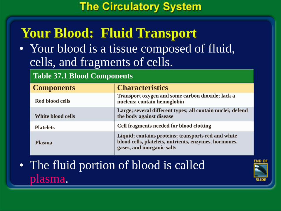
Your Blood: Fluid Transport • Your blood is a tissue composed of fluid,
cells, and fragments of cells.
• The fluid portion of blood is called plasma.
Components Characteristics
Red blood cells Transport oxygen and some carbon dioxide; lack a nucleus; contain hemoglobin
White blood cells
Platelets
Plasma
Large; several different types; all contain nuclei; defend the body against disease
Cell fragments needed for blood clotting
Liquid; contains proteins; transports red and white blood cells, platelets, nutrients, enzymes, hormones, gases, and inorganic salts
Table 37.1 Blood Components

Section 37.2 Summary – pages 975-984
Your Blood: Fluid Transport
• Blood cells-both red and white-and cell fragments are suspended in plasma.
• Plasma is straw colored and makes up about 55 percent of the total volume of blood.
Section 37.2 Summary – pages 975-984
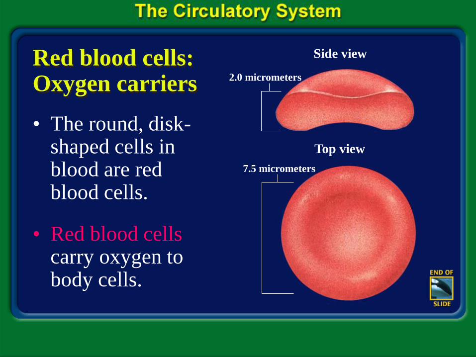
Red blood cells: Oxygen carriers
• The round, disk-shaped cells in blood are red blood cells.
• Red blood cells carry oxygen to body cells.
Side view
Top view
2.0 micrometers
7.5 micrometers
Section 37.2 Summary – pages 975-984
Red blood cells: Oxygen carriers
• They make up 44 percent of the total volume of your blood, and are produced in the red bone marrow of your ribs, humerus, femur sternum, and other long bones.
• Red blood cells remain active in the bloodstream for about 120 days, then they break down and are removed as waste.

Section 37.2 Summary – pages 975-984
Red blood cells: Oxygen carriers
• Old red blood cells are destroyed in your spleen, an organ of the lymphatic system, and in your liver.

Section 37.2 Summary – pages 975-984
Oxygen in the blood
• Red blood cells are equipped with an iron-containing protein molecule called hemoglobin (HEE muh gloh bun).

Section 37.2 Summary – pages 975-984
White blood cells: Infection fighters
• White blood cells play a major role in protecting your body from foreign substances and from microscopic organisms that cause disease.
• They make up only one percent of the total volume of your blood.
White Blood Cells
Section 37.2 Summary – pages 975-984
Blood clotting
• Your blood contains small cell fragments called platelets, which help blood clot after an injury.
• Platelets help link together a sticky network of protein fibers called fibrin, which forms a web over the wound that traps escaping blood cells.
Section 37.2 Summary – pages 975-984
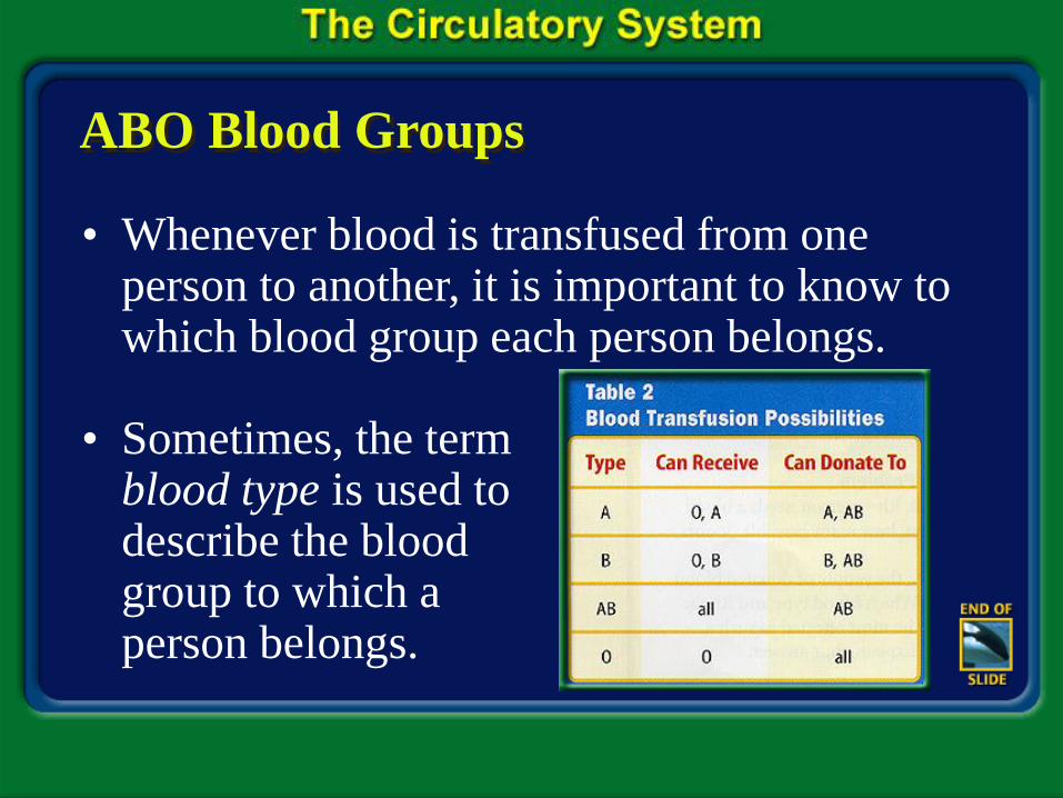
ABO Blood Groups
• Whenever blood is transfused from one person to another, it is important to know to which blood group each person belongs.
• Sometimes, the term blood type is used to describe the blood group to which a person belongs.
Section 37.2 Summary – pages 975-984
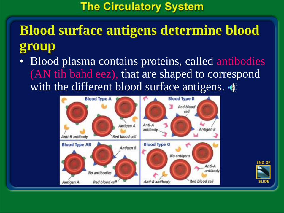
Blood surface antigens determine blood group • Blood plasma contains proteins, called antibodies
(AN tih bahd eez), that are shaped to correspond with the different blood surface antigens.

Section 37.2 Summary – pages 975-984
Rh factor
• Another characteristic of red blood cells involves the presence or absence of an antigen called RH, or Rhesus factor.
• Rh factor is an inherited characteristic.
• People are Rh positive (Rh+) if they have the Rh antigen factor on their red blood cells.
• They are Rh negative (Rh-) if they don’t.
Section 37.2 Summary – pages 975-984
Rh factor
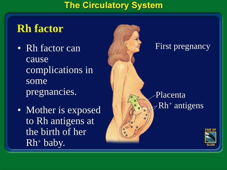
• Rh factor can cause complications in some pregnancies.
• Mother is exposed to Rh antigens at the birth of her Rh+ baby.
First pregnancy
Placenta
Rh+ antigens

Section 37.2 Summary – pages 975-984
Rh factor
• Mother makes anti-Rh+ antibodies.
• During the mother’s next pregnancy, Rh antibodies can cross the placenta and endanger the fetus.
Anti-Rh+ antibodies
Possible subsequent pregnancies
Section 37.2 Summary – pages 975-984
Rh factor
• Prevention of this problem is possible. When the Rh+ fetus is 28 weeks old, and again shortly after the Rh+ baby is born, the Rh- mother is given a substance that prevents the production of Rh antibodies in her blood.
• As a result, the next fetus will not be in danger.
Section 37.2 Summary – pages 975-984
Your Blood Vessels: Pathways of Circulation
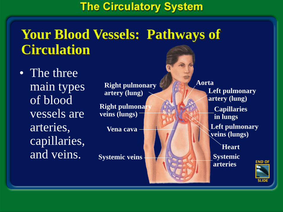
• The three main types of blood vessels are arteries, capillaries, and veins.
Aorta
Left pulmonary artery (lung)
Capillaries in lungs
Left pulmonary veins (lungs)
Heart
Systemic arteries
Systemic veins
Vena cava
Right pulmonary veins (lungs)
Right pulmonary artery (lung)
Section 37.2 Summary – pages 975-984
Your Blood Vessels: Pathways of Circulation
• Arteries are large, thick-walled, muscular, elastic blood vessels that carry blood away from the heart.
• The blood that they carry is under great pressure.
• As the heart contracts, it pushes blood through the arteries.
Section 37.2 Summary – pages 975-984
Your Blood Vessels: Pathways of Circulation
• Blood surges through the arteries in pulses that correspond with the rhythm of the heartbeat.
• After the arteries branch off from the heart, they divide into smaller arteries that in turn divide into even smaller vessels called arterioles.

Section 37.2 Summary – pages 975-984
Your Blood Vessels: Pathways of Circulation
• Arterioles (ar TEER ee ohlz) enter tissues, where they branch into the smallest blood vessels, the capillaries.
Artery Capillary
Section 37.2 Summary – pages 975-984
Your Blood Vessels: Pathways of Circulation
• Capillaries (KA puh ler eez) are microscopic blood vessels with walls that are only one cell thick.
• Capillaries form a dense network that reaches virtually every cell in the body.

Section 37.2 Summary – pages 975-984
Your Blood Vessels: Pathways of Circulation
• Thin capillary walls enable nutrients and gases to diffuse easily between blood cells and surrounding tissue cells.
• As blood leaves the tissues, the capillaries join to form slightly larger vessels called venules.

Section 37.2 Summary – pages 975-984
Your Blood Vessels: Pathways of Circulation
• The venules merge to form veins, the large blood vessels that carry blood from the tissues back toward the heart.
Vein Capillary

Section 37.2 Summary – pages 975-984
Your Blood Vessels: Pathways of Circulation
• In some veins, especially those in your arms and legs, blood travels uphill against gravity.
• Blood in veins is not under pressure as great as that in the arteries.

Section 37.1 Summary – pages 971-974
Click image to view movie.
Your Heart: The Vital Pump
Section 37.2 Summary – pages 975-984
Your Heart: The Vital Pump
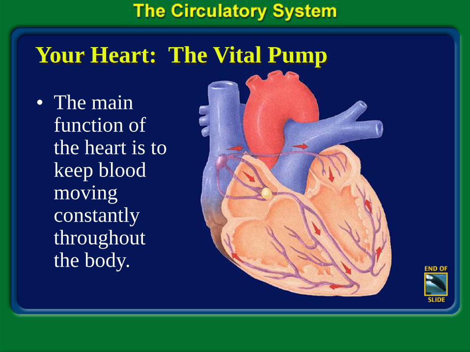
• The main function of the heart is to keep blood moving constantly throughout the body.
Section 37.2 Summary – pages 975-984
Your Heart: The Vital Pump
• The two upper chambers of the heart are the atria.
• All mammalian hearts, including yours, have four chambers.
• The two lower chambers are the ventricles.
• The walls of each atrium are thinner and less muscular than those of each ventricle.

Section 37.2 Summary – pages 975-984
Blood’s path through the heart
• The left atrium receives oxygen-rich blood from the lungs through four pulmonary veins.
Inferior vena cava Left lung
Right lung
Capillaries
Pulmonary vein
Pulmonary artery
Superior vena cava
Aorta
RA
LA
LV
RV
Section 37.2 Summary – pages 975-984
Blood’s path through the heart
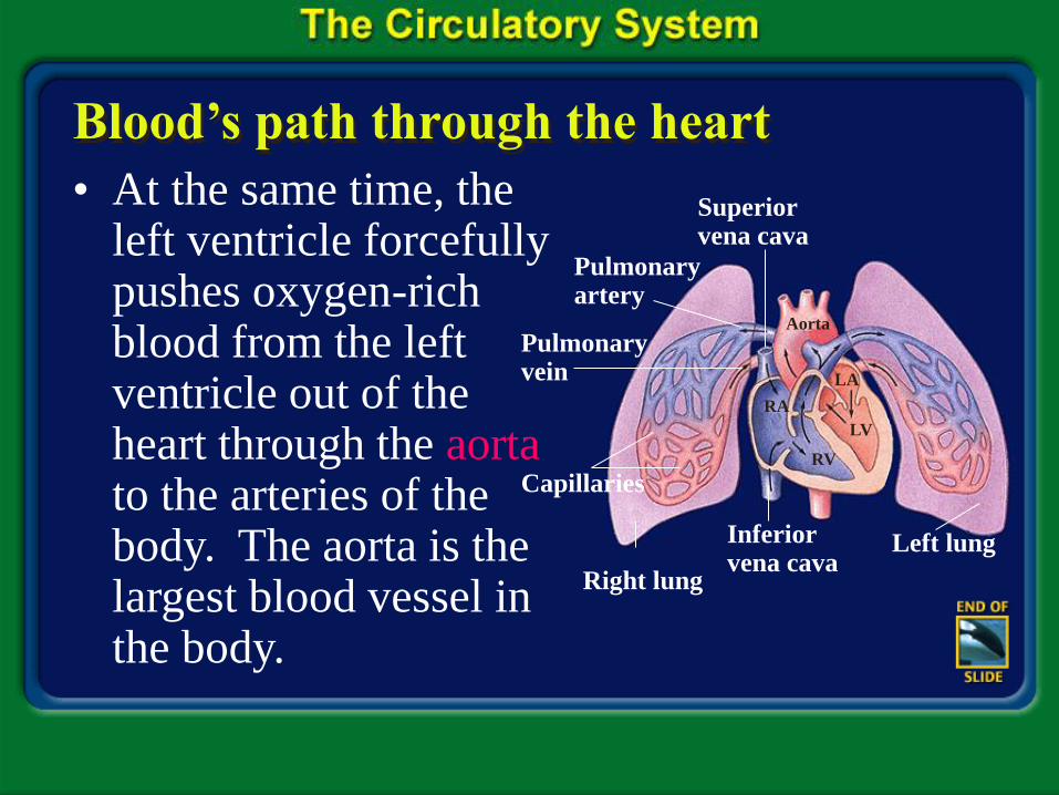
• At the same time, the left ventricle forcefully pushes oxygen-rich blood from the left ventricle out of the heart through the aorta to the arteries of the body. The aorta is the largest blood vessel in the body.
Inferior vena cava
Left lung
Right lung
Capillaries
Pulmonary vein
Pulmonary artery
Superior vena cava
Aorta
RA
LA
LV
RV

Section 37.2 Summary – pages 975-984
The passage of blood
Inferior vena cava Left lung
Right lung
Capillaries
Pulmonary vein
Pulmonary artery
Superior vena cava
Aorta
RA
LA
LV
RV

Section 37.2 Summary – pages 975-984
Heartbeat regulation
• The heart rate is set by the pacemaker, a bundle of nerve cells located at the top of the right atrium.
• This pacemaker generates an electrical impulse that spreads over both atria.
Sinoatrial node
(Pacemaker)
Atrioventricular node

Section 37.2 Summary – pages 975-984
Heartbeat regulation
Ventricular depolarization
Atrial depolarization
Ventricular repolarization
Vo
ltag
e (m
V) 1.0-
0.5-
0-
-0.5-
0 0.1 0.2 0.3
Seconds

Section 37.2 Summary – pages 975-984
Heartbeat regulation
Click image to view movie.
Section 37.2 Summary – pages 975-984
Blood pressure
• Blood pressure is the force that the blood exerts on the blood vessels.
• Blood pressure rises and falls as the heart contracts and then relaxes.
• Blood pressure rises sharply when the ventricles contract, pushing blood through the arteries.

Section 37.2 Summary – pages 975-984
Blood pressure
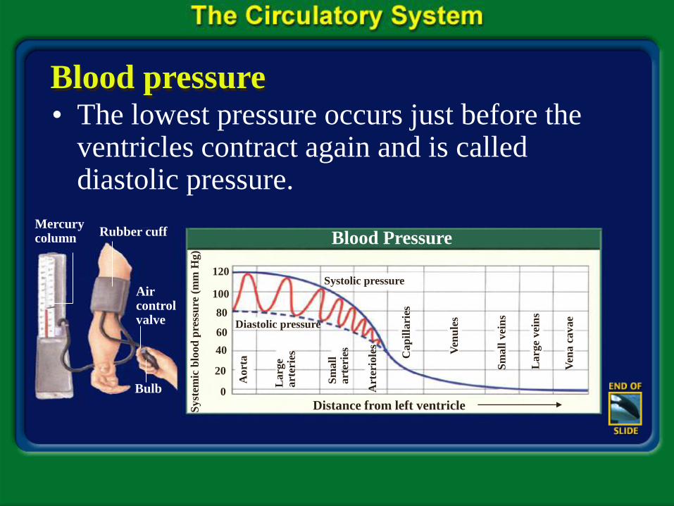
• The high pressure is called systolic pressure.
• Blood pressure then drops dramatically as the ventricles relax.
Section 37.2 Summary – pages 975-984
Blood pressure • The lowest pressure occurs just before the
ventricles contract again and is called diastolic pressure.
Blood Pressure Rubber cuff Mercury column
Air control valve
Bulb
Syst
emic
blo
od
pre
ssu
re (
mm
Hg)
120
100
80
60
40
20
0
Aort
a
Larg
e a
rter
ies
Sm
all
art
erie
s
Art
erio
les
Cap
illa
ries
Ven
ule
s
Sm
all
vei
ns
Larg
e vei
ns
Ven
a c
avae
Distance from left ventricle
Systolic pressure
Diastolic pressure
Section 37.2 Summary – pages 975-984
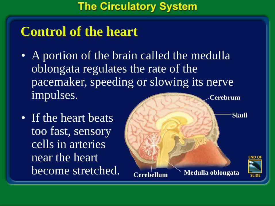
Control of the heart
• A portion of the brain called the medulla oblongata regulates the rate of the pacemaker, speeding or slowing its nerve impulses.
• If the heart beats too fast, sensory cells in arteries near the heart become stretched.
Skull
Cerebrum
Cerebellum Medulla oblongata
Section 37.2 Summary – pages 975-984
Control of the heart
• These cells send a signal to the medulla oblongata, which in turn sends signals that slow the pacemaker.
• If the heart slows down too much, blood pressure in the arteries drops, signaling the medulla oblongata to speed up the pacemaker and increase the heart rate.