Embed Size (px)
Citation preview
BACKGROUND
Perimetry has been the gold standard diagnostic modality of
glaucoma care for many years, though recently it has begun to share
that spotlight with OCT. Much of the literature that has solidified
perimetry’s place in glaucoma management uses the Humphrey®
Field Analyzer from ZEISS, which has changed very little since the
introduction of SITA™ (Swedish Interactive Thresholding Algorithm).
Visual field technology has lagged behind the rapid advancement in
OCT technology up until now. The ZEISS Humphrey Field Analyzer 3
(HFA3) combines upgrades in hardware and software to improve
both patient care and workflow efficiency.
ZEISS HUMPHREY FIELD ANALYZER TODAY
HFA3 – the Machine
The current iteration of the ZEISS Humphrey Field Analyzer is the
HFA3 with software version 1.4. This machine will be very familiar
to all those who used the older versions of HFA™. The HFA3 looks
like a smaller and sleeker version of the HFA that many of us have
in our offices or remember from the clinics where we trained. The
smaller footprint is a welcome element of the upgrade in an eyecare
world that is trending towards multiple space-consuming diagnostic
instruments. A new touchscreen interface simplifies navigation
of the responsive and nimble software. One significant hardware
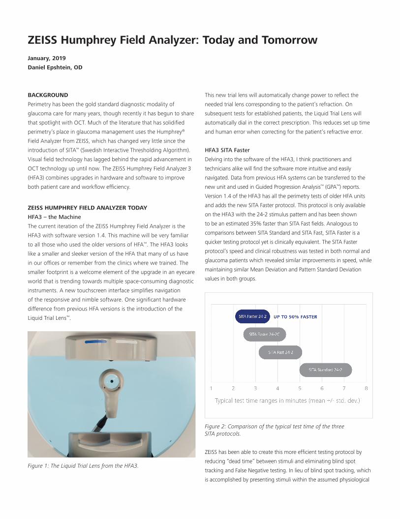
difference from previous HFA versions is the introduction of the
Liquid Trial Lens™.
Figure 1: The Liquid Trial Lens from the HFA3.
This new trial lens will automatically change power to reflect the
needed trial lens corresponding to the patient’s refraction. On
subsequent tests for established patients, the Liquid Trial Lens will
automatically dial in the correct prescription. This reduces set up time
and human error when correcting for the patient’s refractive error.
HFA3 SITA Faster
Delving into the software of the HFA3, I think practitioners and
technicians alike will find the software more intuitive and easily
navigated. Data from previous HFA systems can be transferred to the
new unit and used in Guided Progression Analysis™ (GPA™) reports.
Version 1.4 of the HFA3 has all the perimetry tests of older HFA units
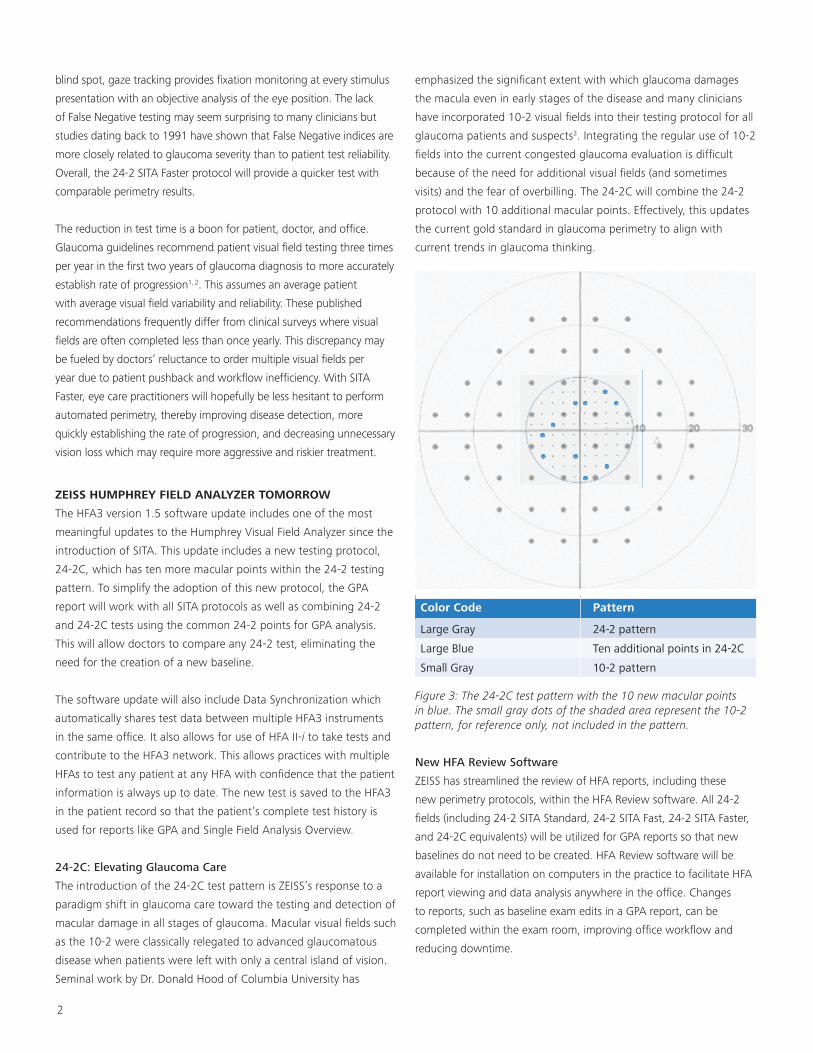
and adds the new SITA Faster protocol. This protocol is only available
on the HFA3 with the 24-2 stimulus pattern and has been shown
to be an estimated 35% faster than SITA Fast fields. Analogous to
comparisons between SITA Standard and SITA Fast, SITA Faster is a
quicker testing protocol yet is clinically equivalent. The SITA Faster
protocol’s speed and clinical robustness was tested in both normal and
glaucoma patients which revealed similar improvements in speed, while
maintaining similar Mean Deviation and Pattern Standard Deviation
values in both groups.
Figure 2: Comparison of the typical test time of the three SITA protocols.
ZEISS has been able to create this more efficient testing protocol by
reducing “dead time” between stimuli and eliminating blind spot
tracking and False Negative testing. In lieu of blind spot tracking, which
is accomplished by presenting stimuli within the assumed physiological
A
ZEISS Humphrey Field Analyzer: Today and TomorrowJanuary, 2019
Daniel Epshtein, OD
2
blind spot, gaze tracking provides fixation monitoring at every stimulus
presentation with an objective analysis of the eye position. The lack
of False Negative testing may seem surprising to many clinicians but
studies dating back to 1991 have shown that False Negative indices are
more closely related to glaucoma severity than to patient test reliability.
Overall, the 24-2 SITA Faster protocol will provide a quicker test with
comparable perimetry results.
The reduction in test time is a boon for patient, doctor, and office.
Glaucoma guidelines recommend patient visual field testing three times
per year in the first two years of glaucoma diagnosis to more accurately
establish rate of progression1, 2. This assumes an average patient
with average visual field variability and reliability. These published
recommendations frequently differ from clinical surveys where visual
fields are often completed less than once yearly. This discrepancy may
be fueled by doctors’ reluctance to order multiple visual fields per
year due to patient pushback and workflow inefficiency. With SITA
Faster, eye care practitioners will hopefully be less hesitant to perform
automated perimetry, thereby improving disease detection, more
quickly establishing the rate of progression, and decreasing unnecessary
vision loss which may require more aggressive and riskier treatment.
ZEISS HUMPHREY FIELD ANALYZER TOMORROW
The HFA3 version 1.5 software update includes one of the most
meaningful updates to the Humphrey Visual Field Analyzer since the
introduction of SITA. This update includes a new testing protocol,
24-2C, which has ten more macular points within the 24-2 testing
pattern. To simplify the adoption of this new protocol, the GPA
report will work with all SITA protocols as well as combining 24-2
and 24-2C tests using the common 24-2 points for GPA analysis.
This will allow doctors to compare any 24-2 test, eliminating the
need for the creation of a new baseline.
The software update will also include Data Synchronization which
automatically shares test data between multiple HFA3 instruments
in the same office. It also allows for use of HFA II-i to take tests and
contribute to the HFA3 network. This allows practices with multiple
HFAs to test any patient at any HFA with confidence that the patient
information is always up to date. The new test is saved to the HFA3
in the patient record so that the patient’s complete test history is
used for reports like GPA and Single Field Analysis Overview.
24-2C: Elevating Glaucoma Care
The introduction of the 24-2C test pattern is ZEISS’s response to a
paradigm shift in glaucoma care toward the testing and detection of
macular damage in all stages of glaucoma. Macular visual fields such
as the 10-2 were classically relegated to advanced glaucomatous
disease when patients were left with only a central island of vision.
Seminal work by Dr. Donald Hood of Columbia University has
emphasized the significant extent with which glaucoma damages
the macula even in early stages of the disease and many clinicians
have incorporated 10-2 visual fields into their testing protocol for all
glaucoma patients and suspects3. Integrating the regular use of 10-2
fields into the current congested glaucoma evaluation is difficult
because of the need for additional visual fields (and sometimes
visits) and the fear of overbilling. The 24-2C will combine the 24-2
protocol with 10 additional macular points. Effectively, this updates
the current gold standard in glaucoma perimetry to align with
current trends in glaucoma thinking.
Color Code Pattern
Large Gray 24-2 pattern
Large Blue Ten additional points in 24-2C
Small Gray 10-2 pattern
Figure 3: The 24-2C test pattern with the 10 new macular points in blue. The small gray dots of the shaded area represent the 10-2 pattern, for reference only, not included in the pattern.
New HFA Review Software
ZEISS has streamlined the review of HFA reports, including these
new perimetry protocols, within the HFA Review software. All 24-2
fields (including 24-2 SITA Standard, 24-2 SITA Fast, 24-2 SITA Faster,
and 24-2C equivalents) will be utilized for GPA reports so that new
baselines do not need to be created. HFA Review software will be
available for installation on computers in the practice to facilitate HFA
report viewing and data analysis anywhere in the office. Changes
to reports, such as baseline exam edits in a GPA report, can be
completed within the exam room, improving office workflow and
reducing downtime.
3
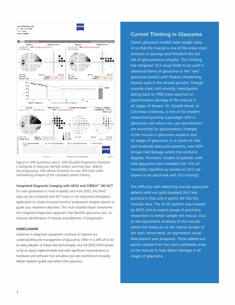
Figure 4: GPA Summary report. GPA (Guided Progression Analysis) is designed to help you identify where, and how fast, defects are progressing. GPA allows transition to new SITA tests while maintaining analysis of the complete patient history.
Integrated Diagnostic Imaging with HFA3 and CIRRUS™ HD-OCT
For next generation in multi-modality care from ZEISS, the HFA3
data can be combined with OCT scans in the Glaucoma Workplace
application to create structure-function progression analysis reports to
guide your treatment decisions. This multi-faceted report streamlines
the integrated diagnostics approach that benefits glaucoma care, to
improve identification of disease and detection of progression.
CONCLUSION
Advances in diagnostic equipment continue to improve our
understanding and management of glaucoma. Often it is difficult to be
an early adopter of these new technologies, but the ZEISS HFA3 proves
to be an easily implementable tool with significant improvements in
hardware and software that will allow eye care practitioners to easily
deliver research grade care within their practices.
Current Thinking in GlaucomaClassic glaucoma models have taught many of us that the macula is one of the areas most resistant to damage and therefore the last site of glaucomatous atrophy. This thinking has relegated 10-2 visual fields to be used in advanced forms of glaucoma or the “rare” glaucoma patient with fixation threatening disease early in the disease process. Though scarcely cited until recently, investigators dating back to 1969 have reported on glaucomatous damage to the macula in all stages of disease. Dr. Donald Hood, of Columbia University, is one of the modern researchers pushing a paradigm shift in glaucoma care where eye care practitioners are searching for glaucomatous changes in the macula in glaucoma suspects and all stages of glaucoma. In a cohort of mild and moderate glaucoma patients, over 50% of eyes had damage within the central 6 degrees. Perimetric studies of patients with mild glaucoma have revealed that 15% of hemifields classified as normal on 24-2 are shown to be abnormal with 10-2 testing4.
The difficulty with detecting macular glaucoma defects with our gold standard 24-2 test protocol is that only 4 points fall into the macular area. The 24-2C pattern was created by ZEISS and an expert group of perimetry researchers to better sample the macula. Due to the asymmetric anatomy of the macula, where the fovea sits at the inferior border of the optic nerve head, an asymmetric visual field pattern was proposed. These added test points sample from the most vulnerable areas of the macula to help detect damage in all stages of glaucoma.
1 Nouri-Mahdavi K, Zarei R, Caprioli J. Influence of visual field
testing frequency on detection of glaucoma progression with
trend analyses. Archives Ophthalmol. 2011 Dec;129(12):1521-7.
2 Chauhan BC, Garway-Heath DF, Goni FJ, et al. Practical
recommendations for measuring rates of visual field change in
glaucoma. Br J Ophthalmol. 2008 Apr;92(4):569-73.
3 Hood, Donald C. et al. Glaucomatous damage of the macula.
Progress in retinal and eye research. 32 (2013): 1-21.
4 Lifferth A, Fisher B, Stursma A, et al. 10-2 Visual Field Testing: A
Tool for All Glaucoma Stages. Review of Optometry. 2017 Jul, 15.
EN_3
1_20
0_01
48I
HFA
.107
89
ww
w.z
eiss
.com
/med
. CZ
-I/2
019
Onl
y fo
r sal
e in
sel
ecte
d co
untr
ies.
Not
all
prod
ucts
, ser
vice
s or
offe
rs a
re a
ppro
ved
or o
ffere
d in
eve
ry m
arke
t and
app
rove
d la
belin
g an
d in
stru
ctio
ns m
ay v
ary
from
one
cou
ntry
to a
noth
er.
The
stat
emen
ts o
f the
aut
hor r
eflec
t his
per
sona
l opi
nion
s an
d ex
perie
nces
and
do
not n
eces
saril
y re
flect
the
opin
ions
of a
ny in
stitu
tion
with
whi
ch h
e is
affi
liate
d.
The
auth
or h
as a
con
tract
ual r
elat
ions
hip
with
Car
l Zei
ss M
edite
c, In
c. a
nd h
as re
ceiv
ed fi
nanc
ial c
ompe
nsat
ion
© C
arl Z
eiss
Med
itec,
Inc.
, 201
9. A
ll rig
hts
rese
rved
.
References