Embed Size (px)
Citation preview
1
Zika virus replication in the mosquito Culex quinquefasciatus in
Brazil
D. R. D. Guedes1†, M. H. S. Paiva
2†, M. M. A. Donato
1, P. P. Barbosa
1, L. Krokovsky
1, S. W.
dos S. Rocha1, K. L. A. Saraiva
1, M. M. Crespo
1, R. M. R. Barbosa
1, C. M. F. Oliveira
1, M.
A. V. Melo-Santos1, L. Pena
3, M. T. Cordeiro
3, R. F. de O. França
3, A. L. S, de Oliveira
4, W.
S. Leal5, C. A. Peixoto
1, C. F. J. Ayres
1*.
Affiliations:
1 Department of Entomology, Centro de Pesquisas Aggeu Magalhães, Fundação Oswaldo
Cruz-Pernambuco. Av. Moraes Rego, s/n campus da UFPE, Cidade Universitária, Recife-PE,
Brazil, CEP: 50670-420.
2 Universidade Federal de Pernambuco, Centro Acadêmico do Agreste - Rodovia BR-104, km
59 - Nova Caruaru, Caruaru - PE – Brazil, CEP: 55002-970.
3 Laboratory of Virology and Experimental Therapy (LAVITE), Centro de Pesquisas Aggeu
Magalhães, Fundação Oswaldo Cruz-Pernambuco. Av. Moraes Rego, s/n campus da UFPE,
Cidade Universitária, Recife-PE, Brazil, CEP: 50670-420.
4 Center for Statistics and Geoprocessing, Centro de Pesquisas Aggeu Magalhães, Fundação
Oswaldo Cruz-Pernambuco. Av. Moraes Rego, s/n campus da UFPE, Cidade Universitária,
Recife-PE, Brazil, CEP: 50670-420.
5 Department of Molecular and Cellular Biology, University of California-Davis, Davis, CA,
95616 USA.
* Corresponding author. E-mail: [email protected]
† These authors contributed equally to this work.
Abstract:
Zika virus (ZIKV) is a flavivirus that has recently been associated with increased incidence of
neonatal microcephaly and other neurological disorders. The virus is primarily transmitted by
mosquito bite, although other routes of infection have been implicated in some cases. The
Aedes aegypti mosquito is considered to be the main vector to humans worldwide, but there is
evidence of other mosquito species, including Culex quinquefasciatus, playing a role in the
Brazilian outbreak. To test this hypothesis, we experimentally compared the vectorial
competence of laboratory-reared A. aegypti and C. quinquefasciatus. We found ZIKV in the
All rights reserved. No reuse allowed without permission. (which was not peer-reviewed) is the author/funder, who has granted bioRxiv a license to display the preprint in perpetuity.
The copyright holder for this preprint. http://dx.doi.org/10.1101/073197doi: bioRxiv preprint first posted online Sep. 2, 2016;
2
midgut, salivary glands, and saliva of artificially fed C. quinquefasciatus. Additionally, we
collected ZIKV-infected C. quinquefasciatus from urban areas of high microcephaly
incidence in Recife, Brazil. Take into account; these findings indicate that there may be a
wider range of vectors for ZIKV than anticipated.
Keywords: Zika, microcephaly, Culex, Aedes, vectorial competence, vector control.
Zika is classically considered a mild disease whose symptoms include fever, joint pain, rash
and, in some cases, conjunctivitis (1). However, the Zika outbreak in Brazil has been
associated with an increased incidence of neonatal microcephaly and neurological disorders
(2, 3). Zika virus (ZIKV) is a poorly known, small, enveloped RNA virus with ssRNA (+)
belonging to the Family Flaviviridae. It was first isolated in April 1947 from a rhesus monkey
and in January 1948 from the mosquito species Aedes africanus (4). Since then, several ZIKV
strains have been isolated from many samples, mostly mosquitoes, including species from the
genera Aedes, Mansonia, Anopheles and Culex (5).
The first known Zika epidemic in an urban environment occurred in Micronesia in 2007, with
approximately 73% of the human population on Yap island becoming infected (6).
Intriguingly, although many Aedes mosquitoes were collected in the field and evaluated for
virus detection, no samples were found to be positive for ZIKV (6). Additionally, it is
important to highlight that Aedes aegypti (A. aegypti) is absent from most islands in the
Micronesia archipelago and is very rare on the islands where it is present (6, 7).
There is a global consensus among scientists and health agencies that Aedes spp. are the main
ZIKV vector in urban areas (WHO, 2016). This is in part because vector competence
experiments for ZIKV have been conducted exclusively for species of this genus, mainly A.
aegypti (8, 9). Previous laboratory studies (8, 10) suggested that A. aegypti is a ZIKV vector.
All rights reserved. No reuse allowed without permission. (which was not peer-reviewed) is the author/funder, who has granted bioRxiv a license to display the preprint in perpetuity.
The copyright holder for this preprint. http://dx.doi.org/10.1101/073197doi: bioRxiv preprint first posted online Sep. 2, 2016;
3
Recently, high rates of dissemination and transmission of the ZIKV in A. aegypti has been
observed under laboratory conditions (11). Intriguingly, a few studies show that A. aegypti
and Aedes albopictus populations have low rates of ZIKV transmission (12) or none (13, 14),
but the role of other vectors in the spread of ZIKV has been overlooked. Thus, other mosquito
species, co-existing with A. aegypti in urban areas, could contribute to ZIKV transmission
(15). Here, we report data that support the idea that Culex quinquefasciatus, the most common
mosquito in tropical and subtropical areas, is a potential ZIKV vector. We performed
mosquito vector competence assays under laboratory conditions, comparing both A. aegypti
and C. quinquefasciatus using different virus doses, as well as the detection of ZIKV in wild
C. quinquefasciatus mosquitoes. ZIKV was detected in salivary glands and in the saliva of
artificially fed C. quinquefasciatus mosquitoes, suggesting that this species is a potential
vector for ZIKV transmission. Taken together, our results have implications for vector control
strategies and understanding the epidemiology of ZIKV.
Vector competence assays
Artificial blood feeding assays were performed using two laboratory-reared colonies: RecLab
(A. aegypti) and CqSLab (C. quinquefasciatus) and a field-collected population of A. aegypti
(F1/F2) from the Archipelago of Fernando de Noronha, a district from Pernambuco state,
Northeast Brazil. A local Zika virus strain, isolated from the serum of a patient with Zika
symptoms from Pernambuco State, Brazil, during the 2015 outbreak
(ZIKV/H.sapiens/Brazil/PE243/201), fully characterized (accession number KX197192.1),
was used in vector competence assays.
Seven to ten days-old females were challenged in an artificial feeding, consisted of a Petri
dish covered with Parafilm M®, with a mixture containing equal volumes of defibrinated
rabbit blood and the viral suspension. Here, in each assay, we used two different viral doses:
106 PFU/ml and 10
4 PFU/ml. Mosquitoes were exposed for 90 minutes and, after that, only
All rights reserved. No reuse allowed without permission. (which was not peer-reviewed) is the author/funder, who has granted bioRxiv a license to display the preprint in perpetuity.
The copyright holder for this preprint. http://dx.doi.org/10.1101/073197doi: bioRxiv preprint first posted online Sep. 2, 2016;
4
the engorged females were transferred to another cage and maintained in the infectory room,
under bio safety conditions (BSL2) for 15 days. At 3, 7, 11 and 15 days post infection (dpi),
midguts and salivary glands were dissected individually and transferred to 1.5 ml vials
containing a mosquito diluent (20% of fetal bovine serum in PBS with 50 g/ml
penicillin/streptomycin, 50 g/ml gentamicin and 2.5g/ml fungizone) and stored at -80oC
until further usage. After RNA extraction, samples were assayed by quantitative RT-PCR
(RT-qPCR) and both Infection Rate (IR), which is the proportion of infected midguts, and the
proportion of infected salivary glands (SR), which is the number of positive salivary glands
divided by the total number of salivary glands tested, were calculated for each species on each
dpi. All procedures are described in details in the Supplemental Materials.
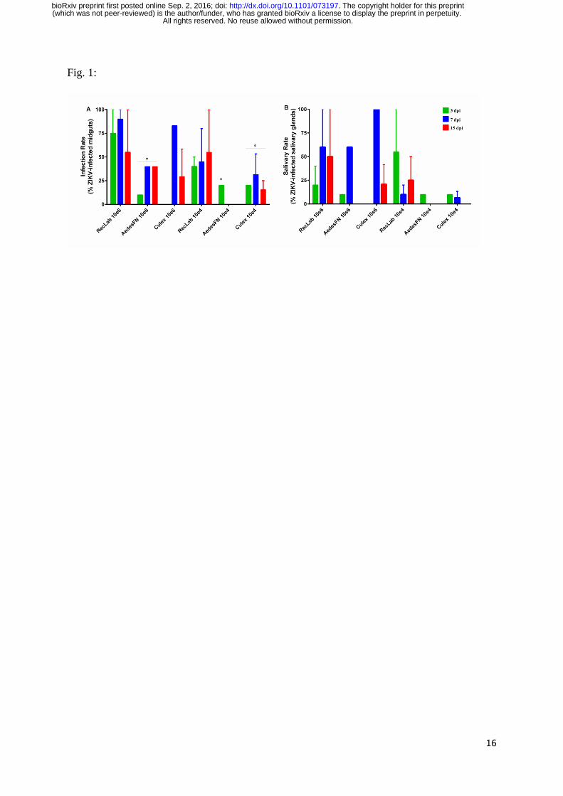
A total of 289 mosquitoes were examined for ZIKV infection by RT-qPCR. Among these
mosquitoes, 130 were A. aegypti RecLab, 60 were A. aegypti FN and 99 were C.
quinquefasciatus. During the extrinsic incubation period, a high mortality rate was observed
in the infected group a few days after blood feeding, with peak mortality observed between 3-
5 dpi. In A. aegypti populations, the mortality rate ranged from 48% to 52%; in C.
quinquefasciatus mosquitoes, it ranged from 33% to 44% (data not shown).
In both species, we detected ZIKV in the midgut at most time-points under study, except in
field-collected A. aegypti (FN) blood fed at a low viral dose (104 PFU/ml). In the salivary
glands of A. aegypti, we detected ZIKV-positive samples at 3 dpi for both viral doses.
Although variations in infection were observed, the IR between Aedes and Culex mosquito’s
species was not statistically significant (Fig 1A). Analysing only Culex species, the
differences in IR were not statistically significant comparing different viral doses (p>0.05).
When a high viral dose (106 PFU/ml) was used during artificial feeding, the SR reached an
average of 60% in A. aegypti RecLab, and 100% in C. quinquefasciatus at 7 dpi, but this
average declined to 20% at 15 dpi in Culex and to 50% in A. aegypti. However, this difference
All rights reserved. No reuse allowed without permission. (which was not peer-reviewed) is the author/funder, who has granted bioRxiv a license to display the preprint in perpetuity.
The copyright holder for this preprint. http://dx.doi.org/10.1101/073197doi: bioRxiv preprint first posted online Sep. 2, 2016;
5
was not significant (p>0.05) (Fig 1B). When a lower viral dose was used, for both A. aegypti
populations (lab and field-caught), we observed that the mosquito lab colony infection rate
was higher than field-caught mosquitoes (Figure 1A p=0.0186), although there were no
differences in SR (Figure 1B p>0.05). The maximum SR in Culex at lower viral dose, was
10% at 3dpi declining to zero after 15 dpi (p>0.05) (Fig. 1B). We also sampled mosquitoes at
11 dpi in the first trial, except for field-collected A. aegypti (FN), which had a SR of 10%;
neither C. quinquefasciatus nor A. aegypti samples from the lab were ZIKV positive (data no
shown).
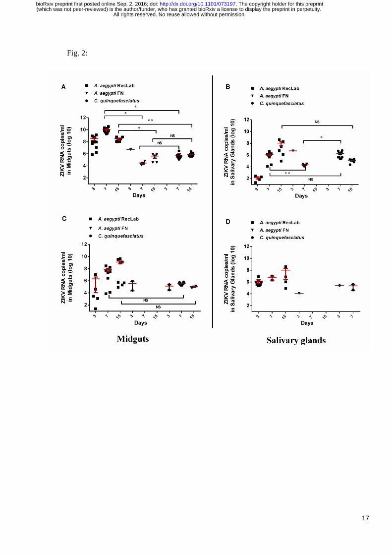
RT-qPCR was used to quantify ZIKV RNA load at the different time-points. In general, viral
RNA copies in A. aegypti RecLab in the midguts and salivary glands varied considerably.
Both A. aegypti FN and C. quinquefasciatus viral copies in target organs (midgut and salivary
glands) remained detectable (Fig. 2A to D). To evaluate ZIKV transmission in saliva for both
species, honey-soaked filter papers (FTA Classic Cards, Whatman®, Maidstone, UK) were
offered to mosquitoes to feed upon 8-14 dpi. At 9-12 dpi, ZIKV RNA was detected in saliva
of both A. aegypti and C. quinquefasciatus species (Fig. 3). When a high viral dose (106) was
used, the amount of viral RNA copies expectorated during salivation in both Aedes and Culex
were similar at all time-points analysed (p>0.05). However, when the mosquitoes were
challenged with a low viral dose (104), A. aegypti expectorated more RNA viral copies than
Culex (p=0.0473).
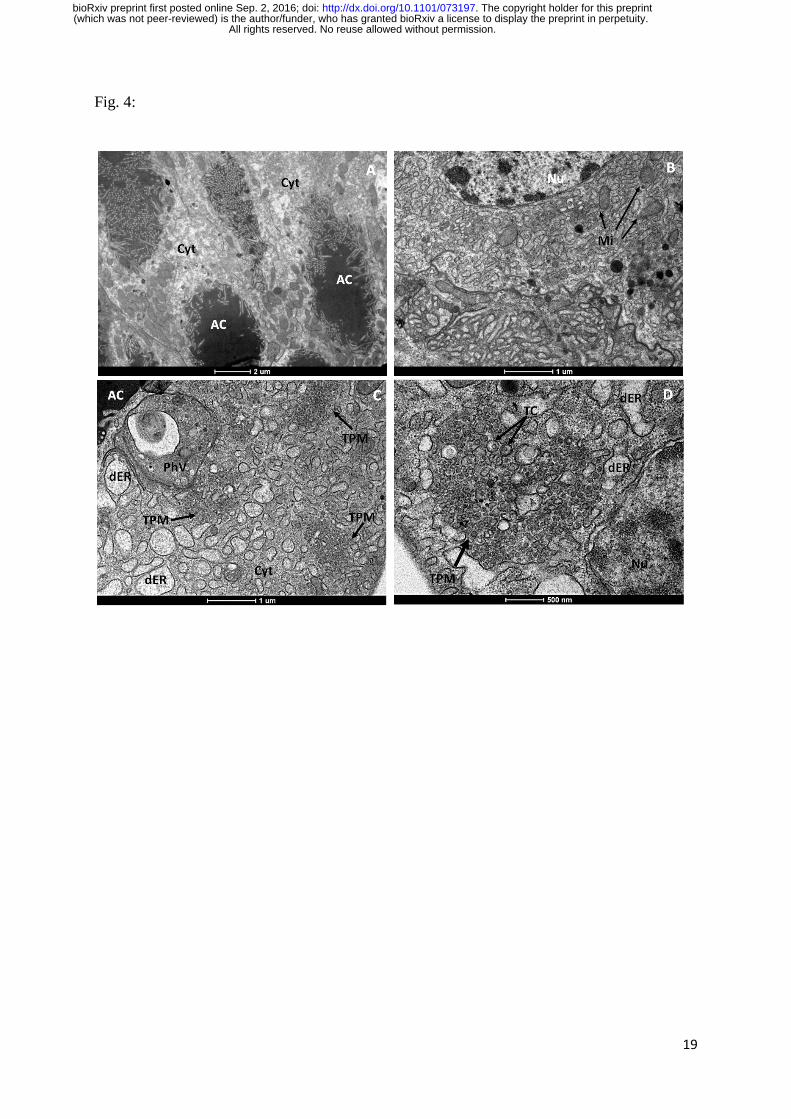
Transmission Electron Microscopy
To further confirm our results from RT-qPCR, we performed a transmission electron
microscopy from dissected salivary glands from C. quinquesfaciatus infected mosquitoes. The
morphological organization of C. quinquefasciatus salivary glands showed an electron-dense
apical cavity, displaying membrane projections extending from the wall (Fig. 4 A,B). ZIKV
infected salivary acinar cells of C. quinquefasciatus showed signs of cytopathic disruptions,
All rights reserved. No reuse allowed without permission. (which was not peer-reviewed) is the author/funder, who has granted bioRxiv a license to display the preprint in perpetuity.
The copyright holder for this preprint. http://dx.doi.org/10.1101/073197doi: bioRxiv preprint first posted online Sep. 2, 2016;
6
including cisternae in the endoplasmic reticulum and tubular proliferated membranes,
organized in several patches within a single cell (Fig. 4C, D). Mature ZIKV particles of 40-50
nm in diameter, composed of a central electrodense core (~30 nm in diameter) surrounded by
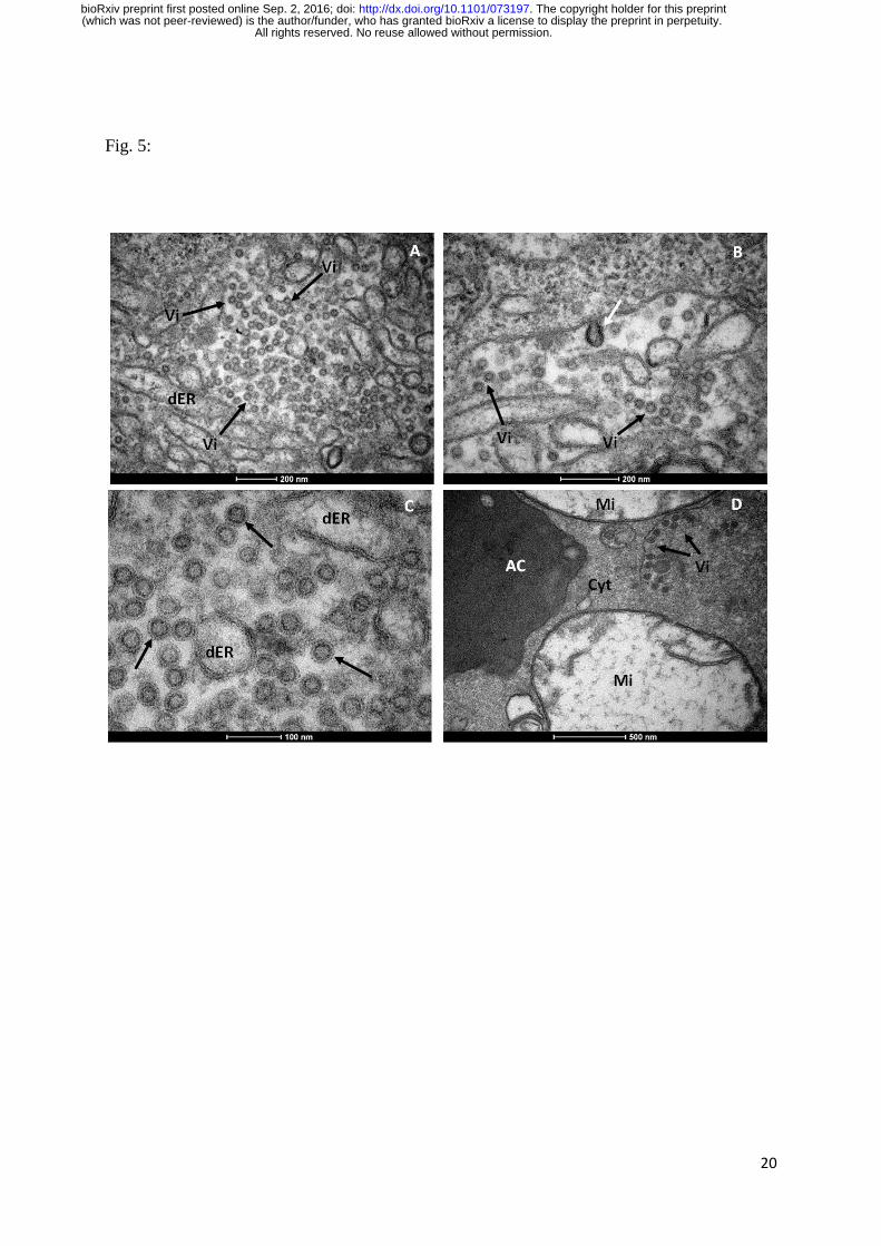
a viral envelope, were observed inside the dilated endoplasmic reticulum (Fig. 5A to D). In
some regions, viral envelope formation is shown to arise from endoplasmic membrane (Fig.
5B). Some ZIKV virions were observed proximal to the apical cavity of the salivary cell.
Mitochondria also showed severe damage, including complete loss of cristae (Fig. 5D). In
summary, transmission electron microscopy analysis confirmed that C. quinquefasciatus
mosquitoes are permissive to ZIKV infection, since viral particles were detected at the
salivary glands of artificially fed mosquitoes.
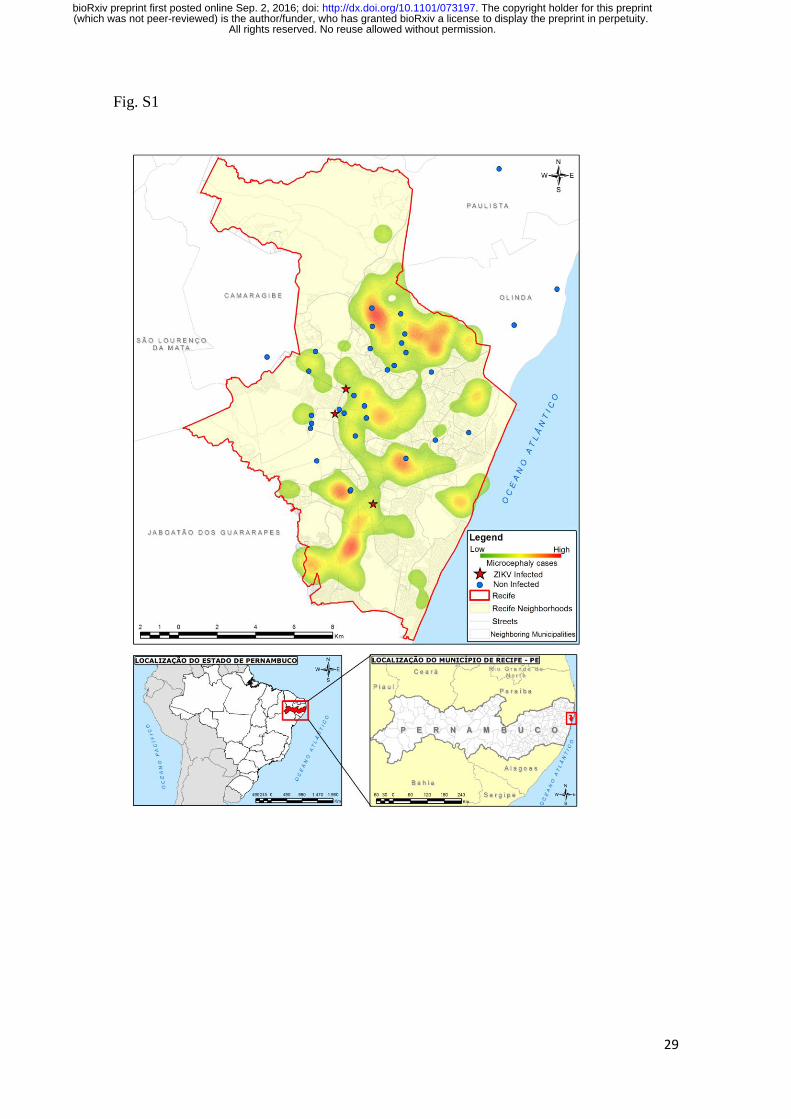
ZIKV detection in field-caught C. quinquefasciatus
Lastly, we conducted ZIKV surveillance (February to May 2016) with mosquitoes collected
with a battery-operated aspirator device (Horst®
) from residences inhabited by individuals
with clinical symptoms of zika fever. Field-collected mosquitoes were sorted by place of
collection, species, sex, feeding status (engorged and not engorged) and grouped in pools of
up to 10 mosquitoes. A total of 1,496 adult C. quinquefasciatus and 408 A. aegypti female
mosquitoes were collected from different sites in the Metropolitan Region of Recife (Fig. S1).
These mosquito pools were grinded in Leibovitz medium supplemented with 5% FBS. These
samples were separated into two aliquots, one for RT-qPCR and the other for virus isolation.
From 270 pooled-samples of adult female C. quinquefasciatus and 117 pools of A. aegypti
mosquitoes assayed by RT-qPCR, three Culex and two Aedes pools were positive for ZIKV.
Interestingly, two out of the three positive Culex samples were not blood-fed, whereas
concerning Aedes pools, the two positive pools for ZIKV were fed. The cycle threshold (Ct)
of Culex positive pools when screened by RT-qPCR were 37.6 (sample 5), 38.0 (sample 17)
All rights reserved. No reuse allowed without permission. (which was not peer-reviewed) is the author/funder, who has granted bioRxiv a license to display the preprint in perpetuity.
The copyright holder for this preprint. http://dx.doi.org/10.1101/073197doi: bioRxiv preprint first posted online Sep. 2, 2016;
7
and 38.15 (sample 163). Concerning Aedes pools, Cts were 37.5 (sample 3) and 37.9 (sample
7). Minimum infection rate (MIR - number of positive pools divided by the total of specimens
assayed multiplied by 1000) were calculated for both species. For C. quinquefasciatus, MIR
was 2.0 and concerning A. aegypti, MIR was 4.9. In an attempt to isolate ZIKV from field-
caught Culex mosquitoes, we inoculated in African Green Monkey kidney cells, samples from
two positive pools with the lowest cycle thresholds. Indeed, ZIKV was isolated from these
samples, thus unambiguously demonstrating that this species was carrying active ZIKV
particles in Recife, Brazil. Two ZIKV-positive isolates from field-caught Culex mosquitoes
were submitted to Sanger and MinION platforms. Sanger sequencing resulted in low quality
sequences and only a partial fragment was acquired from MinION sequencing, probably as a
direct result of low viral titers. This partial sequence enabled us to confirm the virus identity.
Sequence was deposited at GenBank, and accession number is still to be provided.
Discussion
Our work has associated a second mosquito genus in ZIKV transmission cycle in North-
eastern Brazil. We showed that, C. quinquefasciatus, also known as the southern house
mosquito, which is the most common mosquito in urban areas in Brazil, is susceptible to
infection with ZIKV during experimental blood feeding; moreover, we found that ZIKV has
an active replication cycle in the salivary glands and being subsequently released in the saliva.
In addition, we were able to detect ZIKV circulating in wild C. quinquefasciatus collected
from Recife.
Although it is widely assumed that A. aegypti is the main ZIKV vector, previous vector
competence studies are inconclusive. In the present study, a low dose of ZIKV (104 PFU/ml)
was used for comparison with the higher doses used in previous studies (11, 12, 14). We
All rights reserved. No reuse allowed without permission. (which was not peer-reviewed) is the author/funder, who has granted bioRxiv a license to display the preprint in perpetuity.
The copyright holder for this preprint. http://dx.doi.org/10.1101/073197doi: bioRxiv preprint first posted online Sep. 2, 2016;
8
found that both A. aegypti and C. quinquefasciatus can be experimentally infected by ZIKV
even at low doses and that ZIKV virus was subsequently detected in the saliva.
Cornet et al. (10) concluded that not all infected mosquitoes could transmit the virus and
could not always transmit it, in contrast to the idea that once infected, a mosquito would
transmit virus for its entire life. This finding suggests that a time window for vector-borne
ZIKV transmission may exist. We found that after 11 dpi, most samples were negative for
ZIKV (apart from one positive salivary gland of A. aegypti given 106 PFU/ml), thus our
maximum time point analysis was set to 15 days post infection. However, Boorman &
Porterfield (8) reported that virus replication resumed at 15-20 dpi and ZIKV remained
present in Aedes mosquitoes for up to 60 days.
To confirm that the virus detected in the salivary glands by RT-qPCR was being released in
saliva during consecutive blood meals, we followed up the viral load from days 8 to 14 post-
infection using filter paper cards. This strategy of viral RNA detection directly from FTA
cards has been employed in previous studies for arbovirus surveillance (16, 17). In the present
study, we successfully detected ZIKV RNA copies in cards from A. aegypti and C.
quinquefasciatus populations. This result demonstrates that in addition to being susceptible to
ZIKV infection, allowing virus replication in the salivary glands, both species are capable of
effectively transmit ZIKV.
RT-qPCR results were confirmed by transmission electron microscopy. The general mature
ZIKV morphology observed on the salivary glands confirmed previous ultrastructural studies
(18-20). In salivary glands cells, ZIKV replication causes cytopathic effects by 7 dpi. Similar
results have been shown for West Nile virus (WNV) (21, 22), although we did not directly
observe ZIKV nucleocapsids budding from endoplasmic reticulum membranes nor from the
tubular proliferated membrane (21). The fact that we found salivary glands positive for ZIKV
All rights reserved. No reuse allowed without permission. (which was not peer-reviewed) is the author/funder, who has granted bioRxiv a license to display the preprint in perpetuity.
The copyright holder for this preprint. http://dx.doi.org/10.1101/073197doi: bioRxiv preprint first posted online Sep. 2, 2016;
9
when the midgut of the same mosquito was negative indicates that mosquitoes may be
clearing viral infection in the midgut while virus replication continues in the salivary glands.
This finding has implications for the analytical methods employed in vector competence
studies.
Currently, there is a lack of studies investigating Culex vectorial competence for ZIKV. Most
studies have targeted only Aedes species, and only a few studies have compared different
species (including Culex) regarding natural infection rates. Surprisingly, Diallo et al. (5)
observed a higher minimum infection rate for Culex perfuscus (10x higher) than for A.
aegypti. Positive A. aegypti samples have always been reported at very low infection rates,
even in areas with high human ZIKV infection rates, such as Malaysia (23). Indeed, in
Micronesia (6), and French Polynesia, ZIKV was not detected in wild-caught Aedes spp.
mosquitoes during outbreaks. It is interesting that in all of these areas, C. quinquefasciatus is
an abundant mosquito species that may have also played an undetected role in ZIKV
transmission. Furthermore, A. aegypti and C. quinquefasciatus have completely different
behaviours regarding feeding periods and breeding site preferences.
Thus, our findings indicate that vector control strategies may need to be re-examined since
reducing A. aegypti populations may not lead to an overall reduction in ZIKV transmission if
Culex populations are slight affected by Aedes specific control measures. To the moment,
there is no broad ongoing program for C. quinquefasciatus control in Brazil, although Recife,
Olinda and Jaboatão dos Guararapes, three municipalities in Recife Metropolitan Region,
have undertaken specific control of C. quinquefasciatus to control lymphatic filariasis
transmission locally (24).
Viral transmission via C. quinquefasciatus is not a new concept; this species is the major
vector of West Nile virus in North America (25), along with Japanese encephalitis virus (26)
All rights reserved. No reuse allowed without permission. (which was not peer-reviewed) is the author/funder, who has granted bioRxiv a license to display the preprint in perpetuity.
The copyright holder for this preprint. http://dx.doi.org/10.1101/073197doi: bioRxiv preprint first posted online Sep. 2, 2016;
10
and equine encephalitis virus (27). Our present study indicates that C. quinquefasciatus
mosquitoes may be involved in ZIKV transmission in Recife. Thus, it is now necessary to
understand the contributions of each species in transmission to target each one properly. In
conclusion, considering its high abundance in urban environments and its anthropophilic
behaviour in Brazil (28-30), C. quinquefasciatus may be a vector for ZIKV in this region.
References and Notes
1. C. Chang, K. Ortiz, A. Ansari, M. E. Gershwin, The Zika outbreak of the 21st century.
Journal of autoimmunity 68, 1-13 (2016).
2. M. T. Cordeiro, L. J. Pena, C. A. Brito, L. H. Gil, E. T. Marques, Positive IgM for
Zika virus in the cerebrospinal fluid of 30 neonates with microcephaly in Brazil.
Lancet 387, 1811-1812 (2016).
3. A. F. Moron et al., Microcephaly associated with maternal Zika virus infection. BJOG
: an international journal of obstetrics and gynaecology 123, 1265-1269 (2016).
4. G. W. Dick, S. F. Kitchen, A. J. Haddow, Zika virus. I. Isolations and serological
specificity. Transactions of the Royal Society of Tropical Medicine and Hygiene 46,
509-520 (1952).
5. D. Diallo et al., Zika virus emergence in mosquitoes in southeastern Senegal, 2011.
PloS one 9, e109442 (2014).
6. M. R. Duffy et al., Zika virus outbreak on Yap Island, Federated States of Micronesia.
The New England journal of medicine 360, 2536-2543 (2009).
7. H. M. Savage et al., Epidemic of dengue-4 virus in Yap State, Federated States of
Micronesia, and implication of Aedes hensilli as an epidemic vector. The American
journal of tropical medicine and hygiene 58, 519-524 (1998).
8. J. P. Boorman, J. S. Porterfield, A simple technique for infection of mosquitoes with
viruses; transmission of Zika virus. Transactions of the Royal Society of Tropical
Medicine and Hygiene 50, 238-242 (1956).
9. M. I. Li, P. S. Wong, L. C. Ng, C. H. Tan, Oral susceptibility of Singapore Aedes
(Stegomyia) aegypti (Linnaeus) to Zika virus. PLoS neglected tropical diseases 6,
e1792 (2012).
10. M. Cornet, Y. Robin, C. Adam, M. Valade, M. A. Calvo, Comparison between
experimental transmission of yellow fever and zika viruses in Aedes aegypti. Cah
ORSTOM ser Ent med et Parasitol 17, 7 (1979).
11. H. L. Dutra et al., Wolbachia Blocks Currently Circulating Zika Virus Isolates in
Brazilian Aedes aegypti Mosquitoes. Cell host & microbe 19, 771-774 (2016).
12. T. Chouin-Carneiro et al., Differential Susceptibilities of Aedes aegypti and Aedes
albopictus from the Americas to Zika Virus. PLoS neglected tropical diseases 10,
e0004543 (2016).
13. W. G. Bearcroft, Zika virus infection experimentally induced in a human volunteer.
Transactions of the Royal Society of Tropical Medicine and Hygiene 50, 442-448
(1956).
All rights reserved. No reuse allowed without permission. (which was not peer-reviewed) is the author/funder, who has granted bioRxiv a license to display the preprint in perpetuity.
The copyright holder for this preprint. http://dx.doi.org/10.1101/073197doi: bioRxiv preprint first posted online Sep. 2, 2016;
11
14. C. T. Diagne et al., Potential of selected Senegalese Aedes spp. mosquitoes (Diptera:
Culicidae) to transmit Zika virus. BMC infectious diseases 15, 492 (2015).
15. C. F. Ayres, Identification of Zika virus vectors and implications for control. The
Lancet. Infectious diseases 16, 278-279 (2016).
16. E. J. Flies, C. Toi, P. Weinstein, S. L. Doggett, C. R. Williams, Converting Mosquito
Surveillance to Arbovirus Surveillance with Honey-Baited Nucleic Acid Preservation
Cards. Vector borne and zoonotic diseases 15, 397-403 (2015).
17. S. Hall-Mendelin et al., Exploiting mosquito sugar feeding to detect mosquito-borne
pathogens. Proceedings of the National Academy of Sciences of the United States of
America 107, 11255-11259 (2010).
18. T. M. Bell, E. J. Field, H. K. Narang, Zika virus infection of the central nervous
system of mice. Archiv fur die gesamte Virusforschung 35, 183-193 (1971).
19. J. Mlakar et al., Zika Virus Associated with Microcephaly. The New England journal
of medicine 374, 951-958 (2016).
20. L. Rosen, Overwintering mechanisms of mosquito-borne arboviruses in temperate
climates. The American journal of tropical medicine and hygiene 37, 69S-76S (1987).
21. Y. A. Girard, V. Popov, J. Wen, V. Han, S. Higgs, Ultrastructural study of West Nile
virus pathogenesis in Culex pipiens quinquefasciatus (Diptera: Culicidae). Journal of
medical entomology 42, 429-444 (2005).
22. Y. A. Girard et al., Salivary gland morphology and virus transmission during long-
term cytopathologic West Nile virus infection in Culex mosquitoes. The American
journal of tropical medicine and hygiene 76, 118-128 (2007).
23. N. J. Marchette, R. Garcia, A. Rudnick, Isolation of Zika virus from Aedes aegypti
mosquitoes in Malaysia. The American journal of tropical medicine and hygiene 18,
411-415 (1969).
24. K. D. Chalegre et al., Detection of an allele conferring resistance to Bacillus
sphaericus binary toxin in Culex quinquefasciatus populations by molecular
screening. Applied and environmental microbiology 75, 1044-1049 (2009).
25. R. S. Nasci et al., West Nile virus isolates from mosquitoes in New York and New
Jersey, 1999. Emerging infectious diseases 7, 626-630 (2001).
26. S. Sucharit, K. Surathin, S. R. Shrestha, Vectors of Japanese encephalitis virus (JEV):
species complexes of the vectors. The Southeast Asian journal of tropical medicine
and public health 20, 611-621 (1989).
27. Z. Wang et al., Vector competence of five common mosquito species in the People's
Republic of China for Western equine encephalitis virus. Vector borne and zoonotic
diseases 12, 605-608 (2012).
28. R. A. G. B. Consoli, R. Lourenco-de-Oliveira, in Principais mosquitos de importância
sanitária no Brasil. (Editora Fiocruz, Rio de Janeiro, 1994), chap. 3, pp. 57-154.
29. O. P. Forattini, C. Gomes Ade, D. Natal, I. Kakitani, D. Marucci, [Food preferences of
Culicidae mosquitoes in the Ribeira valley, Sao Paulo, Brazil]. Revista de saude
publica 21, 171-187 (1987).
30. L. Regis et al., Integrated control measures against Culex quinquefasciatus, the vector
of filariasis in Recife. Memorias do Instituto Oswaldo Cruz 90, 115-119 (1995).
31. L. B. Amorim, Thesis, Universidade Federal de Pernambuco, (2013).
32. L. B. Amorim, E. Helvecio, C. M. de Oliveira, C. F. Ayres, Susceptibility status of
Culex quinquefasciatus (Diptera: Culicidae) populations to the chemical insecticide
temephos in Pernambuco, Brazil. Pest management science 69, 1307-1314 (2013).
33. A. P. Araujo, M. A. Melo-Santos, S. O. Carlos, E. M. Rios, L. Regis, Evaluation of an
experimental product based on Bacillus thurigiensis sorovar. israelensis against Aedes
aegypti larvae (Diptera: Culicidae). Biol Control 41, 339-347 (2007).
All rights reserved. No reuse allowed without permission. (which was not peer-reviewed) is the author/funder, who has granted bioRxiv a license to display the preprint in perpetuity.
The copyright holder for this preprint. http://dx.doi.org/10.1101/073197doi: bioRxiv preprint first posted online Sep. 2, 2016;
12
34. R. H. Scheuermann, Zika virus: designate standardized names. Nature 531, 173
(2016).
35. M. I. Salazar, J. H. Richardson, I. Sanchez-Vargas, K. E. Olson, B. J. Beaty, Dengue
virus type 2: replication and tropisms in orally infected Aedes aegypti mosquitoes.
BMC microbiology 7, 9 (2007).
36. L. Lambrechts et al., Genetic specificity and potential for local adaptation between
dengue viruses and mosquito vectors. BMC evolutionary biology 9, 160 (2009).
37. R. S. Lanciotti et al., Genetic and serologic properties of Zika virus associated with an
epidemic, Yap State, Micronesia, 2007. Emerging infectious diseases 14, 1232-1239
(2008).
38. M. A. Donato et al., Chronic treatment with Sildenafil has no effect on
folliculogenesis or fertility in C57BL/6 and C57BL/6 knockout for iNOS mice. Tissue
& cell 47, 515-525 (2015).
39. V. T. Chow et al., Monitoring of dengue viruses in field-caught Aedes aegypti and
Aedes albopictus mosquitoes by a type-specific polymerase chain reaction and cycle
sequencing. The American journal of tropical medicine and hygiene 58, 578-586
(1998).
All rights reserved. No reuse allowed without permission. (which was not peer-reviewed) is the author/funder, who has granted bioRxiv a license to display the preprint in perpetuity.
The copyright holder for this preprint. http://dx.doi.org/10.1101/073197doi: bioRxiv preprint first posted online Sep. 2, 2016;
13
Acknowledgements:
This work was supported in part by the Fundação de Amparo à Pesquisa do Estado de
Pernambuco (FACEPE; APQ-1608-2.13/15 and APQ-0085-2.13/16 to C.F.J.A.) and the
National Institute of Allergy and Infectious Diseases of the National Institutes of Health
(R01AI095514 and 1R21AI128931-01 to W.S.L.) C.F.J.A. and C.A.P. are supported by
productivity fellowship from the Brazilian National Council for Research and Development
(CNPq). We thank the staff of the insectary at Aggeu Magalhães Research Center for
technical assistance, the Program for Technological Development in Tools for Health
(PDTIS-FIOCRUZ) for allowing us to use their facilities, and the staff of the Pernambuco
State Health Department for sharing recent data on microcephaly and assisting in
surveillance.
Supplementary materials:
Materials and Methods
Figure S1
References 31-39
All rights reserved. No reuse allowed without permission. (which was not peer-reviewed) is the author/funder, who has granted bioRxiv a license to display the preprint in perpetuity.
The copyright holder for this preprint. http://dx.doi.org/10.1101/073197doi: bioRxiv preprint first posted online Sep. 2, 2016;
14
Figure Legends
Fig. 1: Experimental infection of ZIKV in laboratory-reared A. aegypti and C.
quinquefasciatus collected 3 (green bars), 7 (blue bars) and 15 days post infection (dpi) (red
bars). (A) Proportions of ZIKV-positive midguts at each sampling point (average mosquitoes
per group = 10 for two replicates). (B) Proportions of ZIKV-positive salivary glands at each
sampling point (average of mosquitoes per group = 10 for two replicates). Significance was
determined using one-way ANOVA with Tukey’s multiple comparison test (* p < 0.05).
Fig. 2: Quantification of RNA viral copy number in the midguts and salivary glands of A.
aegypti and C. quinquefasciatus mosquitoes experimentally fed with blood containing ZIKV
at 106 PFU/ml (A, B) and 10
4 PFU/ml (C, D). Squares represent A. aegypti (RecLab)
population, inverted triangles represent A. aegypti (FN) population and circles represent C.
quinquefasciatus. Significance is shown in the bars and was determined using an unpaired t-
test (* p < 0.05, ** p < 0.01).
Fig. 3: Quantification of ZIKV in A. aegypti and C. quinquefasciatus saliva expectorated onto
FTA cards 9 - 12 days post infection (dpi). Green bars show A. aegypti (RecLab) population
blood-fed with ZIKV at 106 PFU/ml, solid blue bars show C. quinquefasciatus population
blood-fed with ZIKV at 106 PFU/ml, red bars show A. aegypti (RecLab) population blood-fed
with ZIKV at 104 PFU/ml and open blue bars show C. quinquefasciatus population blood-fed
with ZIKV at 104 PFU/ml. Significance was determined by an unpaired t-test (* p < 0.05).
Fig. 4: (A-B) Ultrathin sections of uninfected C. quinquefasciatus salivary gland. (A) Shows
the electrodense content of the apical cavity (AC) with membrane projections extended from
the wall. (B) Uninfected acinar salivary gland cell showing Nu, nucleus; Cyt, cytoplasm; ER,
endoplasmic reticulum; Mi, mitochondria. (C-D) Cytopathic effects of salivary glands cells
infected with ZIKV showing several patches of tubular proliferated membrane (TPM),
All rights reserved. No reuse allowed without permission. (which was not peer-reviewed) is the author/funder, who has granted bioRxiv a license to display the preprint in perpetuity.
The copyright holder for this preprint. http://dx.doi.org/10.1101/073197doi: bioRxiv preprint first posted online Sep. 2, 2016;
15
distended endoplasmic reticulum (dER) and a phagolysozome-like vacuole (PhV). Cyt, cell
cytoplasm; TC; thread-like centers.
Fig. 5: Mature ZIKV particles inside C. quinquefasciatus salivary gland cell. (A) Numerous
ZIKV within dilated endoplasmic reticulum (dER). (B) Envelope formation from
endoplasmic membrane (white arrow). (C) Showing enveloped virus particles with
electrodense cores. (D) Viral particles accumulated proximal the acinar cavity (arrows), note
damaged mitochondria. Cyt, cell cytoplasm; AC, acinar cavity, Mi, mitochondrion; Vi,
virion (s).
All rights reserved. No reuse allowed without permission. (which was not peer-reviewed) is the author/funder, who has granted bioRxiv a license to display the preprint in perpetuity.
The copyright holder for this preprint. http://dx.doi.org/10.1101/073197doi: bioRxiv preprint first posted online Sep. 2, 2016;
16
Fig. 1:
All rights reserved. No reuse allowed without permission. (which was not peer-reviewed) is the author/funder, who has granted bioRxiv a license to display the preprint in perpetuity.
The copyright holder for this preprint. http://dx.doi.org/10.1101/073197doi: bioRxiv preprint first posted online Sep. 2, 2016;
17
Fig. 2:
All rights reserved. No reuse allowed without permission. (which was not peer-reviewed) is the author/funder, who has granted bioRxiv a license to display the preprint in perpetuity.
The copyright holder for this preprint. http://dx.doi.org/10.1101/073197doi: bioRxiv preprint first posted online Sep. 2, 2016;
18
Fig. 3:
All rights reserved. No reuse allowed without permission. (which was not peer-reviewed) is the author/funder, who has granted bioRxiv a license to display the preprint in perpetuity.
The copyright holder for this preprint. http://dx.doi.org/10.1101/073197doi: bioRxiv preprint first posted online Sep. 2, 2016;
19
Fig. 4:
All rights reserved. No reuse allowed without permission. (which was not peer-reviewed) is the author/funder, who has granted bioRxiv a license to display the preprint in perpetuity.
The copyright holder for this preprint. http://dx.doi.org/10.1101/073197doi: bioRxiv preprint first posted online Sep. 2, 2016;
20
Fig. 5:
All rights reserved. No reuse allowed without permission. (which was not peer-reviewed) is the author/funder, who has granted bioRxiv a license to display the preprint in perpetuity.
The copyright holder for this preprint. http://dx.doi.org/10.1101/073197doi: bioRxiv preprint first posted online Sep. 2, 2016;
20
Supplementary Materials for
Zika virus replication in the mosquito Culex quinquefasciatus in
Brazil
Duschinka Ribeiro Duarte Guedes, Marcelo Henrique Santos Paiva, Mariana Matos Aragão
Donato, Priscilla Paschoal Barbosa, Larissa Krokovsky, Sura Wanessa dos Santos Rocha,
Karina Lidianne Alcantara Saraiva, Mônica Maria Crespo, Rosângela Maria Rodrigues Barbosa,
Cláudia Maria Fontes Oliveira, Maria Alice Varjal Melo-Santos, Lindomar Pena, Marli Tenório
Cordeiro, Rafael Freitas de Oliveira França, André Luiz Sá de Oliveira, Walter Soares Leal,
Christina Alves Peixoto, Constância Flávia Junqueira Ayres.
Corresponding author. E-mail: [email protected]
All rights reserved. No reuse allowed without permission. (which was not peer-reviewed) is the author/funder, who has granted bioRxiv a license to display the preprint in perpetuity.
The copyright holder for this preprint. http://dx.doi.org/10.1101/073197doi: bioRxiv preprint first posted online Sep. 2, 2016;
21
Material and Methods
Mosquitoes
The present study was conducted using two laboratory colonies of mosquitoes and field-
collected specimens of Aedes aegypti (F1-F2) from the Archipelago of Fernando de
Noronha, a district of Pernambuco state (PE). Culex quinquefasciatus (formerly known
as C. pipiens quinquefasciatus) mosquitoes originated from eggs (rafts) collected in
Peixinhos, a neighborhood from Recife, PE, Brazil in 2009. This colony (CqSLab) was
founded with approximatelly 500 field collected rafts (about 200,000 mosquitoes), and
analysis of 16 microsatelitte loci showed that levels of genetic variation found in the
CqSLab was similar to those found in field-caught mosquitoes (31). The A. aegypti
laboratory colony (RecLab) was established with approximately 1,000 specimens
collected in Graças, a neighborhood from Recife Metropolitan Region and maintained
in the insectary of CPqAM/FIOCRUZ since 1996, under standard conditions: 26 ± 2°C,
65–85% relative humidity, 12/12 light/dark cycle. More information regarding the two
laboratory colonies are described elsewhere (32, 33). The mosquitoes were kept in the
insectary of the Department of Entomology, FIOCRUZ/PE, under standard conditions
described above. Larvae were maintained in plastic trays filled with potable water and
were fed solely on cat food (Friskies®
), while adults were given access to a 10% sucrose
solution until they were administered defibrinated rabbit blood infected with the ZIKV.
Virus strain
Experimental infections of mosquitoes with ZIKV were conducted using the ZIKV
BRPE243/2015 strain derived from the serum of a patient with an acute maculopapular
rash in Pernambuco State, Brazil, during the 2015 outbreak. This strain was named
ZIKV/H. sapiens/Brazil/PE243/2015 strain, according to the nomenclature described by
All rights reserved. No reuse allowed without permission. (which was not peer-reviewed) is the author/funder, who has granted bioRxiv a license to display the preprint in perpetuity.
The copyright holder for this preprint. http://dx.doi.org/10.1101/073197doi: bioRxiv preprint first posted online Sep. 2, 2016;
22
Scheuermann (34). Following isolation, the virus was passed once on A. albopictus
C6/36 cells. Viral stocks were then produced in Vero cells and stored at −80°C until
use. Prior to oral infection, the stock viral titer was calculated via plaque assay on Vero
cells and reached 106 plaque-forming units per millilitre (PFU/mL).
Artificial feeding
We conducted two artificial-feeding assays using a viral stock concentration of 106
PFU/ml and a 100-fold diluted viral stock. Of note, in the first artificial feeding assay,
frozen virus sample was mixed with defibrinated rabbit blood. In the second assay,
ZIKV BRPE243 was first grown in Vero cells at a multiplicity of infection (MOI) of 0.5
for 4-5 days. Subsequently, the cell culture flasks were frozen at -80°C, thawed at 37°C
twice, and then mixed with defibrinated rabbit blood in a 1:1 proportion. Seven- to ten-
day-old female mosquitoes that were under a 10% sugar solution were starved for 18
hours prior to artificial feeding. Mosquitoes were exposed to an infectious blood meal
for 90 minutes, as described in Salazar et al. (35), with minor modifications. Briefly,
infectious blood was provided in a membrane-feeding device, placed on each mosquito
cage. The blood feeding was maintained at 37ºC, by using heat packs during the
process. Fully engorged mosquito females were cold anesthetized, transferred to a new
cage and maintained in the infection room for 15 days. Both assays included a control
group fed on uninfected culture cells mixed with defibrinated rabbit blood. After the
infectious blood meal was administered, the mortality rate was estimated daily for each
cage, including that of the control group. Dead mosquitoes were removed from the
cages.
All rights reserved. No reuse allowed without permission. (which was not peer-reviewed) is the author/funder, who has granted bioRxiv a license to display the preprint in perpetuity.
The copyright holder for this preprint. http://dx.doi.org/10.1101/073197doi: bioRxiv preprint first posted online Sep. 2, 2016;
23
RNA extraction and virus detection
Four to fifteen mosquitoes were dissected in order to collect midgut and salivary glands
at 3, 7 and 15 dpi. Tissues were individually transferred to a 1.5 ml DNAse/RNAse free
microtube containing 300 l of mosquito diluent (36) and stored at -80ºC until further
usage. Each tissue was ground with sterile micropestles and RNA extraction was
performed with 100 µl of the homogenate. The TRizol® method (Invitrogen, Waltham,
MA) was performed, according to the manufacturer’s instructions with modifications as
follows. Tissues homogenate (100 l) was mixed with 200 l of Trizol, homogenized
by vortexing for 15 seconds and incubated for 5 minutes at room temperature.
Chloroform (100 l) was added to the mixture and the homogenization was performed
by shaking tubes vigorously for 15 seconds by hand. Mixture was then incubated at
room temperature for 2-3 minutes. Samples were centrifuged at 12,000 x g for 15
minutes at 4ºC. Aqueous phase of each sample was removed and transferred to a new
tube containing 250 l of 100% isopropanol. Mixture was incubated at room
temperature for 10 minutes and then centrifuged at 12,000 x g for 10 minutes at 4ºC.
Supernatant was removed and RNA pellet was washed with 300 l of 75% ethanol.
Samples were homogenized briefly then centrifuged at 7,500 x g for 5 minutes at 4ºC.
Supernatant was discarded and RNA was then air dried for 15 minutes. RNA pellet was
resuspended in 30 l of RNAse free water. After RNA resuspension, samples were
treated with DNAse (TurboTM
DNase, Ambion) according to the manufacture’s
protocol.
Virus detection was performed by quantitative RT-PCR (RT-qPCR) in an ABI Prism
7500 SDS Real-Time system (Applied BioSystems, Foster City, CA, USA), using the
QuantiNova Probe RT-PCR kit (Qiagen, Hilden, Germany). RT-qPCR was performed
All rights reserved. No reuse allowed without permission. (which was not peer-reviewed) is the author/funder, who has granted bioRxiv a license to display the preprint in perpetuity.
The copyright holder for this preprint. http://dx.doi.org/10.1101/073197doi: bioRxiv preprint first posted online Sep. 2, 2016;
24
in a 20 µl reaction volume containing 5 µl of extracted RNA, 2x QuantiNova Probe RT-
PCR Master Mix, 0.2 µl QuantiNova Probe RT Mix, 0.1 µl ROX Reference Dye, 100
µM of each primer (stock) and 25 µM of the probe (stock). Primers, probe and PCR
conditions were first described in Lanciotti et al. (37) and each sample tested in
duplicates. RT-qPCR cycling followed a single cycle of reverse transcription for 15
minutes at 45°C, 5 minutes at 95°C for reverse transcriptase inactivation and DNA
polymerase activation, and then 45 cycles of 5 seconds at 95°C and of 45 seconds at
60°C (annealing-extension step). The amount of viral RNA from each sample was
estimated by the comparison of cycle threshold values (Ct) to the standard curve for
every RT-qPCR assay. The standard curve consisted of different dilutions of previously
titrated ZIKV BRPE243/2015 RNA. Mosquitoes collected immediately after artificial
feeding were used as positive controls, while control mosquitoes fed on uninfective
blood and RT-PCR reactions containing no RNA represented negative controls.
Fluorescence was analyzed at the end of the amplifications. Positive samples were used
to calculate vector competence parameters, such as: infection rate (IR) which is the
number of positive midguts divided by the total number of midgut tested; and
proportion of infected salivary glands (SR), which is the number of positive salivary
glands divided by the total number of salivary glands tested.
Collection of virus-infected mosquito saliva
To confirm if the virus detected by RT-qPCR within the salivary glands could be
released during blood feeding meals, we assayed ZIKV in saliva samples. During 8-14
dpi, mosquitoes from each group were exposed to honey-soaked FTA Classic Cards
(Whatman®
, Maidstone, UK) to collect mosquito saliva. Each FTA card was prepared in
a sterilized Petri dish and soaked in approximately 10 g of anti-bacterial honey (Manuka
Honey Blend, Arataki Honey Ltd, New Zealand) and 1 ml of blue food dye for 2 hours.
All rights reserved. No reuse allowed without permission. (which was not peer-reviewed) is the author/funder, who has granted bioRxiv a license to display the preprint in perpetuity.
The copyright holder for this preprint. http://dx.doi.org/10.1101/073197doi: bioRxiv preprint first posted online Sep. 2, 2016;
25
The blue food dye was used to determine if the mosquitoes had fed on the FTA cards.
After 24 hours of exposure, each card was placed in a 15 ml falcon tube and stored at –
80ºC until further use. To extract the RNA, cards were individually placed in a 2 ml
microtube with 600 l of UltraPureTM DNase/RNase-Free Distilled Water
(ThermoFisher Scientific®
, Massachusetts, USA). These eluted samples were kept on
ice and vortexed 5 times for 10 seconds each. This process was repeated for 20 minutes.
RNA was recovered from the FTA cards using the TRIzol® method and was used to
detect ZIKV, as described previously.
Transmission Electron Microscopy
Salivary glands of C. quinquefasciatus were dissected on 7 dpi, fixed for 2 hours in a
solution containing 2.5% glutaraldehyde and 4% paraformaldehyde in 0.1 M cacodylate
buffer solution. After fixation, the samples were washed twice in the same buffer and
post-fixed in a solution containing 1% osmium tetroxide, 2 mM calcium chloride and
0.8% potassium ferricyanide in 0.1 M cacodylate buffer, pH 7.2, dehydrated in acetone
as previously reported (38) and embedded in Fluka Epoxy Embedding kit (Fluka
Chemie AG, Switzerland). Polymerization was performed at 60ºC for 24 h. Ultrathin
sections (70 nm) were placed on 300-mesh nickel grids, counterstained with 5% uranyl
acetate and lead citrate, and examined using a transmission electron microscope (Tecnai
Spirit Biotwin, FEI).
Statistical analysis
Infection rate (IR) and proportion of infected salivary glands (SR) were calculated for
each species at different time points. Infection rate corresponded to the number of
positive midguts divided by the total number of mosquitoes assayed. Proportion of
infected salivary gland corresponded to the number of positive salivary glands divided
All rights reserved. No reuse allowed without permission. (which was not peer-reviewed) is the author/funder, who has granted bioRxiv a license to display the preprint in perpetuity.
The copyright holder for this preprint. http://dx.doi.org/10.1101/073197doi: bioRxiv preprint first posted online Sep. 2, 2016;
26
by the total number of salivary glands assayed. Differences in both infection and
transmission rates between species and viral load were analysed using GraphPad Prism®
software v.5.02. This software was used to plot graphics and to compare viral genome
quantification values between different time-points, tissues and samples using an
unpaired t-test and one-way ANOVA with Tukey’s multiple comparison tests.
ZIKV detection, viral isolation and sequencing in field collected mosquitoes
Mosquito samples were collected in the metropolitan region of Recife, from February to
May 2016, in two distinct types of location: premises where zika cases were notified
and at public Emergency Care Units (ECU). The Pernambuco Secretary of Health
personnel carried out collections at ECUs, and our own fieldwork team collected
mosquitoes in the premises. Both sets of collections were performed using a battery-
operated aspirator (Horst®). Samples were sent alive to the Aggeu Magalhães Research
Center (CPqAM), anesthetized at 4ºC, morphologically identified, sorted by species,
locality, sex, feeding status (engorged and not engorged), pooled (1–10
individuals/pool) and preserved at –80°C until assayed for RNA extraction and qRT-
PCR as described above. The minimum infection rate (MIR) for ZIKV in adults
captured in the field was calculated as: (number of positive pools for ZIKV/total
number of mosquitoes tested) x 1000 (39).
Positive samples were assayed for virus isolation in Vero cells as follows. In a tissue
culture tube (TPP®), 1 ml of a 5 x 10
5 cells/ml suspension in MEM medium were
seeded for 24 h to form a monolayer. After that, the MEM medium was discarded and 1
ml of the filter homogenate (100 l of positive homogenate + 900 l of MEM medium)
was inoculated in the cells. After 1h for virus adsorption, 1ml of fresh medium was
added to the tissue culture tubes and they were then incubated at 37ºC in 5% CO2
atmosphere until the detection of cytopathic effect. ZIKV positive samples collected
All rights reserved. No reuse allowed without permission. (which was not peer-reviewed) is the author/funder, who has granted bioRxiv a license to display the preprint in perpetuity.
The copyright holder for this preprint. http://dx.doi.org/10.1101/073197doi: bioRxiv preprint first posted online Sep. 2, 2016;
27
from field mosquitoes were sequenced. Amplicons were generated by RT-PCR using
Cloned AMV Reverse Transcriptase (Invitrogen, Waltham, MA). cDNAs were
submitted to PCR reactions using primers targeting a 500 bp region spanning the
capsid-envelope region of the virus (FW: 5’-CAATATGCTAAAACGCGGAGT-3 and
REV: 5’-GGTTCCGTACACAACCCAAG-3’), under the following conditions: 94°C
for 5 minutes, followed by 30 cycles of 94°C for 1 minute, 60°C for 1 minute and 72°C
for 2 minutes, with a final extension of 72°C for 10 minutes. RT-PCR products were
submitted to Sanger sequencing in an ABI 3500xL (Applied Biosystems, Carlsbad,
CA). Sequences were edited and analyzed using CodonCode Aligner, v.3.7.1
(CodonCode Corporation, Dedham, MA). Both RNAs from ZIKV-positive isolates
were also submitted to the NGS (Next-Generation Sequencing) platform: MinION
(Nanopore, Oxford, UK). MinION sequencing was performed according to
manufacturer’s protocol.
Spatial analysis
We georeferenced the points from where mosquitoes were collected using WGS-84
(World Geodesic System), a GPS receiver navigation and processed the data in a QGIS
software. We generated a geographical database and performed a kernel density analysis
based on the spatial distribution of reported cases of microcephaly registered by the
Health Department of Pernambuco. The illustrated map shows an overlay between the
location of the mosquito sampling, and the Kernel density map of reported cases of
microcephaly from August 2015 to March 2016.
All rights reserved. No reuse allowed without permission. (which was not peer-reviewed) is the author/funder, who has granted bioRxiv a license to display the preprint in perpetuity.
The copyright holder for this preprint. http://dx.doi.org/10.1101/073197doi: bioRxiv preprint first posted online Sep. 2, 2016;
28
Figure Legend
Fig. S1: Kernel density map of microcephaly reported cases versus Point Map of the
mosquito collection sites (with positive and negative Culex samples for the presence of
ZIKV).
All rights reserved. No reuse allowed without permission. (which was not peer-reviewed) is the author/funder, who has granted bioRxiv a license to display the preprint in perpetuity.
The copyright holder for this preprint. http://dx.doi.org/10.1101/073197doi: bioRxiv preprint first posted online Sep. 2, 2016;
29
Fig. S1
All rights reserved. No reuse allowed without permission. (which was not peer-reviewed) is the author/funder, who has granted bioRxiv a license to display the preprint in perpetuity.
The copyright holder for this preprint. http://dx.doi.org/10.1101/073197doi: bioRxiv preprint first posted online Sep. 2, 2016;