1
Muscular System
Poudre High School
Human Anatomy/Physiology
Mr. Bradley

2
I. The Muscular SystemI. The Muscular System
Copyright © 2003 Pearson Education, Inc. publishing as Benjamin Cummings
Muscles are responsible for all types of body movement
1. Muscle cells are elongated (muscle cell = muscle fiber)
2. Contraction of muscles is due to the movement of microfilaments
3. All muscles share some terminologya. Prefix myo refers to muscleb. Prefix mys refers to musclec. Prefix sarco refers to flesh
A. Characteristics of MusclesA. Characteristics of Muscles
3
http://academic.wsc.edu/faculty/jatodd1/351/muscle_gross_anatomy.JPG

4
4. Made up of tissue with the ability to
a. Excitability – ability to receive and respond to stimuli
b. Contractility – ability to shorten and thicken
c. Extensibility – ability to stretch d. Elasticity – ability to return to the original
shape

5
1. Skeletal Muscle
a. striated
b. voluntary
c. Multi-nucleated – fibers in bundles
d. 1-40 mm long, 10-100 microns thick
e. 42% of male body weight, 36% in females
General Characteristics:a. sarcoplasm – cytoplasm of muscle fibers
b. sarcolemma – plasma membrane covering the muscle fibers
c. location – any muscle attached to bones as well as in the tongue, the pharynx, and certain muscles of the eye
d. functions – locomotion, posture, transport of blood and lymph, and heat production (85% of the body’s heat)
B. Three basic types of muscle:
Description:
http://www.nlm.nih.gov/medlineplus/ency/images/ency/fullsize/19917.jpg

6
http://www.meddean.luc.edu/lumen/MedEd/Histo/HistoImages/hl3A-45.jpg
7
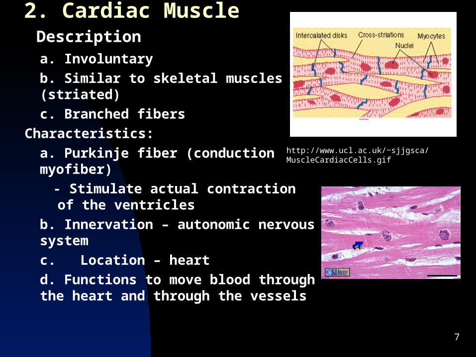
2. Cardiac Muscle
a. Involuntary
b. Similar to skeletal muscles (striated)
c. Branched fibers
Characteristics:
a. Purkinje fiber (conduction myofiber)
- Stimulate actual contraction of the ventricles
b. Innervation – autonomic nervous system
c. Location – heart
d. Functions to move blood through the heart and through the vessels
Description
http://www.ucl.ac.uk/~sjjgsca/MuscleCardiacCells.gif
8
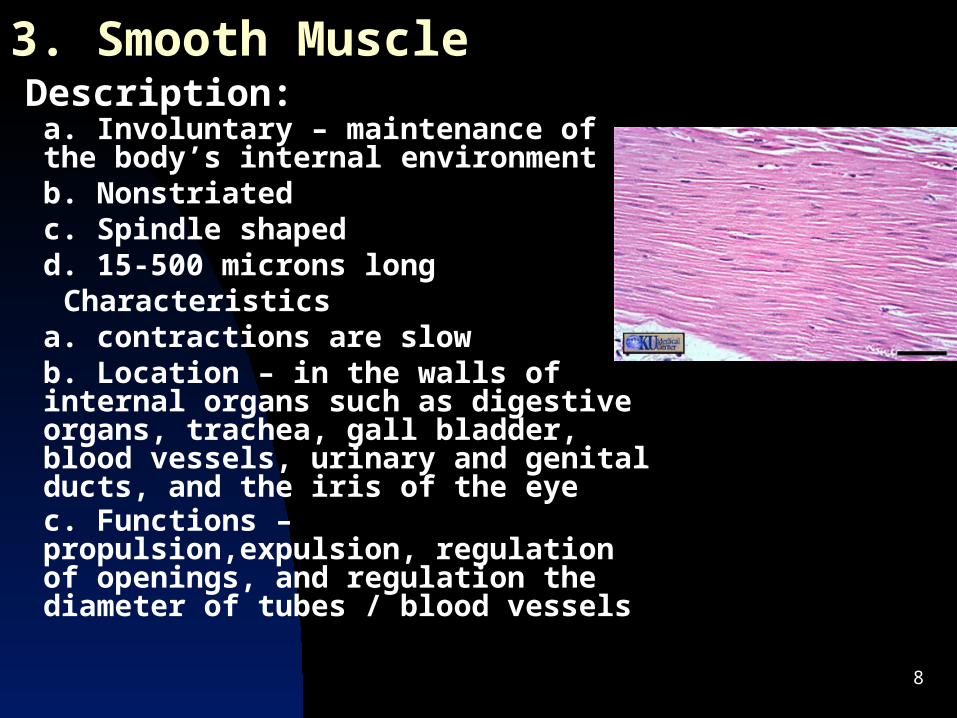
3. Smooth Muscle
a. Involuntary – maintenance of the body’s internal environmentb. Nonstriatedc. Spindle shaped d. 15-500 microns long
Characteristicsa. contractions are slowb. Location – in the walls of internal organs such as digestive organs, trachea, gall bladder, blood vessels, urinary and genital ducts, and the iris of the eyec. Functions – propulsion,expulsion, regulation of openings, and regulation the diameter of tubes / blood vessels
Description:
9
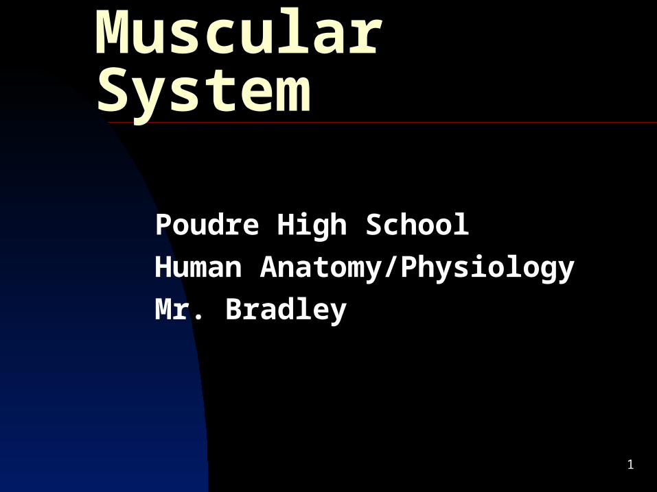
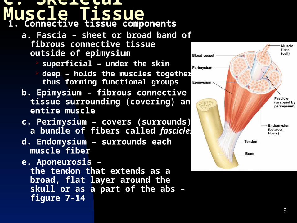
C. Skeletal Muscle Tissue1. Connective tissue components
a. Fascia – sheet or broad band of fibrous connective tissue outside of epimysium
superficial – under the skin deep – holds the muscles together
thus forming functional groups
b. Epimysium – fibrous connective tissue surrounding (covering) an entire muscle
c. Perimysium – covers (surrounds) a bundle of fibers called fascicles
d. Endomysium – surrounds each muscle fiber
e. Aponeurosis –the tendon that extends as a broad, flat layer around the skull or as a part of the abs – figure 7-14

10
2. Must also have:
Action Potential – the electric current stimulation necessary for contraction
Nerve impulse – causes the stimulation
Muscle action potential – the result
b. Blood supply to bring nutrients and oxygen, as well as to carry wastes
Generally there is one artery along with 1 or 2 veins to accompany each nerve
a. Nerve supply
http://biology.clc.uc.edu/fankhauser/Labs/Anatomy_&_Physiology/A&P202/Nerve_Histology/motor_end_plate_400x_PC231459_labeled.JPG
11
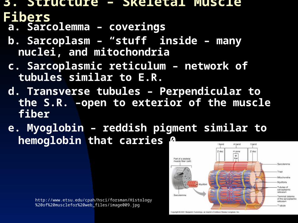
3. Structure – Skeletal Muscle Fibers a. Sarcolemma – coveringsb. Sarcoplasm – “stuff” inside – many nuclei,
and mitochondriac. Sarcoplasmic reticulum – network of tubules
similar to E.R.d. Transverse tubules – Perpendicular to the
S.R. –open to exterior of the muscle fibere. Myoglobin – reddish pigment similar to
hemoglobin that carries 02
http://www.etsu.edu/cpah/hsci/forsman/Histology%20of%20musclefor%20web_files/image009.jpg

12

13
http://highered.mcgraw-hill.com/sites/0072437316/student_view0/chapter42/animations.html#
Sarcomere shortening
http://lessons.harveyproject.org/development/muscle/swstfast.html
Functional Unit of Sarcomere
http://www.blackwellpublishing.com/matthews/myosin.html
Neuromuscular endplate
http://www.brookscole.com/chemistry_d/templates/student_resources/shared_resources/animations/muscles/muscles.html
Muscle Contraction
http://msjensen.cehd.umn.edu/1135/Links/Animations/Flash/0010-swf_action_potenti.swf
If this link doesn’t connect, cut and paste the address
Troponin/Tropomyosin

14
Fibers - continuedf. Myofibrils – Cylindrical structures that run longitudinally through the fibers and contain: thin myofilaments – Made mostly of actin (protein)
and small amounts of tropomyosin and troponin thick myofilaments – made mostly of myosin
(protein) Shaped like a golf club Heads are called cross-bridges

15
D. Microscopic Anatomy of Skeletal MuscleD. Microscopic Anatomy of Skeletal Muscle
Copyright © 2003 Pearson Education, Inc. publishing as Benjamin Cummings
1. Basic organization of the sarcomere
a. Thick filaments = myosin filaments
Composed of the protein myosin
Has ATPase enzymes

16
Microscopic Anatomy of Skeletal MuscleMicroscopic Anatomy of Skeletal Muscle
Copyright © 2003 Pearson Education, Inc. publishing as Benjamin Cummings
b. Thin filaments = actin filaments
Composed of the protein actin

17
Microscopic Anatomy of Skeletal MuscleMicroscopic Anatomy of Skeletal Muscle
Copyright © 2003 Pearson Education, Inc. publishing as Benjamin Cummings
2. Myofibril
a. Bundles of myofilaments
b. Myofibrils are aligned to give distrinct bands
I band = light band
A band = dark band
18
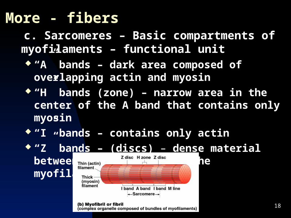
More - fibers c. Sarcomeres – Basic compartments of
myofilaments – functional unit “A” bands – dark area composed of overlapping
actin and myosin “H” bands (zone) – narrow area in the center of
the A band that contains only myosin “I” bands – contains only actin “Z” bands – (discs) – dense material between
sarcomeres where the myofilaments attach

19
Microscopic Anatomy of Skeletal MuscleMicroscopic Anatomy of Skeletal Muscle
Copyright © 2003 Pearson Education, Inc. publishing as Benjamin Cummings
3. Sarcomere
a. Contractile unit of a muscle fiber

20
Microscopic Anatomy of Skeletal MuscleMicroscopic Anatomy of Skeletal Muscle
Copyright © 2003 Pearson Education, Inc. publishing as Benjamin Cummings
b. Myosin filaments have heads (extensions, or cross bridges)
c. Myosin and actin overlap somewhat

21
Microscopic Anatomy of Skeletal MuscleMicroscopic Anatomy of Skeletal Muscle
Copyright © 2003 Pearson Education, Inc. publishing as Benjamin Cummings
d. At rest, there is a bare zone that lacks actin filaments
e. Sarcoplasmic reticulum (SR) – for storage of calcium

22
E. Muscle Contraction
1. Sliding filament mechanism –
a. The myofilaments slide inward towards the center of the sarcomere so the sarcomere shortens but the myofilaments DO NOT!
b. Sufficient Ca2+ and adequate energy are required (ATP)

23
2. The Sliding Filament Theory of 2. The Sliding Filament Theory of Muscle ContractionMuscle Contraction
Copyright © 2003 Pearson Education, Inc. publishing as Benjamin Cummings
a. Activation by nerve causes myosin heads (crossbridges) to attach to binding sites on the thin filament
b. Myosin heads then bind to the next site of the thin filament

24
The Sliding Filament Theory of Muscle The Sliding Filament Theory of Muscle ContractionContraction
Copyright © 2003 Pearson Education, Inc. publishing as Benjamin Cummings
c. This continued action causes a sliding of the myosin along the actin
d. The result is that the muscle is shortened (contracted)
http://highered.mcgraw-hill.com/sites/0072437316/student_view0/chapter42/animations.html#
25
3. Neuromuscular Junction (Motor-end plate)
a. Consists of a motor neuron and the muscle fibers it stimulates
Precise movements require 10:1 or less fiber to MEP ratio Gross movements may have as many as 2000:1 ratio
Synaptic vesicles – Sacs at the end of the axon of a neuron
Synaptic cleft – The space between the axon and the sarcolemma of the the muscle fiber
Acetylcholine(ACh) – A neurotransmitter that is released as a result of Ca2+ from the interstitial fluid
Changes the permeability (especially to Na+) Carries the impulse to receptors on sarcolemma
Cholinesterase(AChE) – found in the synaptic cleft Deactivates ACh within 1/500 of a second by breaking it
into acetate and choline thus preventing continuous stimulation

26http://psyche.knu.ac.kr/notebook/images/ch5fi05.jpg

27
Neuromuscular Junction – cont.
28
4. Physiology of contractiona. The relaxed muscle is low in Ca2+ because the S.R. has
calcium active pumps to remove it from the sarcoplasm.
b. When the action potential travels along the sarcolemma Ca2+ channels open in the S.R. and a flood of Ca2+ move into the sarcoplasm around the thick and thin myofilaments
c. Ca2+ combine with troponin changing it’s shape thus the troponin –tropomyosin complex move away from the bonding sites on actin (exposed)
d. ATP attaches to the myosin crossbridges
e. A portion of each myosin head acts as ATPase (enzyme) which splits ATP into ADP+P
f. The myosin head becomes energized
29
Physiology of contraction – cont.
g. This causes the myosin heads to bind to the binding sites on the the troponin- tropomyosin complex
h. Now the heads pivot causing the “power stroke” of muscle contraction.
i. As the thin filaments draw past the thick filaments they release ADP
j. ATP reforms and returns to the myosin crossbridges and the myosin detaches from the actin . ( about ½ of the crossbrides are bound and ½ are preparing to bind at any one time)
* * As long as ATP is available and the Ca2+ is high the contraction will repeat.

30
5. Energy for muscle contraction ATP is the immediate energy source but the muscle only
contains enough for 5-6 seconds of sustained activity.
a. Creatine Phosphate – high energy molecule found in muscles that can break down into creatine, phosphate, and energy to form enough ATP to last for 15 more seconds.
b. Now we must access glucose for energy - respiration Glucose is stored as glycogen in the muscle
Glycogen is broken into 2 pyruvic acids(glycolysis) anaerobically (no O2 needed) – this requires 2 ATP but it produces 4 ATP – 30-40 seconds of energy -
31
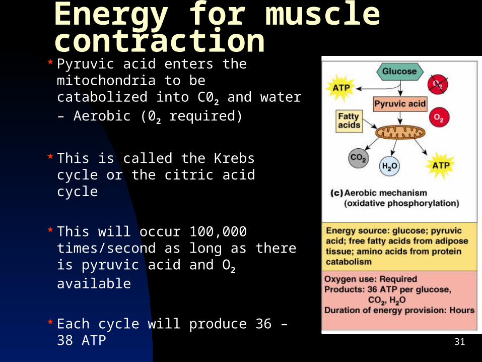
Energy for muscle contraction Pyruvic acid enters the
mitochondria to be catabolized into C02 and water – Aerobic (02 required)
This is called the Krebs cycle or the citric acid cycle
This will occur 100,000 times/second as long as there is pyruvic acid and O2 available
Each cycle will produce 36 – 38 ATP

32
F. Relaxation 2 changes permit the muscle to relax
1. ACh is broken by AChE which prevents the action potential by stopping Ca2+ release
2. Calcium active transport pumps remove the Ca2+ from the sarcoplasm rapidly
At death Ca2+ leaks and causes rigor mortis – no ATP to release the actin from the myosin
Recommended