3,350+OPEN ACCESS BOOKS
108,000+INTERNATIONAL
AUTHORS AND EDITORS115+ MILLION
DOWNLOADS
BOOKSDELIVERED TO
151 COUNTRIES
AUTHORS AMONG
TOP 1%MOST CITED SCIENTIST
12.2%AUTHORS AND EDITORS
FROM TOP 500 UNIVERSITIES
Selection of our books indexed in theBook Citation Index in Web of Science™
Core Collection (BKCI)
Chapter from the book Toxicity and Drug TestingDownloaded from: http://www.intechopen.com/books/toxicity-and-drug-testing
PUBLISHED BY
World's largest Science,Technology & Medicine
Open Access book publisher
Interested in publishing with IntechOpen?Contact us at [email protected]
11
Paraquat, Between Apoptosis and Autophagy
Rosa A. González-Polo, José M. Bravo-San Pedro, Rubén Gómez-Sánchez, Elisa Pizarro-Estrella, Mireia Niso-Santano and José M. Fuentes
Centro de Investigación Biomédica en Red Sobre Enfermedades Neurodegenerativas (CIBERNED),
Departamento de Bioquímica y Biología Molecular y Genética, E. Enfermería y TO, Universidad de Extremadura, Cáceres
Spain
1. Introduction
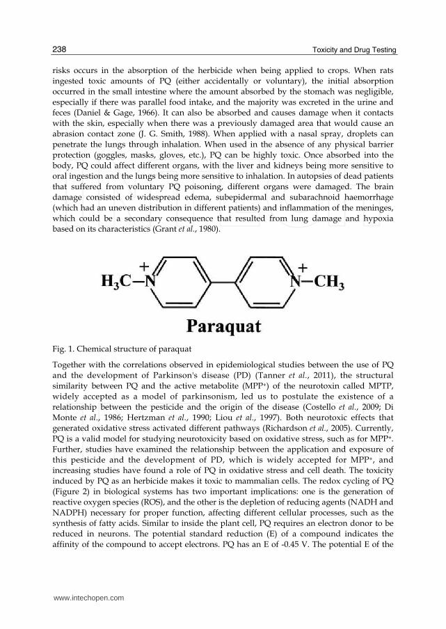
Paraquat (PQ, methyl viologen), 1,1'-dimethyl-4,4'-bipyridinium (Figure 1), is a commonly used, potent herbicide. It was first synthesised in 1882 by Weidel and Russo, as recorded by Hadley in his review of 1979 (Haley, 1979), and its redox properties were discovered by Michaelis and Hill in 1933 (Haley, 1979). Initially, PQ was used as an indicator of oxidation-reduction because in the absence of molecular oxygen, donating an electron to paraquat (PQ2+) generated a monocationically stable violet or blue form that is commonly known as methyl viologen (Dinis-Oliveira et al., 2008). However, its properties as an herbicide were not discovered until 1955, and in 1962, it was introduced into global markets. The PQ is registered and used in approximately 100 countries worldwide and is the second most commonly used herbicide in the world after gliphosate. Despite this, its use is currently banned in the European Union (EU), but the import of products from outside the EU for patients who have been treated with PQ has not. In its recommended rating of "pesticides by risk," WHO (World Health Organization) considers composite PQ to be moderately toxic (Category II) (World Health Organization 2004). The ECB (European Chemicals Bureau) classifies PQ as being very toxic (R26) by inhalation, toxic (R25) orally and moderately toxic (R24) dermally. PQ is included in the family of herbicides called bipyridines. It is an herbicide that is non-selective and functions systemically through contact without acting on the leaves of green plants. Among its advantages, it is rapidly absorbed by the leaves of plants that have been sprayed, but clay soil causes it to be biologically inactive. Its action on plants has been shown to occur on chloroplasts and is based on its redox cycle.
PQ interferes with photosynthesis at the level of photosystem I. At this point, PQ blocks the
flow of electrons from ferredoxin and NADP+ so that electrons from photosystem I would
reduce PQ, which transfers divalent cations (normal state) to monovalent cations (reduced
state). The monovalent cation reduces oxygen to the superoxide radical (O2-), which is
produced by the loss of activity of the chloroplasts and the subsequent cell damage that
leads to plant death. There is controversy about the use of PQ in agriculture because
herbicides are toxic to humans and the environment, especially when not taking the proper
precautions. Specifically, in addition to the adverse effects on humans, one of the greatest
www.intechopen.com
Toxicity and Drug Testing
238
risks occurs in the absorption of the herbicide when being applied to crops. When rats
ingested toxic amounts of PQ (either accidentally or voluntary), the initial absorption
occurred in the small intestine where the amount absorbed by the stomach was negligible,
especially if there was parallel food intake, and the majority was excreted in the urine and
feces (Daniel & Gage, 1966). It can also be absorbed and causes damage when it contacts
with the skin, especially when there was a previously damaged area that would cause an
abrasion contact zone (J. G. Smith, 1988). When applied with a nasal spray, droplets can
penetrate the lungs through inhalation. When used in the absence of any physical barrier
protection (goggles, masks, gloves, etc.), PQ can be highly toxic. Once absorbed into the
body, PQ could affect different organs, with the liver and kidneys being more sensitive to
oral ingestion and the lungs being more sensitive to inhalation. In autopsies of dead patients
that suffered from voluntary PQ poisoning, different organs were damaged. The brain
damage consisted of widespread edema, subepidermal and subarachnoid haemorrhage
(which had an uneven distribution in different patients) and inflammation of the meninges,
which could be a secondary consequence that resulted from lung damage and hypoxia
based on its characteristics (Grant et al., 1980).
Fig. 1. Chemical structure of paraquat
Together with the correlations observed in epidemiological studies between the use of PQ
and the development of Parkinson's disease (PD) (Tanner et al., 2011), the structural
similarity between PQ and the active metabolite (MPP+) of the neurotoxin called MPTP,
widely accepted as a model of parkinsonism, led us to postulate the existence of a
relationship between the pesticide and the origin of the disease (Costello et al., 2009; Di
Monte et al., 1986; Hertzman et al., 1990; Liou et al., 1997). Both neurotoxic effects that
generated oxidative stress activated different pathways (Richardson et al., 2005). Currently,
PQ is a valid model for studying neurotoxicity based on oxidative stress, such as for MPP+.
Further, studies have examined the relationship between the application and exposure of
this pesticide and the development of PD, which is widely accepted for MPP+, and
increasing studies have found a role of PQ in oxidative stress and cell death. The toxicity
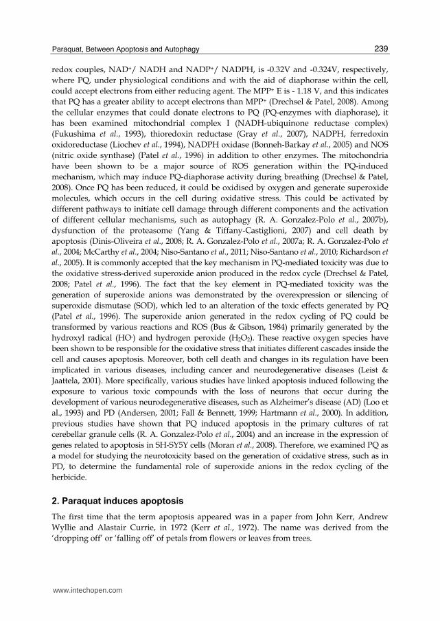
induced by PQ as an herbicide makes it toxic to mammalian cells. The redox cycling of PQ
(Figure 2) in biological systems has two important implications: one is the generation of
reactive oxygen species (ROS), and the other is the depletion of reducing agents (NADH and
NADPH) necessary for proper function, affecting different cellular processes, such as the
synthesis of fatty acids. Similar to inside the plant cell, PQ requires an electron donor to be
reduced in neurons. The potential standard reduction (E) of a compound indicates the
affinity of the compound to accept electrons. PQ has an E of -0.45 V. The potential E of the
www.intechopen.com
Paraquat, Between Apoptosis and Autophagy
239
redox couples, NAD+/ NADH and NADP+/ NADPH, is -0.32V and -0.324V, respectively,
where PQ, under physiological conditions and with the aid of diaphorase within the cell,
could accept electrons from either reducing agent. The MPP+ E is - 1.18 V, and this indicates
that PQ has a greater ability to accept electrons than MPP+ (Drechsel & Patel, 2008). Among
the cellular enzymes that could donate electrons to PQ (PQ-enzymes with diaphorase), it
has been examined mitochondrial complex I (NADH-ubiquinone reductase complex)
(Fukushima et al., 1993), thioredoxin reductase (Gray et al., 2007), NADPH, ferredoxin
oxidoreductase (Liochev et al., 1994), NADPH oxidase (Bonneh-Barkay et al., 2005) and NOS
(nitric oxide synthase) (Patel et al., 1996) in addition to other enzymes. The mitochondria
have been shown to be a major source of ROS generation within the PQ-induced
mechanism, which may induce PQ-diaphorase activity during breathing (Drechsel & Patel,
2008). Once PQ has been reduced, it could be oxidised by oxygen and generate superoxide
molecules, which occurs in the cell during oxidative stress. This could be activated by
different pathways to initiate cell damage through different components and the activation
of different cellular mechanisms, such as autophagy (R. A. Gonzalez-Polo et al., 2007b),
dysfunction of the proteasome (Yang & Tiffany-Castiglioni, 2007) and cell death by
apoptosis (Dinis-Oliveira et al., 2008; R. A. Gonzalez-Polo et al., 2007a; R. A. Gonzalez-Polo et
al., 2004; McCarthy et al., 2004; Niso-Santano et al., 2011; Niso-Santano et al., 2010; Richardson et
al., 2005). It is commonly accepted that the key mechanism in PQ-mediated toxicity was due to
the oxidative stress-derived superoxide anion produced in the redox cycle (Drechsel & Patel,
2008; Patel et al., 1996). The fact that the key element in PQ-mediated toxicity was the
generation of superoxide anions was demonstrated by the overexpression or silencing of
superoxide dismutase (SOD), which led to an alteration of the toxic effects generated by PQ
(Patel et al., 1996). The superoxide anion generated in the redox cycling of PQ could be
transformed by various reactions and ROS (Bus & Gibson, 1984) primarily generated by the
hydroxyl radical (HO-) and hydrogen peroxide (H2O2). These reactive oxygen species have
been shown to be responsible for the oxidative stress that initiates different cascades inside the
cell and causes apoptosis. Moreover, both cell death and changes in its regulation have been
implicated in various diseases, including cancer and neurodegenerative diseases (Leist &
Jaattela, 2001). More specifically, various studies have linked apoptosis induced following the
exposure to various toxic compounds with the loss of neurons that occur during the
development of various neurodegenerative diseases, such as Alzheimer’s disease (AD) (Loo et
al., 1993) and PD (Andersen, 2001; Fall & Bennett, 1999; Hartmann et al., 2000). In addition,
previous studies have shown that PQ induced apoptosis in the primary cultures of rat
cerebellar granule cells (R. A. Gonzalez-Polo et al., 2004) and an increase in the expression of
genes related to apoptosis in SH-SY5Y cells (Moran et al., 2008). Therefore, we examined PQ as
a model for studying the neurotoxicity based on the generation of oxidative stress, such as in
PD, to determine the fundamental role of superoxide anions in the redox cycling of the
herbicide.
2. Paraquat induces apoptosis
The first time that the term apoptosis appeared was in a paper from John Kerr, Andrew
Wyllie and Alastair Currie, in 1972 (Kerr et al., 1972). The name was derived from the
‘dropping off’ or ‘falling off’ of petals from flowers or leaves from trees.
www.intechopen.com
Toxicity and Drug Testing
240
Apoptosis, or programmed cell death, is characterised by several morphological features,
such as DNA degradation into oligonucleosomal fragments, chromatin condensation,
reduction in nuclear and cellular fractions, phosphatidylserine exposure on the outward-
facing side of the plasma membrane and preservation of organelle structure and plasma
membrane integrity, which leads to the generation of apoptotic bodies, or vesicles in the
cytoplasm containing tightly packed organelles with or without nuclear fragments. This
type of cell death contrasts with necrosis, which is uncontrolled, accidental and pathological
cell death. However, these two cell death pathways have been shown to crosstalk, which has
been described as the “apoptosis-necrosis continuum” (Zeiss, 2003).
Fig. 2. Redox cycle of paraquat
Previous studies demonstrating the regulation of apoptosis came from Caenorhabditis elegans where 131 of the 1090 somatic cells in C. elegans were under the control of programmed cell death. In 1985, the Horvitz’s lab discovered four important genes (Ced-3, Ced-4, Ced-9, Egl-1) that are involved in the regulation of cell death (Fixsen et al., 1985). Previous studies have shown that apoptosis was necessary to define whether cells should live or die. However, there are other forms of programmed cell death and other possible mechanisms that have not yet been discovered (Debnath et al., 2005; Formigli et al., 2000; Sperandio et al., 2000). Several mechanisms of cell death have already been shown to be involved in maintaining the balance between life and death (Boya et al., 2005; Lum et al., 2005; Ravikumar et al., 2006). Apoptosis most commonly occurs during development and aging as a homeostatic mechanism. Although it has been shown to be used as a defence mechanism, such as when cells become damaged, during an immune response (Norbury & Hickson, 2001), or as a pathological process in cancer and autoimmune lymphoproliferative syndrome in which apoptosis was suppressed and led to the development and progression of tumours (Kerr et al., 1994; Worth et al., 2006), neurodegenerative diseases, autoimmune diseases and ischaemia-associated injury where there is excessive apoptosis (Ethell & Buhler, 2003; Freude et al., 2000; C. J. Li et al., 1995). Traditionally, apoptosis has been considered to be an irreversible process; however, several reports have demonstrated that these apoptotic cells could be rescued from programmed cell death (Geske et al., 2001; Hoeppner et al., 2001; Reddien et al., 2001).
www.intechopen.com
Paraquat, Between Apoptosis and Autophagy
241
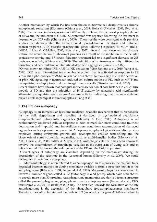
The mechanisms of apoptosis are highly complex and involve an energy-dependent cascade
of molecular events. Apoptosis can be initiated by a variety of stimuli, but previous studies
have shown that there are two main apoptotic pathways: the death receptor or extrinsic
pathway and the mitochondrial or intrinsic pathway. However, these two pathways are
connected, and some molecules of one pathway have been shown to influence the other
pathway (Igney & Krammer, 2002). Further, a third pathway was found in T cells and had
been shown to occur through cytotoxicity and perforin-granzyme-dependent cell death (via
granzyme A or granzyme B). Basically, the extrinsic pathway is activated by the death
receptors (DR), which are localised on the cell surface, through the recognition of their
specific ligands. In comparison, the intrinsic pathway is initiated after several intracellular
triggers, called “stress signals”, such as cytoskeleton disruption, hypoxia, DNA damage,
macromolecular synthesis inhibition and endoplasmic reticulum stress, which induce the
mitochondria to release pro-apoptotic factors into the cytosol. However, both pathways and
the granzyme B pathway terminate in the execution pathway that activates caspases
(cysteine-aspartic acid proteases), and they have been shown to be responsible for
developing the well-known features of apoptosis (Amarante-Mendes & Green, 1999). In
contrast, the granzyme A pathway is a caspase-independent cell death pathway, which has
been shown to act in parallel (Martinvalet et al., 2005). The cell’s decision has been shown to
be determined by the Bcl-2 protein family. The regulation of the Bcl-2 family of proteins is
important (Gross et al., 1999).
It is well known that the toxicity of PQ was due to the production of ROS (Bus & Gibson,
1984; Mollace et al., 2003), which has been partially generated by xanthine oxidase (Kitazawa
et al., 1991; Sakai et al., 1995). Therefore, it has been shown that PQ could induce apoptotic
cell death in cerebellar granule cells using this xanthine oxidase system (R. A. Gonzalez-Polo
et al., 2004). In addition, it has been reported that PQ induced apoptosis in other animal
models, such as human lung epithelial cells (Cappelletti et al., 1998), PC12 cells (X. Li & Sun,
1999), mouse 32D cells (Fabisiak et al., 1997), primary mesencephalic cells and dopaminergic
neuronal cells (Gomez-Sanchez et al., 2010; Peng et al., 2004).
PQ has structural homology to MPP+, which has been linked to PD in epidemiological studies (Hertzman et al., 1990; Liou et al., 1997). In this vein, PINK1-silenced neuroblastoma cells were more sensitive and exhibited increased apoptosis compared with control cells following PQ treatment (Gegg et al., 2009). Silencing DJ-1 in neuroblastoma cells induced apoptotic cell death, and the treatment with PQ increased apoptosis (R. Gonzalez-Polo et al., 2009). The cytotoxic actions of PQ have been shown to involve oxidative stress by producing superoxide anions through the mitochondrial electron transport chain (Dinis-Oliveira et al., 2006; McCormack et al., 2002). PQ has been shown to be reduced by mitochondrial complex I and, thus, impair the respiration complex that led to the generation of ROS to induce selective neurodegeneration in dopaminergic neurons in the substantia nigra pars compacta (Fei et al., 2008) and apoptosis by activating different intracellular pathways. PQ has been shown to induce apoptosis through the mitochondrial intrinsic pathway associated with p53 (Yang & Tiffany-Castiglioni, 2008). JNK proteins have been implicated in dopaminergic neuronal death induced by rotenone, PQ and 6-hydroxydopamine (6-OHDA) (Choi et al., 1999; Klintworth et al., 2007; Newhouse et al., 2004; Niso-Santano et al., 2006). PQ activates cell death through JNK and its downstream target c-Jun (Peng et al. 2004) and induces high levels of pro-apoptotic Bcl-2 family members (Bak, Bid, BNip3 and Noxa) in conjunction with cytochrome c release and caspase-3 activation (Fei et al., 2008).
www.intechopen.com
Toxicity and Drug Testing
242
Another mechanism by which PQ has been shown to activate cell death involves chronic endoplasmic reticulum (ER) stress (Chinta et al., 2008; Holtz & O'Malley, 2003; Ryu et al., 2002). The increase in the expression of GRP family proteins, the increased phosphorylation of eIF2a and the induction of GADD153 expression was reported following PQ treatment in dopaminergic N27 cells (Chinta et al., 2008) . These results were consistent with previous studies that demonstrated the transcriptional upregulation of ER stress and unfolded protein response (UPR)-specific proapoptotic genes following exposure to MPP+ and 6-OHDA (Holtz & O'Malley, 2003; Ryu et al., 2002). Several neurodegenerative diseases feature the accumulation of abnormal proteins as a result of the inhibition of the cellular proteasome activity and ER stress. Paraquat treatment led to a significant decrease in 20S proteasome activity (Chinta et al., 2008). The inhibition of proteasome activity initiated the formation and accumulation of ubiquitinated protein aggregates (Lam et al., 2002). PQ was shown to induce IRE1/ASK1/JNK activation (Niso-Santano et al., 2010; Yang et al., 2009). IRE1 is an ER-resident transmembrane protein that is activated in response to ER stress. IRE1 phosphorylates ASK1, which has been shown to play a key role in the activation of p38/JNK signalling in neurotoxin-induced cell culture models of PD, such as MPTP and paraquat-induced apoptosis in dopaminergic neuronal cells (Niso-Santano et al., 2010). Recent studies have shown that paraquat induced acetylation of core histones in cell culture models of PD and that the inhibition of HAT activity by anacardic acid significantly attenuated paraquat-induced caspase-3 enzyme activity, indicating that histone acetylation played a role in paraquat-induced apoptosis (Song et al.).
3. PQ induces autophagy
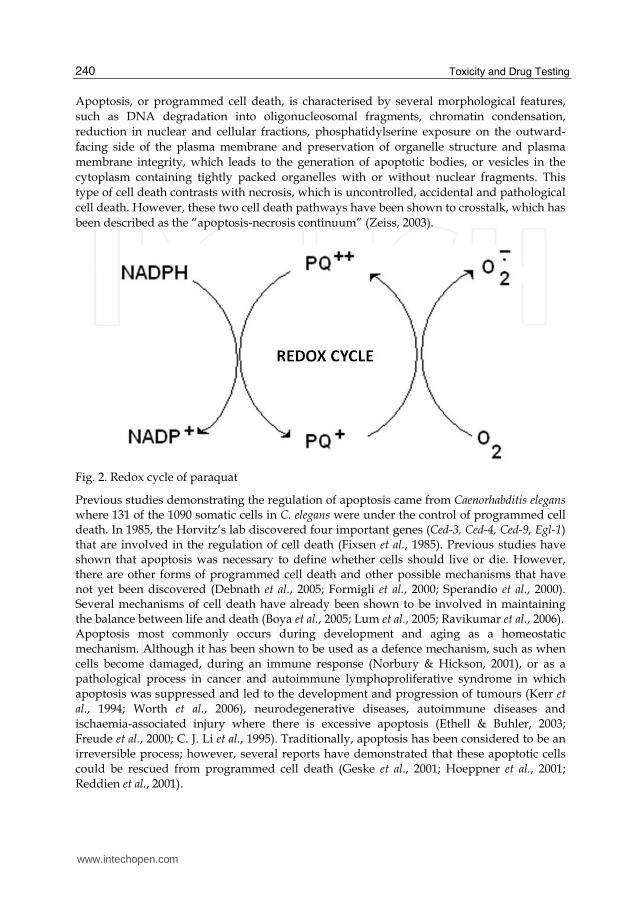
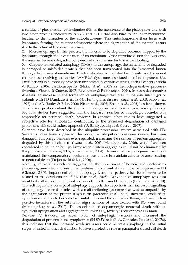
Autophagy is an intracellular lysosome-mediated catabolic mechanism that is responsible for the bulk degradation and recycling of damaged or dysfunctional cytoplasmic components and intracellular organelles (Klionsky & Emr, 2000). Autophagy is an evolutionarily conserved cellular response to both extracellular stress conditions (nutrient deprivation and hypoxia) and intracellular stress conditions (accumulation of damaged organelles and cytoplasmic components). Autophagy is a physiological degradative process employed during embryonic growth and development, cellular remodelling and the biogenesis of some subcellular organelles, such as multi-lamellar bodies (Filonova et al., 2000; Hariri et al., 2000; Sattler & Mayer, 2000). Autophagic cell death has been shown to involve the accumulation of autophagic vacuoles in the cytoplasm of dying cells and in mitochondrial dilation and the enlargement of the ER and the Golgi apparatus. Different types of autophagy are classified depending on the mechanism driving the degradation of the substrate in the lysosomal lumen (Klionsky et al., 2007). We could distinguish three types of autophagy: 1. Macroautophagy: is often referred to as “autophagy”. In this process, the material to be degraded becomes trapped in double-membrane vesicles to form a structure known as the autophagosome (Baba et al., 1994; Fengsrud et al., 1995). Macroautophagy has been shown to involve a number of genes called ATGs (autophagy-related genes), which have been shown to encode more than 30 proteins. Autophagosome membranes are derived from a structure called the pre-autophagosome, phagophore or early autophagosome (Fengsrud et al., 1995; Mizushima et al., 2001; Suzuki et al., 2001). The first step towards the formation of the late autophagosome is the expansion of the phagophore (pre-autophagosome) membrane. Therefore, the carbon terminus of the protein LC3 (encoded by the gene ATG8) is attached to
www.intechopen.com
Paraquat, Between Apoptosis and Autophagy
243
a residue of phosphatidyl-ethanolamine (PE) in the membrane of the phagophore and with two other proteins encoded by ATG12 and ATG5 that also bind to the inner membrane, leading to the formation of the autophagosome. This autophagosome then fuses with lysosomes, forming the autophagolysosome where the degradation of the material occurs due to the action of lysosomal enzymes. 2. Microautophagy: In this process, the material to be degraded becomes trapped by the lysosomes through the invagination of its membrane. Once introduced into the lysosome, the material becomes degraded by lysosomal enzymes similar to macroautophagy. 3. Chaperone-mediated autophagy (CMA): In this autophagy, the material to be degraded is damaged or misfolded protein that has been translocated into the lysosomal lumen through the lysosomal membrane. This translocation is mediated by cytosolic and lysosomal chaperones, involving the carrier LAMP-2A (lysosome-associated membrane protein 2A). Dysfunctions in autophagy have been implicated in various diseases, such as cancer (Kondo & Kondo, 2006), cardiomyopathy (Nakai et al., 2007) or neurodegenerative processes (Martinez-Vicente & Cuervo, 2007; Ravikumar & Rubinsztein, 2004). In neurodegenerative diseases, an increase in the formation of autophagic vacuoles in the substantia nigra of patients with PD (Anglade et al., 1997), Huntington's disease (Kegel et al., 2000; Sapp et al., 1997) and AD (Butler & Bahr, 2006; Nixon et al., 2005; Zheng et al., 2006) has been shown. This raises questions about the role of autophagy in these neurodegenerative processes. Previous studies have suggested that the increased number of autophagic vacuoles was responsible for neuronal death; however, in contrast, other studies have suggested a protective role for autophagy, contributing to the increased degradation of damaged proteins, which could induce apoptosis (U. Bandyopadhyay & Cuervo, 2007). Changes have been described in the ubiquitin-proteasome system associated with PD. Several studies have suggested that once the ubiquitin-proteasome system has been damaged, autophagy becomes over-regulated, increasing the number of protein aggregates degraded by this mechanism (Iwata et al., 2005; Massey et al., 2006), which has been considered to be the default pathway when protein aggregates could not be eliminated by the proteasome (Olanow, 2007; Rideout et al., 2004). However, if the pathogenic insult was maintained, this compensatory mechanism was unable to maintain cellular balance, leading to neuronal death (Trojanowski & Lee, 2000). Recently, converging evidence suggests that the impairment of homeostatic mechanisms processing unwanted and misfolded proteins plays a central role in the pathogenesis in PD (Olanow, 2007). Impairment of the autophagy-lysosomal pathway has been shown to be related to the development of PD (Pan et al., 2008). Activation of autophagy was also identified within peripheral blood mononuclear cells from PD patients (Prigione et al., 2010). This self-regulatory concept of autophagy supports the hypothesis that increased signalling of autophagy occurred in mice with a malfunctioning lysosome that was accompanied by the aggregation of the protein α-synuclein (Meredith et al., 2002). Increased levels of α-synuclein were reported in both the frontal cortex and the ventral midbrain, and α-synuclein positive inclusions in the substantia nigra neurons of mice treated with PQ were found (Manning-Bog et al., 2002). The association of dopaminergic neuronal death with α-synuclein upregulation and aggregation following PQ toxicity is relevant as a PD model. Because PQ induced the accumulation of autophagic vacuoles and increased the degradation of proteins in the cytoplasm of SH-SY5Y cells (R. A. Gonzalez-Polo et al., 2007a), this indicates that the increased oxidative stress could activate autophagy in the initial stages of mitochondrial dysfunction to have a protective role in paraquat-induced cell death
www.intechopen.com
Toxicity and Drug Testing
244
(R. A. Gonzalez-Polo et al., 2007a, 2007b). Moreover, our group has shown that PQ exposure induced an early reticulum stress response that was correlated with the adaptive activation of autophagy, characterised by the accumulation of autophagic vacuoles, activation of beclin-1, accumulation of LC3-II, p62 degradation, and mammalian target of rapamycin dephosphorylation (R. A. Gonzalez-Polo et al., 2007a, 2007b; Niso-Santano et al., 2011). This response was increased in cells that overexpressed wild-type (WT) ASK1 (apoptosis signal kinase 1) protein. In this model, the inhibition of autophagy caused an exacerbation of the apoptosis induced by ASK1 WT overexpression with or without PQ. These results suggest that autophagy has an important role in the cell death/survival events produced by PQ and ASK1 that contribute to neuronal degeneration. Therefore, increased autophagy might be a new strategy for the treatment of
neurodegenerative diseases (Menzies et al., 2006). It is encouraging to consider enhancing
the autophagic capacity as a therapeutic strategy in the prevention of neurodegeneration
because studies have shown that the abnormal regulation of autophagic pathways may lead
to apoptosis and cell death (Walls et al., 2010).
Fig. 3. Schematic representation of the different types of autophagy
4. Paraquat and Parkinson´s disease-related proteins
As previously described, PD is characterised by the selective degeneration of dopaminergic neurons. The aetiology of PD is unknown but has a multifactorial origin that involves both genetic and environmental factors. The interaction of both factors was, in part, involved in the selective death of dopaminergic neurons observed in PD. Apart from the studies that have identified human mutations as a basis for disease, the high number of individuals with
www.intechopen.com
Paraquat, Between Apoptosis and Autophagy
245
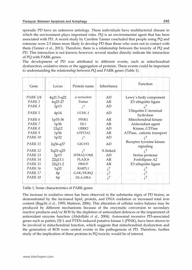
sporadic PD have an unknown aetiology. These individuals have multifactorial disease in which the environment plays important roles. PQ is an environmental agent that has been associated with PD. A recent study by Caroline Tanner concluded that people using PQ and rotenone were 2.5 times more likely to develop PD than those who were not in contact with them (Tanner et al., 2011). Therefore, there is a relationship between the toxicity of PQ and PD. This interaction is not known; however, several studies directly indicate the interaction of PQ with PARK genes. The development of PD was attributed to different events, such as mitochondrial dysfunction, oxidative stress or the aggregation of proteins. These events could be important to understanding the relationship between PQ and PARK genes (Table 1).
Gene
Locus
Protein name
Inheritance
Function
PARK 1/4
4q21.3-q22
-synuclein
AD
Lewy`s body component
PARK 2 6q25-27 Parkin AR E3 ubiquitin ligase PARK 3 2p13 ¿? AD ¿?
PARK 5 4p14 UCHL-1 AD Ubiquitin C-terminal
hydrolase PARK 6 1p35-36 PINK1 AR Mitochondrial kinase PARK 7 1p36 DJ-1 AR Antioxidant agent PARK 8 12q12 LRRK2 AD Kinase, GTPase PARK 9 1p36 ATP13A2 AR ATPase, cationic transport
PARK 10 1p32 ¿? AD ¿?
PARK 11 2q36-q37 GIGYF2 AD Receptor tyrosine kinase
signaling PARK 12 Xq21-q25 ¿? X-linked ¿? PARK 13 2p13 HTRA2/OMI AD Serine protease PARK 14 22q13.1 PLA2G6 AR Fosfolipase A2 PARK 15 22q11.2 FBXO7 AR E3 ubiquitin ligase PARK 16 1q32 RAB7L1 ¿? ¿? PARK 17 4p GAK/DGKQ ¿? ¿? PARK 18
6p
HLA-DRA
¿?
¿?
Table 1. Some characteristics of PARK genes
The increase in oxidative stress has been observed in the substantia nigra of PD brains, as demonstrated by the increased lipid, protein, and DNA oxidation or increased total iron content (Bagchi et al., 1995; Mattson, 2006). This alteration of cellular redox balance may be produced by different mechanisms because of the enzymatic conversion to secondary reactive products and/or ROS by the depletion of antioxidant defences or the impairment of antioxidant enzyme function (Abdollahi et al., 2004). Autosomal recessive PD-associated genes such as parkin, DJ-1 and PTEN-induced putative kinase 1 (PINK), have been shown to be involved in mitochondrial function, which suggests that mitochondrial dysfunction and the generation of ROS were central events in the pathogenesis of PD. Therefore, further study of the implication of these proteins in PQ toxicity would be of interest.
www.intechopen.com
Toxicity and Drug Testing
246
In contrast, the misfolding and aggregation of proteins is another pathway of cell toxicity in PD. The failure of α-synuclein (PD-related protein) clearance by the ubiquitin-proteasome system UPS (ubiquitin proteosome system) led to its accumulation over time and to the formation of fibrillar aggregates and Lewy bodies. In this vein, there is a relationship between PQ toxicity and PD because exposure to PQ has been shown to induce proteasome dysfunction and α-synuclein aggregation (Ding & Keller, 2001; Fei et al., 2008; Goers et al., 2003; Manning-Bog et al., 2002; Yang & Tiffany-Castiglioni, 2007). Therefore, there is a relationship between the toxicity exerted by PQ and different PARK genes.
4.1 α-synuclein (PARK1, PARK4) and paraquat
Lewy bodies (LBs) are abnormal aggregates of protein that develop inside the nerve cells in PD. The presence of α-synuclein in these aggregates has been shown to play an important role in the formation of LBs (Masliah et al., 2000; Spillantini et al., 1997). The mechanisms that promote intraneuronal α-synuclein assembly remain poorly understood. Missense mutations (A53T, A30P and E46K) or multiplications (duplications and triplications) in the α-synuclein gene (PARK1/4) caused autosomal-dominant parkinsonism (Polymeropoulos et al., 1997), but it is still unclear whether fibrils of aggregated α-synuclein, as found in LBs, have a causative role in the more common forms of PD or could be a marker for the underlying pathogenetic process. α-synuclein has three common forms, monomers, dimers, and protofibrils, and it is thought that an excess of the protofibril forms inhibited UPS in vitro (McNaught et al., 2001) and in vivo (Dyllick-Brenzinger et al., 2010). α-synuclein protofibrils have been shown to directly lead to oxidative stress that could further impair UPS by reducing ATP levels, inhibiting the proteasome and by the oxidation of parkin. Studies have indicated that the interaction of environmental factors with alterations in α-synuclein might be involved in the aetiology of PD. The interaction of α-synuclein with PQ toxicity has been extensively examined. PQ has been shown to potentiate α-synuclein-induced toxicity (Norris et al., 2007). PQ preferentially binds to the partially folded α-synuclein intermediate because PQ has been shown to induce a conformational change in α-synuclein and significantly increase the rate of the formation of α-synuclein fibrils in vitro (Uversky et al., 2001). In vivo, rodent studies have shown that the administration of PQ induced an increase in α-synuclein levels in the brain. These results suggest that the upregulation of α-synuclein as a result of toxic insult and the direct interactions between the protein and environmental agents are potential mechanisms leading to α-synuclein pathology in neurodegenerative disorders (Manning-Bog et al., 2002).
4.2 PINK1/PARKIN (PARK6/PARK2) and paraquat
Another hallmark PD characteristic is mitochondrial dysfunction. In post-mortem analysis in the substantia nigra, some patients with PD showed complex I deficiency (Schapira et al., 1989). In addition, the oxidative stress was higher in patients with parkinsonism (Jenner, 2003). In this sense, PINK1 (PARK6) and Parkin (PARK2) are 2 genes related to PD that may be involved in the regulation of mitochondrial homeostasis. Parkin mutations were first linked to an autosomal recessive juvenile-onset form of PD in Japanese families (Kitada et al., 1998; Matsumine et al., 1997). Numerous parkin mutations have been described, including deletions, multiplications and missense mutations (Hattori & Mizuno, 2004). Parkin protein acts as an E3 ubiquitin protein ligase in the UPS (Shimura et al., 2000). Ubiquitination of proteins is essential to start to proteasomal protein degradation.
www.intechopen.com
Paraquat, Between Apoptosis and Autophagy
247
Therefore, parkin mutations should lead to an incorrect ubiquitination, blocking the degradation of the protein and leading to protein accumulation. Mutant parkin has been shown to impair mitochondrial function and morphology in human fibroblasts and to sensitise the cells to an insult with PQ, producing higher levels of oxidised proteins in the Parkin-mutant samples than in controls (Grunewald et al., 2010). PQ has also been demonstrated to induce alterations in parkin solubility and result in its intracellular aggregation (C. Wang et al., 2005). PINK1 is a serine/threonine kinase capable of autophosphorylation. This protein has an N-terminal mitochondrial targeting signal (MTS), is synthesised as a full-length version (FL) and is processed into at least two cleaved forms (∆1 and ∆2) (W. Lin & Kang, 2008). PINK1 is considered to be a mitochondrial protein with a role in protecting against oxidative stress and apoptosis in in vitro models (Valente et al., 2004). Mutations in PINK1 have been associated with autosomal recessive PD (Valente et al., 2004) and with PINK1 KO flies with motor deficits and disorganised mitochondrial morphology (Clark et al., 2006). For the link between PINK1 and the toxicity of PQ, studies using silencer PINK1 have shown an increase in oxidative stress and ATP depletion and a higher sensitivity to PQ (Gegg et al., 2009). Similar results have been observed in studies that examined PINK1 nonsense and missense mutations (Grunewald et al., 2009).
4.3 DJ-1 (PARK7) and paraquat
DJ-1 is a small protein that belongs to the ThiJ/PfpI protein superfamily (S. Bandyopadhyay & Cookson, 2004) that was initially identified as an oncogene that interacted with H-Ras (Nagakubo et al., 1997). The involvement of DJ-1 in neurodegeneration was found when it was discovered that the DJ-1 gene (PARK7) was the cause of autosomal recessive PD in a Dutch family (Bonifati et al., 2003). Different pathogenic mutations have been identified in the PARK7 gene, including truncation, exonic deletions and homozygous and heterozygous missense mutations (Hague et al., 2003). L166P is the most dramatic point mutation, whereas other mutations, such as A104T and M26I, have a weaker destabilising effect on the protein structure. The L166P mutation is located in the centre of α-helix 7, which is a major part of the hydrophobic patch. This mutation has been shown to destabilise the dimeric structure of DJ-1 by promoting the unfolding of its C-terminal region, resulting in rapid degradation (Miller et al., 2003; Moore et al., 2003). However, the frequency of DJ-1 mutations was low, with it being estimated at approximately 1-2 % in early onset PD. The physiological function of DJ-1 is unclear, but it may have a role in protecting against mitochondrial damage in response to oxidative stress (Canet-Aviles et al., 2004). The link between DJ-1 and PQ exposure has been correlated with autophagy and the apoptotic process. An active role for DJ-1 in the autophagic response produced by PQ has been suggested. In a study using transfected cells exposed to PQ and DJ-1-specific siRNA, an inhibition of the autophagic events induced by the herbicide, the increased sensitisation during PQ-induced apoptotic cell death and the exacerbation of apoptosis in the presence of the autophagy inhibitor 3-methyladenine (R. A. Gonzalez-Polo et al., 2009) had been shown. Interestingly, PQ-induced toxicity and proteasome dysfunction was potentiated in a DJ-1 deficiency (Lavara-Culebras & Paricio, 2007; Menzies et al., 2005). In another study using DJ-1 null cells from the DJ-1(-/-) mouse embryos, DJ-1 null cells showed a resistance to PQ-induced apoptosis, including reduced poly (ADP-ribose) polymerase and procaspase-3. Therefore, DJ-1 could be important to maintain mitochondrial complex I, and complex I could be a key target in the interaction of PQ toxicity and DJ-1 in PD (Kwon et al., 2011). In
www.intechopen.com
Toxicity and Drug Testing
248
DJ-1-deficient mice treated with PQ, decreased proteasome activities and increased ubiquitinated protein levels were found, and these pathologies were not observed in brain regions of normal mice treated with PQ (Yang et al., 2007). In another mouse study, the loss of DJ-1 increased the sensitivity to oxidative insults but did not produce neurodegeneration. Similar results have been found when analysing Drosophila melanogaster mutants for the DJ-1 orthologous genes, DJ-1alpha and DJ-1beta, that resulted in increased sensitivity to PQ insults, reduced lifespan and motor impairments. However, these mutations did not lead to dopaminergic neuronal loss (Lavara-Culebras & Paricio, 2007)
4.4 LRRK2 (PARK8) and paraquat
In 2002, PARK8 gene mutations were discovered as a major genetic cause associated with
hereditary parkinsonism (Paisan-Ruiz et al., 2004). The PARK8 gene was associated with PD
in studies of a Japanese Sagamihara family who responded positively to treatment with L-
DOPA, which had parkinsonism that presented with an unknown aetiology of the disease
(Funayama et al., 2002). Other studies examined two additional families (German and
Canadian) who also had an autosomal dominant, late-onset parkinsonism (Zimprich et al.,
2004).
In the LRRK2 structure, two functional domains, kinase and GTPase domains, were shown
to be present. The G2019S mutation was present in the kinase domain specific to the binding
site for Mg2+ (Kachergus et al., 2005). This mutation facilitates the access of the kinase
domain to its substrates, which increases autophosphorylation 2.5-fold the phosphorylation
of other substrates, such as myelin basic protein (MBP), 3-fold for the LRRK2
autophosphorylation without the presence of this mutation (Jaleel et al., 2007; West et al.,
2005), which is responsible for the increased toxicity of this molecule (Greggio et al., 2006). In
the GTPase domain, the R1441C has been the most studied mutation, and there is
controversy as to the influence of GTPase mutations on the kinase activity that was
observed in some studies in which the increase was similar (Guo et al., 2007) or had no
change (Jaleel et al., 2007).
LRRK2 has been shown to play different roles in the cell; however, little information is available. Based on the data we found from the protein interactions, there was a relationship between LRRK2 and cytoskeletal reorganisation (Gandhi et al., 2008), maintenance functions and cell morphology (Plowey et al., 2008), protein transport through synaptic vesicles (Shin et al., 2008), and the ubiquitination process (Ko et al., 2009). There have also been studies that relate LRRK2 and apoptosis (Ho et al., 2009). Previous studies have shown a relationship between LRRK2 and other PD-related proteins, such as parkin (Ng et al., 2009; W. W. Smith et al., 2005), PINK-1 and DJ-1 (Venderova et al., 2009) or α-synuclein (X. Lin et al., 2009). The interaction of LRRK2 with PQ is not clear. Studies in Drosophila melanogaster in which the deletion of kinase domain of LRRK2 did not induce a higher sensitivity to the PQ stimulus has been shown (D. Wang et al., 2008). In contrast, in Caenorhabditis elegans studies, the expression of human LRRK2 protein protected against PQ, which increased nematode survival in response to agents that cause mitochondrial dysfunction. However, protection by G2019S, R1441C, or kinase-dead LRRK2 was less effective than wild-type LRRK2 (Saha et al., 2009). In another study with Caenorhabditits elegans, PINK1 mutant genes have been observed in a minor mitochondrial length and increased PQ sensitivity of the nematode. Moreover, the mutants also displayed defects in axonal outgrowth of a pair of canal-associated neurons. We demonstrated that in the absence of lrk-1 (the C. elegans homologue
www.intechopen.com
Paraquat, Between Apoptosis and Autophagy
249
of human LRRK2), all phenotypic aspects of PINK1 loss-of-function mutants were suppressed (Samann et al., 2009)
5. Conclusion
PQ has been suggested as a potential aetiological factor for the development of PD. We have demonstrated that PQ was able to induce cell death by activating apoptotic machinery. However, PQ also displayed characteristics of autophagy, a degradative mechanism involved in the recycling and turnover of cytoplasmic constituents from eukaryotic cells. Finally, the cells suffered apoptotic death when the PQ remained. Whereas caspase inhibition retarded cell death, autophagy inhibition increased apoptotic cell death induced by PQ. These findings suggest a relationship between autophagy and apoptotic cell death following paraquat exposition and allows us to further investigate and increase our knowledge regarding the toxicity of paraquat and its relationship with the origin of PD.
6. Acknowledgments
Jose M. Bravo-San Pedro was supported by a Junta de Extremadura predoctoral fellowship. Mireia Niso-Santano was supported as a postdoctoral contract of the University of Extremadura. Ruben Gómez-Sánchez was supported by a Spanish Ministerio de Educación predoctoral fellowship. Rosa-Ana González-Polo was supported by a “Miguel Servet” contract (ISCIII, Ministerio de Ciencia e Innovación, Spain). Dr. González-Polo receives research support from ISCIII (Ministerio de Ciencia e Innovación, Spain (CP08/00010, PI11/00040). Dr. José M. Fuentes receives research support from the Ministerio de Ciencia e Innovación, Spain (SAF2010-14993), FUNDESALUD (PRIS10013) and Consejería, Economía, Comercio e Innovación Junta de Extremadura (GR10054).
7. References
Abdollahi, M., Ranjbar, A., Shadnia, S., Nikfar, S., & Rezaie, A. (2004). Pesticides and oxidative stress: a review. Med Sci Monit, 10(6), Jun,pp. RA141-147
Amarante-Mendes, G. P., & Green, D. R. (1999). The regulation of apoptotic cell death. Braz J Med Biol Res, 32(9), Sep,pp. 1053-1061
Andersen, J. K. (2001). Does neuronal loss in Parkinson's disease involve programmed cell death? Bioessays, 23(7), Jul,pp. 640-646
Anglade, P., Vyas, S., Javoy-Agid, F., Herrero, M. T., Michel, P. P., Marquez, J., Mouatt-Prigent, A., Ruberg, M., Hirsch, E. C., & Agid, Y. (1997). Apoptosis and autophagy in nigral neurons of patients with Parkinson's disease. Histol Histopathol, 12(1), Jan,pp. 25-31
Baba, M., Takeshige, K., Baba, N., & Ohsumi, Y. (1994). Ultrastructural analysis of the autophagic process in yeast: detection of autophagosomes and their characterization. J Cell Biol, 124(6), Mar,pp. 903-913
Bagchi, D., Bagchi, M., Hassoun, E. A., & Stohs, S. J. (1995). In vitro and in vivo generation of reactive oxygen species, DNA damage and lactate dehydrogenase leakage by selected pesticides. Toxicology, 104(1-3), Dec 15,pp. 129-140
Bandyopadhyay, S., & Cookson, M. R. (2004). Evolutionary and functional relationships within the DJ1 superfamily. BMC Evol Biol, 4Feb 19,pp. 6
www.intechopen.com
Toxicity and Drug Testing
250
Bandyopadhyay, U., & Cuervo, A. M. (2007). Chaperone-mediated autophagy in aging and neurodegeneration: lessons from alpha-synuclein. Exp Gerontol, 42(1-2), Jan-Feb,pp. 120-128
Bonifati, V., Rizzu, P., van Baren, M. J., Schaap, O., Breedveld, G. J., Krieger, E., Dekker, M. C., Squitieri, F., Ibanez, P., Joosse, M., van Dongen, J. W., Vanacore, N., van Swieten, J. C., Brice, A., Meco, G., van Duijn, C. M., Oostra, B. A., & Heutink, P. (2003). Mutations in the DJ-1 gene associated with autosomal recessive early-onset parkinsonism. Science, 299(5604), Jan 10,pp. 256-259
Bonneh-Barkay, D., Langston, W. J., & Di Monte, D. A. (2005). Toxicity of redox cycling pesticides in primary mesencephalic cultures. Antioxid Redox Signal, 7(5-6), May-Jun,pp. 649-653
Boya, P., Gonzalez-Polo, R. A., Casares, N., Perfettini, J. L., Dessen, P., Larochette, N., Metivier, D., Meley, D., Souquere, S., Yoshimori, T., Pierron, G., Codogno, P., & Kroemer, G. (2005). Inhibition of macroautophagy triggers apoptosis. Mol Cell Biol, 25(3), Feb,pp. 1025-1040
Bus, J. S., & Gibson, J. E. (1984). Paraquat: model for oxidant-initiated toxicity. Environ Health Perspect, 55Apr,pp. 37-46
Butler, D., & Bahr, B. A. (2006). Oxidative stress and lysosomes: CNS-related consequences and implications for lysosomal enhancement strategies and induction of autophagy. Antioxid Redox Signal, 8(1-2), Jan-Feb,pp. 185-196
Canet-Aviles, R. M., Wilson, M. A., Miller, D. W., Ahmad, R., McLendon, C., Bandyopadhyay, S., Baptista, M. J., Ringe, D., Petsko, G. A., & Cookson, M. R. (2004). The Parkinson's disease protein DJ-1 is neuroprotective due to cysteine-sulfinic acid-driven mitochondrial localization. Proc Natl Acad Sci U S A, 101(24), Jun 15,pp. 9103-9108
Cappelletti, G., Maggioni, M. G., & Maci, R. (1998). Apoptosis in human lung epithelial cells: triggering by paraquat and modulation by antioxidants. Cell Biol Int, 22(9-10), 671-678
Clark, I. E., Dodson, M. W., Jiang, C., Cao, J. H., Huh, J. R., Seol, J. H., Yoo, S. J., Hay, B. A., & Guo, M. (2006). Drosophila pink1 is required for mitochondrial function and interacts genetically with parkin. Nature, 441(7097), Jun 29,pp. 1162-1166
Costello, S., Cockburn, M., Bronstein, J., Zhang, X., & Ritz, B. (2009). Parkinson's disease and residential exposure to maneb and paraquat from agricultural applications in the central valley of California. Am J Epidemiol, 169(8), Apr 15,pp. 919-926
Chinta, S. J., Rane, A., Poksay, K. S., Bredesen, D. E., Andersen, J. K., & Rao, R. V. (2008). Coupling endoplasmic reticulum stress to the cell death program in dopaminergic cells: effect of paraquat. Neuromolecular Med, 10(4), 333-342
Choi, W. S., Yoon, S. Y., Oh, T. H., Choi, E. J., O'Malley, K. L., & Oh, Y. J. (1999). Two distinct mechanisms are involved in 6-hydroxydopamine- and MPP+-induced dopaminergic neuronal cell death: role of caspases, ROS, and JNK. J Neurosci Res, 57(1), Jul 1,pp. 86-94
Daniel, J. W., & Gage, J. C. (1966). Absorption and excretion of diquat and paraquat in rats. Br J Ind Med, 23(2), Apr,pp. 133-136
Debnath, J., Baehrecke, E. H., & Kroemer, G. (2005). Does autophagy contribute to cell death? Autophagy, 1(2), Jul,pp. 66-74
www.intechopen.com
Paraquat, Between Apoptosis and Autophagy
251
Di Monte, D., Sandy, M. S., Ekstrom, G., & Smith, M. T. (1986). Comparative studies on the mechanisms of paraquat and 1-methyl-4-phenylpyridine (MPP+) cytotoxicity. Biochem Biophys Res Commun, 137(1), May 29,pp. 303-309
Ding, Q., & Keller, J. N. (2001). Proteasome inhibition in oxidative stress neurotoxicity: implications for heat shock proteins. J Neurochem, 77(4), May,pp. 1010-1017
Dinis-Oliveira, R. J., Duarte, J. A., Sanchez-Navarro, A., Remiao, F., Bastos, M. L., & Carvalho, F. (2008). Paraquat poisonings: mechanisms of lung toxicity, clinical features, and treatment. Crit Rev Toxicol, 38(1), 13-71
Dinis-Oliveira, R. J., Remiao, F., Carmo, H., Duarte, J. A., Navarro, A. S., Bastos, M. L., & Carvalho, F. (2006). Paraquat exposure as an etiological factor of Parkinson's disease. Neurotoxicology, 27(6), Dec,pp. 1110-1122
Drechsel, D. A., & Patel, M. (2008). Role of reactive oxygen species in the neurotoxicity of environmental agents implicated in Parkinson's disease. Free Radic Biol Med, 44(11), Jun 1,pp. 1873-1886
Dyllick-Brenzinger, M., D'Souza, C. A., Dahlmann, B., Kloetzel, P. M., & Tandon, A. (2010). Reciprocal effects of alpha-synuclein overexpression and proteasome inhibition in neuronal cells and tissue. Neurotox Res, 17(3), Apr,pp. 215-227
Ethell, D. W., & Buhler, L. A. (2003). Fas ligand-mediated apoptosis in degenerative disorders of the brain. J Clin Immunol, 23(6), Nov,pp. 439-446
Fabisiak, J. P., Kagan, V. E., Ritov, V. B., Johnson, D. E., & Lazo, J. S. (1997). Bcl-2 inhibits selective oxidation and externalization of phosphatidylserine during paraquat-induced apoptosis. Am J Physiol, 272(2 Pt 1), Feb,pp. C675-684
Fall, C. P., & Bennett, J. P., Jr. (1999). Characterization and time course of MPP+ -induced apoptosis in human SH-SY5Y neuroblastoma cells. J Neurosci Res, 55(5), Mar 1,pp. 620-628
Fei, Q., McCormack, A. L., Di Monte, D. A., & Ethell, D. W. (2008). Paraquat neurotoxicity is mediated by a Bak-dependent mechanism. J Biol Chem, 283(6), Feb 8,pp. 3357-3364
Fengsrud, M., Roos, N., Berg, T., Liou, W., Slot, J. W., & Seglen, P. O. (1995). Ultrastructural and immunocytochemical characterization of autophagic vacuoles in isolated hepatocytes: effects of vinblastine and asparagine on vacuole distributions. Exp Cell Res, 221(2), Dec,pp. 504-519
Filonova, L. H., Bozhkov, P. V., Brukhin, V. B., Daniel, G., Zhivotovsky, B., & von Arnold, S. (2000). Two waves of programmed cell death occur during formation and development of somatic embryos in the gymnosperm, Norway spruce. J Cell Sci, 113 Pt 24Dec,pp. 4399-4411
Fixsen, W., Sternberg, P., Ellis, H., & Horvitz, R. (1985). Genes that affect cell fates during the development of Caenorhabditis elegans. Cold Spring Harb Symp Quant Biol, 5099-104
Formigli, L., Papucci, L., Tani, A., Schiavone, N., Tempestini, A., Orlandini, G. E., Capaccioli, S., & Orlandini, S. Z. (2000). Aponecrosis: morphological and biochemical exploration of a syncretic process of cell death sharing apoptosis and necrosis. J Cell Physiol, 182(1), Jan,pp. 41-49
Freude, B., Masters, T. N., Robicsek, F., Fokin, A., Kostin, S., Zimmermann, R., Ullmann, C., Lorenz-Meyer, S., & Schaper, J. (2000). Apoptosis is initiated by myocardial ischemia and executed during reperfusion. J Mol Cell Cardiol, 32(2), Feb,pp. 197-208
www.intechopen.com
Toxicity and Drug Testing
252
Fukushima, T., Yamada, K., Isobe, A., Shiwaku, K., & Yamane, Y. (1993). Mechanism of cytotoxicity of paraquat. I. NADH oxidation and paraquat radical formation via complex I. Exp Toxicol Pathol, 45(5-6), Oct,pp. 345-349
Funayama, M., Hasegawa, K., Kowa, H., Saito, M., Tsuji, S., & Obata, F. (2002). A new locus for Parkinson's disease (PARK8) maps to chromosome 12p11.2-q13.1. Ann Neurol, 51(3), Mar,pp. 296-301
Gandhi, P. N., Wang, X., Zhu, X., Chen, S. G., & Wilson-Delfosse, A. L. (2008). The Roc domain of leucine-rich repeat kinase 2 is sufficient for interaction with microtubules. J Neurosci Res, 86(8), Jun,pp. 1711-1720
Gegg, M. E., Cooper, J. M., Schapira, A. H., & Taanman, J. W. (2009). Silencing of PINK1 expression affects mitochondrial DNA and oxidative phosphorylation in dopaminergic cells. PLoS One, 4(3), e4756
Geske, F. J., Lieberman, R., Strange, R., & Gerschenson, L. E. (2001). Early stages of p53-induced apoptosis are reversible. Cell Death Differ, 8(2), Feb,pp. 182-191
Goers, J., Manning-Bog, A. B., McCormack, A. L., Millett, I. S., Doniach, S., Di Monte, D. A., Uversky, V. N., & Fink, A. L. (2003). Nuclear localization of alpha-synuclein and its interaction with histones. Biochemistry, 42(28), Jul 22,pp. 8465-8471
Gomez-Sanchez, R., Bravo-San Pedro, J. M., Niso-Santano, M., Soler, G., Fuentes, J. M., & Gonzalez-Polo, R. A. (2010). The neuroprotective effect of talipexole from paraquat-induced cell death in dopaminergic neuronal cells. Neurotoxicology, 31(6), Dec,pp. 701-708
Gonzalez-Polo, R., Niso-Santano, M., Moran, J. M., Ortiz-Ortiz, M. A., Bravo-San Pedro, J. M., Soler, G., & Fuentes, J. M. (2009). Silencing DJ-1 reveals its contribution in paraquat-induced autophagy. J Neurochem, 109(3), May,pp. 889-898
Gonzalez-Polo, R. A., Niso-Santano, M., Ortiz-Ortiz, M. A., Gomez-Martin, A., Moran, J. M., Garcia-Rubio, L., Francisco-Morcillo, J., Zaragoza, C., Soler, G., & Fuentes, J. M. (2007a). Inhibition of paraquat-induced autophagy accelerates the apoptotic cell death in neuroblastoma SH-SY5Y cells. Toxicol Sci, 97(2), Jun,pp. 448-458
Gonzalez-Polo, R. A., Niso-Santano, M., Ortiz-Ortiz, M. A., Gomez-Martin, A., Moran, J. M., Garcia-Rubio, L., Francisco-Morcillo, J., Zaragoza, C., Soler, G., & Fuentes, J. M. (2007b). Relationship between autophagy and apoptotic cell death in human neuroblastoma cells treated with paraquat: could autophagy be a "brake" in paraquat-induced apoptotic death? Autophagy, 3(4), Jul-Aug,pp. 366-367
Gonzalez-Polo, R. A., Rodriguez-Martin, A., Moran, J. M., Niso, M., Soler, G., & Fuentes, J. M. (2004). Paraquat-induced apoptotic cell death in cerebellar granule cells. Brain Res, 1011(2), Jun 18,pp. 170-176
Grant, H., Lantos, P. L., & Parkinson, C. (1980). Cerebral damage in paraquat poisoning. Histopathology, 4(2), Mar,pp. 185-195
Gray, J. P., Heck, D. E., Mishin, V., Smith, P. J., Hong, J. Y., Thiruchelvam, M., Cory-Slechta, D. A., Laskin, D. L., & Laskin, J. D. (2007). Paraquat increases cyanide-insensitive respiration in murine lung epithelial cells by activating an NAD(P)H:paraquat oxidoreductase: identification of the enzyme as thioredoxin reductase. J Biol Chem, 282(11), Mar 16,pp. 7939-7949
Greggio, E., Jain, S., Kingsbury, A., Bandopadhyay, R., Lewis, P., Kaganovich, A., van der Brug, M. P., Beilina, A., Blackinton, J., Thomas, K. J., Ahmad, R., Miller, D. W., Kesavapany, S., Singleton, A., Lees, A., Harvey, R. J., Harvey, K., & Cookson, M. R.
www.intechopen.com
Paraquat, Between Apoptosis and Autophagy
253
(2006). Kinase activity is required for the toxic effects of mutant LRRK2/dardarin. Neurobiol Dis, 23(2), Aug,pp. 329-341
Gross, A., McDonnell, J. M., & Korsmeyer, S. J. (1999). BCL-2 family members and the mitochondria in apoptosis. Genes Dev, 13(15), Aug 1,pp. 1899-1911
Grunewald, A., Gegg, M. E., Taanman, J. W., King, R. H., Kock, N., Klein, C., & Schapira, A. H. (2009). Differential effects of PINK1 nonsense and missense mutations on mitochondrial function and morphology. Exp Neurol, 219(1), Sep,pp. 266-273
Grunewald, A., Voges, L., Rakovic, A., Kasten, M., Vandebona, H., Hemmelmann, C., Lohmann, K., Orolicki, S., Ramirez, A., Schapira, A. H., Pramstaller, P. P., Sue, C. M., & Klein, C. (2010). Mutant Parkin impairs mitochondrial function and morphology in human fibroblasts. PLoS One, 5(9), e12962
Guo, L., Gandhi, P. N., Wang, W., Petersen, R. B., Wilson-Delfosse, A. L., & Chen, S. G. (2007). The Parkinson's disease-associated protein, leucine-rich repeat kinase 2 (LRRK2), is an authentic GTPase that stimulates kinase activity. Exp Cell Res, 313(16), Oct 1,pp. 3658-3670
Hague, S., Rogaeva, E., Hernandez, D., Gulick, C., Singleton, A., Hanson, M., Johnson, J., Weiser, R., Gallardo, M., Ravina, B., Gwinn-Hardy, K., Crawley, A., St George-Hyslop, P. H., Lang, A. E., Heutink, P., Bonifati, V., & Hardy, J. (2003). Early-onset Parkinson's disease caused by a compound heterozygous DJ-1 mutation. Ann Neurol, 54(2), Aug,pp. 271-274
Haley, T. J. (1979). Review of the toxicology of paraquat (1,1'-dimethyl-4,4'-bipyridinium chloride). Clin Toxicol, 14(1), 1-46
Hariri, M., Millane, G., Guimond, M. P., Guay, G., Dennis, J. W., & Nabi, I. R. (2000). Biogenesis of multilamellar bodies via autophagy. Mol Biol Cell, 11(1), Jan,pp. 255-268
Hartmann, A., Hunot, S., Michel, P. P., Muriel, M. P., Vyas, S., Faucheux, B. A., Mouatt-Prigent, A., Turmel, H., Srinivasan, A., Ruberg, M., Evan, G. I., Agid, Y., & Hirsch, E. C. (2000). Caspase-3: A vulnerability factor and final effector in apoptotic death of dopaminergic neurons in Parkinson's disease. Proc Natl Acad Sci U S A, 97(6), Mar 14,pp. 2875-2880
Hattori, N., & Mizuno, Y. (2004). Pathogenetic mechanisms of parkin in Parkinson's disease. Lancet, 364(9435), Aug 21-27,pp. 722-724
Hertzman, C., Wiens, M., Bowering, D., Snow, B., & Calne, D. (1990). Parkinson's disease: a case-control study of occupational and environmental risk factors. Am J Ind Med, 17(3), 349-355
Ho, C. C., Rideout, H. J., Ribe, E., Troy, C. M., & Dauer, W. T. (2009). The Parkinson disease protein leucine-rich repeat kinase 2 transduces death signals via Fas-associated protein with death domain and caspase-8 in a cellular model of neurodegeneration. J Neurosci, 29(4), Jan 28,pp. 1011-1016
Hoeppner, D. J., Hengartner, M. O., & Schnabel, R. (2001). Engulfment genes cooperate with ced-3 to promote cell death in Caenorhabditis elegans. Nature, 412(6843), Jul 12,pp. 202-206
Holtz, W. A., & O'Malley, K. L. (2003). Parkinsonian mimetics induce aspects of unfolded protein response in death of dopaminergic neurons. J Biol Chem, 278(21), May 23,pp. 19367-19377
www.intechopen.com
Toxicity and Drug Testing
254
Igney, F. H., & Krammer, P. H. (2002). Death and anti-death: tumour resistance to apoptosis. Nat Rev Cancer, 2(4), Apr,pp. 277-288
Iwata, A., Christianson, J. C., Bucci, M., Ellerby, L. M., Nukina, N., Forno, L. S., & Kopito, R. R. (2005). Increased susceptibility of cytoplasmic over nuclear polyglutamine aggregates to autophagic degradation. Proc Natl Acad Sci U S A, 102(37), Sep 13,pp. 13135-13140
Jaleel, M., Nichols, R. J., Deak, M., Campbell, D. G., Gillardon, F., Knebel, A., & Alessi, D. R. (2007). LRRK2 phosphorylates moesin at threonine-558: characterization of how Parkinson's disease mutants affect kinase activity. Biochem J, 405(2), Jul 15,pp. 307-317
Jenner, P. (2003). Oxidative stress in Parkinson's disease. Ann Neurol, 53 Suppl 3S26-36; discussion S36-28
Kachergus, J., Mata, I. F., Hulihan, M., Taylor, J. P., Lincoln, S., Aasly, J., Gibson, J. M., Ross, O. A., Lynch, T., Wiley, J., Payami, H., Nutt, J., Maraganore, D. M., Czyzewski, K., Styczynska, M., Wszolek, Z. K., Farrer, M. J., & Toft, M. (2005). Identification of a novel LRRK2 mutation linked to autosomal dominant parkinsonism: evidence of a common founder across European populations. Am J Hum Genet, 76(4), Apr,pp. 672-680
Kegel, K. B., Kim, M., Sapp, E., McIntyre, C., Castano, J. G., Aronin, N., & DiFiglia, M. (2000). Huntingtin expression stimulates endosomal-lysosomal activity, endosome tubulation, and autophagy. J Neurosci, 20(19), Oct 1,pp. 7268-7278
Kerr, J. F., Winterford, C. M., & Harmon, B. V. (1994). Apoptosis. Its significance in cancer and cancer therapy. Cancer, 73(8), Apr 15,pp. 2013-2026
Kerr, J. F., Wyllie, A. H., & Currie, A. R. (1972). Apoptosis: a basic biological phenomenon with wide-ranging implications in tissue kinetics. Br J Cancer, 26(4), Aug,pp. 239-257
Kitada, T., Asakawa, S., Hattori, N., Matsumine, H., Yamamura, Y., Minoshima, S., Yokochi, M., Mizuno, Y., & Shimizu, N. (1998). Mutations in the parkin gene cause autosomal recessive juvenile parkinsonism. Nature, 392(6676), Apr 9,pp. 605-608
Kitazawa, Y., Matsubara, M., Takeyama, N., & Tanaka, T. (1991). The role of xanthine oxidase in paraquat intoxication. Arch Biochem Biophys, 288(1), Jul,pp. 220-224
Klintworth, H., Newhouse, K., Li, T., Choi, W. S., Faigle, R., & Xia, Z. (2007). Activation of c-Jun N-terminal protein kinase is a common mechanism underlying paraquat- and rotenone-induced dopaminergic cell apoptosis. Toxicol Sci, 97(1), May,pp. 149-162
Klionsky, D. J., Cuervo, A. M., Dunn, W. A., Jr., Levine, B., van der Klei, I., & Seglen, P. O. (2007). How shall I eat thee? Autophagy, 3(5), Sep-Oct,pp. 413-416
Klionsky, D. J., & Emr, S. D. (2000). Autophagy as a regulated pathway of cellular degradation. Science, 290(5497), Dec 1,pp. 1717-1721
Ko, H. S., Bailey, R., Smith, W. W., Liu, Z., Shin, J. H., Lee, Y. I., Zhang, Y. J., Jiang, H., Ross, C. A., Moore, D. J., Patterson, C., Petrucelli, L., Dawson, T. M., & Dawson, V. L. (2009). CHIP regulates leucine-rich repeat kinase-2 ubiquitination, degradation, and toxicity. Proc Natl Acad Sci U S A, 106(8), Feb 24,pp. 2897-2902
Kondo, Y., & Kondo, S. (2006). Autophagy and cancer therapy. Autophagy, 2(2), Apr-Jun,pp. 85-90
Kwon, H. J., Heo, J. Y., Shim, J. H., Park, J. H., Seo, K. S., Ryu, M. J., Han, J. S., Shong, M., Son, J. H., & Kweon, G. R. (2011). DJ-1 mediates paraquat-induced dopaminergic neuronal cell death. Toxicol Lett, 202(2), Apr 25,pp. 85-92
www.intechopen.com
Paraquat, Between Apoptosis and Autophagy
255
Lam, Y. A., Lawson, T. G., Velayutham, M., Zweier, J. L., & Pickart, C. M. (2002). A proteasomal ATPase subunit recognizes the polyubiquitin degradation signal. Nature, 416(6882), Apr 18,pp. 763-767
Lavara-Culebras, E., & Paricio, N. (2007). Drosophila DJ-1 mutants are sensitive to oxidative stress and show reduced lifespan and motor deficits. Gene, 400(1-2), Oct 1,pp. 158-165
Leist, M., & Jaattela, M. (2001). Four deaths and a funeral: from caspases to alternative mechanisms. Nat Rev Mol Cell Biol, 2(8), Aug,pp. 589-598
Li, C. J., Friedman, D. J., Wang, C., Metelev, V., & Pardee, A. B. (1995). Induction of apoptosis in uninfected lymphocytes by HIV-1 Tat protein. Science, 268(5209), Apr 21,pp. 429-431
Li, X., & Sun, A. Y. (1999). Paraquat induced activation of transcription factor AP-1 and apoptosis in PC12 cells. J Neural Transm, 106(1), 1-21
Lin, W., & Kang, U. J. (2008). Characterization of PINK1 processing, stability, and subcellular localization. J Neurochem, 106(1), Jul,pp. 464-474
Lin, X., Parisiadou, L., Gu, X. L., Wang, L., Shim, H., Sun, L., Xie, C., Long, C. X., Yang, W. J., Ding, J., Chen, Z. Z., Gallant, P. E., Tao-Cheng, J. H., Rudow, G., Troncoso, J. C., Liu, Z., Li, Z., & Cai, H. (2009). Leucine-rich repeat kinase 2 regulates the progression of neuropathology induced by Parkinson's-disease-related mutant alpha-synuclein. Neuron, 64(6), Dec 24,pp. 807-827
Liochev, S. I., Hausladen, A., Beyer, W. F., Jr., & Fridovich, I. (1994). NADPH: ferredoxin oxidoreductase acts as a paraquat diaphorase and is a member of the soxRS regulon. Proc Natl Acad Sci U S A, 91(4), Feb 15,pp. 1328-1331
Liou, H. H., Tsai, M. C., Chen, C. J., Jeng, J. S., Chang, Y. C., Chen, S. Y., & Chen, R. C. (1997). Environmental risk factors and Parkinson's disease: a case-control study in Taiwan. Neurology, 48(6), Jun,pp. 1583-1588
Loo, D. T., Copani, A., Pike, C. J., Whittemore, E. R., Walencewicz, A. J., & Cotman, C. W. (1993). Apoptosis is induced by beta-amyloid in cultured central nervous system neurons. Proc Natl Acad Sci U S A, 90(17), Sep 1,pp. 7951-7955
Lum, J. J., Bauer, D. E., Kong, M., Harris, M. H., Li, C., Lindsten, T., & Thompson, C. B. (2005). Growth factor regulation of autophagy and cell survival in the absence of apoptosis. Cell, 120(2), Jan 28,pp. 237-248
Manning-Bog, A. B., McCormack, A. L., Li, J., Uversky, V. N., Fink, A. L., & Di Monte, D. A. (2002). The herbicide paraquat causes up-regulation and aggregation of alpha-synuclein in mice: paraquat and alpha-synuclein. J Biol Chem, 277(3), Jan 18,pp. 1641-1644
Martinez-Vicente, M., & Cuervo, A. M. (2007). Autophagy and neurodegeneration: when the cleaning crew goes on strike. Lancet Neurol, 6(4), Apr,pp. 352-361
Martinvalet, D., Zhu, P., & Lieberman, J. (2005). Granzyme A induces caspase-independent mitochondrial damage, a required first step for apoptosis. Immunity, 22(3), Mar,pp. 355-370
Masliah, E., Rockenstein, E., Veinbergs, I., Mallory, M., Hashimoto, M., Takeda, A., Sagara, Y., Sisk, A., & Mucke, L. (2000). Dopaminergic loss and inclusion body formation in alpha-synuclein mice: implications for neurodegenerative disorders. Science, 287(5456), Feb 18,pp. 1265-1269
Massey, A. C., Kaushik, S., Sovak, G., Kiffin, R., & Cuervo, A. M. (2006). Consequences of the selective blockage of chaperone-mediated autophagy. Proc Natl Acad Sci U S A, 103(15), Apr 11,pp. 5805-5810
www.intechopen.com
Toxicity and Drug Testing
256
Matsumine, H., Saito, M., Shimoda-Matsubayashi, S., Tanaka, H., Ishikawa, A., Nakagawa-Hattori, Y., Yokochi, M., Kobayashi, T., Igarashi, S., Takano, H., Sanpei, K., Koike, R., Mori, H., Kondo, T., Mizutani, Y., Schaffer, A. A., Yamamura, Y., Nakamura, S., Kuzuhara, S., Tsuji, S., & Mizuno, Y. (1997). Localization of a gene for an autosomal recessive form of juvenile Parkinsonism to chromosome 6q25.2-27. Am J Hum Genet, 60(3), Mar,pp. 588-596
Mattson, M. P. (2006). Neuronal life-and-death signaling, apoptosis, and neurodegenerative disorders. Antioxid Redox Signal, 8(11-12), Nov-Dec,pp. 1997-2006
McCarthy, S., Somayajulu, M., Sikorska, M., Borowy-Borowski, H., & Pandey, S. (2004). Paraquat induces oxidative stress and neuronal cell death; neuroprotection by water-soluble Coenzyme Q10. Toxicol Appl Pharmacol, 201(1), Nov 15,pp. 21-31
McCormack, A. L., Thiruchelvam, M., Manning-Bog, A. B., Thiffault, C., Langston, J. W., Cory-Slechta, D. A., & Di Monte, D. A. (2002). Environmental risk factors and Parkinson's disease: selective degeneration of nigral dopaminergic neurons caused by the herbicide paraquat. Neurobiol Dis, 10(2), Jul,pp. 119-127
McNaught, K. S., Olanow, C. W., Halliwell, B., Isacson, O., & Jenner, P. (2001). Failure of the ubiquitin-proteasome system in Parkinson's disease. Nat Rev Neurosci, 2(8), Aug,pp. 589-594
Menzies, F. M., Ravikumar, B., & Rubinsztein, D. C. (2006). Protective roles for induction of autophagy in multiple proteinopathies. Autophagy, 2(3), Jul-Sep,pp. 224-225
Menzies, F. M., Yenisetti, S. C., & Min, K. T. (2005). Roles of Drosophila DJ-1 in survival of dopaminergic neurons and oxidative stress. Curr Biol, 15(17), Sep 6,pp. 1578-1582
Meredith, G. E., Totterdell, S., Petroske, E., Santa Cruz, K., Callison, R. C., Jr., & Lau, Y. S. (2002). Lysosomal malfunction accompanies alpha-synuclein aggregation in a progressive mouse model of Parkinson's disease. Brain Res, 956(1), Nov 22,pp. 156-165
Miller, D. W., Ahmad, R., Hague, S., Baptista, M. J., Canet-Aviles, R., McLendon, C., Carter, D. M., Zhu, P. P., Stadler, J., Chandran, J., Klinefelter, G. R., Blackstone, C., & Cookson, M. R. (2003). L166P mutant DJ-1, causative for recessive Parkinson's disease, is degraded through the ubiquitin-proteasome system. J Biol Chem, 278(38), Sep 19,pp. 36588-36595
Mizushima, N., Yamamoto, A., Hatano, M., Kobayashi, Y., Kabeya, Y., Suzuki, K., Tokuhisa, T., Ohsumi, Y., & Yoshimori, T. (2001). Dissection of autophagosome formation using Apg5-deficient mouse embryonic stem cells. J Cell Biol, 152(4), Feb 19,pp. 657-668
Mollace, V., Iannone, M., Muscoli, C., Palma, E., Granato, T., Rispoli, V., Nistico, R., Rotiroti, D., & Salvemini, D. (2003). The role of oxidative stress in paraquat-induced neurotoxicity in rats: protection by non peptidyl superoxide dismutase mimetic. Neurosci Lett, 335(3), Jan 2,pp. 163-166
Moore, D. J., Zhang, L., Dawson, T. M., & Dawson, V. L. (2003). A missense mutation (L166P) in DJ-1, linked to familial Parkinson's disease, confers reduced protein stability and impairs homo-oligomerization. J Neurochem, 87(6), Dec,pp. 1558-1567
Moran, J. M., Gonzalez-Polo, R. A., Ortiz-Ortiz, M. A., Niso-Santano, M., Soler, G., & Fuentes, J. M. (2008). Identification of genes associated with paraquat-induced toxicity in SH-SY5Y cells by PCR array focused on apoptotic pathways. J Toxicol Environ Health A, 71(22), 1457-1467
www.intechopen.com
Paraquat, Between Apoptosis and Autophagy
257
Nagakubo, D., Taira, T., Kitaura, H., Ikeda, M., Tamai, K., Iguchi-Ariga, S. M., & Ariga, H. (1997). DJ-1, a novel oncogene which transforms mouse NIH3T3 cells in cooperation with ras. Biochem Biophys Res Commun, 231(2), Feb 13,pp. 509-513
Nakai, A., Yamaguchi, O., Takeda, T., Higuchi, Y., Hikoso, S., Taniike, M., Omiya, S., Mizote, I., Matsumura, Y., Asahi, M., Nishida, K., Hori, M., Mizushima, N., & Otsu, K. (2007). The role of autophagy in cardiomyocytes in the basal state and in response to hemodynamic stress. Nat Med, 13(5), May,pp. 619-624
Newhouse, K., Hsuan, S. L., Chang, S. H., Cai, B., Wang, Y., & Xia, Z. (2004). Rotenone-induced apoptosis is mediated by p38 and JNK MAP kinases in human dopaminergic SH-SY5Y cells. Toxicol Sci, 79(1), May,pp. 137-146
Ng, C. H., Mok, S. Z., Koh, C., Ouyang, X., Fivaz, M. L., Tan, E. K., Dawson, V. L., Dawson, T. M., Yu, F., & Lim, K. L. (2009). Parkin protects against LRRK2 G2019S mutant-induced dopaminergic neurodegeneration in Drosophila. J Neurosci, 29(36), Sep 9,pp. 11257-11262
Niso-Santano, M., Bravo-San Pedro, J. M., Gomez-Sanchez, R., Climent, V., Soler, G., Fuentes, J. M., & Gonzalez-Polo, R. A. (2011). ASK1 overexpression accelerates paraquat-induced autophagy via endoplasmic reticulum stress. Toxicol Sci, 119(1), Jan,pp. 156-168
Niso-Santano, M., Gonzalez-Polo, R. A., Bravo-San Pedro, J. M., Gomez-Sanchez, R., Lastres-Becker, I., Ortiz-Ortiz, M. A., Soler, G., Moran, J. M., Cuadrado, A., & Fuentes, J. M. (2010). Activation of apoptosis signal-regulating kinase 1 is a key factor in paraquat-induced cell death: modulation by the Nrf2/Trx axis. Free Radic Biol Med, 48(10), May 15,pp. 1370-1381
Niso-Santano, M., Moran, J. M., Garcia-Rubio, L., Gomez-Martin, A., Gonzalez-Polo, R. A., Soler, G., & Fuentes, J. M. (2006). Low concentrations of paraquat induces early activation of extracellular signal-regulated kinase 1/2, protein kinase B, and c-Jun N-terminal kinase 1/2 pathways: role of c-Jun N-terminal kinase in paraquat-induced cell death. Toxicol Sci, 92(2), Aug,pp. 507-515
Nixon, R. A., Wegiel, J., Kumar, A., Yu, W. H., Peterhoff, C., Cataldo, A., & Cuervo, A. M. (2005). Extensive involvement of autophagy in Alzheimer disease: an immuno-electron microscopy study. J Neuropathol Exp Neurol, 64(2), Feb,pp. 113-122
Norbury, C. J., & Hickson, I. D. (2001). Cellular responses to DNA damage. Annu Rev Pharmacol Toxicol, 41367-401
Norris, E. H., Uryu, K., Leight, S., Giasson, B. I., Trojanowski, J. Q., & Lee, V. M. (2007). Pesticide exposure exacerbates alpha-synucleinopathy in an A53T transgenic mouse model. Am J Pathol, 170(2), Feb,pp. 658-666
Olanow, C. W. (2007). The pathogenesis of cell death in Parkinson's disease--2007. Mov Disord, 22 Suppl 17Sep,pp. S335-342
Paisan-Ruiz, C., Jain, S., Evans, E. W., Gilks, W. P., Simon, J., van der Brug, M., Lopez de Munain, A., Aparicio, S., Gil, A. M., Khan, N., Johnson, J., Martinez, J. R., Nicholl, D., Carrera, I. M., Pena, A. S., de Silva, R., Lees, A., Marti-Masso, J. F., Perez-Tur, J., Wood, N. W., & Singleton, A. B. (2004). Cloning of the gene containing mutations that cause PARK8-linked Parkinson's disease. Neuron, 44(4), Nov 18,pp. 595-600
Pan, T., Kondo, S., Le, W., & Jankovic, J. (2008). The role of autophagy-lysosome pathway in neurodegeneration associated with Parkinson's disease. Brain, 131(Pt 8), Aug,pp. 1969-1978
www.intechopen.com
Toxicity and Drug Testing
258
Patel, M., Day, B. J., Crapo, J. D., Fridovich, I., & McNamara, J. O. (1996). Requirement for superoxide in excitotoxic cell death. Neuron, 16(2), Feb,pp. 345-355
Peng, J., Mao, X. O., Stevenson, F. F., Hsu, M., & Andersen, J. K. (2004). The herbicide paraquat induces dopaminergic nigral apoptosis through sustained activation of the JNK pathway. J Biol Chem, 279(31), Jul 30,pp. 32626-32632
Plowey, E. D., Cherra, S. J., 3rd, Liu, Y. J., & Chu, C. T. (2008). Role of autophagy in G2019S-LRRK2-associated neurite shortening in differentiated SH-SY5Y cells. J Neurochem, 105(3), May,pp. 1048-1056
Polymeropoulos, M. H., Lavedan, C., Leroy, E., Ide, S. E., Dehejia, A., Dutra, A., Pike, B., Root, H., Rubenstein, J., Boyer, R., Stenroos, E. S., Chandrasekharappa, S., Athanassiadou, A., Papapetropoulos, T., Johnson, W. G., Lazzarini, A. M., Duvoisin, R. C., Di Iorio, G., Golbe, L. I., & Nussbaum, R. L. (1997). Mutation in the alpha-synuclein gene identified in families with Parkinson's disease. Science, 276(5321), Jun 27,pp. 2045-2047
Prigione, A., Piazza, F., Brighina, L., Begni, B., Galbussera, A., Difrancesco, J. C., Andreoni, S., Piolti, R., & Ferrarese, C. (2010). Alpha-synuclein nitration and autophagy response are induced in peripheral blood cells from patients with Parkinson disease. Neurosci Lett, 477(1), Jun 14,pp. 6-10
Ravikumar, B., Berger, Z., Vacher, C., O'Kane, C. J., & Rubinsztein, D. C. (2006). Rapamycin pre-treatment protects against apoptosis. Hum Mol Genet, 15(7), Apr 1,pp. 1209-1216
Ravikumar, B., & Rubinsztein, D. C. (2004). Can autophagy protect against neurodegeneration caused by aggregate-prone proteins? Neuroreport, 15(16), Nov 15,pp. 2443-2445
Reddien, P. W., Cameron, S., & Horvitz, H. R. (2001). Phagocytosis promotes programmed cell death in C. elegans. Nature, 412(6843), Jul 12,pp. 198-202
Richardson, J. R., Quan, Y., Sherer, T. B., Greenamyre, J. T., & Miller, G. W. (2005). Paraquat neurotoxicity is distinct from that of MPTP and rotenone. Toxicol Sci, 88(1), Nov,pp. 193-201
Rideout, H. J., Lang-Rollin, I., & Stefanis, L. (2004). Involvement of macroautophagy in the dissolution of neuronal inclusions. Int J Biochem Cell Biol, 36(12), Dec,pp. 2551-2562
Ryu, E. J., Harding, H. P., Angelastro, J. M., Vitolo, O. V., Ron, D., & Greene, L. A. (2002). Endoplasmic reticulum stress and the unfolded protein response in cellular models of Parkinson's disease. J Neurosci, 22(24), Dec 15,pp. 10690-10698
Saha, S., Guillily, M. D., Ferree, A., Lanceta, J., Chan, D., Ghosh, J., Hsu, C. H., Segal, L., Raghavan, K., Matsumoto, K., Hisamoto, N., Kuwahara, T., Iwatsubo, T., Moore, L., Goldstein, L., Cookson, M., & Wolozin, B. (2009). LRRK2 modulates vulnerability to mitochondrial dysfunction in Caenorhabditis elegans. J Neurosci, 29(29), Jul 22,pp. 9210-9218
Sakai, M., Yamagami, K., Kitazawa, Y., Takeyama, N., & Tanaka, T. (1995). Xanthine oxidase mediates paraquat-induced toxicity on cultured endothelial cell. Pharmacol Toxicol, 77(1), Jul,pp. 36-40
Samann, J., Hegermann, J., von Gromoff, E., Eimer, S., Baumeister, R., & Schmidt, E. (2009). Caenorhabditits elegans LRK-1 and PINK-1 act antagonistically in stress response and neurite outgrowth. J Biol Chem, 284(24), Jun 12,pp. 16482-16491
www.intechopen.com
Paraquat, Between Apoptosis and Autophagy
259
Sapp, E., Schwarz, C., Chase, K., Bhide, P. G., Young, A. B., Penney, J., Vonsattel, J. P., Aronin, N., & DiFiglia, M. (1997). Huntingtin localization in brains of normal and Huntington's disease patients. Ann Neurol, 42(4), Oct,pp. 604-612
Sattler, T., & Mayer, A. (2000). Cell-free reconstitution of microautophagic vacuole invagination and vesicle formation. J Cell Biol, 151(3), Oct 30,pp. 529-538
Schapira, A. H., Cooper, J. M., Dexter, D., Jenner, P., Clark, J. B., & Marsden, C. D. (1989). Mitochondrial complex I deficiency in Parkinson's disease. Lancet, 1(8649), Jun 3,pp. 1269
Shimura, H., Hattori, N., Kubo, S., Mizuno, Y., Asakawa, S., Minoshima, S., Shimizu, N., Iwai, K., Chiba, T., Tanaka, K., & Suzuki, T. (2000). Familial Parkinson disease gene product, parkin, is a ubiquitin-protein ligase. Nat Genet, 25(3), Jul,pp. 302-305
Shin, N., Jeong, H., Kwon, J., Heo, H. Y., Kwon, J. J., Yun, H. J., Kim, C. H., Han, B. S., Tong, Y., Shen, J., Hatano, T., Hattori, N., Kim, K. S., Chang, S., & Seol, W. (2008). LRRK2 regulates synaptic vesicle endocytosis. Exp Cell Res, 314(10), Jun 10,pp. 2055-2065
Smith, J. G. (1988). Paraquat poisoning by skin absorption: a review. Hum Toxicol, 7(1), Jan,pp. 15-19
Smith, W. W., Pei, Z., Jiang, H., Moore, D. J., Liang, Y., West, A. B., Dawson, V. L., Dawson, T. M., & Ross, C. A. (2005). Leucine-rich repeat kinase 2 (LRRK2) interacts with parkin, and mutant LRRK2 induces neuronal degeneration. Proc Natl Acad Sci U S A, 102(51), Dec 20,pp. 18676-18681
Song, C., Kanthasamy, A., Jin, H., Anantharam, V., & Kanthasamy, A. G. Paraquat induces epigenetic changes by promoting histone acetylation in cell culture models of dopaminergic degeneration. NeurotoxicologyJul 12,pp.
Sperandio, S., de Belle, I., & Bredesen, D. E. (2000). An alternative, nonapoptotic form of programmed cell death. Proc Natl Acad Sci U S A, 97(26), Dec 19,pp. 14376-14381
Spillantini, M. G., Schmidt, M. L., Lee, V. M., Trojanowski, J. Q., Jakes, R., & Goedert, M. (1997). Alpha-synuclein in Lewy bodies. Nature, 388(6645), Aug 28,pp. 839-840
Suzuki, K., Kirisako, T., Kamada, Y., Mizushima, N., Noda, T., & Ohsumi, Y. (2001). The pre-autophagosomal structure organized by concerted functions of APG genes is essential for autophagosome formation. EMBO J, 20(21), Nov 1,pp. 5971-5981
Tanner, C. M., Kamel, F., Ross, G. W., Hoppin, J. A., Goldman, S. M., Korell, M., Marras, C., Bhudhikanok, G. S., Kasten, M., Chade, A. R., Comyns, K., Richards, M. B., Meng, C., Priestley, B., Fernandez, H. H., Cambi, F., Umbach, D. M., Blair, A., Sandler, D. P., & Langston, J. W. (2011). Rotenone, paraquat, and Parkinson's disease. Environ Health Perspect, 119(6), Jun,pp. 866-872
Trojanowski, J. Q., & Lee, V. M. (2000). "Fatal attractions" of proteins. A comprehensive hypothetical mechanism underlying Alzheimer's disease and other neurodegenerative disorders. Ann N Y Acad Sci, 92462-67
Uversky, V. N., Li, J., & Fink, A. L. (2001). Pesticides directly accelerate the rate of alpha-synuclein fibril formation: a possible factor in Parkinson's disease. FEBS Lett, 500(3), Jul 6,pp. 105-108
Valente, E. M., Salvi, S., Ialongo, T., Marongiu, R., Elia, A. E., Caputo, V., Romito, L., Albanese, A., Dallapiccola, B., & Bentivoglio, A. R. (2004). PINK1 mutations are associated with sporadic early-onset parkinsonism. Ann Neurol, 56(3), Sep,pp. 336-341
Venderova, K., Kabbach, G., Abdel-Messih, E., Zhang, Y., Parks, R. J., Imai, Y., Gehrke, S., Ngsee, J., Lavoie, M. J., Slack, R. S., Rao, Y., Zhang, Z., Lu, B., Haque, M. E., & Park,
www.intechopen.com
Toxicity and Drug Testing
260
D. S. (2009). Leucine-Rich Repeat Kinase 2 interacts with Parkin, DJ-1 and PINK-1 in a Drosophila melanogaster model of Parkinson's disease. Hum Mol Genet, 18(22), Nov 15,pp. 4390-4404
Walls, K. C., Ghosh, A. P., Franklin, A. V., Klocke, B. J., Ballestas, M., Shacka, J. J., Zhang, J., & Roth, K. A. (2010). Lysosome dysfunction triggers Atg7-dependent neural apoptosis. J Biol Chem, 285(14), Apr 2,pp. 10497-10507
Wang, C., Ko, H. S., Thomas, B., Tsang, F., Chew, K. C., Tay, S. P., Ho, M. W., Lim, T. M., Soong, T. W., Pletnikova, O., Troncoso, J., Dawson, V. L., Dawson, T. M., & Lim, K. L. (2005). Stress-induced alterations in parkin solubility promote parkin aggregation and compromise parkin's protective function. Hum Mol Genet, 14(24), Dec 15,pp. 3885-3897
Wang, D., Tang, B., Zhao, G., Pan, Q., Xia, K., Bodmer, R., & Zhang, Z. (2008). Dispensable role of Drosophila ortholog of LRRK2 kinase activity in survival of dopaminergic neurons. Mol Neurodegener, 33
West, A. B., Moore, D. J., Biskup, S., Bugayenko, A., Smith, W. W., Ross, C. A., Dawson, V. L., & Dawson, T. M. (2005). Parkinson's disease-associated mutations in leucine-rich repeat kinase 2 augment kinase activity. Proc Natl Acad Sci U S A, 102(46), Nov 15,pp. 16842-16847
Worth, A., Thrasher, A. J., & Gaspar, H. B. (2006). Autoimmune lymphoproliferative syndrome: molecular basis of disease and clinical phenotype. Br J Haematol, 133(2), Apr,pp. 124-140
Yang, W., Chen, L., Ding, Y., Zhuang, X., & Kang, U. J. (2007). Paraquat induces dopaminergic dysfunction and proteasome impairment in DJ-1-deficient mice. Hum Mol Genet, 16(23), Dec 1,pp. 2900-2910
Yang, W., & Tiffany-Castiglioni, E. (2007). The bipyridyl herbicide paraquat induces proteasome dysfunction in human neuroblastoma SH-SY5Y cells. J Toxicol Environ Health A, 70(21), Nov,pp. 1849-1857
Yang, W., & Tiffany-Castiglioni, E. (2008). Paraquat-induced apoptosis in human neuroblastoma SH-SY5Y cells: involvement of p53 and mitochondria. J Toxicol Environ Health A, 71(4), 289-299
Yang, W., Tiffany-Castiglioni, E., Koh, H. C., & Son, I. H. (2009). Paraquat activates the IRE1/ASK1/JNK cascade associated with apoptosis in human neuroblastoma SH-SY5Y cells. Toxicol Lett, 191(2-3), Dec 15,pp. 203-210
Zeiss, C. J. (2003). The apoptosis-necrosis continuum: insights from genetically altered mice. Vet Pathol, 40(5), Sep,pp. 481-495
Zheng, L., Marcusson, J., & Terman, A. (2006). Oxidative stress and Alzheimer disease: the autophagy connection? Autophagy, 2(2), Apr-Jun,pp. 143-145
Zimprich, A., Biskup, S., Leitner, P., Lichtner, P., Farrer, M., Lincoln, S., Kachergus, J., Hulihan, M., Uitti, R. J., Calne, D. B., Stoessl, A. J., Pfeiffer, R. F., Patenge, N., Carbajal, I. C., Vieregge, P., Asmus, F., Muller-Myhsok, B., Dickson, D. W., Meitinger, T., Strom, T. M., Wszolek, Z. K., & Gasser, T. (2004). Mutations in LRRK2 cause autosomal-dominant parkinsonism with pleomorphic pathology. Neuron, 44(4), Nov 18,pp. 601-607
www.intechopen.com
Toxicity and Drug TestingEdited by Prof. Bill Acree
ISBN 978-953-51-0004-1Hard cover, 528 pagesPublisher InTechPublished online 10, February, 2012Published in print edition February, 2012
InTech EuropeUniversity Campus STeP Ri Slavka Krautzeka 83/A 51000 Rijeka, Croatia Phone: +385 (51) 770 447 Fax: +385 (51) 686 166www.intechopen.com
InTech ChinaUnit 405, Office Block, Hotel Equatorial Shanghai No.65, Yan An Road (West), Shanghai, 200040, China
Phone: +86-21-62489820 Fax: +86-21-62489821
Modern drug design and testing involves experimental in vivo and in vitro measurement of the drugcandidate's ADMET (adsorption, distribution, metabolism, elimination and toxicity) properties in the earlystages of drug discovery. Only a small percentage of the proposed drug candidates receive governmentapproval and reach the market place. Unfavorable pharmacokinetic properties, poor bioavailability andefficacy, low solubility, adverse side effects and toxicity concerns account for many of the drug failuresencountered in the pharmaceutical industry. Authors from several countries have contributed chaptersdetailing regulatory policies, pharmaceutical concerns and clinical practices in their respective countries withthe expectation that the open exchange of scientific results and ideas presented in this book will lead toimproved pharmaceutical products.
How to referenceIn order to correctly reference this scholarly work, feel free to copy and paste the following:
Rosa A. González-Polo, José M. Bravo-San Pedro, Rubén Gómez-Sánchez, Elisa Pizarro-Estrella, MireiaNiso-Santano and José M. Fuentes (2012). Paraquat, Between Apoptosis and Autophagy, Toxicity and DrugTesting, Prof. Bill Acree (Ed.), ISBN: 978-953-51-0004-1, InTech, Available from:http://www.intechopen.com/books/toxicity-and-drug-testing/paraquat-between-apoptosis-and-autophagy
Recommended