When Plants Thrive in the Body16 Mycotic InfectionSummary
The hypha is the basic morphologic element of mul-ticellular fungi in the vegetative phase. It is a multi-branched tubular structure subdivided by transversesepta. These structures form a network known as amycelium. The unicellular hyphae are oval to round,but often adhere together in the form of hypha-likechains (pseudo-hyphae). Most of the fungi thatcause disease in humans have only slight pathogenicpotential and can only invade tissue in an immuno-compromised host or after destruction of compet-ing bacterial flora. These are known as opportunisticpathogens. Tissue destruction by mycotic patho-gens is partially attributable to toxic fungal productsthat cause disease by mechanisms that are not al-ways well understood. It is also partially attributableto abnormal immune reactions. Antigens of the fun-gus capsule stimulate a population of B lymphocytes
to form antibodies. This leads to precipitating andcomplement-binding antibodies, whose presenceaids in diagnosing these disorders." The spores of saprophytic fungi such as Asper-gillus, Candida, Coccidioides, and Penicillium causeallergic hypersensitivity reactions in predisposed pa-tients and lead to mycotic allergies. A cell-mediatedtype IV hypersensitivity reaction also plays a decisiverole in combating mycotic infections, as does un-compromised granulocyte function." Histologic findings of a “ruthlessly proliferative”mycelium that does not respect tissue septa, organcapsules, or vascular walls are common to all infec-tious diseases caused by mycelium-forming fungi(mycoses). The mycelium grows through thesestructures and typically exhibits a ring-like or sphe-rical pattern of proliferation.
Skin Mycoses (Superficial Mycoses)
Pathogenesis: Forms of mycosis are differen-tiated according to the pathogen and depth ofpenetration in the tissue.
— Superficial epidermal mycosis is infestation of thehorny layer of the epidermis with fungus organisms(not dermatophytes or fungi that produce deepertypes of mycosis).
— Cutaneous mycosis refers to infestation of the entireepidermis and/or hair with fungal organisms (pri-marily dermatophytes, which cause dermatophyto-sis, and Candida, which causes candidosis).
Dermatophytoses
Pathogens (dermatophytes): They only infecttissue containing large amounts of keratinsuch as the epidermis (Epidermophyton flocco-sum), hair (Trichophyton rubrum), and nails(Trichophyton mentagrophytes).
Pathogenesis: Dermatophytoses are the onlyfungal infections that are spread by human-to-human or animal-to-human contact.
Pathogen identification: All dermatophytes are hypho-mycetes and form septated hyphae in the skin lesionsthey create. These hyphae will be positive in a periodicacid-Schiff reaction (PAS).
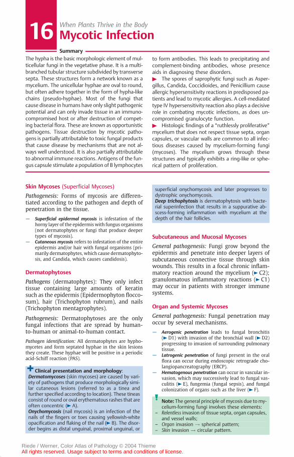
Clinical presentation and morphology:Dermatomycoses (skin mycoses) are caused by vari-ety of pathogens that produce morphologically simi-lar cutaneous lesions (referred to as a tinea andfurther specified according to location). These tineasconsist of round or oval erythematous rashes that areoften concentric ( A).Onychomycosis (nail mycosis) is an infection of thenails of the fingers or toes causing yellowish-whiteopacification and flaking of the nail ( B). The disor-der begins as distal unguinal, proximal unguinal, or
superficial onychomycosis and later progresses todystrophic onychomycosis.Deep trichophytosis is dermatophytosis with bacte-rial superinfection that results in a suppurative ab-scess-forming inflammation with mycelium at thedepth of the hair follicles.
Subcutaneous and Mucosal Mycoses
General pathogenesis: Fungi grow beyond theepidermis and penetrate into deeper layers ofsubcutaneous connective tissue through skinwounds. This results in a focal chronic inflam-matory reaction around the mycelium ( C2);granulomatous inflammatory reactions ( C1)may occur in patients with stronger immunesystems.
Organ and Systemic Mycoses
General pathogenesis: Fungal penetration mayoccur by several mechanisms.
— Aerogenic penetration leads to fungal bronchitis( D1) with invasion of the bronchial wall ( D2)progressing to invasion of surrounding pulmonarytissue.
— Latrogenic penetration of fungi present in the oralflora can occur during endoscopic retrograde cho-langiopancreatography (ERCP).
— Hematogenous penetration can occur in vascular in-vasion, which may successively lead to fungal vas-culitis ( E), fungemia (fungal sepsis), and fungalcolonization of organs such as the liver ( F).
Note: The general principle of mycosis due tomy-celium-forming fungi involves these elements:
– Relentless invasion of tissue septa, organ capsules,and vessel walls;
– Organ invasion ! spherical pattern;– Skin invasion ! circular pattern.
Riede / Werner, Color Atlas of Pathology © 2004 ThiemeAll rights reserved. Usage subject to terms and conditions of license.

C Granulomatous fungal inflammation (PAS) x 15
E Hematogenous fungal infection (vasculitis)(Grocott-silver stain) x 25
A Dermatomycosis
D Aerogenic fungal infection (fungal bronchitis)(Grocott-silver stain) x 25
F Hematogenous fungal infection (fungal sepsis in the liver; PAS) x 75
B Onychomycosis in a black patient
1
2
1
2
Thieme S.257-285.MON 02.06.2004 09:18 Uhr Seite 7
Riede / Werner, Color Atlas of Pathology © 2004 ThiemeAll rights reserved. Usage subject to terms and conditions of license.
Recommended