Suman Sahu et al. European Journal of Pharmaceutical and Medical Research
www.ejpmr.com │ Vol 7, Issue 11, 2020. │ ISO 9001:2015 Certified Journal │ 188
AN UPDATED REVIEW: PREPARATION, EVALUATION AND APPLICATIONS OF
HERBOSOME
Suman Sahu, Rudra Pratap Singh Rajput*, Poonam Verma, Rishabh Sahu, Chandrika Chouhan
Department of Pharmaceutics, Columbia Institute of Pharmacy, Raipur – 493111, Chhattisgarh, India.
Article Received on 24/08/2020 Article Revised on 14/09/2020 Article Accepted on 04/10/2020
INTRODUCTION
Novel Drug Delivery System
Drug delivery is that the methodology or method of
administering a pharmaceutical compound to produce
a therapeutic impact in humans or animals. For the
treatment of human diseases, nasal and respiratory
organ routes of drug delivery square measure gaining
increasing importance. These routes offer promising
alternatives to drug delivery of parenterals significantly
for amide and macromolecule medicine(protein and
peptides). For this purpose, many drug delivery
systems are developed and square measure being
investigated for nasal and respiratory organ delivery.
These contain liposomes, proliposomes, microspheres,
gels, prodrugs, cyclodextrins, among others.
Nanoparticles composed of perishable polymers show
assurance in fulfilling the demanding needs placed on
these delivery systems, like ability to be
transferred into associate aerosol, stability against
forces generated throughout aerosolization,
biocompatibility, targeting of specific sites or cell
populations within the respiratory organ, unleash of the
drug in a very preset manner, and degradation at
intervals a suitable amount of time.[1]
Novel drug delivery systems square measure designed
to produce an non-stop delivery of medication at
predictable Associate in Nursingd duplicatable
mechanics over an extended amount of your time
within the circulation. The potential benefits of this
idea contain minimization of drug associate facet
effects because of controlled therapeutic blood levels
rather than oscillatory blood levels, improved patient
compliance because of reduced frequency of dosing
and therefore the reduction of the entire dose of drug
administered.
Hence, the mix of each sustained unharness and
control unharness properties in an exceedingly
delivery system would additional enhance therapeutic
effectuality.[2]
The Benefits of Novel Drug Delivery System are as
Follows
1. Protection from physical and chemical
degradation.
2. Sustained delivery.
3. Improved tissue macrophages distribution.
4. improvement of stability.
5. improvement of medical specialty activity.
6. Protection from toxicity.
7. enlarged bioavailability.
8. improvement of solubility.[3-4]
Plant Drug Delivery System
In the past few decades, appreciable attention has been
focused on the evolution of a completely unique drug
delivery system (NDDS) for natural herb medicine.
SJIF Impact Factor 6.222
Review Article
ISSN 2394-3211
EJPMR
EUROPEAN JOURNAL OF PHARMACEUTICAL
AND MEDICAL RESEARCH
www.ejpmr.com
ejpmr, 2020,7(11), 188-205
ABSTRACT
The herbal therapeutic agents are widely used to treat and prevent disease and disorders. Recently introduced new
herbal formulation for better action such as herbosomes, liposomes, neosomes, ethosomes, iscoms, cubosomes etc.
Herbosomes have enhanced absorption rate, producing excellent bio-availability, good penetration power. In this
article includes complete description of advantage, dis-advantages, methods of herbosome preparation and
evaluation of herbosome. Different type of techniques used for preparation of herbosome such as solvent
evaporation technique, anti-solvent precipitation technique, rotary evaporation technique, ethanol injection
technique. An evaluation parameters are physicochemical evaluations, microscopical evaluations and in-vitro, in-
vivo evaluations. Through this review article we are concluding that the article would be helpful for the researchers
in terms of the preparation and evaluation of nano technology based drug delivery system.
KEYWORDS: Drug delivery system; Plant drug delivery system; Herbosome; History of herbosomes;
Application; Marketed formulations.
*Corresponding Author: Dr. Rudra Pratap Singh Rajput
Department of Pharmaceutics, Columbia Institute of Pharmacy, Raipur – 493111, Chhattisgarh, India.
Suman Sahu et al. European Journal of Pharmaceutical and Medical Research
www.ejpmr.com │ Vol 7, Issue 11, 2020. │ ISO 9001:2015 Certified Journal │ 189
Conservative indefinite quantity forms, as well as
prolonged-release indefinite quantity forms, they are
unable to satisfy for each holding the drug element at a
definite rate as per directed by the necessities of the
body, at some point of the amount of treatment,
additionally as guiding the phytoconstituents to their
desired target site to get an utmost therapeutic
response. In phytoformulation analysis, developing
nano-sized indefinite quantity forms (polymeric
nanoparticles and nanocapsules, liposomes, solid lipoid
nanoparticles, phytosomes, and nanoemulsion)
contains a variety of benefits for natural herb medicine,
as well as improvement of solubility and
bioavailability, protection from toxicity, improvement
of medical specialty activity, improvement of stability,
rising tissue scavenger cell distribution, sustained
delivery, and protection from physical and chemical
degradation. Thus, the nano-sized Novel drug delivery
systems of flavoring medicine have a possible future
for enhancing the activity and overcoming issues
related to the plant medicines. Liposomes, that area
unit bio decomposable and primarily nontoxic
vehicles, will encapsulate each hydrophilic and
hydrophobic materials.[5-6]
Types of Herbal Novel Drug Delivery System
Several approaches jus in case of recent flavoring drug
delivery system embrace differing kinds of
expressions like mouth-dissolving tablets, liposomes,
phytosomes(herbosomes), pharmacosomes, museums,
nanoparticles, microspheres, transfersomes, ethosomes,
stratum drug delivery system (TDDS), and
proniosomes area unit mentioned.[6]
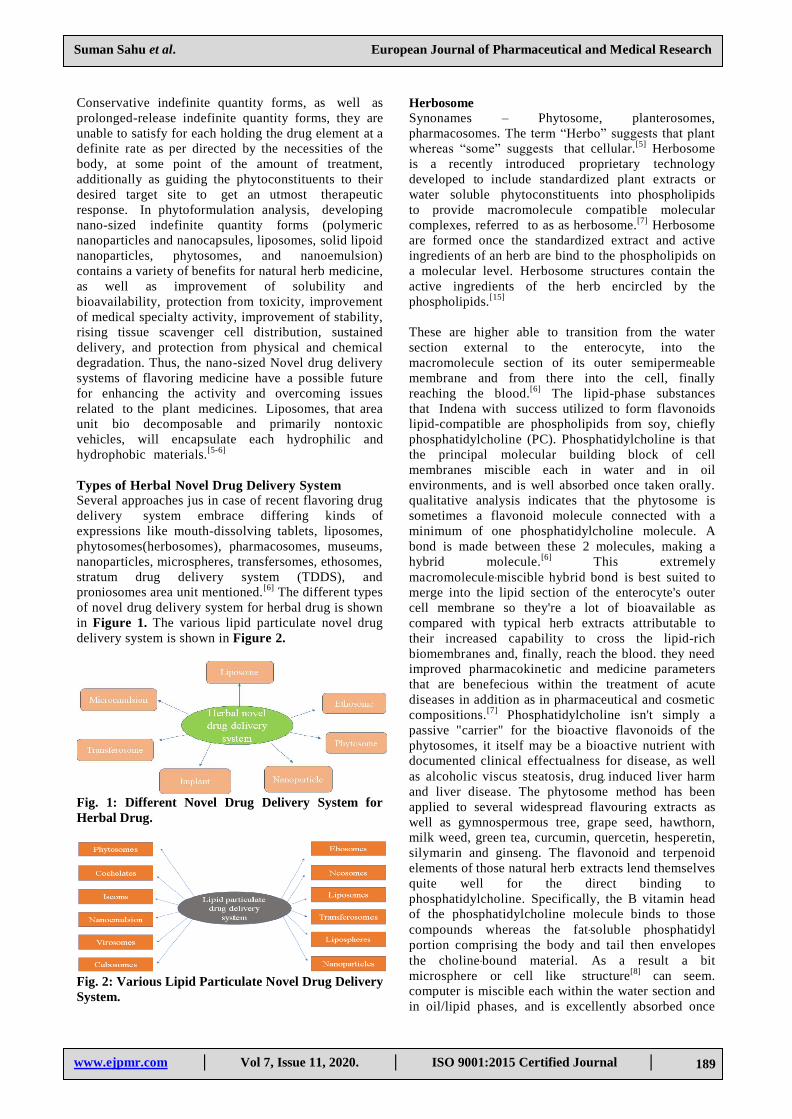
The different types
of novel drug delivery system for herbal drug is shown
in Figure 1. The various lipid particulate novel drug
delivery system is shown in Figure 2.
Fig. 1: Different Novel Drug Delivery System for
Herbal Drug.
Fig. 2: Various Lipid Particulate Novel Drug Delivery
System.
Herbosome
Synonames – Phytosome, planterosomes,
pharmacosomes. The term “Herbo” suggests that plant
whereas “some” suggests that cellular.[5]
Herbosome
is a recently introduced proprietary technology
developed to include standardized plant extracts or
water soluble phytoconstituents into phospholipids
to provide macromolecule compatible molecular
complexes, referred to as as herbosome.[7]
Herbosome
are formed once the standardized extract and active
ingredients of an herb are bind to the phospholipids on
a molecular level. Herbosome structures contain the
active ingredients of the herb encircled by the
phospholipids.[15]
These are higher able to transition from the water
section external to the enterocyte, into the
macromolecule section of its outer semipermeable
membrane and from there into the cell, finally
reaching the blood.[6]
The lipid-phase substances
that Indena with success utilized to form flavonoids
lipid-compatible are phospholipids from soy, chiefly
phosphatidylcholine (PC). Phosphatidylcholine is that
the principal molecular building block of cell
membranes miscible each in water and in oil
environments, and is well absorbed once taken orally.
qualitative analysis indicates that the phytosome is
sometimes a flavonoid molecule connected with a
minimum of one phosphatidylcholine molecule. A
bond is made between these 2 molecules, making a
hybrid molecule.[6]
This extremely
macromolecule ‐miscible hybrid bond is best suited to
merge into the lipid section of the enterocyte's outer
cell membrane so they're a lot of bioavailable as
compared with typical herb extracts attributable to
their increased capability to cross the lipid-rich
biomembranes and, finally, reach the blood. they need
improved pharmacokinetic and medicine parameters
that are benefecious within the treatment of acute
diseases in addition as in pharmaceutical and cosmetic
compositions.[7]
Phosphatidylcholine isn't simply a
passive "carrier" for the bioactive flavonoids of the
phytosomes, it itself may be a bioactive nutrient with
documented clinical effectualness for disease, as well
as alcoholic viscus steatosis, drug. induced liver harm
and liver disease. The phytosome method has been
applied to several widespread flavouring extracts as
well as gymnospermous tree, grape seed, hawthorn,
milk weed, green tea, curcumin, quercetin, hesperetin,
silymarin and ginseng. The flavonoid and terpenoid
elements of those natural herb extracts lend themselves
quite well for the direct binding to
phosphatidylcholine. Specifically, the B vitamin head
of the phosphatidylcholine molecule binds to those
compounds whereas the fat‐soluble phosphatidyl
portion comprising the body and tail then envelopes
the choline ‐bound material. As a result a bit
microsphere or cell like structure[8]
can seem.
computer is miscible each within the water section and
in oil/lipid phases, and is excellently absorbed once
Suman Sahu et al. European Journal of Pharmaceutical and Medical Research
www.ejpmr.com │ Vol 7, Issue 11, 2020. │ ISO 9001:2015 Certified Journal │ 190
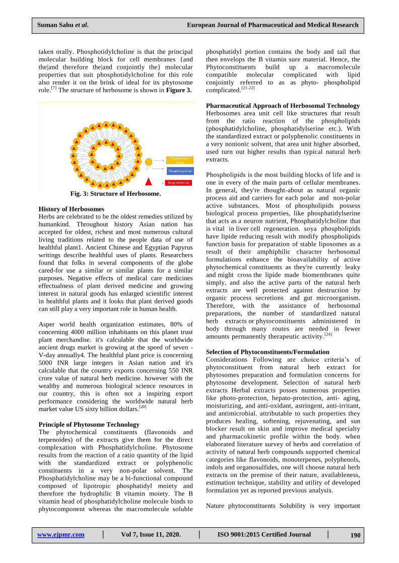
taken orally. Phosphotidylcholine is that the principal
molecular building block for cell membranes {and
the|and therefore the|and conjointly the} molecular
properties that suit phosphotidylcholine for this role
also render it on the brink of ideal for its phytosome
role.[7]
The structure of herbosome is shown in Figure 3.
Fig. 3: Structure of Herbosome.
History of Herbosomes
Herbs are celebrated to be the oldest remedies utilized by
humankind. Throughout history Asian nation has
accepted for oldest, richest and most numerous cultural
living traditions related to the people data of use of
healthful plant1. Ancient Chinese and Egyptian Papyrus
writings describe healthful uses of plants. Researchers
found that folks in several components of the globe
cared-for use a similar or similar plants for a similar
purposes. Negative effects of medical care medicines
effectualness of plant derived medicine and growing
interest in natural goods has enlarged scientific interest
in healthful plants and it looks that plant derived goods
can still play a very important role in human health.
Asper world health organization estimates, 80% of
concerning 4000 million inhabitants on this planet trust
plant merchandise. it's calculable that the worldwide
ancient drugs market is growing at the speed of seven -
V-day annually4. The healthful plant price is concerning
5000 INR large integers in Asian nation and it's
calculable that the country exports concerning 550 INR
crore value of natural herb medicine. however with the
wealthy and numerous biological science resources in
our country, this is often not a inspiring export
performance considering the worldwide natural herb
market value US sixty billion dollars.[20]
Principle of Phytosome Technology
The phytochemical constituents (flavonoids and
terpenoides) of the extracts give them for the direct
complexation with Phosphatidylcholine. Phytosome
results from the reaction of a ratio quantity of the lipid
with the standardized extract or polyphenolic
constituents in a very non-polar solvent. The
Phosphatidylcholine may be a bi-functional compound
composed of lipotropic phosphatidyl moiety and
therefore the hydrophilic B vitamin moiety. The B
vitamin head of phosphatidylcholine molecule binds to
phytocomponent whereas the macromolecule soluble
phosphatidyl portion contains the body and tail that
then envelops the B vitamin sure material. Hence, the
Phytoconstituents build up a macromolecule
compatible molecular complicated with lipid
conjointly referred to as as phyto- phospholipid
complicated.[21-22]
Pharmaceutical Approach of Herbosomal Technology
Herbosomes area unit cell like structures that result
from the ratio reaction of the phospholipids
(phosphatidylcholine, phosphatidylserine etc.). With
the standardized extract or polyphenolic constituents in
a very nonionic solvent, that area unit higher absorbed,
used turn out higher results than typical natural herb
extracts.
Phospholipids is the most building blocks of life and is
one in every of the main parts of cellular membranes.
In general, they're thought-about as natural organic
process aid and carriers for each polar and non-polar
active substances. Most of phospholipids possess
biological process properties, like phosphatidylserine
that acts as a neuron nutrient, Phosphatidylcholine that
is vital in liver cell regeneration. soya phospholipids
have lipide reducing result with modify phospholipids
function basis for preparation of stable liposomes as a
result of their amphiphilic character herbosomal
formulations enhance the bioavailability of active
phytochemical constituents as they're currently leaky
and might cross the lipide made biomembranes quite
simply, and also the active parts of the natural herb
extracts are well protected against destruction by
organic process secretions and gut microorganism.
Therefore, with the assistance of herbosomal
preparations, the number of standardized natural
herb extracts or phytoconstituents administered in
body through many routes are needed in fewer
amounts permanently therapeutic activity.[24]
Selection of Phytoconstituents/Formulation
Considerations Following are choice criteria’s of
phytoconstituent from natural herb extract for
phytosomes preparation and formulation concerns for
phytosome development. Selection of natural herb
extracts Herbal extracts posses numerous properties
like photo-protection, hepato-protection, anti- aging,
moisturizing, and anti-oxidant, astringent, anti-irritant,
and antimicrobial. attributable to such properties they
produces healing, softening, rejuvenating, and sun
blocker result on skin and improve medical specialty
and pharmacokinetic profile within the body. when
elaborated literature survey of herbs and correlation of
activity of natural herb compounds supported chemical
categories like flavonoids, monoterpenes, polyphenols,
indols and organosulfides, one will choose natural herb
extracts on the premise of their nature, availableness,
estimation technique, stability and utility of developed
formulation yet as reported previous analysis.
Nature phytoconstituents Solubility is very important
Suman Sahu et al. European Journal of Pharmaceutical and Medical Research
www.ejpmr.com │ Vol 7, Issue 11, 2020. │ ISO 9001:2015 Certified Journal │ 191
criterion for the event of novel formulations. in keeping
with the character of the phytoconstituents, that's
deliquescent or lipotropic, best appropriate formulation
are often nominated. Selection of dose type for delivery
of phytosomes Suitable sort of type formulation/dosage
form for delivery of phytosomes are often nominated
supported its potential for rising the effectiveness and
potency of bioactive compound. The applying of dose
type cought to improve its effectualness concerning
continuous action of herbs on general result of frame.
The inherent properties of flavorer drug like deliquescent
or hydrophobic, surface characteristics of system like
porousness and charges, degree of biodegradability, and
tonicity; unharness profile and size of the goods needed
of the ultimate formulation got to be taken in to thought.
Phytosome are often developed for each oral yet as
topical use.[21]
Following are few prompt dose forms for
phytosome delivery: Soft gelatin capsules. Hard gelatin
capsules.
Tablets.
Topical dose type
Phospholipids: Phospholipids area unit complicated
lipids that contains one or a lot of phosphate teams.
Phospholipids area unit amphipathic in nature that's
every molecule consists of a deliquescent portion and a
hydrophobic portion therefore tending to create lipide
bilayers.The most thick forms of present alcohol
phospholipids area unit phosphatidyl vitamin B,
phosphatidyl ethanolamine, phosphatidyl amino acid,
phosphatidyl vitamin B, phosphatidyl alcohol and
cardiolipin.[3]
Advantages of Herbosomes
Hydrophilic natural herb extracts absorption is
increased and contains a higher therapeutic result.
Herbosomes deliver the drug at specific site,
therefore low dose is needed to provide
therapeutic result. They are easily developed and
has additional stability than the other natural herb
formulations.
The carrier employed in this formulation
(phosphotidylcholine)has a plus that it's eco-
friendly with biological process worth Drug
defence capability of Herbosomes is high than
compared to the other natural herb formulations.
No advanced techniques don't seem to be needed
within the production of herbosomes and thence
low price is needed for its production Herbosomal
formulations are simply penetrate through the
layer of the skin. thence it will use for
percutaneous delivery.[28]
Herbosomes show higher stability as bond is made
between lipide molecule and phytoconstituent (s).
Dose of phytoconstituents is reduced thanks to
additional bioavailability of phytoconstituents
within the advanced type.
Duration of action is improved.
Herbosomes are easy to manufacture.
Phytoconstituents advanced with phospholipids are
additional stable in stomachic secretion and resist the
action of gut microorganism.
Enhanced porousness of phytoconstituents across the
biological membranes.[24]
Disadvantages of Herbosomes
In herbosomes, phytoconstituents square measure
fast eliminated. it's a brief half-life.
chemical reaction, fusion, leak and oxidization is
undergone by the phospholipids. It's a high price of
production and generally prevalence of aversions to
the herbosomal constituents is also ascertained.
Attributable to their larger size issues will occur
whereas making an attempt to focus on to the
numerous tissues.[24]
Properties of Herbosomes
Physical Properties
Herbosome has lipotropic substances with a
transparent melting point. The melting point of the
ready phytosomes considerably vary from that of the
phytoconstituents and also the lipide, that is thanks to
the chemical interaction between the each moiety and
also the modification in form of the phytosome. The
HP-TLC could be a easy technique used for
characterization of Phytosome. The phytosomes have a
retention issue worth totally different from the
phytoconstituents and phospholipids once they are
eluted with the acceptable solvent system, that
confirms the formation of a newly molecular
entity.[29]
Average size of herbosome vary is 50 nm to a couple of
100 μm. sac(vesicle) size. The sac size and zeta
potential were determined by exploitation Zetasizer
(Horibo instruments). Their sizes vary between fifty
nm to a hundred μm. Smaller vesicles with a mean size
of around 120nm were found to penetrate the skin
higher than larger vesicles on the brink of 1micron.
The results of particle size analysis by lazor device
optical phenomenon as shown in Fig.9 disclosed that
particle sizes varied from <50 nm to 246.98 nm and
also the mean sac size within the MP1B was 191.9nm
(SD ± 67.3nm) and thence it are often mentioned that
the formulation would exhibit a more higher
penetrating ability once developed into a gel.
concerning over 55% particles were found to own
diameter of 216nm[42]
Zeta potential
Zeta potential determined by employing a Horiba SZ
100Z particle size instrument with the measuring mode
of zeta potential. The lead to zeta potential of -25mV
as indicates the steadiness of the advanced. They're
simply soluble in non-polar solvents, insoluble in
water and moderately soluble in fats.[42]
An excess
Suman Sahu et al. European Journal of Pharmaceutical and Medical Research
www.ejpmr.com │ Vol 7, Issue 11, 2020. │ ISO 9001:2015 Certified Journal │ 192
amount of CUR was additional to completely different
oils and surfactants in an exceedingly tightly capped
cone-shaped countinuely raise and was perpetually
agitated at a 100 rpm and thirty seven °C for 24 h in an
exceedingly reciprocatory water tub (Bunsen, India).
Samples were centrifuged (4000 rpm, 15 min) once
twenty four h equilibrium and aliquots of supernatant
were diluted to applicable concentrations with solvent.
The samples were analyzed employing a photometer
(Shimadzu, Japan) at a wavelength of 420 nm with
solvent as a blank.[75]
The apparent solubility decided by adding more than
extract and CP to five metric capacity unit of water or
noctanol in sealed glass containers at temperature (25-
30oC). The liquids was agitated for twenty-four hours
then centrifuged for 20 min at 1,000 rpm to get rid of
more than extract. The supernatant was filtered
through a membrane filter (0.45 μm) then one metric
capacity unit filtrate was diluted with 9 ml capacity
unit of water or n- octanol and these samples were
measured spectrometrically at 268 nm ultraviolet
photometer.[53-55]
Liposomal like structures of micellar form are
shaped once herbosome are treated with water.[24]
Chemical Properties
On the premise of their physical properties and
spectroscopical information, it's been shown that, the
phospholipids-substrate interactionis because of the
formation of H- bond between the polarheads of
phospholipids (i.e. phosphate and ammoniumion
teams) and the polar useful groups of substrate. In
herbosomes, the active principle is anchored to the polar
head of phospholipids, becoming Associate in integral a
part of the membrane. [24-27]
Microscopical Examination
Examination by light-microscopic showed clearly that
altogether formulations of MPC’s there was sac
formation indicating that each one the 3 ways A,B and C
altogether drug: phospholipide ratios of 1:1, 1:2, 1:4
and 1:6 favorably resulted within the formation of
phytosomal complexes. Fig.1 showed spherical form of
the vesicles that were found to be extra diffuse and
showed no aggregation in formulations of 1:1 and 1:6.
Therefore, these 2 were nominated as appropriate
candidates for additional studies.[53]
Standard calibration curve of Ashwagandha in
ultraviolet photometer The ultraviolet absorbance of
Ashwagandha normal solution within the vary of 20-
120μg/ mL capacity unit of drug in phosphate buffered
saline pH scale 7.4 showed dimensionality at λ gamma
hydroxybutyrate 226nm. The dimensionality was
premeditated for absorbance against concentration
with R2 value 0.999 and with the slope equation y=
0.006x +0.012.[51]
X-ray diffraction study - The
powder x- diffraction (PXRD) patterns of (A)
standardized citrus lemon (SCL), (B)
phosphotidylcholine (PC), (C) physical mixture (PM),
and (D) citrus phytosome (CP). Crystalline peaks at
2θ=46.9°, 43.5°, 36.6°, and 28.0°. A diffraction peak
was determined for phosphotidylcholine at 35.5°,
36.4°, and 25.2°. The physical mixture (PM) showed
most of the peaks related to the SCL and laptop. The
diffractogram of the CP unconcealed the
disappearance of most of the crystalline peaks related
to the SCL and in comparison with physical mixture
(PM). These results were in concord with according
studies, wherever the disappearance of the API peaks
was related to the formation of drug lipide
composition.40 The disappearance of SCL crystalline
peak confirms the formation of SCL-phospholipid
composition.[53]
Scanning electron microscope - SEM pictures of
Standardized Citrus Lemon and Citrus P phytosome
are determined. Crystalline state of SCL was
visualised within the SEM photograph as various
crystals in figure 4 (A). In figure 4 (B) the drug was
fully born-again into phyto-phospholipid (CP)
advanced wherever SCL was physically enwrapped by
lipid transmission amorphous nature to the advanced
because of that crystals disappeared. [53]
Differential Scanning Measuring (DSC)
DSC may be a quick, reliable methodology to analyze
the interaction between multiple component and drug
excipient compatibility. These interactions are
determined because the elimination of endothermic
peak, the looks of latest peak, the amendment in peak
form, onset temperature/ temperature, relative peak
space or total heat.42-43 The SCL unconcealed broad
endothermic peaks at 126.48°C and 272.79°C. The
DSC thermogram of CP showed figure vi B provides 2
endothermic peaks at 206.40°C and 244.54°C. thus
from figure vi it absolutely was unconcealed that the
shift of endothermic peak at the distinction of around
25-30°C recommend doable interaction of SCL with
phosphotidylcholine and might account for increased
defense.[53]
Physico-Chemical Properties
As early mentioned, phytosomes are ready by
reaction of ratio quantity of lipoid with the
standardized plant extract as substrate. The
chemical analysis information reveals that the
phopspholipid- substrate interaction is because of
the formation of chemical bond between the polar
head (i.e., phosphate and ammonium ion group)
and therefore the polar functionalities of the
substrate.
The size of phytosome varies from 50 nm to some
100 μm. Example Particle size distribution of
citrus Phytosome The particle size of the ready CP
was carried out using dynamic light-weight
scattering technique. The mean particle size of CP
Suman Sahu et al. European Journal of Pharmaceutical and Medical Research
www.ejpmr.com │ Vol 7, Issue 11, 2020. │ ISO 9001:2015 Certified Journal │ 193
was distributed in an exceedingly slim vary of
233.4 ± 20.0 nm, and polydispersity index was
0.642 ± 0.03. The area at volume (SA/V)
quantitative relation of most particles is
reciprocally proportional to the particle size. Thus,
smaller particles of the CP, having the next SA/V,
create it easier for the entrapped drug to be free
from the phytosome via diffusion and surface
erosion. they need the advantage for the drug
entrapped phytosomes to penetrate into, and
permeate through the physiological drug
barriers.[53,56-58]
Phytosomes once treated with water, they assume
a micellar form resembling liposome and Photon
Correlation qualitative analysis (PCS) reveals
these liposomal structures nonheritable by
Phytosomes.
From the 1HNMR and 13CNMR information, it is
deduced that the fatty chain provides unchanged
signals each in free lipoid and within the
advanced, that indicates that long acyclic chains
are wrapped round the active principle,
manufacturing oleophilic envelope.
Regarding the solubility of phytosomes, the
complexes ar typically freely soluble in aprotic
solvents, moderately soluble in fats, insoluble in
water and comparatively unstable in alcohol.
however the phytosomes of sure oleophilic
phytoconstituents like curcumin has shown
inflated water solubility upon complexation with
phospholipids that has been mentioned later
during this paper.[29]
The apparent solubility of the pure SCL, the physical
mixture of SCL and phosphotidylcholine, and
therefore the ready SCL-PC advanced (CP). it had been
ascertained that the pure SCL had poor liquid
solubility (2.35 μg/mL), and a comparatively higher
solubility in n-Octanol (305 μg/mL). The physical
mixture (PM) increased the solubility of SCL however
this impact was weaker. The ready CP showed a major
increase within the liquid solubility. This increase
within the solubility of the ready advanced could also
be explained by reduced molecular crystallinity of the
drug and therefore the overall amphiphilic nature of
the phytosome.[53,59,60]
Biological Properties
Phytosomes are novel complexes that are higher
absorbed and used, thence they manufacture additional
bioavailability and higher result than the standard
natural herb extract or non-complexed extracts, that
has been stablished by pharmacokinetic studies or by
pharmacodynamic tests in experimental animals and in
human subjects23.
Phytosomes categorical their behaviour in physical or
biological system because their physical size,
membrane porosity, % entrapment, chemical
composition, amount and purity of the materials
used.[29-30]
Anti-oxidant Activity-Reducing power
methodology of Ashwagandha. The reducing
capability of a compound could function a major
indicator of its potential inhibitor activity. at 0.2mg/ml
and 1mg/ml the optical density of crude extract were
0.02, 0.09 and Ashwagandha phytosomes advanced
was zero.09 and 0.24. The results indicate that the
activity of the Ashwagandha extract is very very low
compared to Ashwagandha phytosomes advanced.[51]
Antifungal activity of lawsone Phytosome It was found
that phytosome advanced F1 showed 23 ± 0.39
millimetre zone of inhibition as compared to 19 ± 0.19
mm, 18 ± 0.58 millimetre and 18 ± 0.40 millimetre
zone of inhibition of phytosome advanced F2, plant
drug and normal drug ketoconazole once three days.
The phytosome advanced of lawsone F1 showed higher
antifungal activity as compared to phytosome advanced
F2, plant drug and normal drug. The gels of plant drug
lawsone G1 & G2 and phytosome advanced of lawsone
G3 – G6 were evaluated.[49]
Application of Herbosome
Herbosome are utilized in the treatment of liver
diseases together
with alcoholic internal organ steatosis, drug elicited
liver harm and liver disease.
They are utilized in medicament activity moreover
as in pharmaceutical and cosmetic composition.
They are accustomed treat acute and chronic liver
diseases of harmful metabolic or infective origin or
of chronic nature.
They are used as brain tonic, immunomodulator,
skin improver, antiwrinkle, anti- aging etc.
They are used as antineoplastic and inhibitor, eg-
grape seed.
They are utilized in lipoidemia, vein and skin
problem.
They are used as cancer chemo preventive agent
and accustomed treat benign prostate dysplasia.
They're additionally accustomed treat
cardiovascular disease.[24]
Difference between Herbosomes and Liposomes
Herbosomes
ar anchored through chemical bonds to the polar head of
phopholipid.
herbosomes, phosphatidylcholine and also the
individual plant compound type a 1:1 or 2:1
complicated looking on the substance.

Suman Sahu et al. European Journal of Pharmaceutical and Medical Research
www.ejpmr.com │ Vol 7, Issue 11, 2020. │ ISO 9001:2015 Certified Journal │ 194
Example –
Silybin Phytosome
Panax ginseng Phytosome
Glycyrrhiza Phytosome
Hawthorn Phytosome
Ginkgo Phytosome[7]
Liposomes
In liposomes, the active principle is dissolved within the
medium of activity or within the layers of the membrane.
No chemical bonds are formed. In liposomes, a whole
bunch and thousands of phosphatidylcholine molecules
surround the water soluble molecule.[31]
The structural difference between herbosome and liposome is shown in Figure 4.
Fig. 4: Structural Difference Between Herbosome (a) and Liposome (b).
Liposomes also are ready by mixture appropriate
soluble phytoconstituents in phosphatidylcholine
during a efinite magnitude relation beneath appropriate
conditions. Here no attractive force is created, the
phosphatidylcholine moiety simply anchors the soluble
phytoconstituents as a results of that there could also
be lots of or perhaps thousands of phosphatidylcholine
molecules neighbouring the drug molecule. just in case
of herbosomes the phosphatidylcholine and therefore
the plant constituents type a posh within the fraction
relation 1:1 or 2:1 and therefore the method of
Herbosome formation involves attractive force
formation whereas the liposomes are totally devoid of
the attractive force formation between the
phosphatadylcholine molecule and therefore the
phytoconstituents. because of the lesser composition of
the lipid content just in case of herbosomes the
herbosomes are additional bioavailable and are
absorbed to a higher extent than the liposomes.[32-33]
Methods of herbosome preparation
Herbsomes novel complexes that are ready by reacting
from 3-2 moles however ideally with one mole of
natural or artificial phospholipids like
phosphatidylcholine, phosphatidylethanolamine or
phosphatidylserine with one mole of part like
flavolignans, either alone or within the natural
mixture in aprotic solvents like dioxane or acetone.
The herbosome complicated will be then isolated by
precipitation with non-solvent like open-chain
hydrocarbons or drying up or by spray drying. within
the complicated formation of herbosomes, the
magnitude relation between these 2 moieties is within
the vary from zero.5-2.0 moles. the foremost preferred
magnitude relation of phospholipids to flavonoids is
1:1.[32,34]
The examples and methods of preparation of
herbosomes are shown in.
Suman Sahu et al. European Journal of Pharmaceutical and Medical Research
www.ejpmr.com │ Vol 7, Issue 11, 2020. │ ISO 9001:2015 Certified Journal │ 195
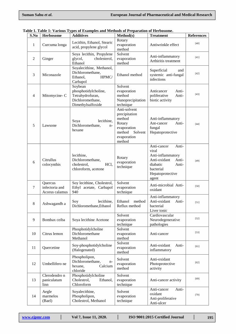
Table 1. Table 1: Various Types of Examples and Methods of Preparation of Herbosome.
S.No Herbosome Additives Method(s) Treatment References
1 Curcuma longa Lecithin, Ethanol, Stearic
acid, propylene glycol
Rotary
evaporation
method
Antiwrinkle effect [40]
2 Ginger
Soya lecithin, Propylene
glycol, cholesterol,
Ethanol
Solvent
evaporation
method
Anti-inflammatory
Arthiritis treatment [41]
3 Miconazole
Soyalecithine, Methanol,
Dichloromethane,
Ethanol, HPMC/
Carbapol
Ethanol method
Superficial and
systemic anti-fungal
infections
[42]
4 Mitomycine- C
Soybean
phosphotidylcholine,
Tetrahydrofuran,
Dichloromethane,
Dimethylsulfoxide
Solvent
evaporation
method
Nanoprecipitation
technique
Anticancer Anti-
poliferative Anti-
biotic activity
[43]
5 Lawsone
Soya lecithine,
Dichloromethane, n-
hexane
Anti-solvent
precipitation
method
Rotary
evaporation
method Solvent
evaporation
method
Anti-inflammatory
Ant-cancer Anti-
fungal
Hepatoprotective
[44]
6 Citrullus
colocynthis
lecithine,
Dichloromethane,
cholestrol, HCl,
chloroform, acetone
Rotary
evaporation
technique
Anti-cancer Anti-
viral
Anti-inflammatory
Anti-oxidant Anti-
diabatic Anti-
bacterial
Hepatoprotective
agent
[49]
7
Quercus
infectoria and
Acorus calamus
Soy lecithine, Cholestrol,
Ethyl acetate, Carbapol
940
Solvent
evaporation
technique
Anti-microbial Anti-
oxidant [50]
8 Ashwagandh a Soy lecithine,
Dichloromethane,Ethanol
Ethanol method
Reflux method
Anti-inflammatory
Anti-oxidant Anti-
bacterial
Liver tonic
[51]
9 Bombax ceiba Soya lecithine Acetone
Solvent
evaporation
technique
Cardiovascular
Neurodegenerative
pathologies
[52]
10 Citrus lemon
Phosphotidylcholine
Dichloromethane
Methanol
Solvent
evaporation
method
Anti-cancer [53]
11 Quercetine Soy-phosphotidylcholine
(Halogenated)
Solvent
evaporation
method
Anti-oxidant Anti-
inflammatory [61]
12 Umbellifero ne
Phospholipon,
Dichloromethane, n-
hexane, Calcium
chloride
Solvent
evaporation
method
Anti-oxidant
Photoprotective
activity
[62]
13
Clerodendro n
paniculatum
linn
Phosphotidylcholine
Cholestrol, Ethanol,
Chloroform
Solvent
evaporation
technique
Anti-cancer activity [69]
14
Aegle
marmelos
(Bael)
Soyalecithine,
Phospholipon,
Cholestrol, Methanol
Solvent
evaporation
technique
Anti-cancer Anti-
oxidant
Anti-proliferative
Anti-ulcer
[70]
Suman Sahu et al. European Journal of Pharmaceutical and Medical Research
www.ejpmr.com │ Vol 7, Issue 11, 2020. │ ISO 9001:2015 Certified Journal │ 196
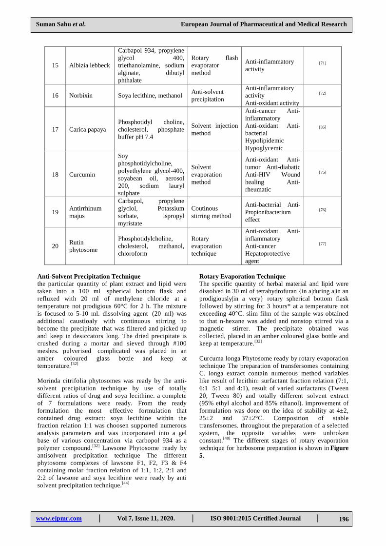
15 Albizia lebbeck
Carbapol 934, propylene
glycol 400,
triethanolamine, sodium
alginate, dibutyl
phthalate
Rotary flash
evaporator
method
Anti-inflammatory
activity [71]
16 Norbixin Soya lecithine, methanol Anti-solvent
precipitation
Anti-inflammatory
activity
Anti-oxidant activity
[72]
17 Carica papaya
Phosphotidyl choline,
cholesterol, phosphate
buffer pH 7.4
Solvent injection
method
Anti-cancer Anti-
inflammatory
Anti-oxidant Anti-
bacterial
Hypolipidemic
Hypoglycemic
[35]
18 Curcumin
Soy
phosphotidylcholine,
polyethylene glycol-400,
soyabean oil, aerosol
200, sodium lauryl
sulphate
Solvent
evaporation
method
Anti-oxidant Anti-
tumor Anti-diabatic
Anti-HIV Wound
healing Anti-
rheumatic
[75]
19 Antirrhinum
majus
Carbapol, propylene
glyclol, Potassium
sorbate, ispropyl
myristate
Coutinous
stirring method
Anti-bacterial Anti-
Propionibacterium
effect
[76]
20 Rutin
phytosome
Phosphotidylcholine,
cholesterol, methanol,
chloroform
Rotary
evaporation
technique
Anti-oxidant Anti-
inflammatory
Anti-cancer
Hepatoprotective
agent
[77]
Anti-Solvent Precipitation Technique
the particular quantity of plant extract and lipid were
taken into a 100 ml spherical bottom flask and
refluxed with 20 ml of methylene chloride at a
temperature not prodigious 60°C for 2 h. The mixture
is focused to 5-10 ml. dissolving agent (20 ml) was
additional caustioaly with continuous stirring to
become the precipitate that was filtered and picked up
and keep in desiccators long. The dried precipitate is
crushed during a mortar and sieved through #100
meshes. pulverised complicated was placed in an
amber coloured glass bottle and keep at
temperature.[32]
Morinda citrifolia phytosomes was ready by the anti-
solvent precipitation technique by use of totally
different ratios of drug and soya lecithine. a complete
of 7 formulations were ready. From the ready
formulation the most effective formulation that
contained drug extract: soya lecithine within the
fraction relation 1:1 was choosen supported numerous
analysis parameters and was incorporated into a gel
base of various concentration via carbopol 934 as a
polymer compound.[32]
Lawsone Phytosome ready by
antisolvent precipitation technique The different
phytosome complexes of lawsone F1, F2, F3 & F4
containing molar fraction relation of 1:1, 1:2, 2:1 and
2:2 of lawsone and soya lecithine were ready by anti
solvent precipitation technique.[44]
Rotary Evaporation Technique
The specific quantity of herbal material and lipid were
dissolved in 30 ml of tetrahydrofuran {in a|during a|in an
prodigiously|in a very} rotary spherical bottom flask
followed by stirring for 3 hours* at a temperature not
exceeding 40°C. slim film of the sample was obtained
to that n-hexane was added and nonstop stirred via a
magnetic stirrer. The precipitate obtained was
collected, placed in an amber coloured glass bottle and
keep at temperature.[32]
Curcuma longa Phytosome ready by rotary evaporation
technique The preparation of transfersomes containing
C. longa extract contain numerous method variables
like result of lecithin: surfactant fraction relation (7:1,
6:1 5:1 and 4:1), result of varied surfactants (Tween
20, Tween 80) and totally different solvent extract
(95% ethyl alcohol and 85% ethanol). improvement of
formulation was done on the idea of stability at 4±2,
25±2 and 37±2°C. Composition of stable
transfersomes. throughout the preparation of a selected
system, the opposite variables were unbroken
constant.[40]
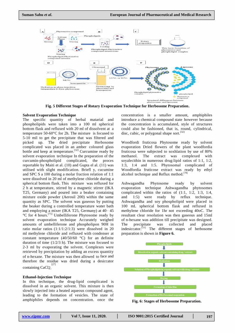
The different stages of rotary evaporation
technique for herbosome preparation is shown in Figure
5.
Suman Sahu et al. European Journal of Pharmaceutical and Medical Research
www.ejpmr.com │ Vol 7, Issue 11, 2020. │ ISO 9001:2015 Certified Journal │ 197
Fig. 5 Different Stages of Rotary Evaporation Technique for Herbosome Preparation.
Solvent Evaporation Technique
The specific quantity of herbal matarial and
phospholipids were taken into a 100 ml spherical
bottom flask and refluxed with 20 ml of dissolvent at a
temperature 50-60°C for 2h. The mixture is focused to
5-10 mil to get the precipitate that was filtered and
picked up. The dried precipitate Herbosome
complicated was placed in an amber coloured glass
bottle and keep at temperature.[32]
Curcumine ready by
solvent evaporation technique In the preparation of the
curcumin-phospholipid complicated, the proces
reportable by Maiti et al. (10) and Gupta el al. (11) was
utilised with slight modification. Briefl y, cucumine
and SPC S a 100 during a molar fraction relation of 1:1
were dissolved in 20 ml of methylene chloride during a
spherical bottom flask. This mixture was refluxed for
2 h at temperature, stirred by a magnetic stirrer (IKA
T25, Germany) and poured into a beaker containing
mixture oxide powder (Aerosil 200) within the same
quantity as SPC. The solvent was gaseous by putting
the beaker during a controlled temperature water bath
and employing a mixer (IKA T25, Germany) at 40– 45
°C for 4 hours.[75]
Umbelliferone Phytosome ready by
solvent evaporation technique Accurately weighed
amounts of umbelliferone and phospholipon 90H in
ratio molar ratios (1:1/1:2/1:3) were dissolved in 20
ml methylene chloride and refluxed with condenser at
constant temperature (40/50/60 °C) for an definite
duration of time (1/2/3 h). The mixture was focused to
2-3 ml by evaporating the solvent. Complexes were
retrieved by precipitation by adding an excess quantity
of n-hexane. The mixture was then allowed to face and
therefore the residue was dried during a desiccator
containing CaCl2.[27]
Ethanol-Injection Technique
In this technique, the drug-lipid complicated is
dissolved in an organic solvent. This mixture is then
slowly injected into a heated aqueous compound agent,
leading to the formation of vesicles. The state of
amphiphiles depends on concentration. once the
concentration is a smaller amount, amphiphiles
introduce a chemical compound state however because
the concentration is accumulated, style of structures
could also be fashioned, that is, round, cylindrical,
disc, cubic, or polygonal shape sort.[32]
Woodfordi fruticosa Phytosome ready by solvent
evaporation Dried flowers of the plant woodfordia
fruticosa were subjected to soxhlation by use of 80%
methanol. The extract was complexed with
soyalecithin in numerous drug:lipid ratios of 1:1, 1:2,
1:3, 1:4 and 1:5. Phytosomal complicated of
Woodfordia fruticose extract was ready by ethyl
alcohol technique and Reflux method.[78]
Ashwagandha Phytosome ready by solvent
evaporation technique Ashwagandha phytosomes
complicated within the ratios of (1:1, 1:2, 1:3, 1:4,
and 1:5) were ready by reflux technique.
Ashwagandha and soy phospholipid were placed in
100 mL spherical bottom flask and refluxed in
methylene chloride for 1hr not exceeding 40oC. The
resultant clear resolution was then gaseous and 15ml
of n-hexane was addition till precipitate was designed.
The precipitate was collected and placed
indesiccator.[51]
The different stages of herbosome
preparation is shown in Figure 6.
Fig. 6: Stages of Herbosome Preparation.

Suman Sahu et al. European Journal of Pharmaceutical and Medical Research
www.ejpmr.com │ Vol 7, Issue 11, 2020. │ ISO 9001:2015 Certified Journal │ 198
Formulation of phytosome
Phytosome complexes are often developed each orally
and locally. so as to get the most effective
performances of this technological innovation each in
terms of formulating manageableness and increased
bioavailability (as acceptable disintegration and
dissolution time of oral forms, for instance).[7]
Soft Gelatin Capsules
The phytosome are often spread in oily vehicles
(vegetable or semi-synthetic oil) to get suspension to
be stuffed in soft gelatin capsules.[21]
Soft gelatin
capsules represent a perfect answer to formulate
herbosome complexes. The Herbosome composition
are often spread in oily vehicles to get suspensions to
be stuffed in soft gelatin capsules. Vegetable or semi-
synthetic oils are often using by this purpose. Indena
mentioned a granulometry of 100% <200 μm to best
perform capsule production. According to Indena’s
knowledge, not all the herbosome complexes behave
within the same manner once spread in oily vehicles
and once the oily suspension is stuffed within the soft
gelatin capsules; for this reasons preliminary
practicableness trials ought to be performed to pick out
the foremost appropriate vehicle.[7]
Garlic soft gel
capsules 500 mg are accessible within the market and
given by oral route. Curcumine soft gelatin 500 mg
capsules are given by oral route.[32]
Hard Gelatin Capsules
The herbosome composition are often developed in
laborious gelatin capsules yet. an direct volumetric
filling method (without precompression) are often
applied, if the apparently small density of the herbosome
composition looks to limit the utmost quantity of
powder that may be stuffed into a capsule (usually less
than 300 mg for a size 0 capsule).With a pistontamp
capsule filling method, however, itis attainable to
extend the number of powder which might be stuffed
in an capsule, however precompression would possibly
have an effect on the disintegration time. Indena
mentioned the cautionaly monitor the connected
parameters throughout product/ method development. A
preliminary dry granulation method is recommended
outline the most effective producing process.[7]
Ashwagandha laborious gelatin capsules 500 mg are
given by oral route. Neem hard gelatin capsules 250
mg are given by oral route.[7]
Tablets
Dry granulation represents the perfect producing
method to get tablets with higher unitary doses and
with appropriate technological and biopharmaceutical
properties. However, thanks to the restricted
flowability, potential gumminess and low apparent
density of the Phytosome. complex, an direct
compression method are often applied just for low
unitary doses; note that whenever an direct
compression method is applied, the herbosome
composition ought to be diluted with 60-70% of
excipients to optimize its technological properties and
to get tablets with acceptable technological and
biopharmaceutical characteristics. On the another
hand, wet granulation ought to be avoided thanks to the
negative result of water and warmness (granulation/
drying) on the constancy of the lipoid complex.[7]
Carica papaya phytosomal tablets 500 mg are given by
oral route Shatavari pill 250 mg are given by oral
route.Neem tablet 300 mg are given by oral route[14]
Topical Dosage Forms
The herbosome composition are often developed
locally yet. the perfect method to include the
herbosome composition in emulsion is to disperse the
phospholipidic composition in an less quantity of the
lipidic part and add it to the already created emulsion
at low temperatures (not over 40°C). The herbosome
complexes are dispersible within the main lipidic
solvents used in topical formulations. just in case of
formulations containing a restricted quantity of lipids,
the herbosome complicated may additionally by spread
into the watery part, and once more further to the
ultimate formulation at temperature less than 40°C[7]
numerous kinds of phytosomes are applied locally like
Curcuma longa physomes[16],
Mitomycine c
phytosomes [19],
ashwagandha Phytosomes [23],
ginger
Phytosomes[17]
etc.
Characterization and evaluation of herbosomes
Organoleptic Properties
Ashwagandha extract was analyzed for their
organoleptic properties like color, Solubility and wave
length maxima of drug. From the results it absolutely
was ended that ashwagandha was found to be soluble
in phosphate buffered saline (Phosphate buffer
solution pH 7.4) and dimethylsulphoxide. The
concentration 100μg/ml of ashwagandha extract in
phosphate buffered saline was found to be 226nm.[7]
Visualization
Visualization of herbosomes is achieved by use of
transmission electron microscopy (TEM) and by
scanning electron microscopy (SEM).[7,8]
Scanning electron microscopy/transmission electron
microscopy: For finding out the surface order of
pharmacosomes these techniques is used [2].
the figure
and size of pharmacosomes is also affected by purity
grade of lipid and also the method variables like speed
of rotation, vaccum applied or the process used.[9]
Pharmacosomes ready by low purity grade lipids
yields greasy product. Pharmacosomes ready by
high purity grades lipids are liable to oxidation
degradation.[36-39]
Solubility
Solubility study is performed by taking associate more
than drug in numerous solvents like water, phosphate
buffer (PH 6.8) acetate buffer (PH 4.5).[32]
Curcumin

Suman Sahu et al. European Journal of Pharmaceutical and Medical Research
www.ejpmr.com │ Vol 7, Issue 11, 2020. │ ISO 9001:2015 Certified Journal │ 199
solubility study An excess amount of CUR was side to
different oils and surfactants during a tightly capped
conical flask and was countinously agitated at a 100
rpm and 37 °C for 24 h in a reciprocating water bath
(Bunsen, India). Samples were centrifuged (4000 rpm,
15 min) when 24 h equilibrium and aliquots of
supernatant were diluted to suitable concentrations
with acetone. The samples were analyzed employing a
photometer (Shimadzu, Japan) at a wavelength of 420
nm with acetone as a blank.[75]
Apparent Solubility of
Citrus Phytosome The apparent solubility estimated by
adding more than extract and CP to 5 millilitre of water
or noctanol in sealed glass containers at temperature
(25-30oC). The liquids were agitated for 24 hr then
centrifuged for 20 min at 1,000 rpm to eliminate extra
extract. The supernatant was filtered through a
membrane filter (0.45 μm) then 1 millilitre filtrate was
diluted with 9 millilitre of water or n-octanol and these
samples were measured spectrometrically at 268 nm
via ultraviolet spectrophotometer.[53]
Melting Point The freezing point of the ready phytosomes
considerably vary from that of the phytoconstituents
and also the lipid, that is the chemical interaction
between the each moiety and also the modification in
figure of the phytosome. The HP-TLC may be a simple
methodology used for characterization of Phytosome.
The phytosomes have a retention influence value
completely different from the phytoconstituents and
phospholipids after they are eluted with the appropriate
solvent system, that confirms the formation of a novel
molecular entity.[36]
Entrapment Efficiency
The Entrapment efficiency of a drug in herbosome is
measured by the centrifugation technique.[7]
Determination of percentage Drug Entrapment
efficiency of miconazole The percentage drug
Entrapment efficiency was estimated by
ultracentrifugation technique.
The% Drug Entrapment efficiency of Miconazole
phospholipid formulations varied within the vary of
54.3% (MP1B) to 70.7 % (MP6C). all formulations,
methodology of loading of the drug into the
phytosomal advanced caused a variation of 2.3 to 4.7%
in drug entrapment and also the results indicated that
methodology C simplified slightly higher Entrapment
of drug. Entrapment of Miconazole improved
considerably by 16.4% because the drug: lipid
provided as relation was improved from 1:1 to 1:6, the
rise in lipid providing a larger hydrophobic matrix for
Entrapment of Miconazole.[42]
Drug Content
The amount of drug will be quantified by a changed
high performance liquid chromatography methodology
or by an appropriate spectroscopically technique.[7,9]
Drug content of miconazole 1 g of the ready gel was
mixed with 100ml of appropriate solvent. Aliquots of
various concentration were ready by appropriate
dilutions once filtering the stock answer and
absorbance was measured. Drug content was
calculated by use of the equation y=0.09x-0.006 that
was obtained by linear regression analysis of
standardisation curve.[42]
Surface Tension Activity Measurement
The Surface phenomenon activity of the drug in Aqueous
solution is measured by the ring methodology during a
DuNouy ring tensiometer[7,10]
Surface morphology of
miconazole Phytosome.
The morphology of the drug loaded phytosomal by
SEM (Scanning electron microscopy). The sample of
blank SRE and MP1B phytosome composition once
examined indicated that the formulation contained
spherical vesicles, uniform in size with absence of
aggregation. The drug loading of miconazole within
the blank SRE Phytosomes is clearly detected. (SEM
of MCZ loaded SRE phytosome).[42]
Determination of % Product Yield
Determination of prcentage yield of phytosome of
Norbixin was calculated by the manual and established
formula:[13,16]
(%) Yield = (Practical yield) × 100 /(Theoretical
yield) Eq.(1)
wherever, Practical yield = the whole weight of the
formulation once completion of experiment.
Theoretical yield = The amount of the load of the
every ingredient that was wont to get the formulation
before continuing the experiment.[72-74]
Particle Size Distribution
to check particle size distribution dispersion of ready
herbosomes will be created in alcoholic solution
(isopropyl alcohol) and analyzed below size
analyser[36]
Particle size distribution of citrus
Phytosome The particle size of the ready CP was
carried outusing dynamic light scattering technique.
The mean particle size of CP was distributed in a very
slight vary of 233.4 ± 20.0 nm and polydispersity index
was 0.642 ± 0.03. The area at volume (SA/V) fraction
relation of most particles is controversy proportional to
the particle size. Thus, smaller particles of the CP,
having the next SA/V, build it easier for the entrapped
drug to be free from the phytosome via diffusion and
surface erosion. they need the advantage for the drug
entrapped phytosomes to penetrate into, and permeate
through the physiological drug barriers.[53,56-58]
Zeta Potential
Zeta potential (ZP) of the ready CUR-SPC advanced
was measured by the use of dynamic light scattering
(DLS) technique (Malvern Zetasizer, Malvern
Instruments, UK). Samples were distributed in water

Suman Sahu et al. European Journal of Pharmaceutical and Medical Research
www.ejpmr.com │ Vol 7, Issue 11, 2020. │ ISO 9001:2015 Certified Journal │ 200
and sonicated for 15 min. when sonication, samples
were diluted with water (1:10) before
measurements.[75]
Zeta potential of meconazole Phytosome Zeta potential
estimated by employing a Horiba SZ 100Z particle
size analyser with the measuring mode of Zeta
potential. The outcome in Zeta potential of -25mV
indicates the steadiness of the compound.[42]
Vesicle Size
The vesicle size and Zeta potential were determined by
use of Zetasizer (Horibo instruments). Their sizes vary
between 50 nm to 100 μm. Smaller vesicles with a
mean size of around 120 nm were found to penetrate
the skin higher than larger vesicles on the brink of
1micron.
The results of particle size analysis by laser diffraction
disclosed that particle sizes varied from <50 nm to
246.98 nm and also the mean vesicle size within the
MP1B was 191.9nm (SD ± 67.3nm) and thus it is
mentioned that the formulation would exhibit a higher
penetrating ability once developed into a gel. regarding
over 55% particles were found to diameter of 216
nm.[42]
Vesicle Stability
The stability of vesicles is determined by assessing
the size and structure of the vesicles over time. The
mean size is measured by DLS and structural changes
are monitored by Transmission electron
microscope[7,11]
Stability of the vesicles estimated by
storing the vesicles atn 4±0.5°C for 6 months and so
measurement their vesicle size and Zeta potential.[40]
Stability studay of Ashwagandha Phytosomes
Ashwagandha phytosome were keep at temperature and
cold temperature for three months and Entrapement
efficiency was estimated Stability studies were
conducted for optimized formulation E3 that showed
higher entrapement efficiency. The results showed no
important changes. so we tend to conclude that the
drug doesn't endure degradation on storage.[51]
Spectroscopic Evaluation
The spectroscopically evaluations are wide utilized so
as to verify the formation of complicated between
phytoconstituents and therefore the lipoid moiety still
on study the corresponding interaction between the
both. [7,12]
1H-Nuclear Magnetic Resonance
The complicated formation between the active
phytoconstituents and therefore the
phosphatidylcholine molecule will be calculable by
this technique. Bombardelli et al., studied the Nuclear
magnetic resonance spectra of herbosome
composition in non- polar solvents. there's a marked
modification in 1H-NMR signal originating from
atoms contain the formation of complicated, with none
summation of the signal peculiar to individual
molecules. The signals from protons belongs to the
phytoconstituents are broadened. In phospholipids
there's broadening of signals whereas the single
corresponding the N-(CH3)3 of B complex undergoes
an up field shift.[7,12]
13C-Nuclear Magnetic Resonance
In the 13
C NMR of the phytoconstituents and therefore
the stoichiometric complicated with the
phosphatidylcholine once recorded in C6D6 at normal
room temperature all the phytoconstituents carbons
were invisible. The signals equivalent to the glycerol
and choline portion are broadened and a few are
shifted, whereas most of the resonance of the fatty acid
chains retains their original sharp line form.[7,13,14]
Nuclear resonance study of umbelliferone H–NMR-
The samples of umbelliferone and composition were
dissolved within the solvent dimethyl sulphoxide and
analysed with a Bruker Avance II 400 NMR
spectroscope (SAIF, Panjab University, Chandigarh).
The spectrum was obtained and compared for the drug
and composition. 13C–NMR-The 13C-NMR spectrum
was taken for confirmation of the interaction between
drug and lipoid and therefore the formation of the
composition. The sample of umbelliferone and
composition was dissolved within the solvent dimethyl
sulphoxide and so analyzed with a Bruker Avance II
400 NMR spectroscope (SAIF, Panjab University,
Chandigarh). The spectrum was obtained and
compared for the drug and complex. [62-65]
Fourier Transform Infrared Spectroscopy (FT-IR)
The formation of the compound will be even be
confirmed by IR spectroscopy by comparison the
spectrum of the compound with the spectrum of the
individual elements and their mechanical mixtures.
FTIR spectrum analysis is additionally a useful tool
for the management of the steadiness of herbosomes
once micro-dispersed in water or once incorporated in
very simple cosmetic gels. From a laboratory point of
view, the stability will be confirmed by comparison the
spectrum of the compound in solid type (herbosomes)
with the spectrum of its micro- dispersion in water
once lyophillization, at totally different times. within
the case of simple formulations, it's necessary to
deduct the spectrum of the excipients (blank) from the
spectrum of the cosmetic type at totally different times,
comparison the remaining spectrum of the compound
itself.[7,13,14]
FT-IR study of citrus lemon Phytosome Fourier
transform infrared spectrum analysis (FTIR) analyses
of the SCL, PC, the physical mixture of SCL with
phosphotidylcholine (PM), and therefore the ready
Citrus phytosome were studied interaction between
SCL and phosphotidylcholine. The FTIR spectrum of
SCL showed broad peak at 3556 cm−1 representing
the open-chain alcoholic (−OH) group, 2900 cm−1
(CH stretching), 1660 cm−1 (C=O stretching), 1559
Suman Sahu et al. European Journal of Pharmaceutical and Medical Research
www.ejpmr.com │ Vol 7, Issue 11, 2020. │ ISO 9001:2015 Certified Journal │ 201
cm−1 (C=C stretching). distinguished peak detected at
1160 cm−1 and 1050 cm−1 generally relates to the
presence of acidic functional group. FTIR spectrum of
phosphotidylcholine disclosed the characteristic
absorption at 2921 and 2850 cm−1 (CH stretching),
1775 cm−1 (C=O stretching), 1235 cm−1 (P=O
stretching), 1081 cm−1 (P–O–C stretching) and 975
cm−1 (C-C-N stretching). The FTIR spectrum of the
ready Citrus phytosome is sort of totally different from
that of SCL and phosphotidylcholine. The peaks at
1660 cm−1, 1559 cm−1 and 3556 cm−1 were
protected by phospholipids. The absorption at 1660
cm−1 shifted to lower field within the spectrum of m
compound, indicating the formation of H- bond and
existence electricity interaction between extract and
lipoid.[53]
FTIR study of meconazole Phytosome FTIR spectrum
analysis reveals any interaction between varied
functional groups present in drug and excipients. within
the present study, the compatibility between the drug,
SRE, Lipid and alternative excipients were evaluated
by use of FTIR peak matching technique. The FTIR
spectra Fig. 6, seven and eight of meconazole, SRE and
MP1B severally were compared. All characteristic
peaks of MCZ and SRE of C-Cl, C=C, C-O, C-N, -O-
H and C=O were maintained within the formulation
and were detected at 638.46cm-1, 1612.54cm-1,
1089.82cm-1, 1375.58cm-1, 3400cm-1 and
1734.06cm-1 severally. No physico-chemical
interaction between MCZ, SRE and Soya lecithin was
detected and MCZ and SRE were present within the
pure type within the formulation.[42]
Differential Scanning Calorimetry (DSC)
Differential scanning calorimetry studies was
conducted via differential scanning calorimetry 60
(Shimadzu DSC60, Japan). Sample was weighed
(2.00-10.00 ± 5 mg) and placed within the sealed
aluminum crimp cell. The sample was scanned at
100C/ min up to 3500C within the atmosphere of N2.
Peak transition onset temperatures were recorded.[36-
38]
Differential scanning calorimetry (DSC) of citrus
lemon Phytosome DSC is a rapid, reliable technique to
analyse the interaction between multiple element and
drug excipient compatibility. These interactions are
detected because the elimination of endothermic peak,
the looks of recent peak, the modification in peak
form, onset temperature/ melting point, relative peak
area or enthalpy. The SCL disclosed broad
endothermic peaks at 126.48°C and 272.79°C. The
DSC thermogram of CP showed figure half dozen B
provides two endothermic peaks at 206.40°C and
244.54°C. it had been disclosed that the shift of
endothermic peak at the variance of around 25-30°C
counsel attainable interaction of SCL with
phosphotidylcholine and may account for increased
entrapment.[53]
High Performance Liquid Chromatography (HPLC)
A 10 μL volume of the standard soln of umbelliferone
and compound was applied by a 100 μL Hamilton
syringe in triplicate to associate degree HPTLC plate
(20×10 cm, 0.2 mm thick pre-coated by colloid 60
F254). The plates were developed in a solvent system
of toluene– methanol (9.5:0.5 v/v) at 25±2 °C
temperature and 40% till a distance of eight cm was
achieved. later development, the plates were dried and
scanned at 331 nm for umbelliferone by using Camag
thin layer chromatography scanner. The peak areas
were found and Rf values were recorded and compared
for the plain drug and compound by using WINCATS
software system.[27]
X-Ray Diffraction Study
X-ray diffraction study of umbelliferone Phytosome
The crystalline behaviour of umbelliferone,
phosphatidylcholine, physical mixture and also the
compound was evaluated by using X-ray powder
diffraction. Diffraction patterns were analysed with the
help of Bruker AXS D8 Advance (sophisticated
analytical instrument facility, STIC. Cochin). The X-
ray generator was allowed to work at 35 mA tube
current and 40 kV tube voltages. The scanning angle
was adjusted within the vary from 3 to 60 ° within the
step scan mode with a step time of 32.8s.[62,66,67]
In-vitro and In-vivo Evaluations
Models of in-vitro and in-vivo evaluations are chosen
on the basis of the expected therapeutic activity of the
biologically active phytoconstituents present within
the Phytosomes.[34]
as an example, in-vitro anti hepato
toxic activity are often assessed by the anti-oxidant
and free radical scavenging activity of the herbosomes.
Example - Stability of pharmacosomes: Stability of the
compound can be studied by correlating the spectrum
of compound at varied points of time within the solid
state with spectrum of a dispersion in water consisting
of tiny particles. – Stability studies of Phytosomal gels
of meconazole Optimized gel formulation of
meconazole was keep at a temperature of 400 C and
40C for 90 days and evaluated for modification in pH,
thickness and drug content.[9]
In-vitro drug release
In vitro drug release is the study of various pH by use
of standard dissolution equipment obtainable for the
purpose. The results are assessed on the based on
understood activity of the active constituents
therapeutically. [32-34]
In vitro Permeation Study
The phytosome compound gel of lawsone measurement
1.5 cm2 was subjected to an ex vivo permeation study
employing a changed Franz diffusion cell (cell
capability 7 ml). basis on literature survey, phosphate
buffer saline pH 7.4 was used as a diffusion media. The
phytosome compound gel of lawsone and plant drug
gel was applied on the dermal surface of one.5 cm2
Suman Sahu et al. European Journal of Pharmaceutical and Medical Research
www.ejpmr.com │ Vol 7, Issue 11, 2020. │ ISO 9001:2015 Certified Journal │ 202
cellulose ester membrane/rat skin. The diffusion media
was constantly stirred with needle formed magnetic
stirrer rotating at a speed of around 300-350
revolutions per minute(rpm). The temperature was
maintained at 32 ± zero.5°C with the help of hot
water. The diffusion was passed for 6 hours. At pre-
determined time intervals (0.5, 1, 2, 4 and 6 hr), 0.5 ml
sample were withdrawn and replaced with a similar
volume of recent phosphate buffer saline having pH 7.4.
Absorbance of the solutions was measured UV
spectrophotometrically at 227 nm. The cumulative %
drug permeation of the phytosome compound gel of
lawsone and plant drug gel were determined. [44-46]
In vivo Anti - Inflammatory Study
Male wistar rats were divided into four teams like
management, inflammation, phytosome gel and plant
drug gel teams. All rats were fed standard rat chow and
were maintained on a 12-hour light/dark cycle.
additionally, rats were acclimated to the procedure of
anti- inflammatory activity daily for one week.
Inflammation was induced in rats by injecting, ml of
carrageenan (1% w/v) beneath the planter region of the
right and left paw. Anti- inflammatory activity was
measured by digital plethysmometer (PM 01 orchid
Scientifics, India.[44,47,48]
CONCLUSION
Herbosome is novel formulations, improves
bioavailability of hydrophilic flavonoids and other similar
compounds through skin and gastrointestinal tract. It can
enhanced absorption rate, producing excellent
bioavailability, good penetration power. It may improve
the in vivo bioavailability of herbal drugs, which in spite
of positive in vitro results fail to deliver a similar response
in vivo. Use of herbal extracts in the form of dentifrice,
medicated gel, local drug delivery systems proved to be
efficient in preventing and treating periodontal disease.
REFERENCES
1. Tiwari Gaurav, Tiwari Ruchi, Shriwastawa
Birendra. Drug delivery system: An updated
review. International Journal of pharmaceutical
Investigation, 2016; 2(1): 230-253.
2. Kumar Vikas, Sharma Arvind, Sharma Ashish.
Recent advances in Novel drug delivery system for
delivery of antihypertensive drug. International
journal of drug development and research, 2011;
3(1): 252-259.
3. Patil Shaktipal, Mhaiskar Amrapali, Mudhada
Dharmendra. A review on Novel drug delivery
system: A recent trend. Intrnational journal of
current Pharmaceutical and clinical research,
2016; 6(2): 89-93.
4. Muller RH, Runge SA. Solid lipid nanoparticles
(SLN) for controlled drug delivery. Harwood
Academic Pub, 1998; 22(7): 219-234.
5. Madina OP, Zhu Y, Kairemo K. Targeted liposomal
drug delivery in cancer. Current Pharmaceutical
Des, 2004; 10: 2981‑9.
6. Sarangi Manoj kumar, Padhi Sasmita. Noval
herbal drug delivery system: An overview.
Archives of Medicine and Health Sciences, 2019;
6(1): 103 -132.
7. Ravi GS, Chandur V, Shabaraya AR, Sanjay K.
Phytosome: An advanced herbal drug delivery
system. World journal of pharmaceutical research
and bioscience, 2015; 4(3): 415-432.
8. Fry DW, White JC, Goldman ID. Rapid separation
of low molecular weight solutes from liposomes
without dilution. Analysis Biochemistry, 1978;
90(2): 809-15.
9. Maffei Facino R, Carini M, Aldini G, Bombardelli
E, Morazzoni P, Morelli R. Free radicals scavenging
action and anti-enzyme activities of procyanidines
from Vitis vinifera. Arzneimittelforschung, 1994;
44(5): 592-601.
10. Van den Bergh BA, Swartzendruber DC, Bos-Van
der Geest A, Hoogstraate JJ, Schrijvers AH, Boddé
HE et al. Development of an optimal protocol for the
ultrastructural examination of skin by transmission
electron microscopy. Journal Microsc, 1997; 187(2):
125-33.
11. Dayan N, Touitou E. Carriers for skin delivery of
trihexyphenidyl HCl: ethosomes vs. liposomes.
Biomaterials, 2000; 21(18): 1879-85.
12. Bombardelli E, Mustich G. Bilobalide-phospholipid
comlex, their uses and formulation containing them.
U. S. 1991.
13. Gabetta B, Zini GF, Pifferi G. Spectroscopic Studies
on Idb-1016 A New Flavanolignan Complex. Plant
Med. 1989; 55: 615.
14. Malandrino S, Pifferi G. Idb-1016 Silybin
Phosphatidylcholine Complex. Drugs Future, 1990;
15: 226-7.
15. Monica G, Naik V Vasu. Herbosomes: A potential
caarriers for the bioavalability enhancement of
herbal extracts. World journal of pharmacy and
pharmaceutical sciences, 2014; 4(1): 1052-1079.
16. Barzaghi N, Crema F, Gatti G, Pifferi G, Perucca E.
Pharmacokinetic studies on IdB 1016, a silybin-
phosphatidylcholine complex, in healthy human
subjects. Europian Journal Drug Metab
Pharmacokinets, 1990; 15(4): 333-8.
17. Lars I, Martin B. Determination of the size
distribution of liposomes by SEC fractionation, and
PCS analysis and enzymatic assay of lipid content.
AAPS Pharm Sci Tech, 2002; 3(2): 9–15.
18. Gupta NK, Dixit VK. Development and evaluation
of vesicular system for curcumin delivery. Arch
Dermatol Research, 2011; 303(2): 89-101.
19. Mukherjie PK. Evaluation of Indian Traditional
Medicine. Drug Information Journal, 2001; 35(2):
623-31.
20. Das Malay k, Kalita Bhupen. Phytosome: An
overview. Journal of pharmaceutical and Scientific
innovation, 2013; 2(3): 7-11.
21. Pawar Harshal Ashok, Bhangale Bhagyashree
Dilip. Journal of bioanalysis and biomedicine,
2015; 7(1): 6-12.
Suman Sahu et al. European Journal of Pharmaceutical and Medical Research
www.ejpmr.com │ Vol 7, Issue 11, 2020. │ ISO 9001:2015 Certified Journal │ 203
22. Comoglio A, Tomasi A, Malandrino S, Poli G,
Albano E. Scavenging effect of silipide, a new
silybin-phospholipid complex, on ethanol-derived
free radicals. Biochem Pharmacology, 1995; 50:
1313-1316.
23. Saha S, Sarma A, Saikia P, Chakrabarty T.
Phytosome: A Brief Overview. Scholars Academic
Journal of Pharmacy, 2013; 2: 12-20.
24. Dewan Nazeem, Dasgupta Debaprotim.
Herbosomes: A new arena of drug delivery. Journal
of pharmacognosy and phytochemistry, 2016;
5(4): 104 -108.
25. Patel J, Patel R, Khambholja K, Patel N. An
overview of phytosomes as an advanced herbal drug
delivery system. Asian Journal Pharmaceutical
Science, 2009; 4: 363-71.
26. Darshan D, Shrivastava S, Kapoor S, Gangawal A.
Phytosome. A novel dosage structure.
Pharmaceutical review, 2007; 6(2): 5-1.
27. Sindhumol PG, Thomas M, Mohanchandran PS.
Phytosome: A novel dosage form for enhancement
of bioavailability of botanicals and neutraceutical.
International Journal of Pharmacy and
Pharmaceutical Sciences, 2010; 2(4): 10-14.
28. Pradeepa Muragadaval, Narayanan Venkateshan,
Cherkuri Sowmya. A Review on Phytosomes:
importance and its application. International
journal of phytopharmacology, 2018; 9(1): 22-28.
29. Tripathy Surendra, Patel Dilip k, Baro Lipika. A
review on phytosomes: Their characterization,
advancement and potential for Transdermal
application. Journal of drug delivery and
therapeutics, 2013; 3(3): 147-152.
30. Franco PG & Bombardelli E, Complex compounds
of bioflavonoids with phospholipids, their
preparation and uses and pharmaceutical and
cosmetic compositions containing them, 1998.
31. Sharma path, Verma Surajpal, Mishri plakshi.
Global need for novel herbal drug formulation.
Intrnational journal of Pharmacognosy and
phytochemical research, 8(9): 1535-1544.
32. Chaudhary Imdadul Haque, Deb pulak, Das Sumit.
A review on formulation and characterization of
herbosome complex. International journal of
current Pharmaceutical research, 2019; 11(4): 42-
46.
33. Bombardelli E, Mustich G. Bilobalide phospholipid
complex, their uses and formulation containing
them, 1991.
34. Prachi U, Omprakash B. Phyto-phospholipid
complex vesicles for phytoconstituents and herbal
extracts: a promising drug delivery system.
International Journal Herbal Med, 2016; 4: 14-20.
35. Patil Swati, Ayare Priyanka. Carica papaya:
Formulation and evaluation of new dosage form
design. International Journal of Pharmaceutical
sciences and research, 2019; 10(4): 1677-1685.
36. Singh Deepak, Upadhyay Prashant. Phytosomes:
An advanced drug delivery system for herbal
drug.Global journal of nanomedicine, 2018; 4(3):
2573 -2374.
37. Dhyani Archana, Juyal Devi. Phytosomes an
advanced herbal drug delivery system. Current
Trends in Biomedical Engineering and Biosciences,
2017; 3(5): 1-2.
38. Rasaie Solmaz, Ghanbarzadeh, Mohammadi
Maryan, Hamishekhar Hamed. Nano phytosomes of
Quercitin: A promising formulation for Fortifiction
of Food Products with Antioxidants. Pharmaceutical
Sciences, 2014; 20: 96-101.
39. Bhupen Kalita, Malay K Das, Sharma Anil. Novel
Phytosome Formulation in Making Herbal Extract
More Effective. Research Journal of Pharmacy and
Technology, 2013; 6(11): 1295-1301.
40. Saraf Swarnalata, Jeswani Gunjan, Kaur Chanchal
Deep. Development of novel herbal cosmetic
cream with curcuma longa extract loaded
transferosomes for Antiwrinkle effect. African
journal of pharmacy and pharmacology, 2011;
5(8): 1054-1062.
41. Pawar Sonali Balasaheb, Ambekar abdul Wahid,
Chemate Satyam. Formulation and
characterization of an Herbal Anti-inflammatory
phytosome of Ginger Extract. Journal of pharma
research, 2018; 7(7): 167-170.
42. Pingali Pransuna Sundari, Srinivas Prathima,
Reddy B. Madhava. Miconazole loaded novel
phytosomal topical gels. World journal of
Pharmacy and Pharmceutical sciences, 2015;
4(10): 2305-2320.
43. Zhenquing Hou, Yang Li. Phytosome Loaded with
Mitomycin C- Soybean Phosphotidylcholine
Complex Developed for Drug Delivery. ACS
Publication, 2012; 10: 90-10.
44. Singh Rudra Pratap, Narke Ramakant. Preparation
and Evaluation of Phytosome of Lawsone.
International Journal of Pharmaceutical Sciences
and Research, 2015; 6(12): 2320-5148.
45. Chandira RM, Pradeep AP and Bhowmik D: Design,
development and formulation of anti-acne
dermatological gel. J. Chem. Pharm. Res, 2010;
2(1): 401-414.
46. Deepak P, Ramesh S and Noor Ahmed VH.
Isolation of eumelanin from sepia officinalis and
investigation of its antimicrobial activity by
ointment formulation. International Journal of
Pharmacy, 2012; 2(2): 67-72.
47. Bachhav YG and Patravale VB. Formulation of
meloxicam gel for topical application; in vitro and in
vivo evaluation. Acta Pharma, 2010; 60: 153-163.
48. Arunachalam G, Chattopadhyay D, Chatterjee S,
Mandal AB, Sur TK and Mandal SC: Evaluation of
anti-inflammatory activity of Alstonia macrophylla
Wall ex A. DC. Leaf extract. Phytomedicine, 2002;
(9): 632–635.
49. Rani alka, Arora Sandeep, Goyal Anju. Citrullus
Colosynthis Phytosomes: Development and
physiochemical characterization. International
Suman Sahu et al. European Journal of Pharmaceutical and Medical Research
www.ejpmr.com │ Vol 7, Issue 11, 2020. │ ISO 9001:2015 Certified Journal │ 204
journal of pharmaceutical sciences and research,
2019; 10(10): 4568-4573.
50. Ganeshan V, Kaithhavalappil SS, Kannappan M.
Formulation and In-vitro anti microbial evaluation
of herbosomal gels of extracts of Quercus
infectoria and Acorus calamus. Asian journal of
research in pharmaceutical sciences and
biotechnology, 2014; 2(2): 47-54.
51. B. Keerthi, Pinghali PS, Shrinivas Dr. Pratima.
Formulation and Evaluation of capsules of
Ashwagandha phytosomes. International journal
of pharmaceutical Sciences review and research,
2014; 29(2): 138-104.
52. Karole Sarita, Kumar Girender, Gupta Shailesh.
Preparation and evaluation of phytosomes
containing ethanolic extracts of leaves of Bombax
Ceiba for hepatoprotective activity. The pharma
innovation journal, 2019; 8(2): 22-26.
53. Udapurkar PP, Bhusnure OG, Kamble SR.
Development and characterization of Citrus limon
phospholipid complex as an effective
phytoconstituent delivery system. International
journal of life science and pharmaceutical
research, 2018; 8(1): 29-41.
54. Devendra SR, Bandana KT, Semalty M, Semalty A,
Badoni P, Rawat MSM, Baicalein- Phospholipid
Complex: A Novel Drug Delivery Technology for
Phytotherapeutics. Curr Drug Disc Tech, 2013;
10(3): 1-9.
55. Xiaoqing C, Yuxia L, Yue J, Aixin S, Wei S,
Zhonghao L, Zhongxi Z. Huperzine A–
phospholipid complex-loaded biodegradable
thermosensitive polymer gel for controlled drug
release. International Journal Pharmceutics. 2012;
433: 102– 11.
56. LeFevre ME, Olivo R, Vanderhoff JW, Joel DD.
Accumulation of latex in Peyer's patches and its
subsequent appearance in villi and mesenteric lymph
nodes. Proc Soc Exp Biol Med, 1978; 159(2): 298–
302.
57. Savic R, Luo L, Eisenberg A, Maysinger D.
Micellarnanocontainers distribute to defined
cytoplasmic organelles. Science, 2003; 300(5619):
615–8.
58. Freitas C, Muller RH. Effect of light and
temperature on zeta potential and physical stability
in solid lipid nanoparticle (SLN™) dispersions.
International Journal Pharm, 1998; 168(2): 221– 9.
59. Shrestha H, Bala R, Arora S. Lipid-Based Drug
Delivery Systems. Journal Pharma, 2014; 1- 10.
60. Sovova H. Apparent Solubility of Natural Products
Extracted with Near-Critical Carbon Dioxide.
American Journal Analytical Chemistry, 2012; 3:
958- 65.
61. Trang NH, Tung Dao BH, Giang Vu TT.
Preparation and evaluation of phytosomes of
Quercetine by solvent evaporation method. Journal
of pharmaceutical research and drug information,
2014; 7(4): 29-35.
62. Ittadwar PA, Puranik PK. Novel Umbelliferone
phytosomes: Develpoement and optimization
using experimental design approachand evaluation
of photoprotective and anti-oxidant activity.
International journal of pharmacy and
pharmaceutical sciences, 2017; 9(1): 1-11.
63. Saharan VA, Agarwal A, Kharb V. Process
optimisation, characterization and evaluation of
resveratrol-phospholipid complexes using box-
behnken statistical design. International Current
Pharmaceutical Journal, 2014; 3: 301-8.
64. Rawat MSM, Singh D, Semalty A, Semalty M.
Emodin-phospholipid complex-A potential of herbal
drug in the novel drug delivery system. Journal
Therm Analalysis Calorim Springer Sci, 2012; 108:
289-98.
65. Pretsch E, Buhlmann P, Badertscher M. Structure
determination of organic compounds. Tables of
Spectral Data, Springer Sci, 2009; 4: 93-108.
66. Dhalwal K, Shinde VM, Mahadik KR, Namdeo AG.
A rapid densitometric method for simultaneous
analysis of umbelliferone, psoralen, and eugenol in
herbal raw materials using HPTLC. J Sep Sci, 2007;
30: 2053.
67. Husain A, Aamir M, Mujeeb M, Siddique NA.
Simultaneous quantification of umbelliferone and
quercetin in polyherbal formulations of Aegle
marmelos by HPTLC. Am Journal PharmTech Res,
2012; 2: 2249-3387.
68. Yue PF, Yuan H, Li X, Yang M, Zhu W. Process
optimisation, characterization and evaluation in vivo
of the oxymatrinephospholipid complex.
International Journal Pharmaceutics, 2010; 387:
139-46.
69. Sundaraganpathy, PN leena. Development and
evaluation of Anti-cancer activity of phytosome
formulated from the root extract of clerodendron
paniculatum Linn. International journal of
pharmacognosy and phytochemical research,
2016; 8(11): 1778-1781.
70. Dhase AS, Saboo SS. Preparation and evaluation
of phytosomes containing methanolic extract of
leaves of Aegle Marmelos (Bael). International
journal of pharm tech research, 2015; 8(6): 231-
240.
71. B Rajesh, Das Sowmy, Pattanayak Dharmjit, M.
Pavani. Formulation design and optimization of
herbal gel containing Albizia Lebbeck bark
extract. International journal of pharmacy and
pharmaceutical sciences, 2014; 6(5): 111 -114.
72. Mund AK, Guru AC, Singh RP. Preparation and
characterization of phytosome of Norbixin. World
journal of pharmacy and pharmaceutical sciences,
2018; 8(1): 1045-1057.
73. Cho YJ, Bae IY, Inglett GE, Lee S. Utilization of
tartary buckwheat bran as a source of rutin and its
effect on the rheological and antioxidant properties
of wheat-based products. Ind Crop Prod, 2014; 61:
211-216.
Suman Sahu et al. European Journal of Pharmaceutical and Medical Research
www.ejpmr.com │ Vol 7, Issue 11, 2020. │ ISO 9001:2015 Certified Journal │ 205
74. Drinkwater JM, Tsao R, Liu R, Defelice C, Wolyn
DJ. Effects of cooking on rutin and glutathione
concentrations and antioxidant activity of green
asparagus (asparagus officinalis) spears. J Funct
Foods, 2015; 12: 342-353.
75. Allam AH, Komeil AH, Abdallah OY. Curcumine
phytosomal soft gel formulation: Development,
optimization and physiochemical characterization.
Acta pharmaceutical journal, 2015; 65: 285-297.
76. Shahtalebi MA, Asghari GR, Shafiee F.
Formulation of herbal gel of Antirrhinum Majus
extract and evaluation of it’s Anti-
propionibacterium acne effects. Advance
biomedical research, 2019; 47: 247-1.
77. Hooresfand Zahra, Ghanbarzadeh Saeed,
Hamishekhar Hamed. Preparation and
characterization of Rutin loaded nanophytosome.
Pharmaceutical sciences, 2015; 21(1): 145-151.
78. K. Rajashekhar, Sundari PJP, shinivas Dr.
Prathima. Development of a Topical Phytosomal
gel of Woodfordia fruticose. Words journal of
pharmacy and pharmaceutical Sciences, 2015;
4(11): 919-932.
Recommended