118 SEER Summary Staging Manual - 2000
ANATOMIC DRAWING OF THE RESPIRATORY TRACT
THE RESPIRATORY TRACT
Trachea
Carina
Rightmain stembronchus Left
main stembronchus
Lobarbronchus
Segmentalbronchus
Bronchiole
Terminalbronchiole
Respiratorybronchiole
Alveolarduct
Alveolus
Alveolar sacs

119SEER Summary Staging Manual - 2000
Auricle
Externalauditorymeatus
Tympanicmembrane
Pharyngotympanic(auditory)
tube
Malleus(head)
Incus
Stapes(limbs)
Semicircular ducts,ampullae, utricle, and saccule
Cochlear nerve
CochleaThe cochlea contains:Helicotrema (center of spiral);Cochlear duct which contains the spiral organ (“Corti”); andScala tympani
ANATOMIC DRAWINGS OF THE NASAL CAVITY AND MIDDLE EAR
SAGITTAL CUT THROUGH THE NASAL AND ORAL CAVITIES
THE EAR AND MIDDLE EAR
Prevertebral fascia
Soft palate
Nasal cavity
Palatine bone
Floor of mouthMandible
Nasopharynx
Hard palate
Uvula
LarynxCricoid cartilage
Oropharynx
HypopharynxGenioglossus muscle
Geniohyoid muscleMylohyoid muscle
Hyoid bone
Vallecula
Esophagus

120 SEER Summary Staging Manual - 2000
NASAL CAVITY AND MIDDLE EARC30.0-C30.1C30.0 Nasal Cavity (excludes nose, NOS C76.0)<>?
C30.1 Middle Ear (tympanic cavity) <><> Laterality must be coded for this site.? For nasal cartilage and nasal septum laterality is coded 0.
SUMMARY STAGE
0 In situ: Noninvasive; intraepithelial(Adeno)carcinoma in a polyp, noninvasive
1 Localized only
Invasive tumor confined to:Nasal cavity:
Meatus (superior, middle, inferior)Nasal chonchae (superior, middle, inferior)Septum
Middle ear:CochleaIncusMalleusSemicircular ducts, NOS:
AmpullaeSacculeUtricle
SeptumStapesTympanic membrane
Localized, NOS
2 Regional by direct extension only
Extension to: Nasal cavity:
Bone of skullChoanaFrontal sinusHard palateNasolacrimal ductNasopharynx
Code 2 continued on next page

121SEER Summary Staging Manual - 2000
NASAL CAVITY AND MIDDLE EARC30.0-C30.1
2 Regional by direct extension only (continued)
Middle ear:Auditory tubeExternal auditory meatusInternal carotid arteryMastoid antrumNasopharynxNerve(s)Pharyngotympanic tubeTemporal bone
3 Regional lymph node(s) involved only
REGIONAL Lymph Nodes
Cervical, NOSInternal jugular, NOS:
Deep cervical, NOS:Upper, NOS:
Jugulodigastric (subdigastric)Mandibular, NOS:
Submandibular (submaxillary)Submental
Mastoid (post-/retro-auricular) for middle earRetropharyngeal
Regional lymph node(s), NOS
4 Regional by BOTH direct extension AND regional lymph node(s) involved
Codes (2) + (3)
5 Regional, NOS
7 Distant site(s)/lymph node(s) involved
Distant lymph node(s)
Further contiguous extensionMiddle ear:
Meninges
Metastasis
9 Unknown if extension or metastasis

122 SEER Summary Staging Manual - 2000

123SEER Summary Staging Manual - 2000
Frontal sinus
Floor of orbit
Sphenoid sinus
Ethmoid sinus
Maxillary sinus
Skull
ANATOMIC DRAWING OF THE SINUSES
THE MAJOR SINUSES

124 SEER Summary Staging Manual - 2000
MAXILLARY SINUSC31.0C31.0 Maxillary sinus (antrum) <><> Laterality must be coded for this site.
SUMMARY STAGE
0 In situ: Noninvasive; intraepithelial
1 Localized only
Invasive tumor confined to mucosa of maxillary antrum (sinus)
Localized, NOS
2 Regional by direct extension only
Extension to :Base of skullCribriform plateEthmoid, NOSEthmoid sinus, anterior or posteriorFloor or medial wall of orbitFloor or posterior wall of maxillary sinusFrontal sinusInfratemporal fossaMaxilla, NOSMiddle nasal meatusNasal cavity, NOS:
FloorLateral wallSeptumTurbinates
NasopharynxOrbital contents including eyePalate, hard or softPalatine bonePterygoid platesPterygomaxillary or temporal fossaSkin of cheekSphenoid
125SEER Summary Staging Manual - 2000
MAXILLARY SINUSC31.0
3 Regional lymph node(s) involved only
REGIONAL Lymph Nodes
Cervical, NOSInternal jugular, NOS:
Deep cervical, NOS:Upper, NOS:
Jugulodigastric (subdigastric)Mandibular, NOS:
Submandibular (submaxillary)Submental
Retropharyngeal
Regional lymph node(s), NOS
4 Regional by BOTH direct extension AND regional lymph node(s) involved
Codes (2) + (3)
5 Regional, NOS
7 Distant site(s)/lymph node(s) involved
Distant lymph node(s)
Further contiguous extension
Metastasis
9 Unknown if extension or metastasis

126 SEER Summary Staging Manual - 2000
ETHMOID SINUSC31.1C31.1 Ethmoid sinus
SUMMARY STAGE
0 In situ: Noninvasive; intraepithelial
1 Localized only
Invasive tumor confined to ethmoid with or without bone erosion (cribriform plate)
Localized, NOS
2 Regional by direct extension only
Extension to :More than one ethmoid sinus
Anterior orbitBase of skullFrontal sinusIntracranial extensionMaxillary sinusNasal cavity, NOS:
FloorLateral wallSeptumTurbinates
NasopharynxOrbital extension including apex of orbitSkin of external noseSphenoid
3 Regional lymph node(s) involved only
REGIONAL Lymph Nodes
Cervical, NOSInternal jugular, NOS:
Deep cervical, NOS:Upper, NOS:
Jugulodigastric (subdigastric)Mandibular, NOS:
Submandibular (submaxillary)Submental
Retropharyngeal
Regional lymph node(s), NOS

127SEER Summary Staging Manual - 2000
ETHMOID SINUSC31.1
4 Regional by BOTH direct extension AND regional lymph node(s) involved
Codes (2) + (3)
5 Regional, NOS
7 Distant site(s)/lymph node(s) involved
Distant lymph node(s)
Further contiguous extension
Metastasis
9 Unknown if extension or metastasis

128 SEER Summary Staging Manual - 2000
ACCESSORY (Paranasal) SINUSES (excluding Maxillary and Ethmoid Sinuses)C31.2-C31.3, C31.8-C31.9C31.2 Frontal sinus <>C31.3 Sphenoid sinusC31.8 Overlapping lesion of accessory sinusesC31.9 Accessory sinus, NOS<> Laterality must be coded for this site.
SUMMARY STAGE
0 In situ: Noninvasive; intraepithelial
1 Localized only
Invasive tumor confined to mucosa in one of the following:Frontal sinusSphenoid sinus
Localized, NOS
2 Regional by direct extension only
Destruction/invasion of:Bone: Bony wall of sinus
Facial bonesMaxillaOrbital structuresPterygoid fossaZygoma
BrainCranial nervesMuscles: Masseter
PterygoidNasal cavity, NOS:
FloorLateral wallSeptumTurbinates
NasopharynxOrbital contents including eyePalateSkinSoft tissue
More than one accessory sinus invaded

129SEER Summary Staging Manual - 2000
ACCESSORY (Paranasal) SINUSES (excluding Maxillary and Ethmoid Sinuses)C31.2-C31.3, C31.8-C31.9
3 Regional lymph node(s) involved only
REGIONAL Lymph Nodes
Cervical, NOSInternal jugular, NOS:
Deep cervical, NOS:Upper, NOS:
Jugulodigastric (subdigastric)Mandibular, NOS:
Submandibular (submaxillary)Submental
Retropharyngeal
Regional lymph node(s), NOS
4 Regional by BOTH direct extension AND regional lymph node(s) involved
Codes (2) + (3)
5 Regional, NOS
7 Distant site(s)/lymph node(s) involved
Distant lymph node(s)
Further contiguous extension
Metastasis
9 Unknown if extension or metastasis

130 SEER Summary Staging Manual - 2000
Medianglossoepiglottic fold
Base of tongue
Epiglottis
Vestibular folds(false vocal cords)
Aryepiglottic fold
Cuneiform tubercle
Corniculate tubercleTracheaInterarytenoid
notch
Vocal ligaments(true vocal cords)
ANATOMIC DRAWINGS OF THE LARYNX
THE VOCAL CORDS AS VIEWEDTHROUGH A LARYNGOSCOPE
ANTERIOR VIEW OF LARYNX POSTERIOR VIEW OF LARYNX
Epiglottis(supraglottis)
Hyoid boneSuperior horn ofthyroid cartilage
Thyrohyoidmembrane
Corniculate cartilageArytenoid cartilage
Superior thyroid notchThyroid cartilage
Vocal cord (vocal ligament)Inferior horn of
thyroid cartilageCricoid cartilage
Trachea
Adapted from an illustration(now the possession of Novartis and available as freeware)
by F. Netter, MD

131SEER Summary Staging Manual - 2000
Definition of Anatomical Limits of the Larynx
Anterior Limit is bounded by the anterior or lingual surface of the suprahyoid epiglottis, thyrohyoidmembrane, the anterior commissure, and the anterior wall of the subglottic region, which is composed ofthe thyroid cartilage, the cricothyroid membrane, and the anterior arch of the cricoid cartilage.
Posterior Lateral Limits include the aryepiglottic folds, the arytenoid region, the interarytenoid space,and the posterior surface of the subglottic space represented by the mucous membrane covering thecricoid cartilage.
Superior Lateral Limits are bounded by the tip and the lateral border of the epiglottis.
Inferior Limits are bounded by a plane passing through the inferior edge of the cricoid cartilage.
The larynx is divided into the following anatomic regions and sites:
Region Site
Supraglottic Ventricular bands (false cords)
Arytenoids
Epiglottis (both lingual and laryngeal aspects) Aryepiglottic foldsInfrahyoid epiglottisSupraglottis:
LeftRight
Suprahyoid epiglottis
Glottic Glottic:Anterior and posterior commissuresTrue vocal cords
Subglottic Right and left walls of the subglottisSubglottis (rima glottidis)
exclusive of the undersurface of the cords

132 SEER Summary Staging Manual - 2000
LARYNX: GLOTTISC32.0 Glottis (intrinsic larynx, laryngeal commisure, true vocal cord, vocal cord, NOS)
SUMMARY STAGE
0 In situ: Noninvasive; intraepithelial
1 Localized only
Confined to glottis, NOSIntrinsic larynxLaryngeal commissure(s):
AnteriorPosterior
Vocal cord(s), NOS:True cord(s), NOSTrue vocal cord(s)
Invasive tumor WITH or WITHOUT## normal vocal cord mobility:One vocal cordBoth vocal cords
Tumor involves adjacent region(s) of larynx:##
SubglottisSupraglottis
False vocal cord(s)Tumor limited to larynx WITH vocal cord fixation##
Involvement of intrinsic muscle(s):##
AryepiglotticCorniculate tubercleCuneform tubercle
ArytenoidCricoarytenoidCricothyroidThyroarytenoidThyroepiglotticVocalis
Localized, NOS
2 Regional by direct extension only
Extension to:Base of tongue###
Hypopharynx, NOS###
Postcricoid areaPre-epiglottic tissuesPyriform sinus (pyriform fossa)Vallecula###

133SEER Summary Staging Manual - 2000
LARYNX: GLOTTISC32.0
3 Regional lymph node(s) involved only
REGIONAL Lymph Nodes (including contralateral or bilateral nodes)
Anterior deep cervical (laterotracheal) (recurrent laryngeal):ParalaryngealParatrachealPrelaryngeal:
Delphian nodePretracheal
Cervical, NOSInternal jugular, NOS:
Deep cervical, NOS:Lower, NOS:###
Jugulo-omohyoid (supraomohyoid)Middle###
Upper, NOS:Jugulodigastric (subdigastric)
Mandibular, NOS:Submandibular (submaxillary)###***
Submental###***
Retropharyngeal###***
Regional lymph node(s), NOS
4 Regional by BOTH direct extension AND regional lymph node(s) involved
Codes (2) + (3)
5 Regional, NOS
Continued on next page

134 SEER Summary Staging Manual - 2000
LARYNX: GLOTTISC32.0
7 Distant site(s)/lymph node(s) involved
Distant lymph node(s):MediastinalSupraclavicular (transverse cervical)Other distant lymph node(s)
Extension to/through:Cervical (upper) esophagusCricoid cartilage**
Extrinsic (strap) muscles:OmohyoidSternohyoidSternothyroidThyrohyoid
OropharynxSkinSoft tissues of neckThyroid cartilage**
Thyroid glandTrachea
Further contiguous extension
Metastasis
9 Unknown if extension or metastasis
## Considered regional in Historic Stage### Considered distant in Historic Stage** Considered regional in 1977 Summary Staging Guide*** Considered distant in 1977 Summary Staging Guide

135SEER Summary Staging Manual - 2000

136 SEER Summary Staging Manual - 2000
LARYNX: SUPRAGLOTTISC32.1C32.1 Supraglottis (extrinsic larynx, laryngeal aspect of aryepiglotic fold, ventricular band,
false vocal cord)
SUMMARY STAGE
0 In situ: Noninvasive; intraepithelial
1 Localized only
Invasive tumor with normal vocal cord mobility confined to:Supraglottis (one subsite):
Aryepiglottic foldArytenoid cartilageCorniculate cartilageCuneiform cartilageEpilarynx, NOSFalse cord(s):
Ventricular band(s)Ventricular cavityVestibular fold
Infrahyoid epiglottisLaryngeal cartilage, NOSLaryngeal (posterior) surface of epiglottisSuprahyoid epiglottis (including tip, lingual {anterior} and laryngeal surfaces)
Impaired vocal cord mobility##
Tumor involves adjacent region(s) of larynxTumor involves more than one subsite of supraglottis WITHOUT fixation or NOSTumor limited to larynx WITH vocal cord fixation##
Localized, NOS
2 Regional by direct extension only
Extension to:Base of tongue including mucosaCricoid cartilageHypopharynx, NOSPostcricoid areaPre-epiglottic tissuesPyriform sinus (pyriform fossa)Vallecula

137SEER Summary Staging Manual - 2000
Tumor involves region outside the supraglottisLARYNX: SUPRAGLOTTISC32.1
3 Regional lymph node(s) involved only
REGIONAL Lymph Nodes (including contralateral or bilateral nodes)
Anterior deep cervical (laterotracheal) (recurrent laryngeal):ParalaryngealParatrachealPrelaryngeal:
Delphian nodePretracheal
Cervical, NOSInternal jugular, NOS:
Deep cervical, NOS:Middle###
Upper, NOS:Jugulodigastric (subdigastric)
Mandibular, NOS:Submandibular (submaxillary)###***
Submental###***
Retropharyngeal###***
Regional lymph node(s), NOS
4 Regional by BOTH direct extension AND regional lymph node(s) involved
Codes (2) + (3)
5 Regional, NOS
Continued on next page
138 SEER Summary Staging Manual - 2000
LARYNX: SUPRAGLOTTISC32.1
7 Distant site(s)/lymph node(s) involved
Distant lymph node(s):MediastinalSupraclavicular (transverse cervical)Other distant lymph node(s)
Extension to/through:Cervical esophagusExtrinsic (strap) muscles:
OmohyoidSternohyoidSternothyroidThyrohyoid
OropharynxSkinSoft tissues of neckThyroid cartilage##**
Thyroid gland
Further contiguous extension
Metastasis
9 Unknown if extension or metastasis
## Considered regional in Historic Stage### Considered distant in Historic Stage** Considered regional in 1977 Summary Staging Guide*** Considered distant in 1977 Summary Staging Guide

139SEER Summary Staging Manual - 2000

SEER Summary Staging Manual - 2000140
LARYNX: SUBGLOTTISC32.2C32.2 Subglottis
SUMMARY STAGE
0 In situ: Noninvasive; intraepithelial
1 Localized onlyInvasive tumor with normal vocal cord mobility confined to subglottisTumor involves adjacent region(s) of larynx##
Vocal cords with normal or impaired mobility##
Tumor limited to larynx WITH vocal cord fixation##
Localized, NOS
2 Regional by direct extension only
Extension to/through:Base of tongue###
Hypopharynx, NOSPostcricoid areaPre-epiglottic tissuesPyriform sinus (pyriform fossa)Vallecula
3 Regional lymph node(s) involved only
REGIONAL Lymph Nodes (including contralateral or bilateral nodes)
Anterior deep cervical (laterotracheal) (recurrent laryngeal):ParalaryngealParatrachealPrelaryngeal:
Delphian nodePretracheal
Cervical, NOSInternal jugular, NOS:
Deep cervical, NOS:Lower, NOS:
Jugulo-omohyoid (supraomohyoid)Middle***
Mandibular, NOS:Submandibular (submaxillary)###***
Submental###***
Retropharyngeal###***
Regional lymph node(s), NOS

141SEER Summary Staging Manual - 2000
LARYNX: SUBGLOTTISC32.2
4 Regional by BOTH direct extension AND regional lymph node(s) involved
Codes (2) + (3)
5 Regional, NOS
7 Distant site(s)/lymph node(s) involved
Distant lymph node(s):MediastinalSupraclavicular (transverse cervical)Other distant lymph node(s)
Extension to:Cervical esophagusCricoid cartilage##**
Extrinsic (strap) muscles:OmohyoidSternohyoidSternothyroidThyrohyoid
OropharynxSkin##
Soft tissues of neckThyroid cartilage**
Thyroid glandTrachea##
Further contiguous extension
Metastasis
9 Unknown if extension or metastasis
## Considered regional in Historic Stage### Considered distant in Historic Stage** Considered regional in 1977 Summary Staging Guide*** Considered distant in 1977 Summary Staging Guide

SEER Summary Staging Manual - 2000142
LARYNX: OVERLAPPING LESION OR NOS (including Laryngeal Cartilage)C32.3, C32.8-C32.9C32.3 Laryngeal cartilageC32.8 Overlapping lesion of larynxC32.9 Larynx, NOS
SUMMARY STAGE
0 In situ: Noninvasive; intraepithelial
1 Localized only
Invasive tumor confined to site of origin
Impaired vocal cord mobilityTumor involves adjacent region(s) of larynxTumor involves more than one subsite without fixation or NOSTumor limited to larynx WITH vocal cord fixation
Localized, NOS
2 Regional by direct extension only
Extension to:Hypopharynx, NOSPostcricoid areaPre-epiglottic tissuesPyriform sinus (pyriform fossa)Vallecula
3 Regional lymph node(s) involved only
REGIONAL Lymph Nodes (including contralateral or bilateral nodes)
Anterior deep cervical (laterotracheal) (recurrent laryngeal):ParalaryngealParatrachealPrelaryngeal:
Delphian nodePretracheal
Cervical, NOSInternal jugular, NOS:
Deep cervical, NOS:Lower, NOS:
Jugulo-omohyoid (supraomohyoid)MiddleUpper, NOS:
Jugulodigastric (subdigastric)
Code 3 continued on next page

143SEER Summary Staging Manual - 2000
LARYNX: OVERLAPPING LESION OR NOS (including Laryngeal Cartilage)C32.3, C32.8-C32.9
3 Regional lymph node(s) involved only (continued)
Mandibular, NOS:Submandibular (submaxillary)###***Submental###***
Retropharyngeal###***
Regional lymph node(s), NOS
4 Regional by BOTH direct extension AND regional lymph node(s) involved
Codes (2) + (3)
5 Regional, NOS
7 Distant site(s)/lymph node(s) involved
Distant lymph node(s):MediastinalSupraclavicular (transverse cervical)Other distant lymph node(s)
Extension to/through:Cervical esophagusCricoid cartilage**
Extrinsic (strap) muscles:OmohyoidSternohyoidSternothyroidThyrohyoid
OropharynxSkinSoft tissues of neckThyroid cartilage**
Thyroid glandTrachea
Further contiguous extension
Metastasis
9 Unknown if extension or metastasis
### Considered distant in Historic Stage** Considered regional in 1977 Summary Staging Guide*** Considered distant in 1977 Summary Staging Guide

SEER Summary Staging Manual - 2000144
ANATOMIC DRAWINGS OF THE TRACHEA, LUNGS AND BRONCHI
Nasal cavity
Esophagus
Trachea
CarinaLeft upper
lobe of lung
Mediastinum
Left lowerlobe of lung
Right upperlobe of lung
Larynx
Right middlelobe of lung
Right lowerlobe of lung
Visceral pleura
Pleura(pleural space)
Parietalpleura
Diaphragm
TRACHEA AND LUNGS
1
443
2 2
5
9
6 6
7 7
8
910
10 108
8
10
1011
1111
11
11
11
11
Regional Lymph Nodes of the Lung1 = Anterior mediastinal2 = Paratracheal3 = Pre- and retro- tracheal4 = Lower paratracheal (azygos)5 = Carinal; subcarinal6 = Paraesophageal7 = Peripulmonary; peripulmonary
ligament nodes8 = Hilar9 = Lobar
10 = Peribronchial 11 = Segmental, subsegmental
REGIONAL LYMPH NODES OF THE LUNGS
145SEER Summary Staging Manual - 2000
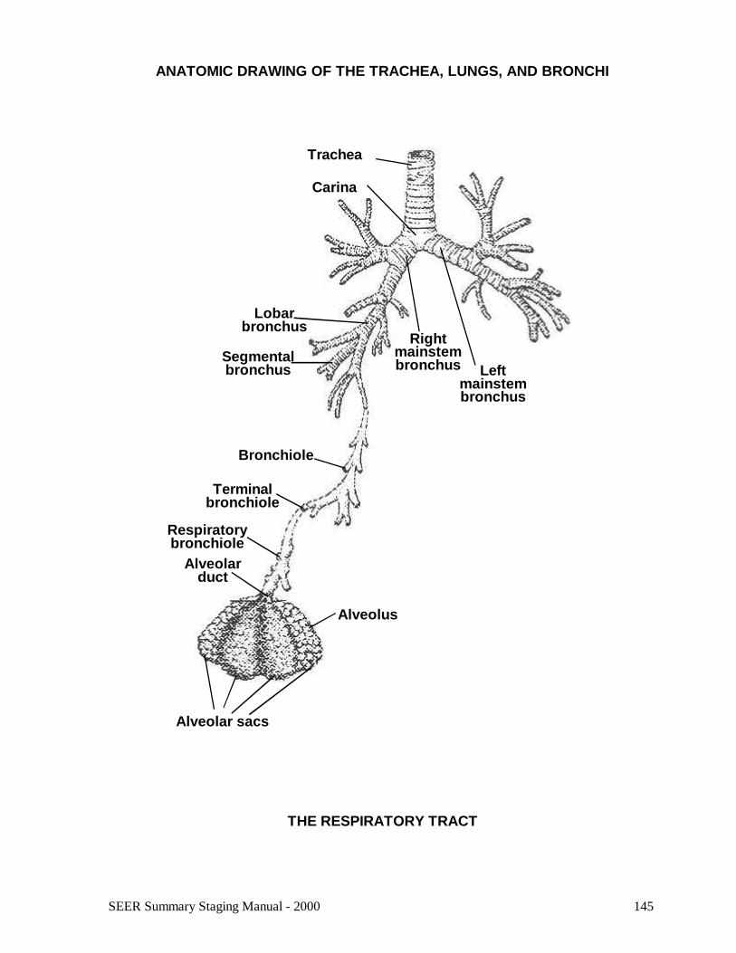
Trachea
Carina
Rightmainstembronchus Left
mainstembronchus
Lobarbronchus
Segmentalbronchus
Bronchiole
Terminalbronchiole
Respiratorybronchiole
Alveolarduct
Alveolus
Alveolar sacs
ANATOMIC DRAWING OF THE TRACHEA, LUNGS, AND BRONCHI
THE RESPIRATORY TRACT

SEER Summary Staging Manual - 2000146
TRACHEAC33.9C33.9 Trachea
SUMMARY STAGE
0 In situ: Noninvasive; intraepithelial
1 Localized only
Invasive tumor confined to trachea
Localized, NOS
2 Regional by direct extension only
Extension to:
Adjacent tissue(s), NOSConnective tissue:
Arch of aortaAzygos vein, rightBrachiocephalic veinCarotid sheathCommon carotid artery(ies)Jugular archPhrenic nervesPretracheal fasciaSubclavian artery(ies)Vagus nerve
Adjacent organs/structure(s):Cricoid cartilageEsophagusPleuraRight and left main bronchiSternumThymusThyroid glandVertebral column

147SEER Summary Staging Manual - 2000
TRACHEAC33.9
3 Regional lymph node(s) involved only
REGIONAL Lymph Nodes
Mediastinal, NOS: Posterior (tracheoesophageal)
ParatrachealPretrachealTracheal, NOS
Regional lymph node(s), NOS
4 Regional by BOTH direct extension AND regional lymph node(s) involved
Codes (2) + (3)
5 Regional, NOS
7 Distant site(s)/lymph node(s) involved
Distant lymph node(s)
Further contiguous extension
Metastasis
9 Unknown if extension or metastasis
SEER Summary Staging Manual - 2000148
BRONCHUS AND LUNGC34.0-C34.3, C34.8-C34.9C34.0 Main bronchus (including carina, hilus of lung) <>C34.1 Upper lobe (including lingula), lung <>C34.2 Middle, lung <>C34.3 Lower lobe, lung <>C34.8 Overlapping lesion of lung <>C34.9 Lung, NOS <><> Laterality must be coded for this site (except carina and hilus of lung).
SUMMARY STAGE
0 In situ: Noninvasive; intraepithelial
1 Localized
Confined to carinaConfined to hilus of lungConfined to the main stem bronchus >2.0 cm from carinaConfined to the main stem bronchus, NOSExtension from other parts of the lung to main stem bronchus >2.0 cm from carina##
Extension from other parts of the lung to main stem bronchus, NOS##
Single tumor confined to one lung
Localized, NOS

149SEER Summary Staging Manual - 2000
BRONCHUS AND LUNGC34.0-C34.3, C34.8-C34.9
2 Regional by direct extension only
Atelectasis/obstructive pneumonitis
Extension to:Blood vessel(s) (major):
Aorta***
Azygos veinPulmonary artery or veinSuperior vena cava (SVC syndrome)
Brachial plexus from superior sulcus###***
Carina from lungChest (thoracic) wall***
Diaphragm***
EsophagusMain stem bronchus <2.0 cm from carinaMediastinum, extrapulmonary or NOSNerve(s):
Cervical sympathetic (Horner’s syndrome)PhrenicRecurrent laryngeal (vocal cord paralysis)Vagus
Pancoast tumor (superior sulcus syndrome)###***
Parietal (mediastinal) pleura***
Parietal pericardium###
Pericardium, NOSPleura, NOSPulmonary ligamentTracheaVisceral pleura
Multiple masses/separate tumor nodule(s) in the SAME lobe#*
Multiple masses/separate tumor nodule(s) in the main stem bronchus
Tumor of main stem bronchus <2.0 cm from carina*
Continued on next page
SEER Summary Staging Manual - 2000150
BRONCHUS AND LUNGC34.0-C34.3, C34.8-C34.9
3 Regional IPSILATERAL regional lymph node(s) involved only
REGIONAL Lymph Nodes
Aortic [above diaphragm], NOS:Peri/para-aortic, NOS:
Ascending aorta (phrenic)Subaortic (aortico-pulmonary window )
BronchialCarinal (tracheobronchial) (tracheal bifurcation)Hilar (bronchopulmonary) (proximal lobar) (pulmonary root)Intrapulmonary, NOS:
InterlobarLobarSegmentalSubsegmental
Mediastinal, NOS:AnteriorPosterior (tracheoesophageal)
PericardialPeri/parabronchialPeri/paraesophagealPeri/paratracheal, NOS:
Azygos (lower peritracheal)Pre- and retrotracheal, NOS:
PrecarinalPulmonary ligamentSubcarinal
Regional lymph node(s), NOS
4 Regional by BOTH direct extension AND IPSILATERAL regional lymph node(s) involved
Codes (2) + (3)
5 Regional, NOS
151SEER Summary Staging Manual - 2000
BRONCHUS AND LUNGC34.0-C34.3, C34.8-C34.9
7 Distant site(s)/node(s) involved
Distant lymph node(s):Cervical, NOSContralateral/bilateral hilar (bronchopulmonary) (proximal lobar) (pulmonary root)Contralateral/bilateral mediastinalScalene (inferior deep cervical), ipsilateral or contralateralSupraclavicular (transverse cervical), ipsilateral or contralateralOther distant lymph node(s)
Extension to:Abdominal organsAdjacent rib##
Contralateral lungContralateral main stem bronchusHeart##
Pericardial effusion (malignant or NOS)Pleural effusion (malignant or NOS)Skeletal muscleSkin of chestSternumVertebra(e)Visceral pericardium##
Further contiguous extension
Separate tumor nodule(s) in different lobe#*
Separate tumor nodule(s) in contralateral lung
Metastasis
9 Unknown if extension or metastasis
Note 1: “Bronchopneumonia” is not the same thing as “obstructive pneumonitis” and should not be coded as such.Note 2: Assume tumor >2 cm from carina if lobectomy, segmental resection, or wedge resection is done.Note 3: If no mention is made of the opposite lung on a chest x-ray, assume it is not involved.Note 4: Ignore pleural effusion which is negative for tumor. Assume that a pleural effusion is negative if a resection is done.Note 5: If at mediastinoscopy/x-ray, the description is “mass,” “adenopathy,” or “enlargement” of the mediastinum or of any ofthe lymph nodes listed under Regional Lymph Nodes (see page 150), assume that at least regional lymph nodes are involved.Note 6: The words “no evidence of spread” and/or “remaining examination negative” are sufficient information to considerregional lymph nodes negative in the absence of any statement about nodes.Note 7: “Vocal cord paralysis,” “superior vena cava syndrome,” and “compression of the trachea or the esophagus” are classifiedas mediastinal lymph node involvement unless there is a statement of involvement by direct extension from the primary tumor.
# Considered localized in Historic Stage## Considered regional in Historic Stage### Considered distant in Historic Stage* Considered localized in 1977 Summary Staging Guide*** Considered distant in 1977 Summary Staging Guide

SEER Summary Staging Manual - 2000152
HEART, MEDIASTINUMC38.0-C38.3, C38.8C38.0 HeartC38.1 Anterior mediastinumC38.2 Posterior mediastinumC38.3 Mediastinum, NOSC38.8 Overlapping lesion of heart, mediastinum and pleura
Note: AJCC includes these sites with soft tissue sarcomas (C47.0-C48.9)
SUMMARY STAGE
1 Localized only
Invasive tumor confined to site of origin
Localized, NOS
2 Regional by direct extension only
Extension to:Adjacent tissue(s), NOSConnective tissueSee definition of connective tissue on page 14.
Adjacent organs/structures:See definition of adjacent organs and structures on page 14.
Heart:Ascending aortaPericardium, NOS:
ParietalVisceral (epicardium)
Vena cavaMediastinum:
Descending aortaEsophagusLarge (named) artery(ies)Large (named) vein(s)Pericardium, NOS:
ParietalVisceral (epicardium)
Phrenic nerve(s)Pleura, NOS:
ParietalVisceral
SternumSympathetic nerve trunk(s)Thoracic ductThymusTracheaVertebra(e)
153SEER Summary Staging Manual - 2000
HEART, MEDIASTINUMC38.0-C38.3, C38.8
3 Regional lymph node(s) involved only
REGIONAL Lymph Nodes
Aortic [above diaphragm], NOS:Peri/para-aortic, NOS:
Ascending aorta (phrenic)Subaortic (aortico-pulmonary window)
Carinal (tracheobronchial) (tracheal bifurcation)Mediastinal, NOS:
AnteriorPosterior (tracheoesophageal)
PericardialPeri/paraesophagealPeri/paratracheal, NOS:
Azygos (lower peritracheal)Pre- and retrotracheal, NOS:
PrecarinalPulmonary ligamentSubcarinal
Regional lymph node(s), NOS
4 Regional by BOTH direct extension AND regional lymph node(s) involved
Codes (2) + (3)
5 Regional, NOS
7 Distant site(s)/lymph node(s) involved
Distant lymph node(s)
Further contiguous extension
Metastasis
9 Unknown if extension or metastasis
Note: Code 0 is not applicable for this scheme.

SEER Summary Staging Manual - 2000154
PLEURAC38.4C38.4 Pleura, NOS (including visceral and parietal) <><> Laterality must be coded for this site.
SUMMARY STAGE
1 Localized only
Invasive tumor (mesothelioma) confined to pleuraIpsilateral parietal and/or visceral pleuraMesothelioma WITH nodule(s) beneath visceral pleural surface
Localized, NOS
2 Regional by direct extension only
Extension to adjacent organs/structure:Chest wallConnective tissueDiaphragmEndothoracic fasciaHeart muscleLung involvement, NOSMediastinal organs or tissuesPericardiumRib
Mesothelioma nodule(s) which have broken through the visceral pleural surface to lung surface
Continued on next page

155SEER Summary Staging Manual - 2000
PLEURAC38.4
3 Regional IPSILATERAL lymph nodes involved only
REGIONAL Lymph Nodes
Aortic [above diaphragm], NOS:Peri/para-aortic, NOS:
Ascending aorta (phrenic)Subaortic (aortico-pulmonary window)
Carinal (tracheobronchial) (tracheal bifurcation)Hilar (bronchopulmonary) (proximal lobar) (pulmonary root)Intrapulmonary, NOS:
InterlobarLobarSegmentalSubsegmental
Mediastinal, NOS:AnteriorPosterior (tracheoesophageal)
PericardialPeri/parabronchialPeri/paraesophagealPeri/paratracheal, NOS:
Azygos (lower peritracheal)Pre- and retrotracheal, NOS:
PrecarinalPulmonary ligamentSubcarinal
Regional lymph node(s), NOS
4 Regional by BOTH direct extension AND regional IPSILATERAL lymph node(s) involved
Codes (2) + (3)
5 Regional, NOS
Continued on next page

SEER Summary Staging Manual - 2000156
PLEURAC38.4
7 Distant site(s)/node(s) involved
Distant lymph node(s):Contralateral/bilateral hilar (bronchopulmonary) (proximal lobar) (pulmonary root)Contralateral/bilateral mediastinalScalene (inferior deep cervical), ipsilateral or contralateralSupraclavicular (transverse cervical), ipsilateral or contralateralOther distant lymph node(s)
Extension to:Cervical (neck) tissuesContralateral lungContralateral pleuraIntra-abdominal organsPeritoneum
Mesothelioma WITH malignant pleural fluidPleural effusion
Further contiguous extension
Metastasis
9 Unknown if extension or metastasis
Note 1: Code 0 is not applicable for this scheme.Note 2: Ignore pleural effusion which is negative for tumor.

157SEER Summary Staging Manual - 2000

SEER Summary Staging Manual - 2000158
OTHER AND ILL-DEFINED RESPIRATORYSITES AND INTRATHORACIC ORGANSC39.0, C39.8-C39.9C39.0 Upper respiratory tract, NOSC39.8 Overlapping lesion of respiratory system and intrathoracic organsC39.9 Ill-defined sites within respiratory system
SUMMARY STAGE
1 Localized only
Invasive tumor confined to site of origin
Localized, NOS
2 Regional by direct extension only
Extension to:Adjacent tissue(s), NOSConnective tissue
See definition of connective tissue on page 14.
Adjacent organs/structures:Descending aortaEsophagusLarge (named) artery(ies)Large (named) vein(s)Pericardium, NOS:
ParietalVisceral (epicardium)
Phrenic nerve(s)Pleura, NOS:
ParietalVisceral
SternumSympathetic nerve trunk(s)Thoracic ductThymusTracheaVertebra(e)

159SEER Summary Staging Manual - 2000
OTHER AND ILL-DEFINED RESPIRATORYSITES AND INTRATHORACIC ORGANSC39.0, C39.8-C39.9
3 Regional lymph node(s) involved only
REGIONAL Lymph Nodes
Aortic [above diaphragm], NOS:Peri/para-aortic, NOS:
Ascending aorta (phrenic)Subaortic (aortico-pulmonary window)
Carinal (tracheobronchial) (tracheal bifurcation)Hilar (bronchopulmonary) (proximal lobar) (pulmonary root)Intrapulmonary, NOS:
InterlobarLobarSegmentalSubsegmental
Mediastinal, NOS:AnteriorPosterior (tracheoesophageal)
PericardialPeri/parabronchialPeri/paraesophagealPeri/paratracheal, NOS:
Azygos (lower peritracheal)Pre- and retrotracheal, NOS:
PrecarinalPulmonary ligamentSubcarinal
Regional lymph node(s), NOS
4 Regional by BOTH direct extension AND regional lymph node(s) involved
Codes (2) + (3)
5 Regional, NOS
7 Distant site(s)/lymph node(s) involved
Distant lymph node(s)
Further contiguous extension
Metastasis
9 Unknown if extension or metastasis
Note: Code 0 is not applicable for this scheme.
Recommended