Fetal AnomaliesAnita Nowak, RDMS, MBA
Manager, ImagingMagee-Womens Hospital of UPMC
AnencephalySpina BifidaCleft LipGastroschisis/OmphaloceleTrisomy 18Conjoined Twins
Common Fetal Anomalies
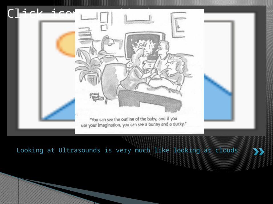
Looking at Ultrasounds is very much like looking at clouds
Click icon to add picture
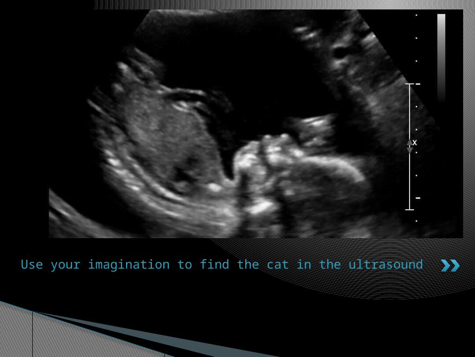
Use your imagination to find the cat in the ultrasound
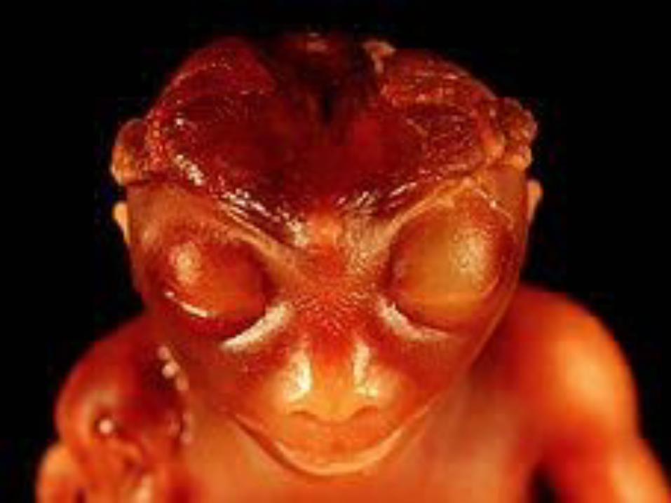
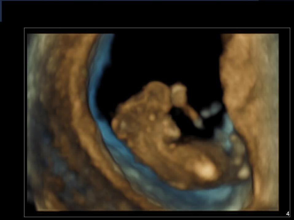
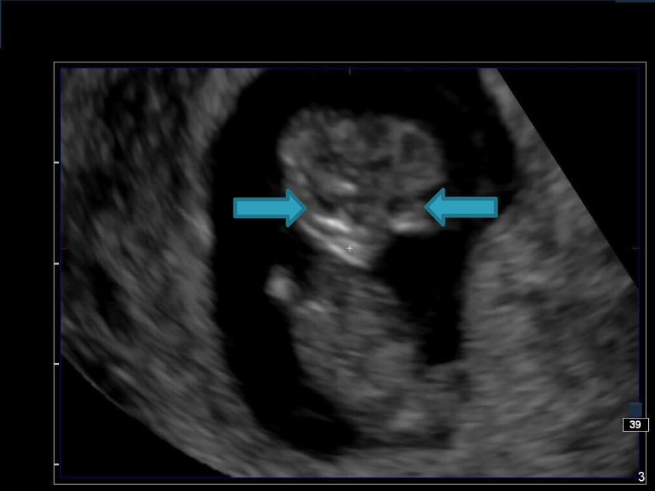
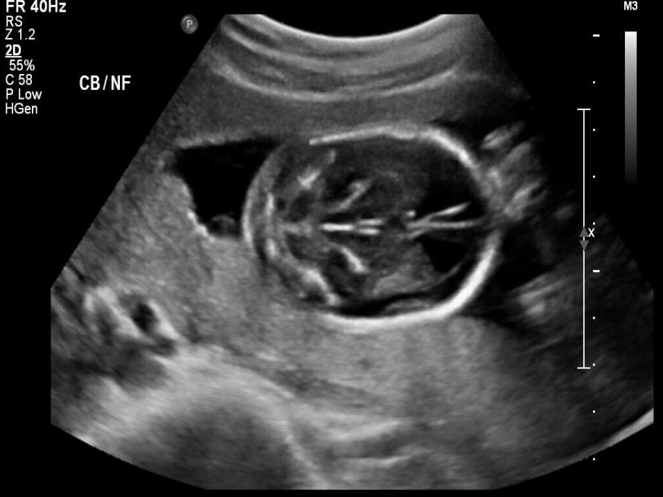
Anencephaly The absence of the cranial vault
If early in pregnancy, brain tissue can be seenHead has an irregular shapeThere is no soft tissue seen above the orbitsFace – eyes appear “frog like”
Ultrasound Findings
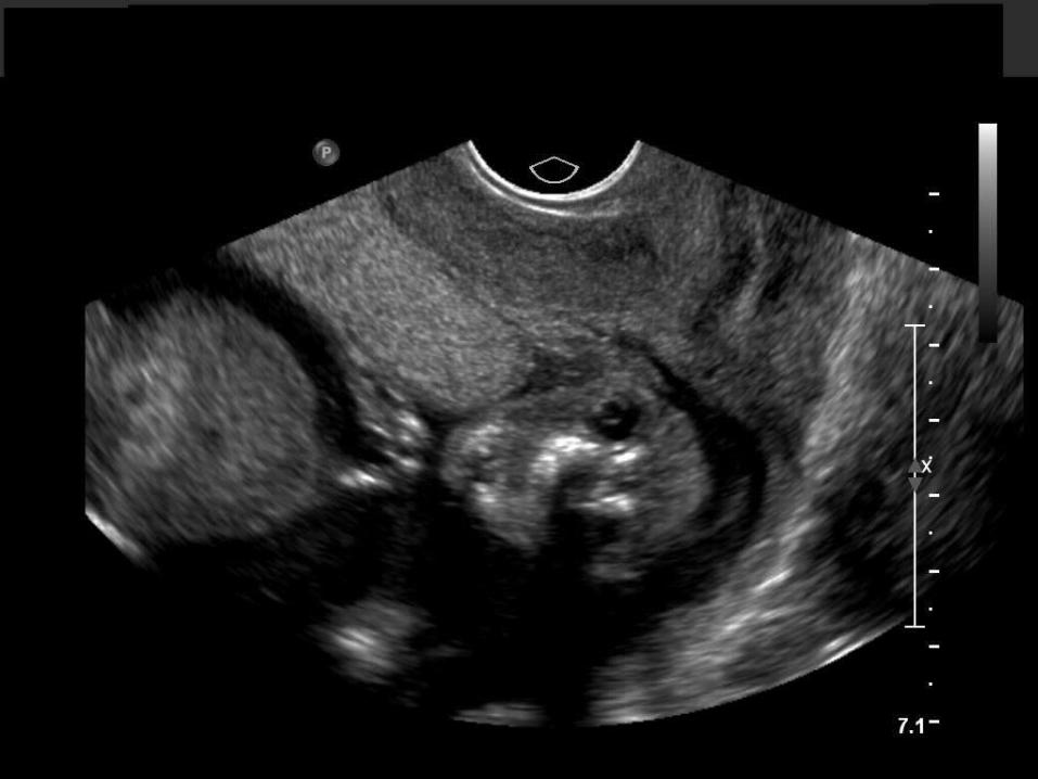
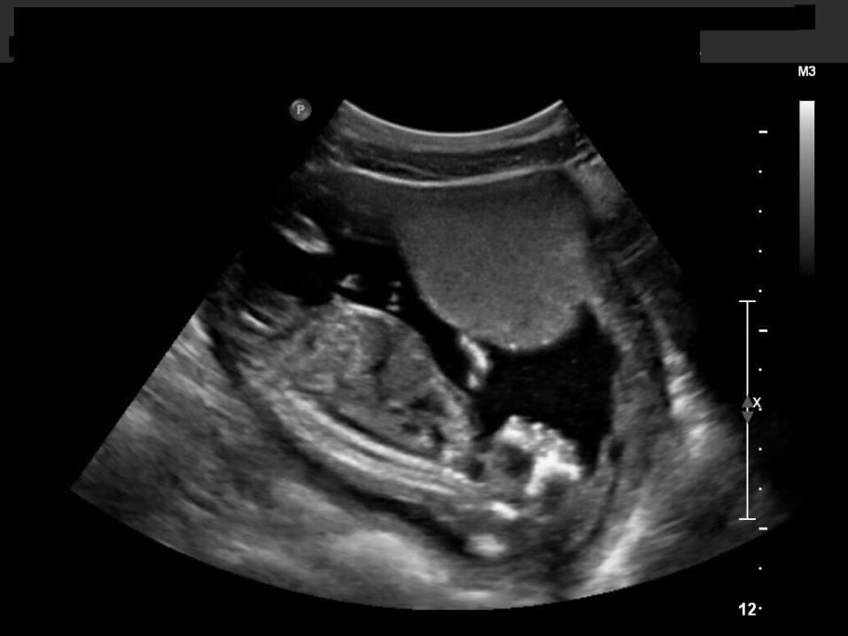
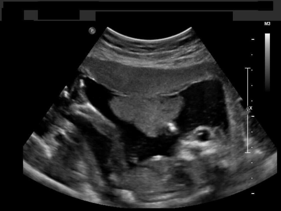
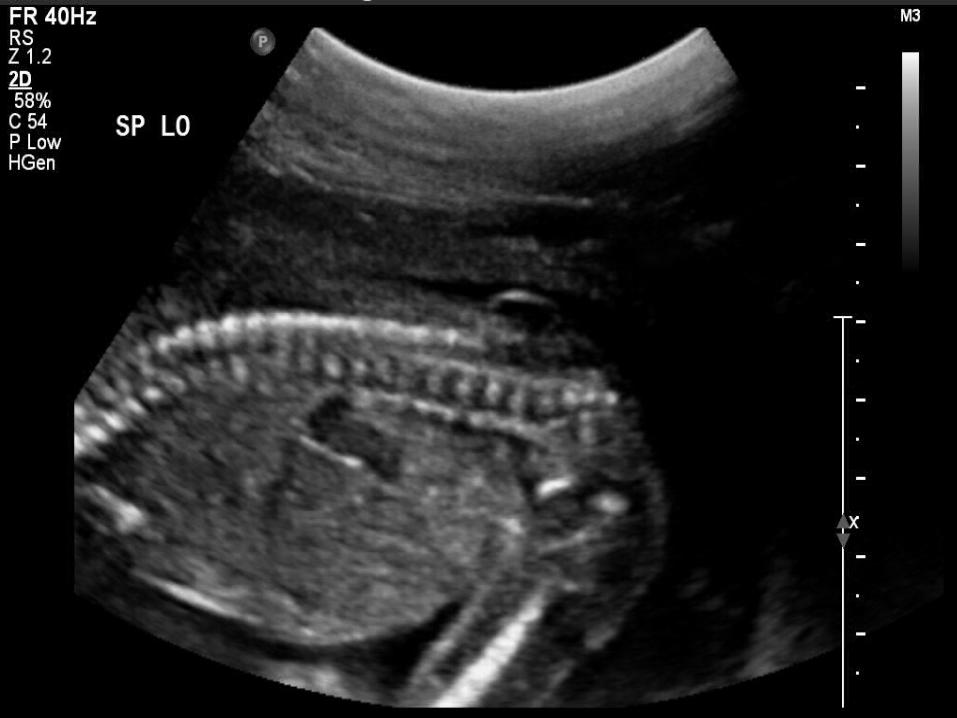
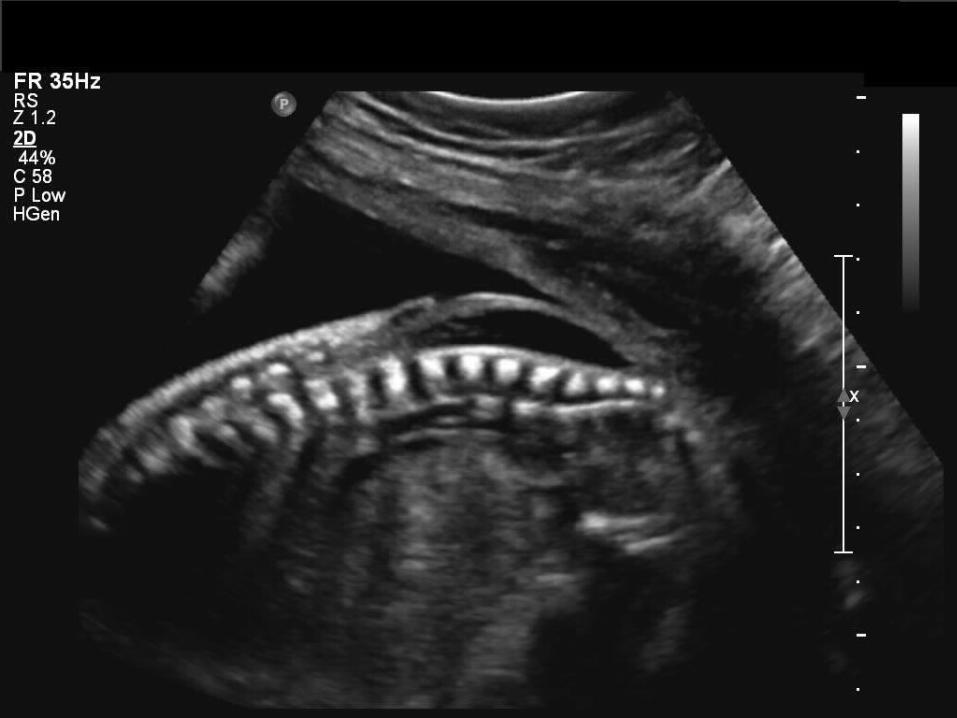
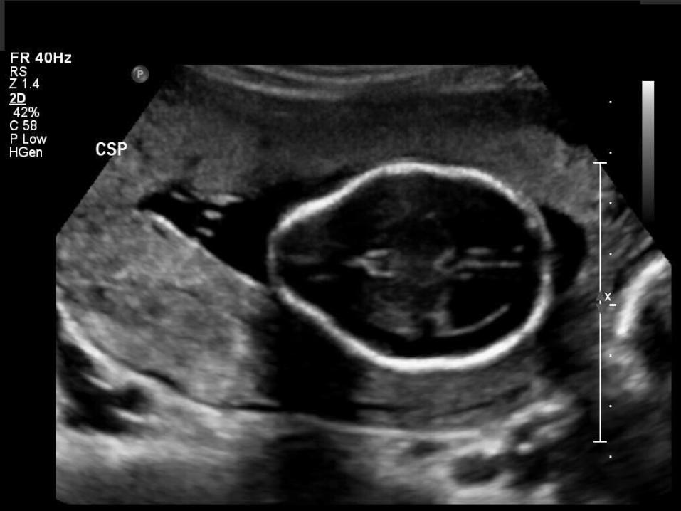
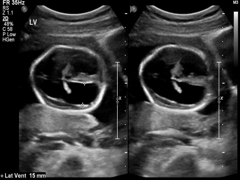
Spina Bifida There are many forms of neural tube
defects, Spina Bifida is the most common of the central nervous system
A midline defect of the vertebrae that results in exposure of the contents of the neural canal
Can be genetic
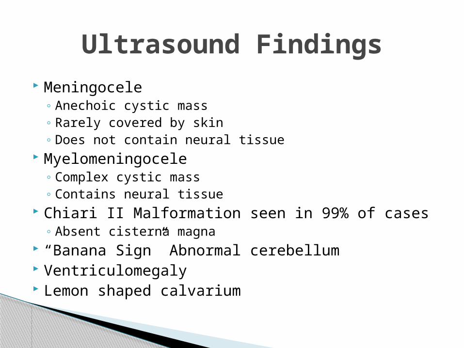
Meningocele◦ Anechoic cystic mass◦ Rarely covered by skin◦ Does not contain neural tissue
Myelomeningocele ◦ Complex cystic mass◦ Contains neural tissue
Chiari II Malformation seen in 99% of cases◦ Absent cisterna magna
“Banana Sign” Abnormal cerebellum Ventriculomegaly Lemon shaped calvarium
Ultrasound Findings
Cleft Lip 2nd most common congenital malformation Estimated to be 1:700 live births 50% both lip and palate are defective Can be caused by both genetic and
environmental factors 97% of the time it is an isolated finding Occurs shortly after 3rd week of gestation when
the grooves that separate the structures that form the primitive oral cavity persist, they would normally be obliterated by normal growth.
Most commonly seen is a unilateral cleft
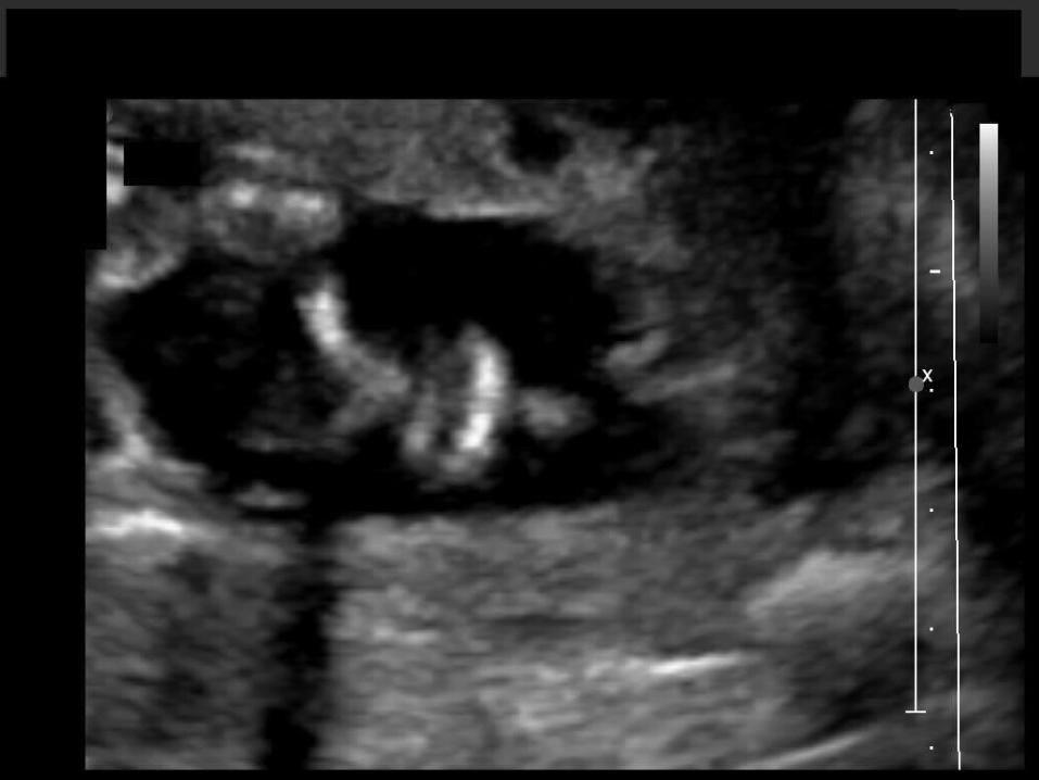
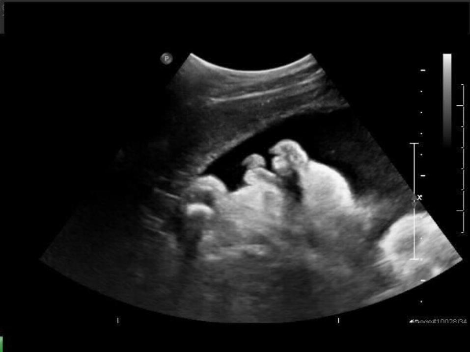
Upper lip defect on nose/mouth view
Ultrasound Findings
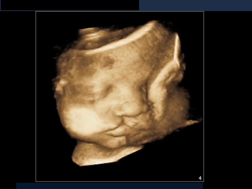
Gastroschisis Gastroschisis is a paraumbilical defect of
the anterior abdominal wall. Incidence ranges from 1:10,000 to 1:15,000 Is not associated with an increased risk of
other anomalies Not usually associated with a chromosomal
abnormality
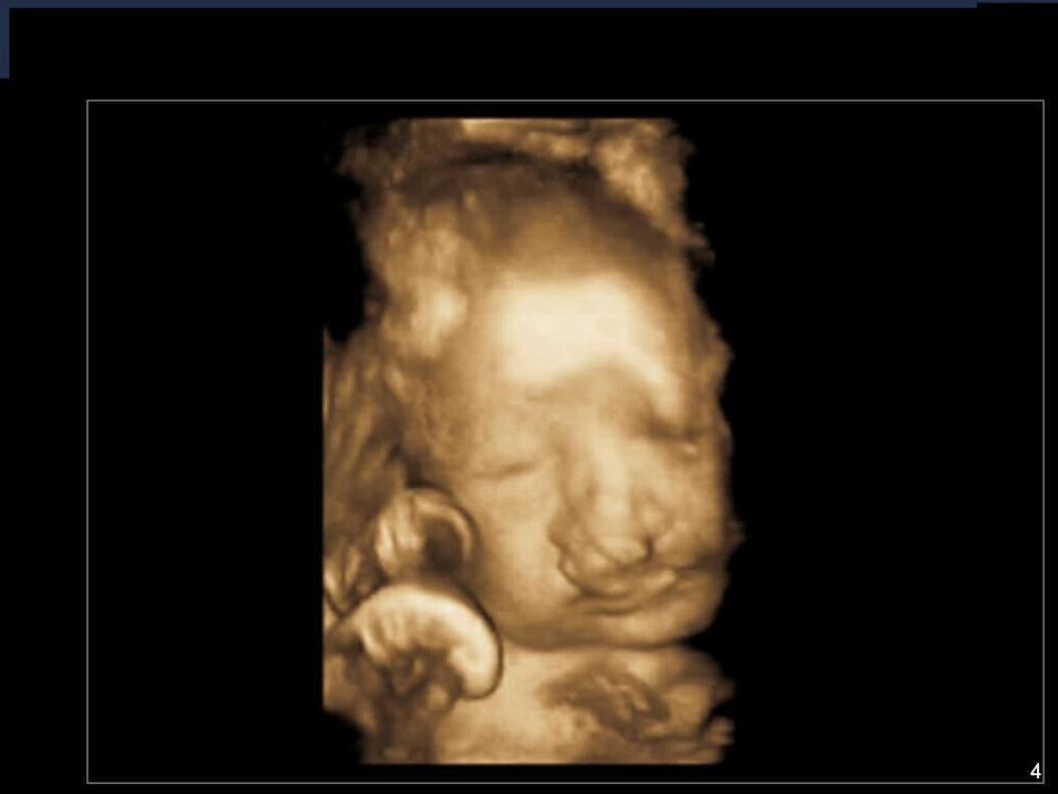
Normal umbilical cord insertion site Small bowel loops seen in the amniotic
cavity No covering membrane over the loops of
bowel Can include stomach and large bowel Majority occur to the right of the umbilical
cord
Ultrasound Findings
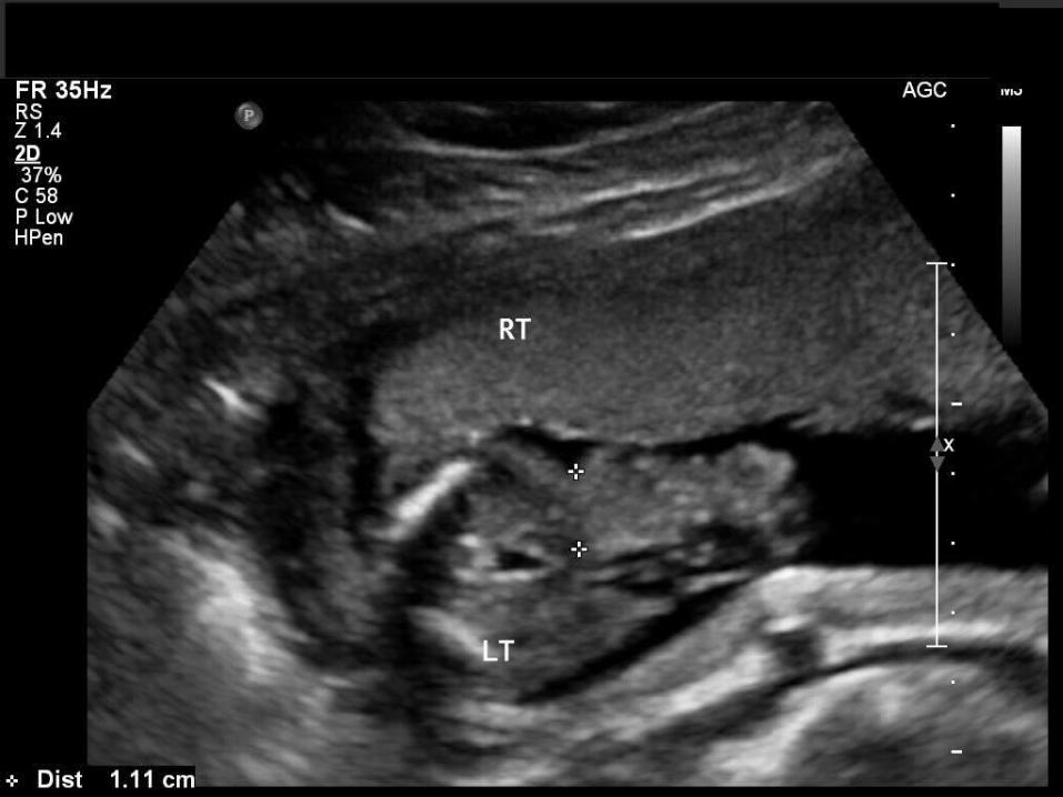
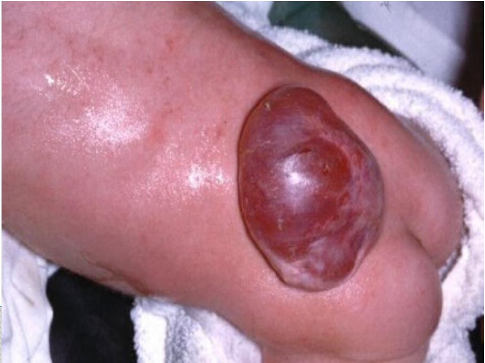
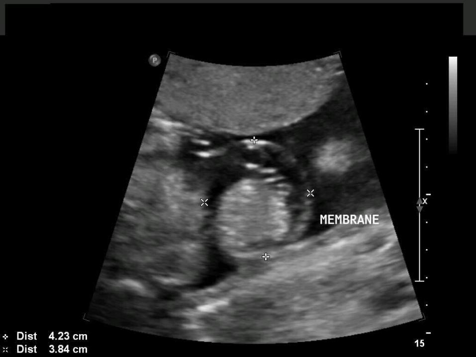
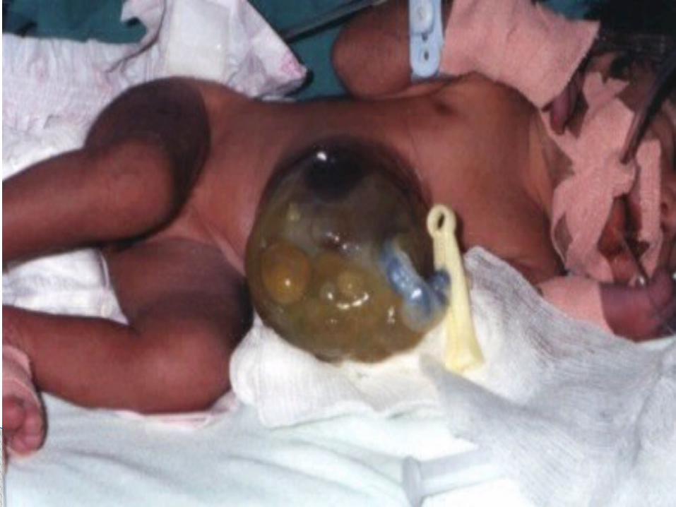
A ventral wall defect where there is herniation of the intraabdominal contents into the base of the umbilical cord
Unlike gastroschisis, there is a membrane covering these contents
Estimated to occur in 1:5800 to 1:5130 Most cases are sporadic Unlike gastroschisis this condition IS often
associated with a chromosomal abnormality
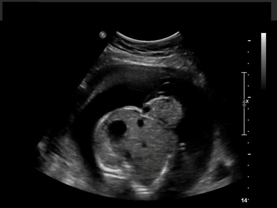
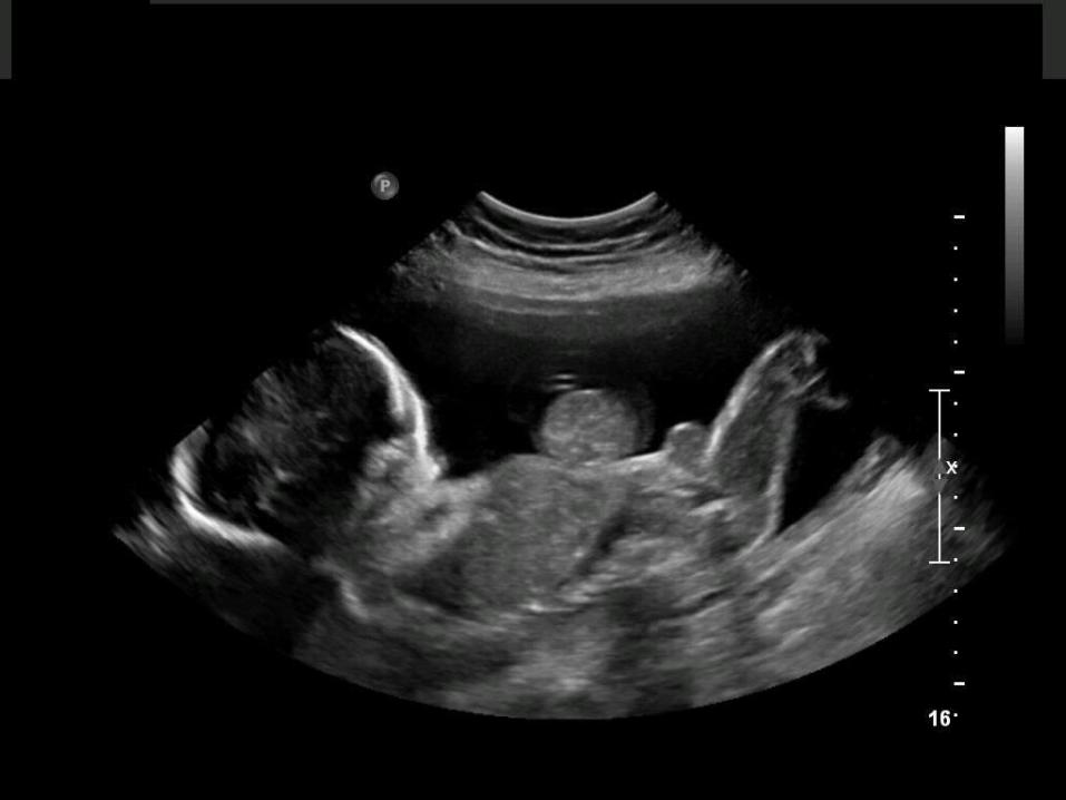
Omphalocele
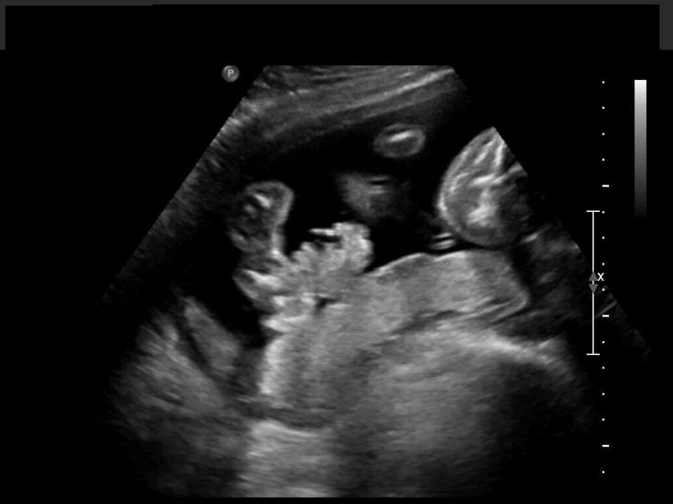
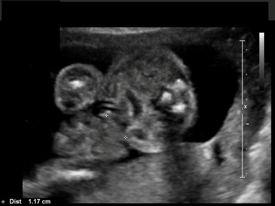
Umbilical cord insertion is typically midline on the mass
Located centrally Typically the contents of the mass are liver
and small bowel; however, other abdominal organs can be present
Ultrasound Findings
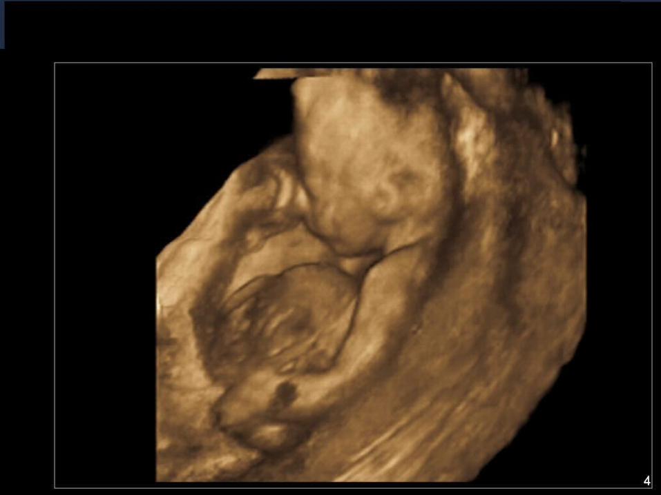
Trisomy 18 Also called Edwards Syndrome There are three 18th chromosomes instead
of two Multiple major anomalies are seen Occurs in approximately 1:2500
pregnancies 50% carried to term will be stillborn Of those that survive, only 10% survive to
their first birthday Not genetic – typically occur sporadically
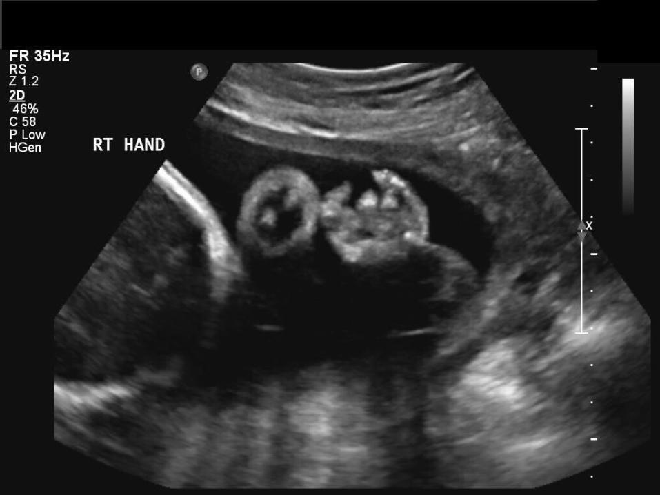
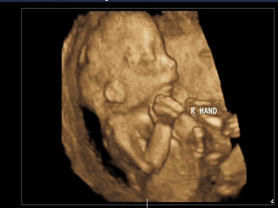
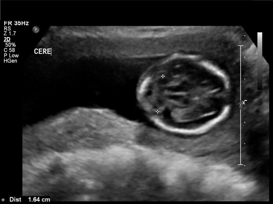
Clenched Hands Choroid plexus cysts “Strawberry” shaped head Intrauterine growth restriction Cardiac defects Micrognathia Low set ears
Ultrasound Findings
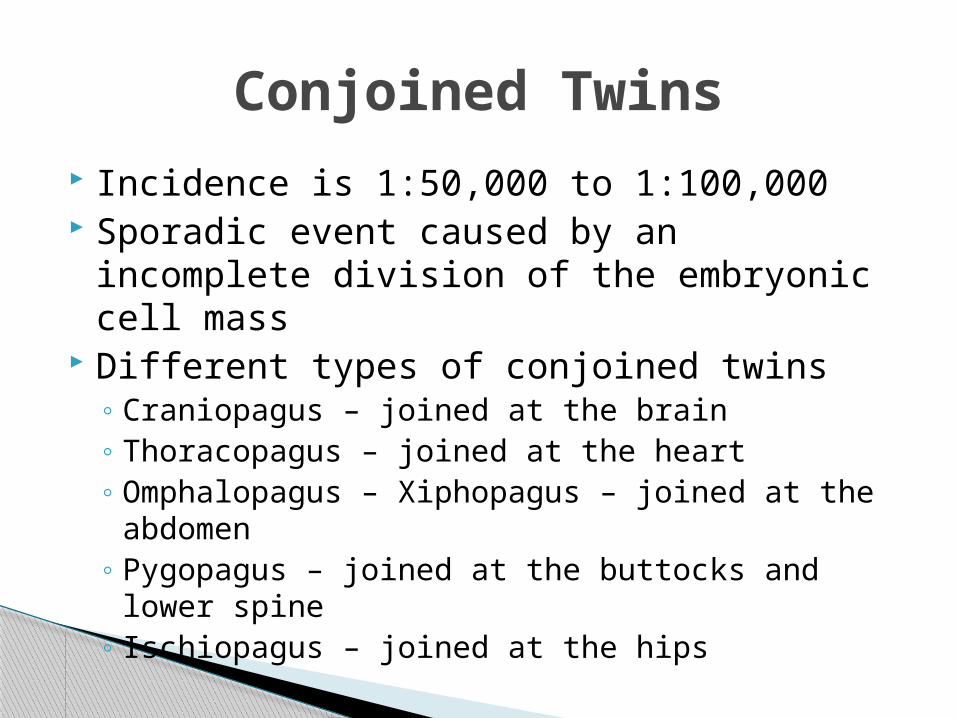
Conjoined Twins Incidence is 1:50,000 to 1:100,000 Sporadic event caused by an incomplete
division of the embryonic cell mass Different types of conjoined twins
◦ Craniopagus – joined at the brain◦ Thoracopagus – joined at the heart◦ Omphalopagus – Xiphopagus – joined at the
abdomen◦ Pygopagus – joined at the buttocks and lower
spine◦ Ischiopagus – joined at the hips
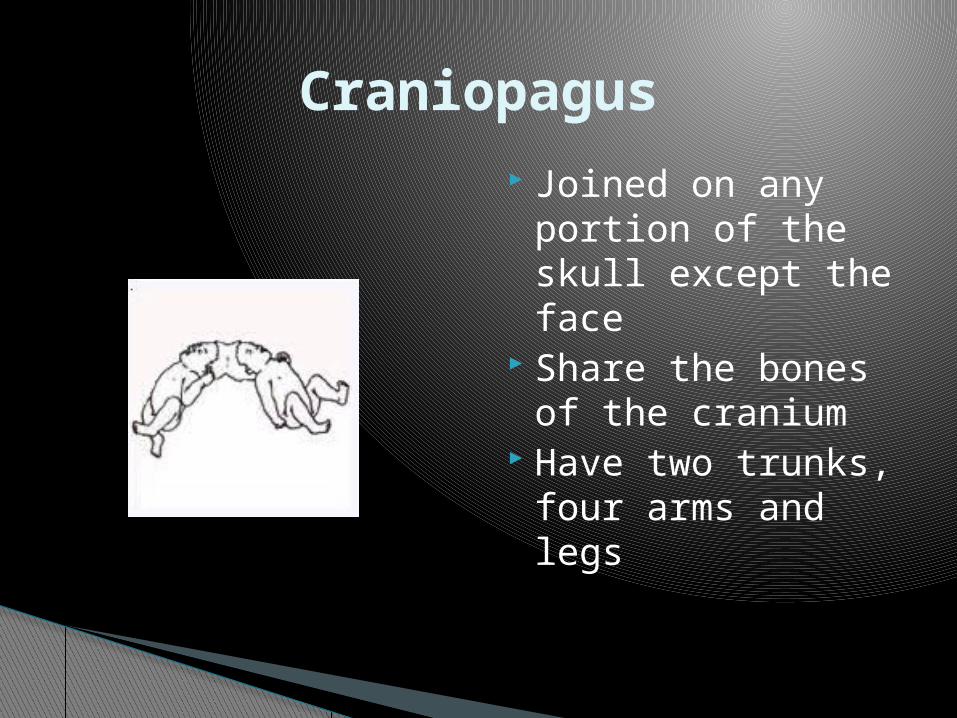
Joined on any portion of the skull except the face
Share the bones of the cranium
Have two trunks, four arms and legs
Craniopagus
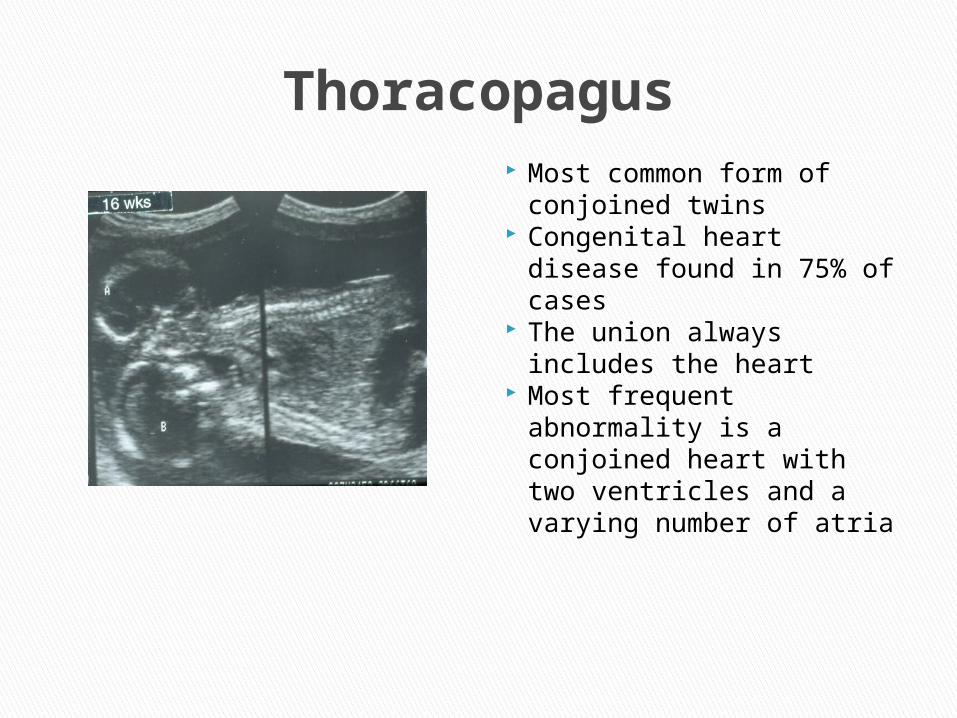
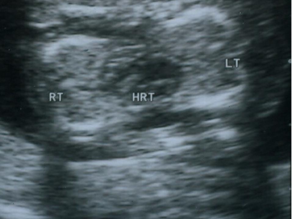
Thoracopagus Most common form of
conjoined twins Congenital heart disease
found in 75% of cases The union always
includes the heart Most frequent
abnormality is a conjoined heart with two ventricles and a varying number of atria

Omphalopagus in the first trimester
Attached in the lower abdomen
Remain facing each other throughout the exam

Joined at the buttocks and lower spine
Face away from each other
Have one anus, two rectums, four arms and legs
Pygopagus

Joined end to end with the spine in a straight line
Four arms and a variable number of legs
Have only one external genitalia
Ischiopagus

Questions??
Recommended