1
ASSESSING THE EYES
Structures
External
� Eyelids
� Extraocular muscles
� Eyelashes
� Lacrimal glands: Lacrimal ducts
� Cornea
� Conjunctiva
� Sclera
� Pupils
� Iris

2

3
Structures
Internal
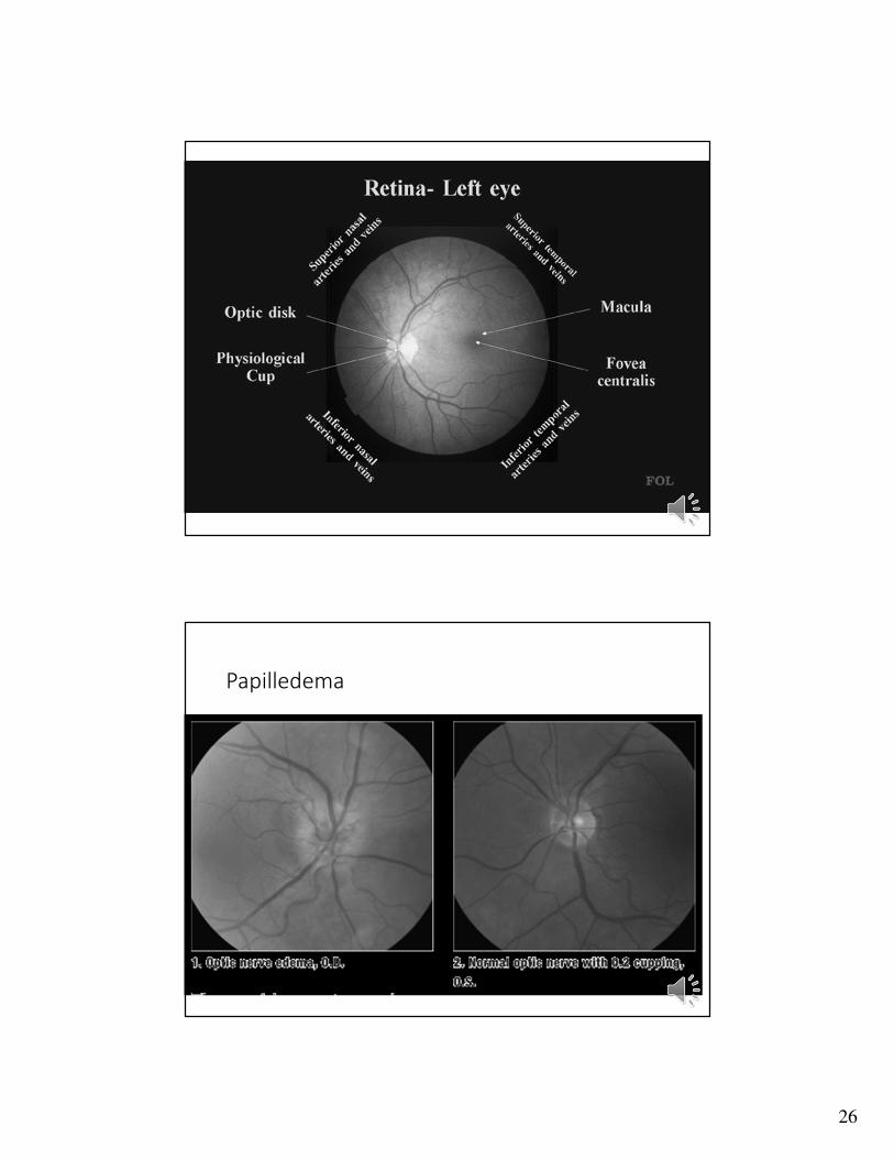
• Optic disc
• Physiological cup
• Retinal arteries
• Retinal veins
• Retina
• Macula

4
Characteristics of normal eye
• Symmetrical
• Full lashes/brows
• Distance between eyelids equal (palpebral fissure)
• Lids w/o edema/lag-‘ptosis’
• Conjunctiva pink or clear, w/o swelling, nodules, or drainage
Physical Assessment�Anatomical Landmarks: visual fields (superior, inferior, nasal, temporal)
�Approach: inspection, palpation, ophthalmoscopy
�Position: sitting
�Tools: visual acuity charts (Snellen), penlight, ophthalmoscope, cotton ball, cotton swab

5
Assessment of the Eye
• Assess Structure and Function:
• Color Vision
• Visual Acuity
• External Eye structures
• Extraocular Muscles
• Visual Fields
• Internal Eye structures
Visual Acuity
Far vision:
Snellen eye chart
Near vision:
read newsprint 13 to 15” from eyes
Color vision:
identify color bars on Snellen or use color plates, or tabs by room doors

6

7
Inspection of External Structures
�Lids and lashes: �color, lesions, edema, symmetry, position and
distribution of lashes
� Lacrimal glands and ducts: �color, edema, excessive tearing or drainage
�Conjunctiva: �color, moisture, lesions, and foreign bodies
�Sclera: �color, moisture, lesions,
Inspection of External Structures
�Cornea: �clarity and abrasions, corneal reflex
�Iris: �Color, size, shape, and symmetry
�Pupils: �Size, shape,
�Reaction to light–direct and consensual
�Test accommodation (CN III)

8

9
Palpation of External Structures
Eye ball: consistency and tenderness
Lacrimal glands and ducts: tenderness and excessive tearing
Eyelids: consistency, masses
� EOM◦ Checking the six cardinal fields of gaze by asking patient to follow your finger with eyes only in a big ‘H’
� Visual fields by confrontation◦ Two methods—when see red pin
◦ Wiggle fingers

10

11
� Darken the room and have the patient look off in the distance
� Switch the ophthalmoscope light and turn the lens disc to the large round beam of white light
� Turn lens disc to the 0 diopter
� Hold the ophthalmoscope in your right hand to examine the patient’s right eye with your right eye; hold it in your left hand to examine the patient’s left eye with your left eye
� Stand directly in front of the patient, 15 inches away, and start at an angle of 15 degrees lateral to the patient’s line of vision
� Shine the beam of light onto the pupil and look for an orange glow; this is the red reflex
� Follow the red reflex and move inward towards the nasal aspect of the visual field
� Red reflex: presence, opacities
� Optic disc and physiologic cup:
�color, size, shape, borders, cup-disc ratio
� Retinal vessels:
�size ratio of arteries and veins, color, arteriole light reflex, crossings
� Retina:
�color, texture, exudates, lesions, hemorrhages, and aneurysms
� Macula and fovea:
� color, size, location, lesions

12

13
Entropion
Ectropion
Chalazion
Hordoleum
Xanthelasma

14
Abnormalities of the eyelids
• Entropion—eyelids and lashes turn in
• Ectropion—eyelids and lashes turn out
• Chalazion—meibomian gland cyst
• Hordeolum (sty)—inflammation or infection of the eyelid margin
• Xanthelasma—cholesterol deposits
Entropion

15
Entropion (lower lid)
Ectropion

16
Ectropion
Chalazion

17
Sty--hordeoleum

18
Xanthelasma
Xanthelasma

19
EXTERNAL EYE DISORDERSArcus Senilis
Cataracts
Pterygium
Pinguecula
CHANGES WITH AGING
• Visual acuity decreases
• Presbyopia
• Altered structures
• Arcus senilus--gray or white arc visible above and below the outer part of the cornea
• Cataracts (some are congenital)
• Pupils decrease in size
• EOMs may have upward gaze impairment

20
Arcus Senilus
Cataract

21
Congenital Cataract
Pterygium

22
Pinguecula
EOM disordersStrabismus
Amblyopia
Esotropia
Exotropia
Tests: Cover test
Hirschbergs
EOMs

23
Strabismus
•Esotropia__eye(s) turn to nose
•Exotropia—eye(s) turn outward or temporal
•Pediatrics: • At birth if fixed esotropia or exotropia—refer
• If intermittent can hold on referral until 12 months
• Generally eyes align together by 4 months
•Tests: EOMs• Cover/uncover
• Hirschberg’s/corneal light reflex
Significant strabismus

24
Ambylopia—Decreased vision in one eye or both eyes—usually due to strabismus or refractive error
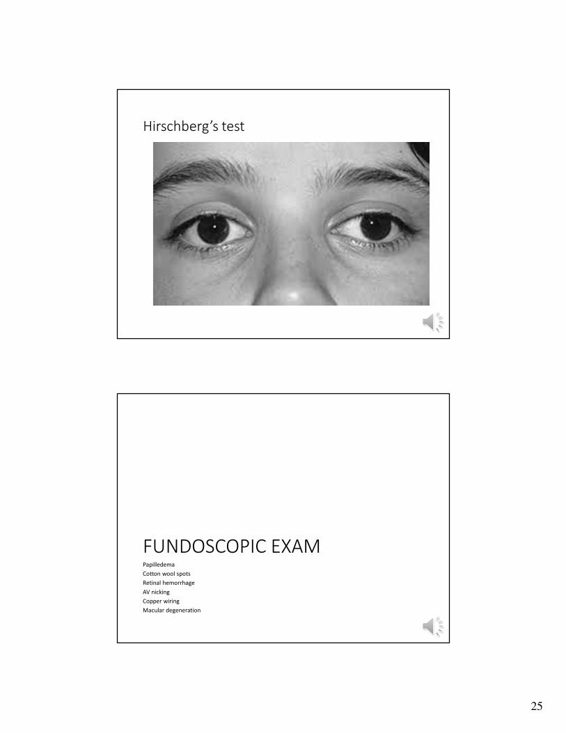
25
Hirschberg’s test
FUNDOSCOPIC EXAMPapilledema
Cotton wool spots
Retinal hemorrhage
AV nicking
Copper wiring
Macular degeneration
26
Papilledema

27
Papilledema
Cotton Wool Spots

28
Cotton wool spots
•Small areas of yellowish white coloration in the retina
•Occur because the blood supply through the retinal vessels and the nerve fibers are injured resulting in swelling and the appearance of a "cotton wool spot. "
•Most common cause diseases that affect retina blood supply•Diabetes
•Hypertension
Microaneurysms

29
Hemorrhages
Retinal Hemorrhage

30
Copper wiring Nicking
Macular Degeneration

31
Macular Degener.
Documentation
• Eyelids, lashes, and eyebrows with normal hair distribution and without lesions
• Sclera clear, conjunctiva pink with no lesions exudate or inflammation
• PERRLA, EOMs intact,
• Visual fields by confrontation intact bilaterally
• Cover/Uncover test negative
• Hirschberg test negative
• Fundi with sharp disc margins
Recommended

![EYES : Meningitis, eyes, inflamed, wild, staring, pupils ...homoeopathybooks.com/Repertory of Concomitant... · Difficult, breathing, closing, eyes, on : Carb-v., Carb-an. EYES :[CONSTITUTION]](https://img.pdfslide.net/doc/110x75/5a9a0bd77f8b9aba4a8d6b57/eyes-meningitis-eyes-inflamed-wild-staring-pupils-of-concomitantdifficult.jpg)

![26.AH, THOSE BLACK EYES! (Akh, Eli Chiornye Glaza)DARK EYES (Ochi Chernye) Dark eyes, passionate eyes, Fiery and beautiful eyes; How I love you, how] fear you, Evidently I met you](https://img.pdfslide.net/doc/110x75/60adc1b670fe6b0a8514df75/26ah-those-black-eyes-akh-eli-chiornye-glaza-dark-eyes-ochi-chernye-dark.jpg)