CLINICAL CASE REPORT
Autosomal recessive bestrophinopathy associatedwith angle-closure glaucoma
C. Crowley • R. Paterson • T. Lamey •
T. McLaren • J. De Roach • E. Chelva •
J. Khan
Received: 16 December 2013 / Accepted: 12 May 2014 / Published online: 24 May 2014
� The Author(s) 2014. This article is published with open access at Springerlink.com
Abstract
Purpose Abnormalities in the BEST1 gene have
recently been recognised as causing autosomal recessive
bestrophinopathy (ARB). ARB has been noted to have a
variable phenotypic presentation, distinct from that of
autosomal dominant Best vitelliform macular dystrophy
(BVMD). Both conditions are associated with deposits in
the retina, a reduced or absent electro-oculography
(EOG) light rise, and the risk of developing angle-
closure glaucoma. Herein, we describe the clinical and
genetic characteristics of a young male diagnosed with
ARB associated with angle-closure glaucoma resulting
from a novel homozygous mutation in BEST1.
Methods All research involved in this case adhered to
the tenets of the Declaration of Helsinki. The proband
underwent slitlamp examination, retinal autofluorescence
imaging and optical coherence tomography after pre-
senting with deteriorating vision. The findings prompted
genetic testing with bi-directional DNA sequencing of
coding and flanking intronic regions of BEST1. The
proband’s family members were subsequently screened.
Results A provisional diagnosis of ARB was made
based on the findings of subretinal and schitic lesions
on fundoscopy and retinal imaging, together with
abnormal EOG and electroretinography. Genetic test-
ing identified a novel homozygous mutation in BEST1,
c.636?1 G[A. Family members were found to carry
one copy of the mutation and had no clinical or
electrophysiological evidence of disease. The proband
was additionally diagnosed with angle-closure glau-
coma requiring topical therapy, peripheral iridotomies
and phacoemulsification.
Conclusions Phenotypic overlap, reduced pene-
trance, variable expressivity and the ongoing discov-
ery of new forms of bestrophinopathies add to the
difficulty in distinguishing these retinal diseases. All
patients diagnosed with ARB or BVMD should be
examined for narrow angles and glaucoma, given their
frequent association with these conditions.
Keywords BEST1 gene � Autosomal recessive
bestrophinopathy � Best vitelliform macular
dystrophy � Angle-closure glaucoma �Electroretinography � Electro-oculography �Intraocular pressure
Introduction
Autosomal recessive bestrophinopathy (ARB) has
recently been described and has a more global influence
on eye development and physiology than autosomal
C. Crowley (&) � R. Paterson � T. Lamey �T. McLaren � J. De Roach � E. Chelva � J. Khan
Department of Medical Technology and Physics, Sir
Charles Gairdner Hospital, Hospital Avenue, Nedlands,
WA 6009, Australia
e-mail: [email protected]
T. Lamey � J. Khan
Centre for Ophthalmology and Visual Science, University
of Western Australia, Nedlands, WA 6009, Australia
123
Doc Ophthalmol (2014) 129:57–63
DOI 10.1007/s10633-014-9444-z
dominant Best vitelliform macular dystrophy (BVMD),
otherwise known as Best disease. We present a case of
ARB associated with narrow-angle glaucoma and a
novel homozygous mutation in BEST1.
Case description
A 26-year-old male presented with deteriorating
vision in both eyes. He had no family history of
eye disorders but had been diagnosed with
possible Stargardt disease at the age of 12 years.
This diagnosis was amended to exudative poly-
morphous vitelliform macular dystrophy two
years later when he underwent electrophysiolog-
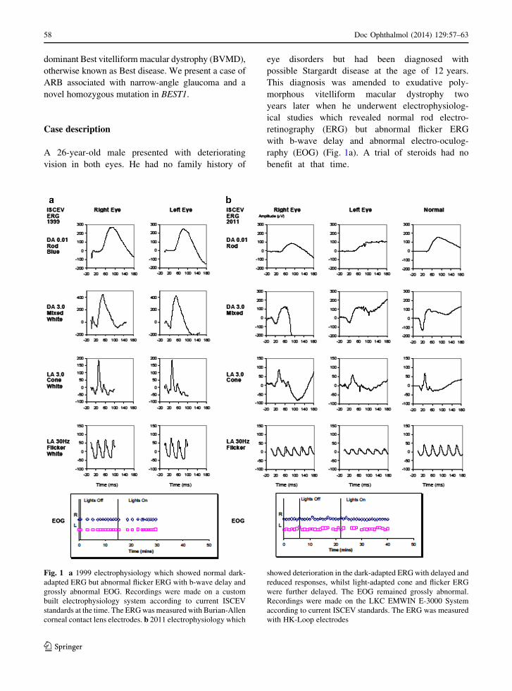
ical studies which revealed normal rod electro-
retinography (ERG) but abnormal flicker ERG
with b-wave delay and abnormal electro-oculog-
raphy (EOG) (Fig. 1a). A trial of steroids had no
benefit at that time.
Fig. 1 a 1999 electrophysiology which showed normal dark-
adapted ERG but abnormal flicker ERG with b-wave delay and
grossly abnormal EOG. Recordings were made on a custom
built electrophysiology system according to current ISCEV
standards at the time. The ERG was measured with Burian-Allen
corneal contact lens electrodes. b 2011 electrophysiology which
showed deterioration in the dark-adapted ERG with delayed and
reduced responses, whilst light-adapted cone and flicker ERG
were further delayed. The EOG remained grossly abnormal.
Recordings were made on the LKC EMWIN E-3000 System
according to current ISCEV standards. The ERG was measured
with HK-Loop electrodes
58 Doc Ophthalmol (2014) 129:57–63
123
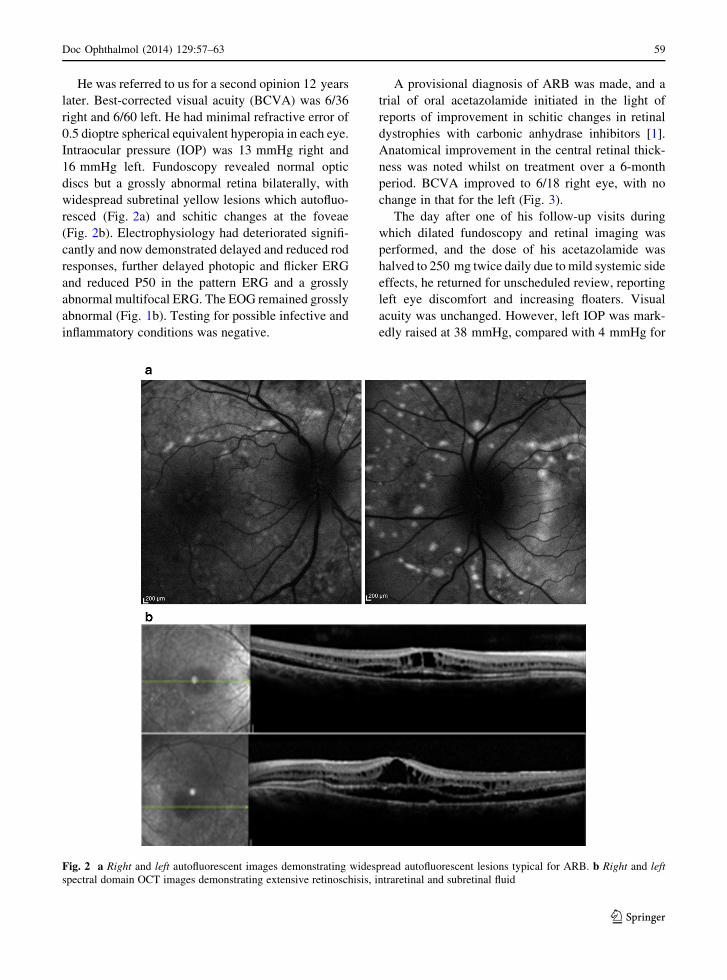
He was referred to us for a second opinion 12 years
later. Best-corrected visual acuity (BCVA) was 6/36
right and 6/60 left. He had minimal refractive error of
0.5 dioptre spherical equivalent hyperopia in each eye.
Intraocular pressure (IOP) was 13 mmHg right and
16 mmHg left. Fundoscopy revealed normal optic
discs but a grossly abnormal retina bilaterally, with
widespread subretinal yellow lesions which autofluo-
resced (Fig. 2a) and schitic changes at the foveae
(Fig. 2b). Electrophysiology had deteriorated signifi-
cantly and now demonstrated delayed and reduced rod
responses, further delayed photopic and flicker ERG
and reduced P50 in the pattern ERG and a grossly
abnormal multifocal ERG. The EOG remained grossly
abnormal (Fig. 1b). Testing for possible infective and
inflammatory conditions was negative.
A provisional diagnosis of ARB was made, and a
trial of oral acetazolamide initiated in the light of
reports of improvement in schitic changes in retinal
dystrophies with carbonic anhydrase inhibitors [1].
Anatomical improvement in the central retinal thick-
ness was noted whilst on treatment over a 6-month
period. BCVA improved to 6/18 right eye, with no
change in that for the left (Fig. 3).
The day after one of his follow-up visits during
which dilated fundoscopy and retinal imaging was
performed, and the dose of his acetazolamide was
halved to 250 mg twice daily due to mild systemic side
effects, he returned for unscheduled review, reporting
left eye discomfort and increasing floaters. Visual
acuity was unchanged. However, left IOP was mark-
edly raised at 38 mmHg, compared with 4 mmHg for
Fig. 2 a Right and left autofluorescent images demonstrating widespread autofluorescent lesions typical for ARB. b Right and left
spectral domain OCT images demonstrating extensive retinoschisis, intraretinal and subretinal fluid
Doc Ophthalmol (2014) 129:57–63 59
123

his right eye. A narrow drainage angle was noted
bilaterally, but it was significantly narrower in the left
eye, with a shallow anterior chamber and ‘volcano
sign’. Subacute angle-closure glaucoma was diag-
nosed and effectively treated initially with topical anti-
hypertensives and peripheral iridotomies. However,
left-sided IOP continued to rise over a few days to
60 mmHg. B-scan ultrasound excluded choroidal
effusion and confirmed plateau iris. His axial length
measured 21.64 mm right and 22.06 mm left, and his
anterior chamber depth was 2.48 mm right and
2.47 mm left. His optic disc was not cupped, with a
cup-to-disc ratio of less than 0.4 bilaterally. Phacoe-
mulsification lens extraction was eventually required
to lower the IOP, and a 24.0 dioptre posterior chamber
lens (A-constant 119.0) was inserted uneventfully.
IOP was controlled at 18 mmHg thereafter but
required continued use of topical prostaglandin and
beta-blocker.
Bi-directional DNA sequencing of coding and
flanking intronic regions of BEST1 revealed that the
patient was homozygous for a novel splice variant,
c.636?1 G[A. This substitution was predicted to be
pathogenic by abolishing the conserved donor splice
site (Mutation Taster; Human Splicing Finder v2.4.1;
NN SPLICE) [2], with potential usage of a cryptic
donor splice site present 294 bp downstream in intron
5 (0.88 NN SPLICE). Cascade family testing identi-
fied this patient’s non-consanguineous parents and
sister as carriers. They were asymptomatic and did not
show signs of disease, with completely normal vision,
fundi, anterior segments and electrophysiology.
Discussion
The BEST1 gene, formally known as VMD2, encodes
bestrophin-1, previously postulated to act as a Ca2?-
activated chloride channel [3], a regulator of voltage-
gated Ca2? channels [4], or a HCO3- channel [5] in
the basolateral membrane of the RPE [6]. It was
recently shown to localise in the endoplasmic reticu-
lum membrane, however [7]. Bestrophin-1 dysfunc-
tion has been associated with defective regulation of
subretinal fluid reabsorption and aberrant phagocyto-
sis of the photoreceptor discs [8]. Over 250 disease-
causing mutations have been identified in the BEST1
gene to date associated with a broad range of
phenotypes, including BVMD, adult vitelliform mac-
ular dystrophy, autosomal dominant vitreoretinochor-
oidopathy (ADVIRC), the MRCS (microcornea, rod-
cone dystrophy, cataract, posterior staphyloma) syn-
drome, retinitis pigmentosa and ARB [9–14].
ARB is thought to result from biallelic functionally
null mutations of the gene, whilst most dominantly
inherited missense mutations have been found to
produce dominant negative effects and so do not
compromise protein synthesis [10, 12, 13]. In vitro
Fig. 3 Right and left spectral domain OCT images demonstrating redistribution of intraretinal and subretinal fluid after 4 months of
oral acetazolamide
60 Doc Ophthalmol (2014) 129:57–63
123
studies using HEK293 cells showed that co-transfec-
tion of the two mutations observed in the compound
heterozygous state in ARB abolished chloride con-
ductance in contrast to co-transfection of a single
mutant with wild-type bestrophin-1 which led to
significantly smaller chloride currents compared to
wild-type bestrophin-1 [9, 15]. This suggests that the
autosomal recessive phenotype only manifests when
bestrophin-1 activity falls below a functional thresh-
old. Davidson et al. [15] also found that different
ARB-associated mutants lead to the same disease
phenotype but through different effects on cellular
processing mechanisms. This finding has implications
for potential gene replacement therapies as the authors
showed that missense mutations associated with
autosomal recessive diseases may have a pathogenic
outcome beyond simple loss of function.
Whilst BVMD is characterised by vitelliform
lesions that typically occur at the macula as a result
of abnormal deposition of lipofuscin in the retinal
pigment epithelium (RPE), ARB is associated with
subretinal deposits occurring predominantly outside
the macula, mainly at the posterior pole and along the
vascular arcades [9, 16–19]. These are often small and
fleck-like or punctate in shape, white or yellow in
colour, and hyperfluoresce on fundus autofluorescence
imaging. Optical coherence tomography (OCT) shows
subretinal and intraretinal fluid accumulation, often at
the maculae [9, 18–21]. Using higher resolution
Fourier-domain OCT, Gerth et al. [17] demonstrated
RPE deposits and significant photoreceptor changes
but preserved inner retinal layers in an 11-year-old boy
with ARB, including thickened, elongated photore-
ceptor outer segments and detachment from the RPE.
Unlike BVMD, ARB is associated with diminished
rod- and cone-driven ERG responses, though it shares
the presence of a severely reduced or absent EOG light
rise with BVMD as well as ADVIRC [9, 10, 16–24].
The expression of BEST1 is higher in the peripheral
RPE than at the macula [25]. This may explain the
more widespread and progressive photoreceptor dys-
function, as well as the predominantly peripheral
location of retinal lesions observed in patients with
ARB. No histopathological data are available due to
the novel description of the ARB phenotype.
Since ARB was first recognised in 2008 by Burgess
et al. [9], at least 35 novel compound heterozygous and
homozygous mutations have been reported to cause
the disorder [9, 16–19, 21, 26–31]. Incomplete
penetrance and variability in expression associated
with some mutations in BEST1, together with the fact
that some phenotypic distinctions may not develop
until later years, make it difficult to distinguish those
resulting in dominant inheritance from those resulting
in recessive inheritance.
A family reported by Schatz et al. in 2006 [32] with
compound heterozygous BEST1 mutations is thought
by some to have represented ARB rather than ‘atyp-
ical’ BVMD [9]. However, the heterozygous carriers
also had ERG and EOG abnormalities, suggesting a
diagnosis of BVMD with reduced penetrance in those
individuals. Sharon et al. [30] recently reported a
novel BEST1 mutation in a Danish family in which the
proband also had a previously reported mutation of the
other allele and a phenotype suggestive of ARB.
However, his mother and sister who were heterozy-
gous for the novel mutation additionally had reduced
EOG light rise which would not be expected with an
autosomal recessive inheritance pattern. Pineiro-Gal-
lego et al. [28] similarly reported on a case with a
homozygous mutation causing what they considered
to be ARB even though heterozygotes in the family
also had abnormal EOG findings. One of the two cases
of ARB reported by Pomares et al. [29] had fundus
findings suggestive of ARB, but the patient had a
normal EOG and no family history given he was
adopted and so his novel homozygous mutation cannot
be confirmed to cause ARB.
Bitner et al. [33] were of the impression that BVMD
can be inherited as an autosomal recessive disease and
distinguished this from ARB where the patients were
homozygous for a novel mutation, had fundus and
electrophysiology findings in keeping with a diagnosis
of BVMD and their unaffected parents each carried
one copy of the same mutation. As shown by
Cascavilla et al. [26] however, it may not be until
later years that the ERG becomes abnormal in cases of
ARB.
In addition to small eyes, reduced axial length and
hyperopia, it is increasingly recognised that bestro-
phinopathies are also strongly associated with anterior
segment abnormalities and a high incidence of
narrow-angle glaucoma [9, 10, 13, 14, 20, 22–24,
34]. The causes behind these associations are still to be
elucidated, but there is evidence to support the
hypothesis that BEST1 is involved through bestro-
phin-1 expression in the RPE in the development of
ocular structures beyond the retina [10]. Wittstrom
Doc Ophthalmol (2014) 129:57–63 61
123
et al. [22] reported that two of four patients with
BVMD from one pedigree exhibited shallow anterior
chambers, and all four patients had hyperopia as well
as reduced axial lengths. Micropthalmos (axial length
B20 mm) was found in two cases, both of whom had
narrow angles and one of whom developed acute
closed-angle glaucoma at the age of 12 years. Low
et al. [34] found that sibling carriers of probands with
BVMD can also have narrow anterior chamber angles,
short axial lengths and a similar risk of angle-closure
glaucoma.
Angle-closure glaucoma has been shown to affect
approximately 50 % of those with ARB. Burgess et al.
[9] found that all seven cases of ARB examined were
hyperopic, and three required surgery for angle-
closure glaucoma. Davidson et al. [20] described two
unrelated cases of ARB, both of whom had angle-
closure glaucoma contributing to their visual loss. All
ten patients with ARB examined by Boon et al. [18]
were hyperopic, and five had shallow anterior cham-
bers and narrow angles for which they underwent
prophylactic laser peripheral iridotomies. One of these
patients with additional short axial lengths underwent
phacoemulsification and intraocular lens implantation
in an effort to deepen the anterior chamber and its
angles for both eyes. Unlike the case we have
presented, this was not enough to control her IOPs
and she went on to have an iris base laser iridoplasty
followed by topical anti-glaucomatous therapy. The
authors postulated that cataract extraction may not
sufficiently open the anterior chamber angles as some
patients with ARB may have a dysgenesis of the
anterior segment that additionally affects the trabec-
ular meshwork.
Although plateau iris has not been described
previously in ARB, a study by Etter et al. [35]
suggests that it has a heritable component, with five of
the ten patients studied with plateau iris syndrome
having at least one affected first-degree family mem-
ber. The authors likened this to an autosomal dominant
pattern of inheritance with incomplete penetrance.
In conclusion, we report a novel BEST1 splice
variant, c.636?1 G[A, identified homozygously in a
proband clinically diagnosed with ARB in association
with plateau iris and narrow-angle glaucoma. A
heterozygous G[C change in this position has been
reported before, together with known pathogenic
variant c.422 G[A (R141H) for a patient clinically
diagnosed with BVMD [36]. The splice variant
reported in the present study is likely to abolish the
conserved 30 splice site of exon 5. Family members
carrying one copy of the mutation had no symptoms or
signs of disease, supporting a recessive inheritance
pattern; however, functional studies are required to
confirm pathogenicity of the variant. We strongly
recommend routine screening for narrow angles and
glaucoma in ARB and BVMD given their frequent
association, as well as close monitoring of IOP even
after peripheral iridotomy and/or phacoemulsification
have been performed.
Acknowledgments The Australian Inherited Retinal Disease
Register is financially supported by Retina Australia. We
gratefully acknowledge the assistance of the Western Australian
DNA Bank (National Health and Medical Research Council
(NHMRC) Enabling Facility) with processing of DNA samples
for this study and the funding provided by an Australian
NHMRC Centres of Research Excellence Grant 1023911
(2012–2016). We would also like to thank Translation of
Genetic Eye Research (ToGER) for its support.
Open Access This article is distributed under the terms of the
Creative Commons Attribution License which permits any use,
distribution, and reproduction in any medium, provided the
original author(s) and the source are credited.
References
1. Iannaccone A, Fung KH, Eyestone ME, Stone EM (2009)
Treatment of adult-onset acute macular retinoschisis in
enhanced-S cone syndrome with oral acetazolamide. Am J
Ophthalmol 147(2):307–312
2. Reese MG, Eeckman FH, Kulp D, Haussler D (1997)
Improved splice site detection in Genie. J Comput Biol
4(3):311–323
3. Sun H, Tsunenari T, Yau KW, Nathans J (2002) The
vitelliform macular dystrophy protein defines a new family
of chloride channels. Proc Natl Acad Sci USA
99:4008–4013
4. Rosenthal R, Bakall B, Kinnick T, Peachey N, Wimmers S,
Wadelius C et al (2006) Expression of bestrophin-1, the
product of the VMD2 gene, modulates voltage-dependent
Ca2? channels in retinal pigment epithelial cells. FASEB J
20:178–180
5. Qu Z, Hartzell HC (2008) Bestrophin Cl- channels are
highly permeable to HCO3. Am J Physiol Cell Physiol
294:C1371–C1377
6. Marmorstein AD, Marmorstein LY, Rayborn M, Wang X,
Hollyfield JG, Pretrukin K (2000) Bestrophin, the product of
the Best vitelliform macular dystrophy gene (VMD2), local-
izes to the basolateral plasma membrane of the retinal pigment
epithelium. Proc Natl Acad Sci USA 97:12758–12763
7. Gomez NM, Tamm ER, Straubeta O (2013) Role of bes-
trophin-1 in store-operated calcium entry in retinal pigment
epithelium. Pflugers Arch 465(4):481–495
62 Doc Ophthalmol (2014) 129:57–63
123
8. Xiao Q, Hartzell HC, Yu K (2010) Bestrophins and retin-
opathies. Pflugers Arch 460:559–569
9. Burgess R, Millar ID, Leroy BP, Urquhart JE, Fearon IM,
De Baere E et al (2008) Biallelic mutation of BEST1 causes
a distinct retinopathy in humans. Am J Hum Genet 1:19–31
10. Boon CJ, Klevering BJ, Leroy BP, Hoyng CB, Keunen JE,
den Hollander AI (2009) The spectrum of ocular phenotypes
caused by mutations in the BEST1 gene. Prog Retin Eye Res
3:187–205
11. Davidson AE, Millar ID, Urquhart JE, Burgess-Mullan R,
Shweikh Y, Parry N et al (2009) Missense mutations in a
retinal pigment epithelium protein, bestrophin-1, cause
retinitis pigmentosa. Am J Hum Genet 85:581–592
12. Petrukhin K, Koisti MJ, Bakall B, Li W, Xie W, Marknell T
et al (1998) Identification of the gene responsible for Best
macular dystrophy. Nat Genet 19:241–247
13. Yardley J, Leroy BP, Hart-Holden N, Lafaut BA, Loeys B,
Messiaen LM et al (2004) Mutations of VMD2 splicing
regulators cause nanophthalmos and autosomal dominant
vitreochoroidopathy (ADVIRC). Invest Ophthalmol Vis Sci
45:3683–3689
14. Reddy MA, Francis PJ, Berry V et al (2003) A clinical and
molecular genetic study of a rare dominantly inherited
syndrome (MRCS) comprising of microcornea, rod-cone
dystrophy, cataract, and posterior staphyloma. Br J Oph-
thalmol 87:197–202
15. Davidson AE, Miller ID, Burgess-Mullan R, Maher GJ,
Urquhart JE, Brown PD et al (2011) Functional character-
ization of bestrophin-1 missense mutations associated with
autosomal recessive bestrophinopathy. Invest Ophthalmol
Vis Sci 52:3730–3736
16. Kinnick TR, Mullins RF, Dev S, Leys M, Mackey DA, Kay
CN et al (2011) Autosomal recessive vitelliform macular
dystrophy in a large cohort of vitelliform macular dystrophy
patients. Retina 3:581–595
17. Gerth C, Zawadzki RJ, Werner JS, Heon E (2009) Detailed
analysis of retinal function and morphology in a patient with
autosomal recessive bestrophinopathy (ARB). Doc Oph-
thalmol 3:239–246
18. Boon CJ, van der Born LI, Visser L, Keunen JE, Bergen B,
Booij JC et al (2013) Autosomal recessive bestrophinopa-
thy. Differential diagnosis and treatment options. Ophthal-
mology 120:809–820
19. Borman AD, Davidson AE, O’ Sullivan J, Thompson DA,
Robson AG, De Baere E et al (2011) Childhood-onset
autosomal recessive bestrophinopathy. Arch Ophthalmol
129(8):1088–1093
20. Davidson AE, Sergouniotis PI, Burgess-Mullan R, Hart-
Holden N, Low S, Foster PJ et al (2010) A synonymous
codon variant in two patients with autosomal recessive
bestrophinopathy alters in vitro splicing of BEST1. Mol Vis
16:2916–2922
21. Iannacone A, Kerr NC, Kinnick TR, Calzada JI, Stone EM
(2011) Autosomal recessive best vitelliform macular dys-
trophy: report of a family and management of early-onset
neovascular complications. Arch Ophthalmol 2:211–217
22. Wittstrom E, Ponjavic V, Bondeson ML, Andreasson S
(2011) Anterior segment abnormalities and angle-closure
glaucoma in a family with a mutation in the BEST1 gene
and best vitelliform macular dystrophy. Ophthalmic Genet
32(4):217–227
23. Bard LA, Cross HE (1975) Genetic counseling of families
with Best macular dystrophy. Trans Sect Ophthalmol Am
Acad Ophthalmol Otolaryngol 79(6):OP865–OP873
24. Sohn EH, Francis PJ, Duncan JL, Weleber RG, Saperstein
DA, Farrell DF, Stone EM (2009) Phenotypic variability
due to a novel Glu292Lys variation in exon 8 of the BEST1
gene causing best macular dystrophy. Arch Ophthalmol
127:913–920
25. Mullins RF, Keuhn MH, Faidley EA, Syed NA, Stone EM
(2007) Differential macular and peripheral expression of
bestrophin in human eyes and its implication for best dis-
ease. Invest Ophthalmol Vis Sci 48:3372–3380
26. Cascavilla ML, Querques G, Stenirri S, Parodi MB, Quer-
ques L, Bandello F (2012) Unilateral vitelliform phenotype
in autosomal recessive bestrophinopathy. Ophthalmic Res
48:146–150
27. Fung A, Yzer S, Allikmets R (2013) Clinical and genetic
misdiagnosis of autosomal recessive bestrophinopathy.
JAMA 131:1651 (Letter to the editor)
28. Pineiro-Gallego T, Alverez M, Pereiro I, Campos S, Sharon
D, Schatz P et al (2011) Clinical evaluation of two con-
sanguineous families with homozygous mutations in
BEST1. Mol Vis 17:1607–1617
29. Pomares E, Bures-Jelstrup A, Ruiz-Nogales S, Corcostegui
B, Gonzalez-Duatre R, Navarro R (2012) Nonsense-medi-
ated decay as the molecular cause for autosomal recessive
bestrophinopathy in two unrelated families. Invest Oph-
thalmol Vis Sci 53:532–537
30. Sharon D, Al-Hamdani S, Engelsberg K, Mizrahi-Meis-
sonnier L, Obolensky B, Banin E et al (2014) Ocular phe-
notype analysis of a family with biallelic mutations in the
BEST1 gene. Am J Ophthalmol 157:697–709
31. Preising MN, Pasquay C, Friedburg C, Bowl W, Jager M,
Andrassi-Darida M et al (2012) Autosomal recessive bets-
trophinopathy (ARB): a clinical and molecular description
of two patients at childhood. Klin Monbl Augenheilkd
229(10):1009–1017
32. Schatz P, Klar J, Andreasson S, Ponjavic V, Dahl N (2006)
Variant phenotype of Best vitelliform macular dystrophy
associated with compound heterozygous mutations in
VMD2. Ophthalmol Genet 2:51–56
33. Bitner H, Mizrahi-Meissonnier L, Griefner G, Erdinest I,
Sharon D, Banin E (2011) A homozygous frameshift
mutation in BEST1 causes the classical form of Best disease
in an autosomal recessive mode. Invest Ophthalmol Vis Sci
52:5332–5338
34. Low S, Davidson AE, Holder GE, Hogg CR, Bhattacharya
SS, Black GC et al (2011) Autosomal dominant Best disease
with an unusual electrooculographic light rise and risk of
angle-closure glaucoma: a clinical and molecular genetic
study. Mol Vis 17:2272–2282
35. Etter JR, Affel EL, Rhee DJ (2006) High prevalence of
plateau iris configuration in family members of patients with
plateau iris syndrome. J Glaucoma 15(5):394–398
36. Kramer F, White K, Pauleikhoff D, Gehrig A, Passmore L,
Rivera A et al (2000) Mutations in the MD2 gene are
associated with juvenile-onset vitelliform macular dystro-
phy (Best disease) and adult vitelliform macular dystrophy
but not age-related macular degeneration. Eur J Hum Genet
8:286–292
Doc Ophthalmol (2014) 129:57–63 63
123
Recommended