J Cutan Pathol 2011: 38: 842–843doi: 10.1111/j.1600-0560.2011.01712.xJohn Wiley & Sons. Printed in Singapore
Copyright © 2011 John Wiley & Sons A/S
Journal ofCutaneous Pathology
Letter to the Editor
Benign cephalic histiocytosis withS-100 protein positivity
Keywords: atypical features, cutaneous, immunohistochemical method, Langerhans cells, S-100 protein
To the Editor,Benign cephalic histiocytosis represents a non-
Langerhans cell (non-X) histiocytosis first describedby Gianotti et al. in 1971.1 It presents inotherwise healthy infants and young children andis characterized by an eruption of red-yellowpapules on the face, particularly the cheeks, andupper extremities. The condition is self-limitedand usually resolves after a few years. It iscritical to distinguish cephalic histiocytosis fromLangerhans cell histiocytosis because of differingprognosis and therapy. Immunohistochemistry isoften utilized to facilitate this distinction. Langerhanscells stereotypically express both CD1a and S-100protein, whereas in contrast the cells of cephalichistiocytosis typically lack expression of both ofthose determinants. In this report, we describe apatient with a typical clinical morphology of cephalichistiocytosis with the unusual occurrence of anS-100-positive immunophenotype.
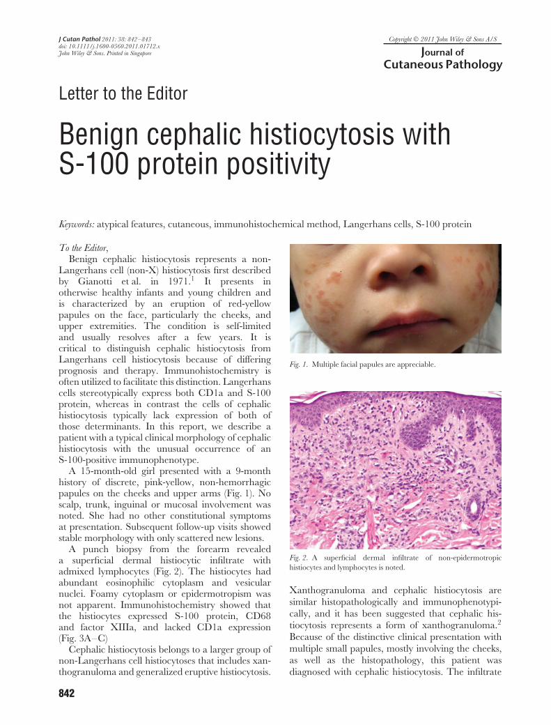
A 15-month-old girl presented with a 9-monthhistory of discrete, pink-yellow, non-hemorrhagicpapules on the cheeks and upper arms (Fig. 1). Noscalp, trunk, inguinal or mucosal involvement wasnoted. She had no other constitutional symptomsat presentation. Subsequent follow-up visits showedstable morphology with only scattered new lesions.
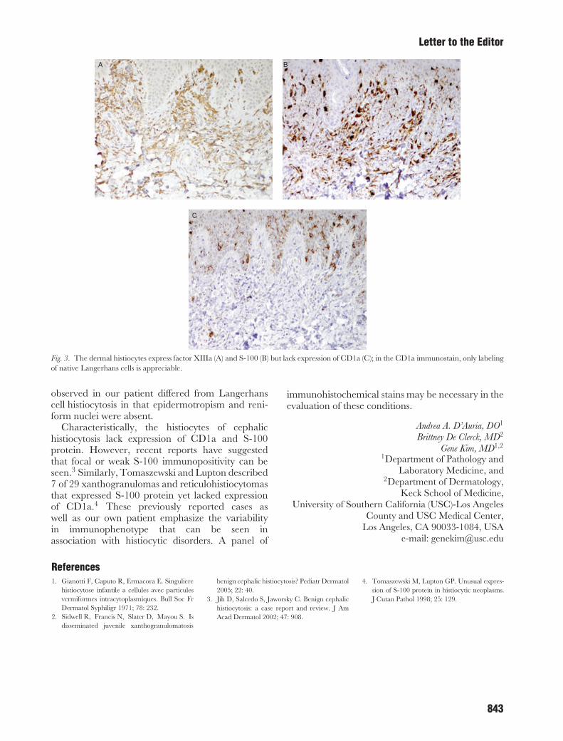
A punch biopsy from the forearm revealeda superficial dermal histiocytic infiltrate withadmixed lymphocytes (Fig. 2). The histiocytes hadabundant eosinophilic cytoplasm and vesicularnuclei. Foamy cytoplasm or epidermotropism wasnot apparent. Immunohistochemistry showed thatthe histiocytes expressed S-100 protein, CD68and factor XIIIa, and lacked CD1a expression(Fig. 3A–C)
Cephalic histiocytosis belongs to a larger group ofnon-Langerhans cell histiocytoses that includes xan-thogranuloma and generalized eruptive histiocytosis.
Fig. 1. Multiple facial papules are appreciable.
Fig. 2. A superficial dermal infiltrate of non-epidermotropichistiocytes and lymphocytes is noted.
Xanthogranuloma and cephalic histiocytosis aresimilar histopathologically and immunophenotypi-cally, and it has been suggested that cephalic his-tiocytosis represents a form of xanthogranuloma.2
Because of the distinctive clinical presentation withmultiple small papules, mostly involving the cheeks,as well as the histopathology, this patient wasdiagnosed with cephalic histiocytosis. The infiltrate
842
Letter to the Editor
A B
C
Fig. 3. The dermal histiocytes express factor XIIIa (A) and S-100 (B) but lack expression of CD1a (C); in the CD1a immunostain, only labelingof native Langerhans cells is appreciable.
observed in our patient differed from Langerhanscell histiocytosis in that epidermotropism and reni-form nuclei were absent.
Characteristically, the histiocytes of cephalichistiocytosis lack expression of CD1a and S-100protein. However, recent reports have suggestedthat focal or weak S-100 immunopositivity can beseen.3 Similarly, Tomaszewski and Lupton described7 of 29 xanthogranulomas and reticulohistiocytomasthat expressed S-100 protein yet lacked expressionof CD1a.4 These previously reported cases aswell as our own patient emphasize the variabilityin immunophenotype that can be seen inassociation with histiocytic disorders. A panel of
immunohistochemical stains may be necessary in theevaluation of these conditions.
Andrea A. D’Auria, DO1
Brittney De Clerck, MD2
Gene Kim, MD1,2
1Department of Pathology andLaboratory Medicine, and
2Department of Dermatology,Keck School of Medicine,
University of Southern California (USC)-Los AngelesCounty and USC Medical Center,
Los Angeles, CA 90033-1084, USAe-mail: [email protected]
References1. Gianotti F, Caputo R, Ermacora E. Singuliere
histiocytose infantile a cellules avec particulesvermiformes intracytoplasmiques. Bull Soc FrDermatol Syphiligr 1971; 78: 232.
2. Sidwell R, Francis N, Slater D, Mayou S. Isdisseminated juvenile xanthogranulomatosis
benign cephalic histiocytosis? Pediatr Dermatol2005; 22: 40.
3. Jih D, Salcedo S, Jaworsky C. Benign cephalichistiocytosis: a case report and review. J AmAcad Dermatol 2002; 47: 908.
4. Tomaszewski M, Lupton GP. Unusual expres-sion of S-100 protein in histiocytic neoplasms.J Cutan Pathol 1998; 25: 129.
843
Recommended