BROOKLYN 3STUDENTS
Bridgette MAIR
Fri 30th Aug 2013Session 3 / Talk 4
13:45 – 13:55
ABSTRACT
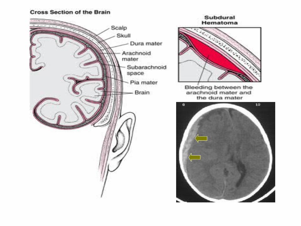
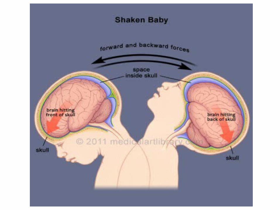
Subdural haematomas are the most common form of traumatic injury and make up
80% of head injury from non-accidental injuries. Diagnosis of subdural haematomas
can be difficult as the child regularly has non-specific symptoms and lacks external
cues. Neuroimaging is a fundamental investigation in obtaining diagnosis of subdural
haematomas. It is therefore essential to understand the role of MRI in demonstrating
subdural haematomas for diagnosis to be achieved, enabling correct management
steps to be ascertained improving the prognosis of paediatrics subjected to non-
accidental injury. The aim of this literature review is to explain the role MRU plays in
diagnosing subdural haematomas in NAI paediatrics. This was completed by reviewing
15 literature articles from 1999 to understand the role it plays and why. It was
concluded that while CT remains the most appropriate initial modality of choice for
suspected NAI cases die to accessibility, speed and ease, MRI is the test of choice to
demonstrate SDH due to its higher level specificity and aging of multiple haematomas
if present.
The role of magnetic resonance imaging in diagnosing subdural
haematomas in non-
accidental injury
paediatrics
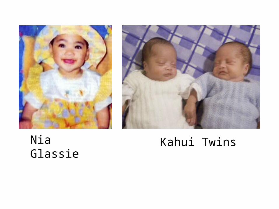
Nia Glassie Kahui Twins
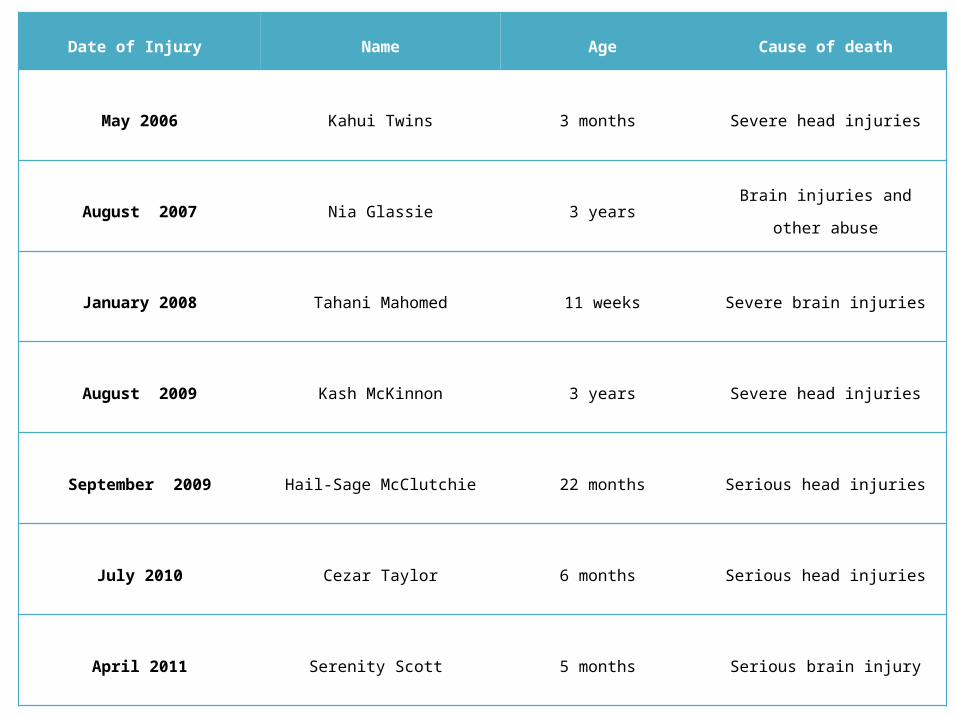
Date of Injury Name Age Cause of death
May 2006 Kahui Twins 3 months Severe head injuries
August 2007 Nia Glassie 3 years Brain injuries and other abuse
January 2008 Tahani Mahomed 11 weeks Severe brain injuries
August 2009 Kash McKinnon 3 years Severe head injuries
September 2009 Hail-Sage McClutchie 22 months Serious head injuries
July 2010 Cezar Taylor 6 months Serious head injuries
April 2011 Serenity Scott 5 months Serious brain injury
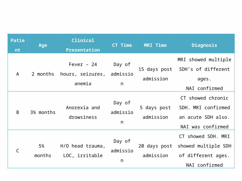
Patient AgeClinical
PresentationCT Time MRI Time Diagnosis
A 2 monthsFever – 24 hours,
seizures, anemia
Day of
admission
15 days post
admission
MRI showed multiple
SDH’s of different ages.
NAI confirmed
B 3½ monthsAnorexia and
drowsiness
Day of
admission
5 days post
admission
CT showed chronic SDH.
MRI confirmed an acute
SDH also. NAI was
confirmed
C 5½ monthsH/O head trauma,
LOC, irritable
Day of
admission
20 days post
admission
CT showed SDH. MRI
showed multiple SDH of
different ages. NAI
confirmed
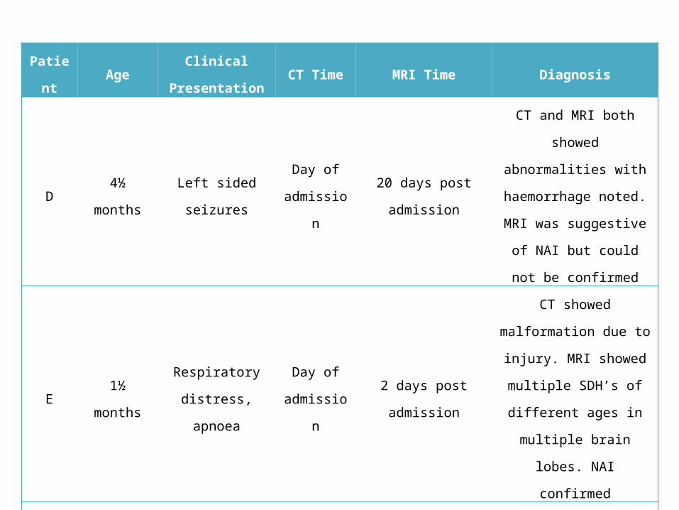
Patient AgeClinical
PresentationCT Time MRI Time Diagnosis
D 4½ monthsLeft sided
seizures
Day of
admission
20 days post
admission
CT and MRI both showed
abnormalities with
haemorrhage noted. MRI
was suggestive of NAI but
could not be confirmed
E 1½ monthsRespiratory
distress, apnoea
Day of
admission
2 days post
admission
CT showed malformation
due to injury. MRI showed
multiple SDH’s of different
ages in multiple brain
lobes. NAI confirmed
F 2 months ApnoeaDay of
admission
6 days post
admission
Skeletal survey was done
on admission showing
numerous fractures. CT
was normal. MRI showed
a chronic SDH. NAI
confirmed
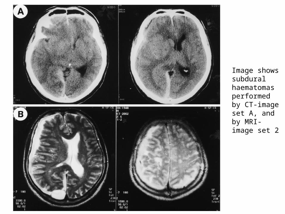
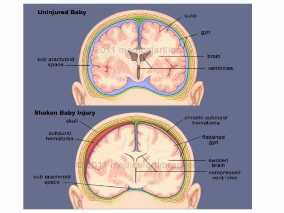
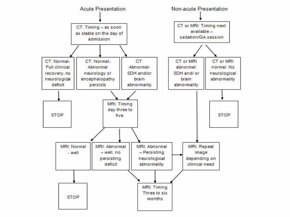
Image shows subdural haematomas performed by CT-image set A, and by MRI-image set 2

AnyQuestions
?
Recommended