Jordi Heijman, Niels Voigt, Stanley Nattel and Dobromir Dobrevand Progression
Cellular and Molecular Electrophysiology of Atrial Fibrillation Initiation, Maintenance,
Print ISSN: 0009-7330. Online ISSN: 1524-4571 Copyright © 2014 American Heart Association, Inc. All rights reserved.is published by the American Heart Association, 7272 Greenville Avenue, Dallas, TX 75231Circulation Research
doi: 10.1161/CIRCRESAHA.114.3022262014;114:1483-1499Circ Res.
http://circres.ahajournals.org/content/114/9/1483World Wide Web at:
The online version of this article, along with updated information and services, is located on the
http://circres.ahajournals.org//subscriptions/
is online at: Circulation Research Information about subscribing to Subscriptions:
http://www.lww.com/reprints Information about reprints can be found online at: Reprints:
document. Permissions and Rights Question and Answer about this process is available in the
located, click Request Permissions in the middle column of the Web page under Services. Further informationEditorial Office. Once the online version of the published article for which permission is being requested is
can be obtained via RightsLink, a service of the Copyright Clearance Center, not theCirculation Researchin Requests for permissions to reproduce figures, tables, or portions of articles originally publishedPermissions:
by guest on May 6, 2014http://circres.ahajournals.org/Downloaded from by guest on May 6, 2014http://circres.ahajournals.org/Downloaded from
1483
Atrial fibrillation (AF) is the most prevalent cardiac ar-rhythmia in the developed world, affecting ≈6 million
people in the United States alone, an incidence that is ex-pected to double by 2030 because of the aging of the popula-tion.1,2 Largely as a major risk factor for embolic stroke and worsening heart failure (HF), AF is associated with significant morbidity and mortality.3 AF is classified as paroxysmal AF
(pAF) when episodes last <7 days and spontaneously con-vert to normal sinus rhythm, persistent AF when lasting ≥7 days, or permanent AF when no further attempts to achieve normal sinus rhythm are made.4 Patients with more advanced stages (long-standing persistent) of AF are generally older and have more comorbidities.5 The progression from paroxys-mal to persistent and permanent forms of AF has pronounced
Compendium
© 2014 American Heart Association, Inc.
Circulation Research is available at http://circres.ahajournals.org DOI: 10.1161/CIRCRESAHA.114.302226
Abstract: Atrial fibrillation (AF) is the most common clinically relevant arrhythmia and is associated with increased morbidity and mortality. The incidence of AF is expected to continue to rise with the aging of the population. AF is generally considered to be a progressive condition, occurring first in a paroxysmal form, then in persistent, and then long-standing persistent (chronic or permanent) forms. However, not all patients go through every phase, and the time spent in each can vary widely. Research over the past decades has identified a multitude of pathophysiological processes contributing to the initiation, maintenance, and progression of AF. However, many aspects of AF pathophysiology remain incompletely understood. In this review, we discuss the cellular and molecular electrophysiology of AF initiation, maintenance, and progression, predominantly based on recent data obtained in human tissue and animal models. The central role of Ca2+-handling abnormalities in both focal ectopic activity and AF substrate progression is discussed, along with the underlying molecular basis. We also deal with the ionic determinants that govern AF initiation and maintenance, as well as the structural remodeling that stabilizes AF-maintaining re-entrant mechanisms and finally makes the arrhythmia refractory to therapy. In addition, we highlight important gaps in our current understanding, particularly with respect to the translation of these concepts to the clinical setting. Ultimately, a comprehensive understanding of AF pathophysiology is expected to foster the development of improved pharmacological and nonpharmacological therapeutic approaches and to greatly improve clinical management. (Circ Res. 2014;114:1483-1499.)
Key Words: atrial fibrillation ■ atrial remodeling ■ calcium ■ electrophysiology
Cellular and Molecular Electrophysiology of Atrial Fibrillation Initiation, Maintenance, and Progression
Jordi Heijman,* Niels Voigt,* Stanley Nattel, Dobromir Dobrev
Circulation Research Compendium on Atrial Fibrillation:
Atrial Fibrillation Compendium: Historical Context and Detailed Translational Perspective on an Important Clinical ProblemThe Clinical Profile and Pathophysiology of Atrial Fibrillation: Relationships Among Clinical Features, Epidemiology, and MechanismsEmerging Directions in the Genetics of Atrial FibrillationCellular and Molecular Electrophysiology of Atrial Fibrillation Initiation, Maintenance, and ProgressionRole of Autonomic Nervous System in Atrial Fibrillation: Pathophysiology and TherapyMathematical Approaches to Understanding and Imaging Atrial Fibrillation: Significance for Mechanisms and ManagementAtrial Fibrillation Therapy Now and in the Future: Drugs, Biologicals, and Ablation
Stanley Nattel, Guest Editor
Original received December 15, 2013; revision received January 13, 2014; accepted January 16, 2014. In February 2014, the average time from submission to first decision for all original research papers submitted to Circulation Research was 13.8 days.
From the Institute of Pharmacology, Faculty of Medicine, University Duisburg-Essen, Essen, Germany (J.H., N.V., D.D.); Department of Medicine, Montreal Heart Institute and Université de Montréal, Montreal, Quebec, Canada (S.N.); and Department of Pharmacology and Therapeutics, McGill University, Montreal, Quebec, Canada (S.N.).
*These authors contributed equally.Correspondence to Dobromir Dobrev, MD, Hufelandstrasse 55, 45122 Essen, Germany. E-mail [email protected]
by guest on May 6, 2014http://circres.ahajournals.org/Downloaded from
1484 Circulation Research April 25, 2014
therapeutic implications, with pAF being more amenable to rhythm control therapy.6
Current pharmacological options have imperfect efficacy and substantial adverse side effects, including drug-induced proar-rhythmia and both cardiac and noncardiac toxicity.7–9 The limit-ed efficacy of current pharmacological treatment options likely results from an incomplete understanding of the pathophysiol-ogy of this complex heart rhythm disorder. Here, we provide a conceptual overview of the factors involved in the initiation, maintenance, and progression of AF. Subsequently, we review the molecular mechanisms identified for each of these compo-nents. Finally, we highlight important gaps in the current under-standing of AF pathophysiology, particularly with respect to the translation of these findings to the clinical setting.
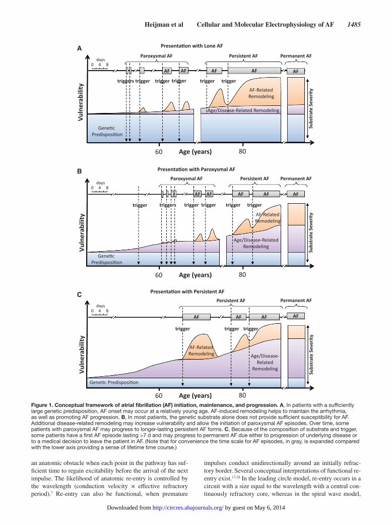
Conceptual FrameworkAF as a Progressive DiseaseThe pathophysiology of AF contains 3 major components: initiation of the arrhythmia, arrhythmia maintenance, and pro-gression toward longer-lasting AF forms (ie, from paroxysmal to persistent/permanent AF).10,11 Each AF episode requires initiation by a trigger acting on a vulnerable substrate. This
vulnerable substrate is at least partly determined by genetic fac-tors.12–14 Several mutations and gene variants have been identi-fied that allow AF initiation in the absence of traditional risk factors (Figure 1A). Although they are rare, and generally limit-ed to isolated families, AF-causing mutations have provided im-portant insights into the ionic mechanisms underlying AF.12 In addition, recent genome-wide association studies have discov-ered several genetic variants associated with an increased risk of AF, identifying novel potential factors contributing to AF.13 However, the exact mechanisms linking genetic loci identified with genome-wide association studies to AF are incompletely understood, because (1) causative genes are often uncertain, and (2) the likely candidates generally have poorly understood functions. Even after including genome-wide association study variants, a large portion of the heritability of AF is uncertain, with large population studies showing that a parental history of AF almost doubles the future AF risk in their offsprings.14 Thus, other currently unknown genetic components also play a role in more common forms of AF.13 Furthermore, genetic variants are unlikely to cause AF directly, but rather provide background vulnerability. When additional risk factors develop over time, because of physiological aging or cardiac remodeling resulting from other cardiovascular and noncardiovascular diseases, an appropriate trigger may then initiate AF (Figure 1B). Common comorbidities that promote a vulnerable substrate for the ini-tiation and maintenance of AF include hypertension, HF, and cardiac valve disease.5 Genetic variants that increase the risk of hypertension, valve disease, and other AF risk factors may, therefore, also augment the risk of AF, even when not directly affecting the atria. A detailed discussion of the relationships among clinical features, epidemiology, and arrhythmogenic mechanisms is provided in another article in this compendium, along with an overview of AF pathophysiology.15
About 5% of patients with pAF progress to persistent forms each year.11 Further progression occurs at increasing rates, with 35% to 40% of patients with persistent AF developing per-manent AF <1 year.4 The progression rate is lowest in young patients without associated heart disease (lone AF), amount-ing to 1% to 3% per year.16 However, there exists a wide vari-ability in AF progression among patients. In some cases, AF initially presents as persistent AF (Figure 1C).11 When AF is maintained, it causes atrial tachycardia–induced remodeling, increasing substrate vulnerability and promoting AF mainte-nance, progression, and stabilization. It must be recognized that Figure 1 is a schematic presentation of our present con-cepts of common forms of AF evolution, and that other clinical forms are possible, for example, recurrent paroxysmal AF that never becomes persistent, and initial presentation with persis-tent AF that is never terminated, whether because treating phy-sicians decide that AF conversion is unnecessary or because successful conversion to sinus rhythm proves impossible.
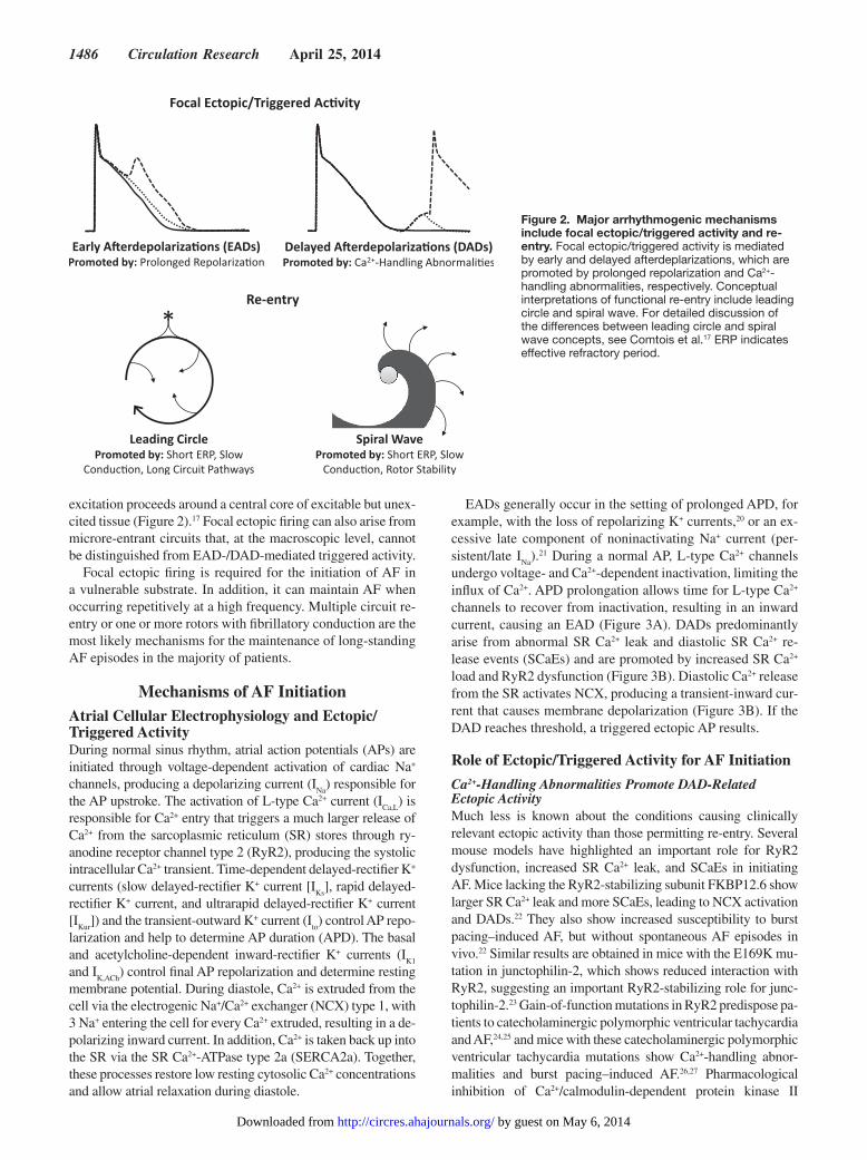
Fundamental AF MechanismsEctopic (triggered) activity and re-entry are major arrhythmo-genic mechanisms in AF (Figure 2).10,17,18 Focal ectopic/trig-gered activity is likely caused by early afterdepolarizations (EADs) and delayed afterdepolarizations (DADs). EADs are favored by delayed repolarization, whereas DADs depend on Ca2+-handling abnormalities.7,8,10,19 Re-entry can occur around
Nonstandard Abbreviations and Acronyms
AF atrial fibrillation
AP action potential
APD action potential duration
cAF chronic atrial fibrillation
CaMKII Ca2+/calmodulin-dependent protein kinase II
CREM cAMP-response element modulator
DAD delayed afterdepolarization
EAD early afterdepolarization
HF heart failure
ICa,L L-type Ca2+ current
IK1 basal inward-rectifier K+ current
IK,ACh acetylcholine-dependent inward-rectifier K+ current
IKs slow delayed-rectifier K+ current
IKur ultrarapid delayed-rectifier K+ current
INa Na+ current
Ito transient-outward K+ current
LA left atrium
NCX Na+/Ca2+ exchanger
pAF paroxysmal atrial fibrillation
PKA protein kinase A
PP1 protein phosphatase type 1
PV pulmonary vein
RA right atrium
RyR2 ryanodine receptor channel type 2
SCaE spontaneous sarcoplasmic reticulum Ca2+ release event
SERCA2a sarcoplasmic reticulum Ca2+-ATPase type 2a
SK small-conductance Ca2+-activated K+
SR sarcoplasmic reticulum
TGFβ1 transforming growth factor β1
TRP transient receptor potential
TRPC3 transient receptor potential canonical-3
by guest on May 6, 2014http://circres.ahajournals.org/Downloaded from
Heijman et al Cellular and Molecular Electrophysiology of AF 1485
an anatomic obstacle when each point in the pathway has suf-ficient time to regain excitability before the arrival of the next impulse. The likelihood of anatomic re-entry is controlled by the wavelength (conduction velocity × effective refractory period).7 Re-entry can also be functional, when premature
impulses conduct unidirectionally around an initially refrac-tory border. Several conceptual interpretations of functional re-entry exist.17,18 In the leading circle model, re-entry occurs in a circuit with a size equal to the wavelength with a central con-tinuously refractory core, whereas in the spiral wave model,
B
C
A
Figure 1. Conceptual framework of atrial fibrillation (AF) initiation, maintenance, and progression. A, In patients with a sufficiently large genetic predisposition, AF onset may occur at a relatively young age. AF-induced remodeling helps to maintain the arrhythmia, as well as promoting AF progression. B, In most patients, the genetic substrate alone does not provide sufficient susceptibility for AF. Additional disease-related remodeling may increase vulnerability and allow the initiation of paroxysmal AF episodes. Over time, some patients with paroxysmal AF may progress to longer-lasting persistent AF forms. C, Because of the composition of substrate and trigger, some patients have a first AF episode lasting >7 d and may progress to permanent AF due either to progression of underlying disease or to a medical decision to leave the patient in AF. (Note that for convenience the time scale for AF episodes, in gray, is expanded compared with the lower axis providing a sense of lifetime time course.)
by guest on May 6, 2014http://circres.ahajournals.org/Downloaded from
1486 Circulation Research April 25, 2014
excitation proceeds around a central core of excitable but unex-cited tissue (Figure 2).17 Focal ectopic firing can also arise from microre-entrant circuits that, at the macroscopic level, cannot be distinguished from EAD-/DAD-mediated triggered activity.
Focal ectopic firing is required for the initiation of AF in a vulnerable substrate. In addition, it can maintain AF when occurring repetitively at a high frequency. Multiple circuit re-entry or one or more rotors with fibrillatory conduction are the most likely mechanisms for the maintenance of long-standing AF episodes in the majority of patients.
Mechanisms of AF InitiationAtrial Cellular Electrophysiology and Ectopic/Triggered ActivityDuring normal sinus rhythm, atrial action potentials (APs) are initiated through voltage-dependent activation of cardiac Na+ channels, producing a depolarizing current (I
Na) responsible for
the AP upstroke. The activation of L-type Ca2+ current (ICa,L
) is responsible for Ca2+ entry that triggers a much larger release of Ca2+ from the sarcoplasmic reticulum (SR) stores through ry-anodine receptor channel type 2 (RyR2), producing the systolic intracellular Ca2+ transient. Time-dependent delayed-rectifier K+ currents (slow delayed-rectifier K+ current [I
Ks], rapid delayed-
rectifier K+ current, and ultrarapid delayed-rectifier K+ current [I
Kur]) and the transient-outward K+ current (I
to) control AP repo-
larization and help to determine AP duration (APD). The basal and acetylcholine-dependent inward-rectifier K+ currents (I
K1
and IK,ACh
) control final AP repolarization and determine resting membrane potential. During diastole, Ca2+ is extruded from the cell via the electrogenic Na+/Ca2+ exchanger (NCX) type 1, with 3 Na+ entering the cell for every Ca2+ extruded, resulting in a de-polarizing inward current. In addition, Ca2+ is taken back up into the SR via the SR Ca2+-ATPase type 2a (SERCA2a). Together, these processes restore low resting cytosolic Ca2+ concentrations and allow atrial relaxation during diastole.
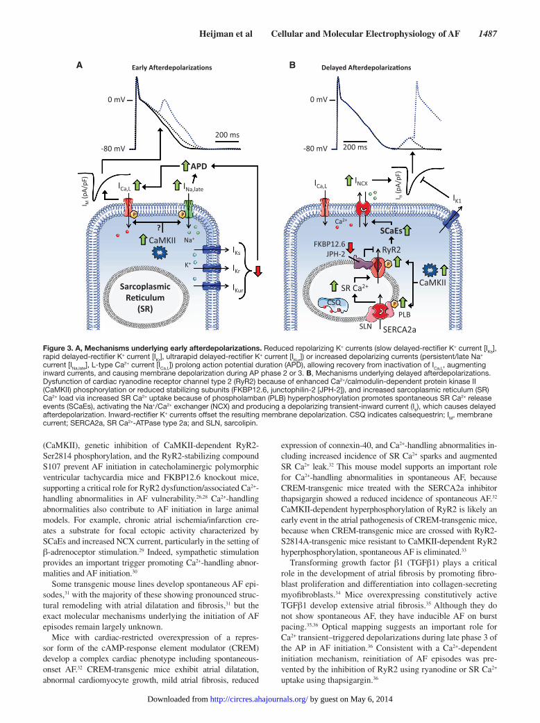
EADs generally occur in the setting of prolonged APD, for example, with the loss of repolarizing K+ currents,20 or an ex-cessive late component of noninactivating Na+ current (per-sistent/late I
Na).21 During a normal AP, L-type Ca2+ channels
undergo voltage- and Ca2+-dependent inactivation, limiting the influx of Ca2+. APD prolongation allows time for L-type Ca2+ channels to recover from inactivation, resulting in an inward current, causing an EAD (Figure 3A). DADs predominantly arise from abnormal SR Ca2+ leak and diastolic SR Ca2+ re-lease events (SCaEs) and are promoted by increased SR Ca2+ load and RyR2 dysfunction (Figure 3B). Diastolic Ca2+ release from the SR activates NCX, producing a transient-inward cur-rent that causes membrane depolarization (Figure 3B). If the DAD reaches threshold, a triggered ectopic AP results.
Role of Ectopic/Triggered Activity for AF Initiation
Ca2+-Handling Abnormalities Promote DAD-Related Ectopic ActivityMuch less is known about the conditions causing clinically relevant ectopic activity than those permitting re-entry. Several mouse models have highlighted an important role for RyR2 dysfunction, increased SR Ca2+ leak, and SCaEs in initiating AF. Mice lacking the RyR2-stabilizing subunit FKBP12.6 show larger SR Ca2+ leak and more SCaEs, leading to NCX activation and DADs.22 They also show increased susceptibility to burst pacing–induced AF, but without spontaneous AF episodes in vivo.22 Similar results are obtained in mice with the E169K mu-tation in junctophilin-2, which shows reduced interaction with RyR2, suggesting an important RyR2-stabilizing role for junc-tophilin-2.23 Gain-of-function mutations in RyR2 predispose pa-tients to catecholaminergic polymorphic ventricular tachycardia and AF,24,25 and mice with these catecholaminergic polymorphic ventricular tachycardia mutations show Ca2+-handling abnor-malities and burst pacing–induced AF.26,27 Pharmacological inhibition of Ca2+/calmodulin- dependent protein kinase II
Figure 2. Major arrhythmogenic mechanisms include focal ectopic/triggered activity and re-entry. Focal ectopic/triggered activity is mediated by early and delayed afterdeplarizations, which are promoted by prolonged repolarization and Ca2+-handling abnormalities, respectively. Conceptual interpretations of functional re-entry include leading circle and spiral wave. For detailed discussion of the differences between leading circle and spiral wave concepts, see Comtois et al.17 ERP indicates effective refractory period.
by guest on May 6, 2014http://circres.ahajournals.org/Downloaded from
Heijman et al Cellular and Molecular Electrophysiology of AF 1487
(CaMKII), genetic inhibition of CaMKII-dependent RyR2-Ser2814 phosphorylation, and the RyR2-stabilizing compound S107 prevent AF initiation in catecholaminergic polymorphic ventricular tachycardia mice and FKBP12.6 knockout mice, supporting a critical role for RyR2 dysfunction/associated Ca2+-handling abnormalities in AF vulnerability.26,28 Ca2+-handling abnormalities also contribute to AF initiation in large animal models. For example, chronic atrial ischemia/infarction cre-ates a substrate for focal ectopic activity characterized by SCaEs and increased NCX current, particularly in the setting of β-adrenoceptor stimulation.29 Indeed, sympathetic stimulation provides an important trigger promoting Ca2+-handling abnor-malities and AF initiation.30
Some transgenic mouse lines develop spontaneous AF epi-sodes,31 with the majority of these showing pronounced struc-tural remodeling with atrial dilatation and fibrosis,31 but the exact molecular mechanisms underlying the initiation of AF episodes remain largely unknown.
Mice with cardiac-restricted overexpression of a repres-sor form of the cAMP-response element modulator (CREM) develop a complex cardiac phenotype including spontaneous- onset AF.32 CREM-transgenic mice exhibit atrial dilatation, abnormal cardiomyocyte growth, mild atrial fibrosis, reduced
expression of connexin-40, and Ca2+-handling abnormalities in-cluding increased incidence of SR Ca2+ sparks and augmented SR Ca2+ leak.32 This mouse model supports an important role for Ca2+-handling abnormalities in spontaneous AF, because CREM-transgenic mice treated with the SERCA2a inhibitor thapsigargin showed a reduced incidence of spontaneous AF.32 CaMKII-dependent hyperphosphorylation of RyR2 is likely an early event in the atrial pathogenesis of CREM-transgenic mice, because when CREM-transgenic mice are crossed with RyR2-S2814A-transgenic mice resistant to CaMKII-dependent RyR2 hyperphosphorylation, spontaneous AF is eliminated.33
Transforming growth factor β1 (TGFβ1) plays a critical role in the development of atrial fibrosis by promoting fibro-blast proliferation and differentiation into collagen-secreting myofibroblasts.34 Mice overexpressing constitutively active TGFβ1 develop extensive atrial fibrosis.35 Although they do not show spontaneous AF, they have inducible AF on burst pacing.35,36 Optical mapping suggests an important role for Ca2+ transient–triggered depolarizations during late phase 3 of the AP in AF initiation.36 Consistent with a Ca2+-dependent initiation mechanism, reinitiation of AF episodes was pre-vented by the inhibition of RyR2 using ryanodine or SR Ca2+ uptake using thapsigargin.36
A B
Figure 3. A, Mechanisms underlying early afterdepolarizations. Reduced repolarizing K+ currents (slow delayed-rectifier K+ current [IKs], rapid delayed-rectifier K+ current [IKr], ultrarapid delayed-rectifier K+ current [IKur]) or increased depolarizing currents (persistent/late Na+ current [INa,late], L-type Ca2+ current [ICa,L]) prolong action potential duration (APD), allowing recovery from inactivation of ICa,L, augmenting inward currents, and causing membrane depolarization during AP phase 2 or 3. B, Mechanisms underlying delayed afterdepolarizations. Dysfunction of cardiac ryanodine receptor channel type 2 (RyR2) because of enhanced Ca2+/calmodulin-dependent protein kinase II (CaMKII) phosphorylation or reduced stabilizing subunits (FKBP12.6, junctophilin-2 [JPH-2]), and increased sarcoplasmic reticulum (SR) Ca2+ load via increased SR Ca2+ uptake because of phospholamban (PLB) hyperphosphorylation promotes spontaneous SR Ca2+ release events (SCaEs), activating the Na+/Ca2+ exchanger (NCX) and producing a depolarizing transient-inward current (Iti), which causes delayed afterdepolarization. Inward-rectifier K+ currents offset the resulting membrane depolarization. CSQ indicates calsequestrin; IM, membrane current; SERCA2a, SR Ca2+-ATPase type 2a; and SLN, sarcolipin.
by guest on May 6, 2014http://circres.ahajournals.org/Downloaded from
1488 Circulation Research April 25, 2014
There is paucity of large animal models showing spontane-ous AF. In dog, pig, goat, and sheep models, AF is generally initiated by burst pacing, with the duration of inducible AF being quantified as an index of the arrhythmia-maintaining substrate. One notable exception is dogs with chronic left ventricular myocardial infarction, which develop spontaneous AF episodes on sympathetic stimulation with tyramine.37 AF was because of Ca2+-dependent late phase 3 EADs around the left atrium (LA)/pulmonary vein (PV) junction.37 Spontaneous AF initiation around the LA/PV junction also occurs in aged rats after glycolytic inhibition.38 In this model, glycolytic inhibition interacts with the fibrotic substrate of the aged atria to amplify Ca2+-handling abnormalities that facilitate EAD-mediated triggered activity.38
Together, these studies support the concept that focal ecto-pic/triggered firing resulting from Ca2+-handling abnormali-ties, particularly in the atrial myocardium surrounding the PVs, may play an important role in the initiation of AF.
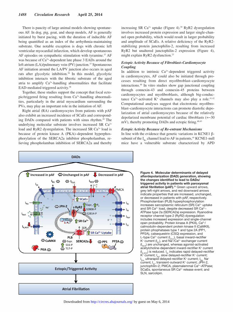
Right atrial (RA) cardiomyocytes from patients with pAF also exhibit an increased incidence of SCaEs and correspond-ing DADs compared with patients with sinus rhythm.39 The underlying molecular substrate involves increased SR Ca2+ load and RyR2 dysregulation. The increased SR Ca2+ load is because of protein kinase A (PKA)–dependent hyperphos-phorylation of the SERCA2a inhibitor phospholamban, re-lieving phospholamban inhibition of SERCA2a and thereby
increasing SR Ca2+ uptake (Figure 4).39 RyR2 dysregulation involves increased protein expression and larger single-chan-nel open probability, which would result in larger probability and amplitude of SCaEs. A relative deficiency of the RyR2-stabilizing protein junctophilin-2, resulting from increased RyR2 but unaltered junctophilin-2 expression (Figure 4), might explain RyR2 dysfunction.23
Ectopic Activity Because of Fibroblast–Cardiomyocyte CouplingIn addition to intrinsic Ca2+-dependent triggered activity in cardiomyocytes, AF could also be initiated through pro-cesses resulting from direct myofibroblast–cardiomyocyte interactions.40 In vitro studies show gap junctional coupling through connexin-43 and connexin-45 proteins between cardiomyocytes and myofibroblasts, although big-conduc-tance Ca2+-activated K+ channels may also play a role.41,42 Computational analyses suggest that electrotonic myofibro-blast–cardiomyocyte interactions can promote diastolic depo-larization of atrial cardiomyocytes because of the relatively depolarized membrane potential of cardiac fibroblasts (≈−30 mV), thereby promoting DADs and ectopic firing.40,43
Ectopic Activity Because of Re-entrant MechanismsIn line with the evidence that genetic variations in KCNE1 β-subunit of the I
Ks channel lead to AF in patients,44 KCNE1-null
mice have a vulnerable substrate characterized by APD
Increased in pAF Unchanged in pAF Decreased in pAF
DADs
Ectopic/Triggered Ac�vity
Atrial Fibrilla�on
Figure 4. Molecular determinants of delayed afterdepolarization (DAD) generation, showing the changes identified to lead to DADs/triggered activity in patients with paroxysmal atrial fibrillation (pAF).50 Green upward arrows, grey left-right arrows, and red downward arrows indicate properties that are increased, unchanged, or decreased in patients with pAF, respectively. Phospholamban (PLB) hyperphosphorylation increases sarcoplasmic reticulum (SR) Ca2+ uptake and SR Ca2+ load, despite decreased SR Ca2+-ATPase type 2a (SERCA2a) expression. Ryanodine receptor channel type 2 (RyR2) dysregulation includes increased expression and single-channel open probability. Protein kinase A (PKA), Ca2+/calmodulin-dependent protein kinase II (CaMKII), protein phophatases type 1 and type 2A (PP1, PP2A), calsequestrin (CSQ) expression, and L-type Ca2+ current (ICa,L), basal inward-rectifier K+ current (IK1), and Na+/Ca2+ exchanger current (INCX) are unchanged, whereas agonist-activated acetylcholine-dependent inward-rectifier K+ current (IK,ACh) is reduced. IKr indicates rapid delayed-rectifier K+ current; IKs, slow delayed-rectifier K+ current; IKur, ultrarapid delayed-rectifier K+ current; INa, Na+ current; Ito, transient-outward K+ current; JPH-2, junctophilin-2; PMCA, plasmalemmal Ca2+-ATPase; SCaEs, spontaneous SR Ca2+ release event; and SLN, sarcolipin.
by guest on May 6, 2014http://circres.ahajournals.org/Downloaded from
Heijman et al Cellular and Molecular Electrophysiology of AF 1489
shortening, with spontaneous AF episodes.45 The molecular mechanisms underlying the initiation of AF were not studied, although APD prolongation with isoprenaline reduces the in-cidence of AF episodes.45
Aged spontaneously hypertensive rats have a pronounced fibrotic substrate promoting AF.46 These rats showed sponta-neous atrial tachyarrhythmias associated with an autonomic imbalance with relative vagal hyperactivity, producing repo-larization shortening and heterogeneity that preceded the oc-currence of arrhythmia.47
Role of the PVsPV sleeves play an important role in the initiation of AF.48 The isolation of PVs prevents AF recurrence in 75% of patients with pAF.49 Both structural and functional properties of the PV cardiomyocyte sleeves contribute to their arrhythmogenic potential.50 The PV sleeves have a tissue architecture consist-ing of discrete fibers with abrupt changes in fiber direction, resulting in reduced electrotonic load and facilitating the development of focal ectopic activity. The identification of molecular mechanisms promoting ectopic activity around the PVs in humans is difficult because of the limited availability of PV tissue from patients,51 but recent results showed no dif-ferences in gene expression profiles of major ion channel sub-units or Ca2+-handling proteins between PV sleeves and LA free wall tissue samples from patients with valvular AF.52 The transcription factor PITX2 is highly expressed around the PVs and is critical for the development of the PV sleeve myocar-dium.53 PITX2 downregulates the nodal gene program, sup-pressing the development of focal ectopic activity around the PVs. Accordingly, reduced PITX2 expression in patients with AF and genetic variants close to the PITX2 gene have been as-sociated with increased AF susceptibility.54,55 Animal studies revealed reduced expression of I
K1 in PV sleeves,56 resulting
in depolarized resting potentials that facilitate ectopic activity. The propensity for SCaEs was increased in PV regions versus LA or RA appendages in some57 but not all studies.58 If hu-man PV sleeve myocytes are vulnerable to SCaEs, this could further explain their importance in AF initiation.
In addition to the re-entry-favoring fiber architecture, effec-tive refractory periods around the PVs are shorter in patients with pAF,59 further increasing the likelihood of a re-entrant circuit around the PVs that can initiate or maintain AF.
Mechanisms of AF MaintenanceMechanisms of Cardiac Conduction and Re-entryRe-entry is promoted by short effective refractory periods and slow impulse conduction. Postrepolarization refractoriness largely results from voltage-dependent inactivation of Na+ channels.60 Atrial Na+ channels have been suggested to have a more negative half-inactivation voltage compared with ventric-ular channels, allowing for greater postrepolarization refracto-riness, particularly in the presence of Na+ channel blockers.60
Conduction velocity is mainly determined by excitatory Na+ current, cardiomyocyte electric coupling through gap junctions, and muscle bundle architecture.8,10,17 Reduced I
Na,
decreased gap junctional coupling, and muscle bundle discon-tinuities resulting from fibrosis all reduce conduction velocity
and promote re-entry. Ca2+-handling abnormalities can also promote AF maintenance through conduction slowing.61 Atrial conduction velocity is reduced in mice with a RyR2 catechol-aminergic polymorphic ventricular tachycardia mutation caus-ing increased SR Ca2+ leak and in mouse hearts with acutely elevated intracellular Ca2+.62 The underlying mechanisms seem to involve both acute Ca2+-dependent inhibition of Na+ chan-nels and chronic downregulation of Nav1.5 expression.63
Experimental Models of Primary Cardiac Conditions Promoting AF Maintenance
APD ChangesIn HF because of 3 to 6 weeks of ventricular tachypacing, I
Ca,L, I
to, and I
Ks are reduced; I
K1 and T-type Ca2+ currents are
unaltered; and NCX current is increased.64 Atrial APD is un-altered at slow rates and slightly prolonged at faster rates.64 Experimental HF also increases the incidence of DADs, likely because of intracellular Ca2+ overload.65 In another study of long-term tachypacing-induced HF, an increase in I
to, largely
unaltered ICa,L
, and reduced IK1
, IKur
, and IKs
were observed, along with a shortening of atrial APD.66 Together, these stud-ies suggest complex time-dependent HF-induced atrial elec-tric remodeling.67 Other models have not been characterized as extensively. AF promotion associated with chronic volume overload in sheep is characterized by APD triangulation and I
Ca,L reduction.68 Endurance exercise training increases vagal
tone, causing heterogeneous APD shortening via increased sensitivity to acetylcholine because of a reduction in regula-tors of G-protein signaling proteins.69 Normal aging is also associated with electric remodeling, including a reduction in I
Ca,L and increased I
to, although other studies have reported a
seemingly contradictory APD prolongation.70
Ca2+-Handling AbnormalitiesExperimental HF increases atrial cardiomyocyte intracellu-lar Ca2+ concentration, Ca2+ transient amplitude, and SR Ca2+ load, promoting SCaEs and triggered activity.71 Underlying molecular mechanisms involve increased CaMKII and protein phosphatase type 1 (PP1) activity, CaMKII-dependent phos-pholamban hyperphosphorylation, reduced RyR2 expression with unaltered phosphorylation, and reduced expression of calsequestrin.71 In addition, tachypacing-induced HF caused degeneration of the T-tubular network, responsible for syn-chronizing Ca2+-induced Ca2+ release from the SR in sheep atrial cardiomyocytes.72
Conduction Abnormalities and Structural RemodelingIncreased atrial fibrosis and atrial dilatation are central fea-tures of atrial structural remodeling in a large number of ani-mal AF models, including ventricular tachypacing–induced HF,65,73–75 endurance exercise training,69 atrial infarction,29 chronic volume overload,68 and aging.70 These changes are as-sociated with re-entry-promoting conduction abnormalities.
Angiotensin II and TGFβ1 are the major profibrotic signal-ing molecules, with additional roles for platelet-derived and connective tissue growth factors.34,76 HF-induced atrial fibrosis is preceded by increased tissue angiotensin II levels and acti-vation of mitogen-activated protein kinases, c-Jun N-terminal kinase, and extracellular signal–related kinase.75 Although
by guest on May 6, 2014http://circres.ahajournals.org/Downloaded from
1490 Circulation Research April 25, 2014
angiotensin-converting enzyme inhibition prevented these changes, atrial fibrosis was only partially reduced, highlight-ing the multifactorial nature of atrial fibrosis.75
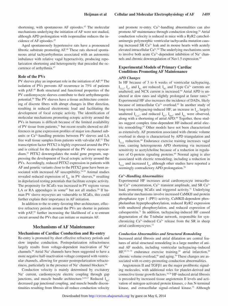
The proliferation of fibroblasts and their differentiation into collagen-secreting myofibroblasts play a critical role in fibrosis (Figure 5), with atrial fibroblasts showing greater fibrotic responses compared with ventricular fibroblasts.77 MicroRNA-21 plays a major role in profibrotic remodeling by reducing Sprouty-1.78 Sprouty-1 is a negative regulator of type 1/2 extracellular signal–related kinase, thereby inhib-iting fibroblast survival and density.78,79 LA microRNA-21 knockdown suppresses atrial fibrosis and AF substrate devel-opment in rats with post-MI HF.79 MicroRNA-29b suppress-es collagen gene expression and is downregulated in canine HF, so microRNA-29 downregulation could contribute to HF-related fibrosis.80
Fibroblast ion channel remodeling may also promote AF. Ca2+ permeable transient receptor potential (TRP) canonical-3 (TRPC3) channels regulate cardiac fibroblast proliferation
and differentiation, likely by mediating fibroblast Ca2+ entry that activates extracellular signal–related kinase signaling.81 TRPC3 expression is increased in atria from patients with AF, goats with electrically maintained AF, and dogs with tachypacing-induced HF, because of reduced repression re-sulting from the downregulation of microRNA-26.81 In con-trast, TRPC3 knockdown decreases canine atrial fibroblast proliferation.81 Kv1.5 seems to be the principal K+ channel α-subunit in fibroblasts, and channel expression is strongly downregulated in HF dogs, thereby promoting fibroblast pro-liferation and suggesting a functional role in HF-induced AF-promoting fibrosis.82 Atrial fibroblasts also express Nav1.5 α-subunits and Na+ currents when differentiated into myofi-broblasts, and the resulting Na+ entry may contribute to their arrhythmogenic potential.83,84
In addition to promoting muscle bundle discontinuities, myofibroblasts can affect atrial cardiomyocytes through paracrine interactions, notably via angiotensin II and TGFβ1 (Figure 5).85 Moreover, myofibroblasts promote re-entry via
Disease-Related Remodeling AF-Related Remodeling
Fibroblast
Fibrosis/Collagen
Deposi�on
Slow/Heterogeneous Conduc�on Focal Ectopic Ac�vity (?)
Re-entry
Paracrine Interac�ons
Ang-II / AldoTGF-β1TNFα
Electrotonic Interac�ons
Atrial Fibrilla�on
Gap-junc�ons
Cardiomyocyte
TRPM7
miR-21
Ang-II
Kv1.5
miR-29b
TRPC3
Nav1.5miR-26
Myofibroblast
Myofibroblast
Figure 5. Arrhythmogenic changes in atrial fibroblasts. Disease- and atrial fibrillation (AF)–related remodeling promotes fibroblast differentiation into myofibroblasts, involving altered expression of several ion channel proteins and microRNAs (miRs). Myofibroblasts facilitate AF maintenance by promoting re-entry through fibrosis/collagen deposition, as well as paracrine and direct electrotonic interactions with cardiomyocytes. Ado indicates aldosterone; Ang-II, angiotensin II; TGFβ1, transforming growth factor β1; TNFα, tumor necrosis factor α; TRPC3, transient receptor potential (TRP) canonical-3; and TRPM, TRP melastatin–related 7.
by guest on May 6, 2014http://circres.ahajournals.org/Downloaded from
Heijman et al Cellular and Molecular Electrophysiology of AF 1491
direct electric interaction with cardiomyocytes (Figure 5),40 by reducing conduction velocity through passive loading and depolarization-induced Na+ channel inactivation.
Conduction abnormalities are also promoted by impaired cell-to-cell coupling via gap junctions. For example, acute atri-al ischemia promotes AF induction by impairing cell-to-cell coupling, causing severe local conduction slowing.86 HF causes connexin-43 dephosphorylation and associated gap junction lateralization.73 However, because recovery from HF normalizes atrial function and connexin properties, but not fi-brosis, conduction abnormalities, or AF persistence, fibrosis is probably the predominant determinant of AF maintenance in experimental HF.73 Accordingly, the gap junction stabilizer rotigaptide suppresses AF in acute atrial ischemia but not HF.87
Clinical Disease–Related Atrial Remodeling Promoting AF MaintenancePatients with valvular heart disease show substantial remodel-ing of cardiac ion channel gene expression, with additional remodeling because of AF.88 Left ventricular systolic dys-function is associated with APD shortening in the presence of unaltered I
Ca,L, I
K1, or sustained outward current, possibly
because of increased Ito.89 In contrast, mitral valve disease
and low left ventricular ejection fraction are associated with reduced I
Ca,L,90 whereas atrial dilatation involves reduced I
Ca,L
and Ito.91 It is likely that such disease-related remodeling pre-
disposes to AF, especially in combination with AF risk factors. In addition, AF can also be mediated by atrial stretch resulting from hypertension, HF, or mitral valve disease. Atrial stretch is a common paradigm in AF-related conditions and might im-portantly contribute to AF-promoting structural remodeling.92
The consequences of atrial pressure and volume overload could also be directly related to these underlying diseases, in-dependent of atrial ion channel remodeling.
AF-Induced Remodeling Promoting AF Maintenance in Animal ModelsIn addition to disease-related remodeling, AF-induced atrial remodeling seems to play a major role in the maintenance, progression, and stabilization of AF.34,93–95
APD ShorteningAtrial tachycardia pacing causes a pronounced reduction in atrial APD associated with reduced I
Ca,L and I
to caused by the
downregulation of the underlying Cav1.2 and Kv4.3 sub-unit expression, an increase in constitutively active I
K,ACh,96
whereas IK1
, rapid delayed-rectifier K+ current, IKs
, IKur
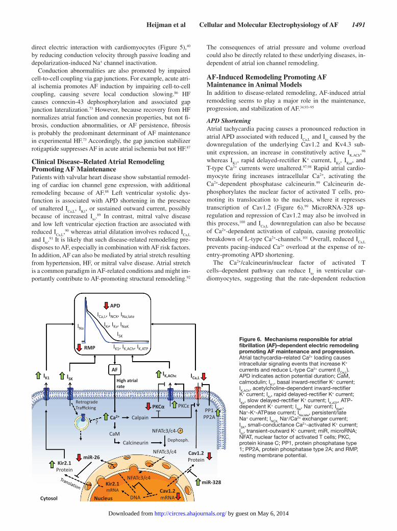
, and T-type Ca2+ currents were unaltered.97,98 Rapid atrial cardio-myocyte firing increases intracellular Ca2+, activating the Ca2+-dependent phosphatase calcineurin.99 Calcineurin de-phosphorylates the nuclear factor of activated T cells, pro-moting its translocation to the nucleus, where it represses transcription of Cav1.2 (Figure 6).99 MicroRNA-328 up-regulation and repression of Cav1.2 may also be involved in this process,100 and I
Ca,L downregulation can also be because
of Ca2+-dependent activation of calpain, causing proteolitic breakdown of L-type Ca2+-channels.101 Overall, reduced I
Ca,L
prevents pacing-induced Ca2+ overload at the expense of re-entry-promoting APD shortening.
The Ca2+/calcineurin/nuclear factor of activated T cells–dependent pathway can reduce I
to in ventricular car-
diomyocytes, suggesting that the rate-dependent reduction
Figure 6. Mechanisms responsible for atrial fibrillation (AF)–dependent electric remodeling promoting AF maintenance and progression. Atrial tachycardia–related Ca2+ loading causes intracellular signaling events that increase K+ currents and reduce L-type Ca2+ current (ICa,L). APD indicates action potential duration; CaM, calmodulin; IK1, basal inward-rectifier K+ current; IK,ACh, acetylcholine-dependent inward-rectifier K+ current; IKr, rapid delayed-rectifier K+ current; IKs, slow delayed-rectifier K+ current; IK,ATP, ATP-dependent K+ current; INa, Na+ current; INaK, Na+-K+-ATPase current; INa,late, persistent/late Na+ current; INCX, Na+/Ca2+ exchanger current; ISK, small-conductance Ca2+-activated K+ current; Ito, transient-outward K+ current; miR, microRNA; NFAT, nuclear factor of activated T cells; PKC, protein kinase C; PP1, protein phosphatase type 1; PP2A, protein phosphatase type 2A; and RMP, resting membrane potential.
by guest on May 6, 2014http://circres.ahajournals.org/Downloaded from
1492 Circulation Research April 25, 2014
in Ito in AF could also be mediated via this pathway.102,103
Interestingly, similar mechanisms are responsible for the rate-dependent upregulation of I
K1: nuclear factor of activated T
cells reduces the expression of the inhibitory microRNA-26, removing translational inhibition of Kir2.1 by microRNA-26 (Figure 6).104 The rate-dependent increase in constitutively active I
K,ACh is also Ca2+-dependent and is at least partly medi-
ated via calpain, which breaks down classical protein kinase C type α-isoforms (Figure 6).105
Several studies have suggested a role for small-conductance Ca2+-activated K+ (SK) currents in AF. The expression of the SK1 subunit and SK channel open probability are enhanced in dogs with atrial tachycardia remodeling, promoting repolar-ization shortening,106 whereas the inhibition of SK channels prolongs atrial repolarization and reduces AF duration in sev-eral animal models.106,107
Ca2+-Handling AbnormalitiesCa2+ transient amplitude is reduced in dogs with atrial tachy-cardia remodeling, contributing to atrial contractile dysfunc-tion.108,109 The reduced Ca2+ transient amplitude feeds back on repolarization, contributing to reduced APD rate depen-dence in AF.110,111 In addition, atrial tachycardia remodeling induces impaired Ca2+ wave propagation to the cell center and is associated with hypophosphorylation-dependent myofila-ment desensitization because of reduced expression of PKA and increased activity of PP1 and CaMKII.110 In contrast, PKA-dependent phosphorylation of RyR2 is increased in dogs with atrial tachypacing, similar to patients with chronic AF (cAF), and is associated with decreased RyR2–FKBP12.6 interaction.112 In goats with persistent AF, PKA-dependent phospholamban phosphorylation is reduced (decreasing SR Ca2+ uptake), whereas CaMKII-dependent RyR2 phosphory-lation is increased (increasing SR Ca2+ leak), reducing SR Ca2+ load and contributing to reduced contractility associated with AF.113 In sheep with persistent AF, the coupling efficiency be-tween RyR and L-type Ca2+ channels is decreased, contribut-ing to reduced SR Ca2+ release and Ca2+ transient amplitude despite normal SR Ca2+ load.114
Conduction Abnormalities and Structural RemodelingLong-term atrial pacing leads to conduction slowing in sev-eral animal models. In canine atrial tachycardia remodeling, reduced conduction velocity is at least partly because of I
Na
downregulation.115 Heterogeneously reduced gap junction coupling because of connexin remodeling can also contribute to atrial conduction slowing. Heterogeneity in connexin-40 distribution correlated with increased AF stability in atrial cardiomyocytes from goats with AF because of repetitive burst pacing.116 Similarly, connexin-40 expression in the PVs is decreased in the canine tachypacing model, possibly be-cause of increased degradation by calpains activated by the Ca2+-loading effects of high atrial rates.117
Although less pronounced than in HF, atrial tachycar-dia remodeling promotes atrial contractile dysfunction and causes atrial dilatation.93 Calpain activation contributes to troponin breakdown and subsequent contractile dysfunc-tion after high-frequency activation.118 Atrial dilatation promotes atrial remodeling and fibrosis through increased
atrial stretch.92 Atrial tachycardia also results in atrial fibro-sis and increased susceptibility to AF, even in the absence of ventricular dysfunction, indicating that a high atrial rate per se can cause fibrosis.119 Recent work has identified com-ponents of the underlying signaling pathways. Serum from tachypaced atrial myocytes promotes fibroblast differentia-tion to collagen-secreting myofibroblasts, through autocrine and paracrine mechanisms.120 Rapid atrial activation in rab-bits produces fibrosis associated with increased angioten-sin II and TGFβ1, activation of the Smad2/3 pathway, and inhibition of the TGFβ1/Smad-mediated fibrosis antagonist Smad7, effects that are prevented by angiotensin II type 1 receptor blockade.121
Tachycardia-induced nuclear factor of activated T cell–me-diated decreases in fibroblast microRNA-26 may also contrib-ute to structural remodeling. Because microRNA-26 represses TRPC3 gene expression, microRNA-26 reductions enhance TRPC3 expression, promoting fibroblast proliferation/myofi-broblast differentiation.81
AF-Maintaining Substrates Resulting from AF-Induced Remodeling in PatientsA comparison of the electrophysiological and molecular char-acteristics of atrial cardiomyocytes from pAF versus patients with long-standing persistent cAF provides strong indications about the AF-promoting consequences of atrial tachycardia remodeling, because patients with pAF had been in normal sinus rhythm for days to weeks at the time of cardiac surgery, whereas patients with cAF had a persistent high atrial rate be-fore and during surgery.
APD ShorteningIn contrast to patients with pAF, atrial myocytes from pa-tients with cAF show decreased APD. Depolarizing I
Ca,L is
consistently reduced in cAF,122–124 likely because of an adap-tive mechanism to protect atrial myocytes from toxic Ca2+ overload resulting from fast rates. Reduced I
Ca,L contributes
both to reduced APD, promoting re-entry, and decreased Ca2+ transient amplitude, reducing atrial contractility. Cav1.2 α
1C-subunit expression is reduced in cAF atrial cardiomyo-
cytes in most but not all studies,125 possibly because of an in-crease in microRNA-328.100 In addition, there is evidence for altered Cav1.2 phosphorylation,124,126 S-nitrosylation,127 and channel subunit breakdown by calpain.128 The complex mo-lecular basis of reduced I
Ca,L in cAF suggests that the precise
mechanisms may differ among patients.Increased inward-rectifier K+ currents also contribute to
APD shortening in cAF. LA IK1
is increased in both pAF and cAF.129 The increase in I
K1 is because of increased pro-
tein expression of underlying Kir2.1 subunits,129,130 probably through a reduction of microRNAs that normally repress Kir2.1 translation104,131 and an enhancement of single-channel open probability.132 The increased single-channel open prob-ability may involve stronger channel dephosphorylation by PP1 and serine/threonine protein phosphatase type 2A in cAF.133 Agonist-activated I
K,ACh is larger in RA than in LA
from patients with sinus rhythm, but is decreased in RA of pAF and cAF because of a reduction in underlying Kir3.1 and Kir3.4 subunits.129,130 Kir3.4, but not Kir3.1, is regulated by
by guest on May 6, 2014http://circres.ahajournals.org/Downloaded from
Heijman et al Cellular and Molecular Electrophysiology of AF 1493
intracellular [Na+],134 resulting in an Na+-dependent increase in agonist-activated I
K,ACh.135 This Na+-dependent regulation
is lost in cAF, possibly because of a more pronounced re-duction of the Na+-sensitive subunit Kir3.4 than Kir3.1, and further reduces I
K,ACh at fast rates with increased intracellular
[Na+].135 IK,ACh
also develops agonist-independent (constitu-tive) activity in cAF.132 The constitutive activity of I
K,ACh in
cAF is promoted by abnormal channel phosphorylation by novel protein kinase C isoforms.133 Computational studies show that increased total inward-rectifier K+ current in cAF is the major contributor to the stabilization of re-entrant ro-tors by shortening APD and hyperpolarizing the resting mem-brane potential.136
There is evidence for increased IKs
in patients with cAF, which might contribute to APD shortening.137,138 The mo-lecular mechanisms underlying increased I
Ks are unknown.
Increased, decreased, and unaltered mRNA levels of the un-derlying KCNQ1 α-subunit have been reported in patients with cAF.88,125 The expression of the KCNE1 β-subunit is re-duced in patients with valvular heart disease, without differ-ences between sinus rhythm and patients with cAF.88
In one study, SK current was increased in cAF atrial car-diomyocytes and augmented by high-frequency depolariz-ing pulses.139 The increase in SK current was prevented by the inhibition of retrograde channel trafficking, suggesting a rate-dependent influence on membrane channel availabil-ity.139 However, another study reported reduced SK channel
expression in cAF atrial cardiomyocytes,140 possibly because of increased microRNA-499, downregulating the SK3 subunit.141
Despite reduced APD at full repolarization, APD at 20% repolarization is generally prolonged.142 This effect is partly because of smaller I
to123 through reduced expression of the un-
derlying Kv4.3 subunit. Ito reduction is more pronounced in LA
than in RA.137 Similarly, IKur
and Kv1.5 subunits are reduced in cAF.137,143,144 I
Kur reduction has indirect effects on other currents,
and the overall impact on APD depends on AP morphology.142 For example, there is evidence that reduced I
Kur can promote
EADs in the presence of sympathetic stimulation.145,146
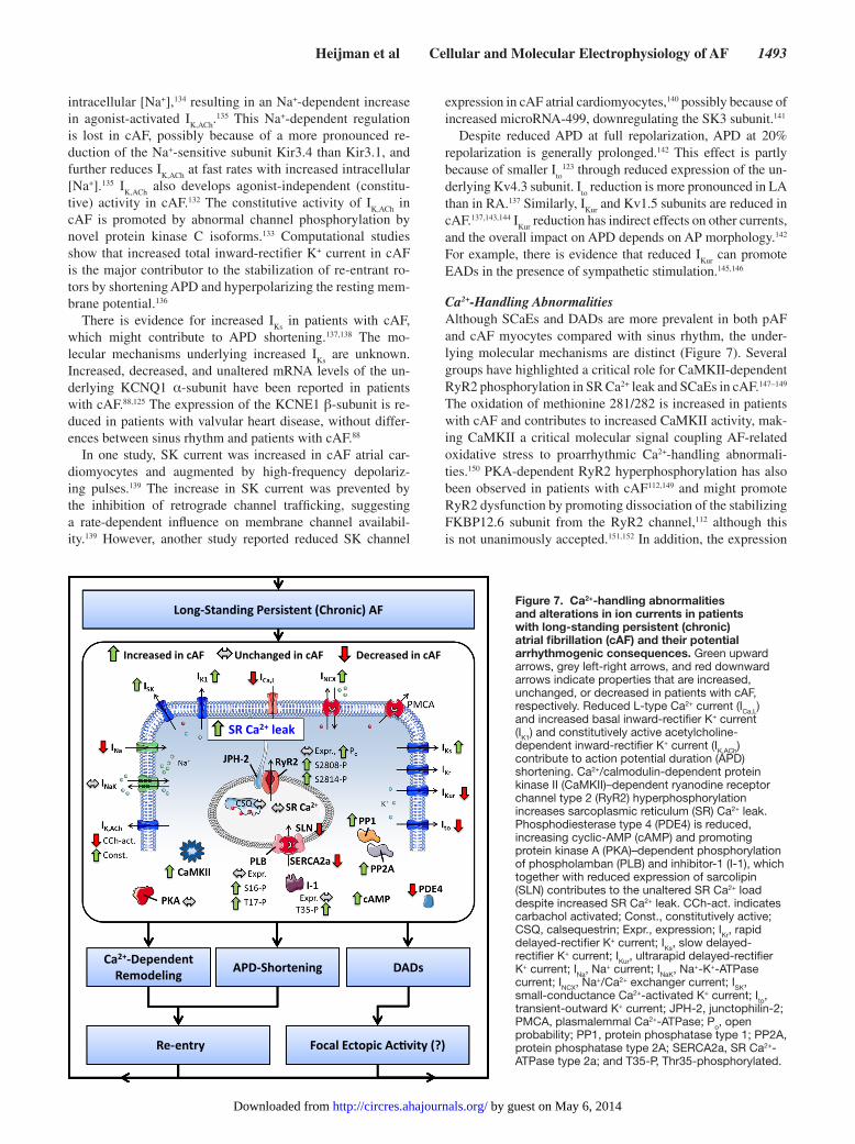
Ca2+-Handling AbnormalitiesAlthough SCaEs and DADs are more prevalent in both pAF and cAF myocytes compared with sinus rhythm, the under-lying molecular mechanisms are distinct (Figure 7). Several groups have highlighted a critical role for CaMKII-dependent RyR2 phosphorylation in SR Ca2+ leak and SCaEs in cAF.147–149 The oxidation of methionine 281/282 is increased in patients with cAF and contributes to increased CaMKII activity, mak-ing CaMKII a critical molecular signal coupling AF-related oxidative stress to proarrhythmic Ca2+-handling abnormali-ties.150 PKA-dependent RyR2 hyperphosphorylation has also been observed in patients with cAF112,149 and might promote RyR2 dysfunction by promoting dissociation of the stabilizing FKBP12.6 subunit from the RyR2 channel,112 although this is not unanimously accepted.151,152 In addition, the expression
-
Figure 7. Ca2+-handling abnormalities and alterations in ion currents in patients with long-standing persistent (chronic) atrial fibrillation (cAF) and their potential arrhythmogenic consequences. Green upward arrows, grey left-right arrows, and red downward arrows indicate properties that are increased, unchanged, or decreased in patients with cAF, respectively. Reduced L-type Ca2+ current (ICa,L) and increased basal inward-rectifier K+ current (IK1) and constitutively active acetylcholine-dependent inward-rectifier K+ current (IK,ACh) contribute to action potential duration (APD) shortening. Ca2+/calmodulin-dependent protein kinase II (CaMKII)–dependent ryanodine receptor channel type 2 (RyR2) hyperphosphorylation increases sarcoplasmic reticulum (SR) Ca2+ leak. Phosphodiesterase type 4 (PDE4) is reduced, increasing cyclic-AMP (cAMP) and promoting protein kinase A (PKA)–dependent phosphorylation of phospholamban (PLB) and inhibitor-1 (I-1), which together with reduced expression of sarcolipin (SLN) contributes to the unaltered SR Ca2+ load despite increased SR Ca2+ leak. CCh-act. indicates carbachol activated; Const., constitutively active; CSQ, calsequestrin; Expr., expression; IKr, rapid delayed-rectifier K+ current; IKs, slow delayed-rectifier K+ current; IKur, ultrarapid delayed-rectifier K+ current; INa, Na+ current; INaK, Na+-K+-ATPase current; INCX, Na+/Ca2+ exchanger current; ISK, small-conductance Ca2+-activated K+ current; Ito, transient-outward K+ current; JPH-2, junctophilin-2; PMCA, plasmalemmal Ca2+-ATPase; Po, open probability; PP1, protein phosphatase type 1; PP2A, protein phosphatase type 2A; SERCA2a, SR Ca2+-ATPase type 2a; and T35-P, Thr35-phosphorylated.
by guest on May 6, 2014http://circres.ahajournals.org/Downloaded from
1494 Circulation Research April 25, 2014
and activity of NCX are increased in cAF, so that SCaEs produce larger transient-inward currents.149 SR Ca2+ load is unaltered in cAF despite the larger SR Ca2+ leak,149 possibly because of increased phospholamban phosphorylation149 or reduced expression of the SERCA2a inhibitor sarcolipin.153,154 The increase in PP1 and protein phosphatase type 2A activity in patients with cAF would be expected to reduce phosphory-lation levels155; however, cAMP levels are increased in cAF,149 possibly because of reduced cAMP-hydrolyzing phospho-diesterase type 4,156 promoting PKA activation. In addition, increased PKA-dependent activation of the PP1 inhibitory protein inhibitor-1, which controls PP1 in the SR compart-ment,157 could explain phospholamban hyperphosphorylation because of local reductions in PP1 activity.155
Conduction Abnormalities and Structural RemodelingEarlier studies showed no change in I
Na or mRNA expression
of the Nav1.5 α-subunit in patients with cAF.158,159 However, recent studies reported reduced peak I
Na in patients with AF
that could contribute to re-entry-promoting conduction slow-ing.160,161 In addition, persistent/late I
Na is increased in some
studies.160 Although the exact functional consequences are presently unknown, patients with early-onset lone AF also ex-hibit a high prevalence of Na+ channel mutations that increase persistent/late I
Na.162
Connexin-40/connexin-43 mRNA and protein expres-sion are altered in patients with AF, potentially contributing to re-entry-promoting conduction abnormalities.163 Reduced connexin-40 expression has been reported in some studies,88,164 whereas others reported increased expression at the transverse cell membrane, promoting heterogeneous conduction, which was reduced by β-adrenoceptor blockade.165
Fibrosis is common in patients with AF,166 and connexin-43 remodeling correlates with atrial fibrosis in patients,167 sug-gesting an interaction between these re-entry-promoting factors. High-density electroanatomic mapping in patients identified conduction abnormalities that correlated with AF progression. Because conduction abnormalities also corre-lated with low electrogram voltage and percentage of com-plex electrograms, it was suggested that conduction slowing was because of AF-related fibrosis.168 There is evidence that fibroblast remodeling can occur as a consequence of AF, thereby promoting AF progression and stabilization. Beside the increase in TRPC3, TRP melastatin–related 7 channels are upregulated in patients with cAF and form a major Ca2+ permeation pathway in human atrial fibroblasts.169 The down-regulation of TRP melastatin–related 7 reduced AF fibroblast differentiation, and the atrial profibrotic effects of TGFβ1 require TRP melastatin–related 7–mediated Ca2+ signals.169 Recent work suggests that fibrocytes (bone marrow–derived fibroblast-like cells) may also be involved in atrial fibrosis of patients with cAF because they display stronger proliferative capacity and higher expression of collagen-I and α-smooth muscle actin.170
Mechanisms of AF ProgressionAs depicted in Figure 1, the progression of AF substrate oc-curs as a result of both AF-related remodeling and remodeling because of age and heart disease. The mechanistic components
of the underlying processes are discussed in detail above. The key components include changes in ion currents that promote re-entry by abbreviating APD/refractory period, alterations in connexin expression, Na+ current decreases and fibrotic remodeling that cause conduction slowing, and changes in Ca2+ handling that induce focal ectopic impulse formation. AF progression can also be because of the evolution of atrial changes caused by underlying cardiac and noncardiac diseas-es, independent of any AF-induced remodeling. For example, AF is an established risk factor for worsening HF,171,172 and the evolution of mixed-type atrial remodeling in patients with HF can create a vicious cycle further accelerating AF progres-sion. In contrast, some patients show limited AF progression, remaining in paroxysmal AF for decades. The mechanisms explaining this heterogeneity in the natural history of AF are at present largely unknown.
Gaps in Current Knowledge and Translational Prospects
Despite the enormous advances in our understanding of the molecular pathophysiology of AF during the past decades, there are still numerous important gaps that need to be ad-dressed. Structural remodeling seems a key for AF stabili-zation and therapy resistance. For many years, researchers focused on quantifying fibrosis as an index of structural re-modeling severity. However, processes such as fat accumu-lation,173 edema, amyloidosis,174 and other still unidentified factors might have great importance for AF progression and stabilization. The dynamic nature and specific pattern of myo-fibroblast–cardiomyocyte interactions is just emerging, and the extent to which they contribute to the initiation and main-tenance of AF is unclear.
The upstream and downstream signaling pathways leading to focal ectopic/triggered firing and AF-maintaining re-entry need precise delineation. The identification of nodal points in atrial cardiomyocyte signaling will be a key to sort out com-mon determinants among pathophysiological contributors. This may help to identify and target key drivers of the fibril-latory process.
Cardiac genomics and proteomics require further explora-tion and clarification. Advanced bioinformatics and compu-tational modeling approaches have the capacity to integrate and synthesize current insights to grapple with the complex-ity of AF. Computational science might play a key transla-tional role in understanding and combating the mechanisms of AF in vivo, because sophisticated multiscale compu-tational modeling can integrate the cellular and molecular processes in the second and third dimensions, providing key insights into the impact of molecular events for AF at the multicellular tissue level.
Although animal models have provided a wealth of informa-tion on AF pathophysiology, they have important limitations.46 Few currently available experimental models show spontane-ous AF occurrence and progression as observed in patients. Therapeutic interventions that are effective in animal models are often unsuccessful in patients, and the interpretation of genetic models may be hindered by complex compensatory phenomena.18,46 Animal models tend to focus on specific iso-lated pathophysiological stressors applied for a relatively short
by guest on May 6, 2014http://circres.ahajournals.org/Downloaded from
Heijman et al Cellular and Molecular Electrophysiology of AF 1495
period of time in the absence of other forms of disease (eg, AF because of experimental hypertension, HF, ischemia, diabetes mellitus, thyroid dysfunction). Clinical AF is often the result of many years of complex pathophysiology including multiple disease conditions, modified by extraneous drug therapy. Thus, the mechanisms observed in much simpler experimental mod-els might operate in complex combinations, or even not at all, in patients with similar clinical conditions. Newer methods in-volving in vivo imaging of structural and functional substrate in patients may hold the key to therapeutic application of fun-damental concepts,175–178 but currently available invasive and noninvasive mapping methods that assess the dynamics of AF in patients do not adequately exploit our knowledge of the cel-lular and molecular pathophysiology of AF.
Importantly, the causes of AF are extremely diverse. Rather than a specific disease, AF is a final end product of a wide range of clinical conditions, as discussed in detail in another article of this compendium.15 The exact combination of in-dividual pathophysiological processes contributing to AF is likely distinct in specific patient subsets.15 Improved under-standing of the connection between causes of AF and cellular mechanisms is required to provide tailored therapies for select patient cohorts.
AcknowledgmentsElements from Servier Medical Art were used in the making of the figures.
Sources of FundingOur work is supported by the European–North American Atrial Fibrillation Research Alliance grant of Fondation Leducq (07CVD03, to D. Dobrev and S. Nattel), the European Network for Translational Research in Atrial Fibrillation (261057, to D. Dobrev), the German Federal Ministry of Education and Research through the Atrial Fibrillation Competence Network (01Gi0204, to D. Dobrev), the Deutsche Forschungsgemeinschaft (Do 769/1-1-3, to D. Dobrev), the DZHK (German Center for Cardiovascular Research, to D. Dobrev), the Canadian Institutes of Health Research (6757 and 44365, to S. Nattel), and the Quebec Heart and Stroke Foundation (to S. Nattel).
DisclosuresNone.
References 1. Colilla S, Crow A, Petkun W, Singer DE, Simon T, Liu X. Estimates of
current and future incidence and prevalence of atrial fibrillation in the U.S. adult population. Am J Cardiol. 2013;112:1142–1147.
2. Go AS, Mozaffarian D, Roger VL, et al; American Heart Association Statistics Committee and Stroke Statistics Subcommittee. Heart disease and stroke statistics–2013 update: a report from the American Heart Association. Circulation. 2013;127:e6–e245.
3. Camm AJ, Lip GY, De Caterina R, Savelieva I, Atar D, Hohnloser SH, Hindricks G, Kirchhof P; ESC Committee for Practice Guidelines (CPG). 2012 focused update of the ESC Guidelines for the management of atrial fibrillation: an update of the 2010 ESC Guidelines for the management of atrial fibrillation. Developed with the special contribution of the European Heart Rhythm Association. Eur Heart J. 2012;33:2719–2747.
4. Camm AJ, Al-Khatib SM, Calkins H, Halperin JL, Kirchhof P, Lip GY, Nattel S, Ruskin J, Banerjee A, Blendea D, Guasch E, Needleman M, Savelieva I, Viles-Gonzalez J, Williams ES. A proposal for new clinical concepts in the management of atrial fibrillation. Am Heart J. 2012;164:292–302.e1.
5. Chiang CE, Naditch-Brûlé L, Murin J, Goethals M, Inoue H, O’Neill J, Silva-Cardoso J, Zharinov O, Gamra H, Alam S, Ponikowski P, Lewalter
T, Rosenqvist M, Steg PG. Distribution and risk profile of paroxysmal, persistent, and permanent atrial fibrillation in routine clinical practice: in-sight from the real-life global survey evaluating patients with atrial fibrilla-tion international registry. Circ Arrhythm Electrophysiol. 2012;5:632–639.
6. Calkins H, Kuck KH, Cappato R, et al. 2012 HRS/EHRA/ECAS Expert Consensus Statement on Catheter and Surgical Ablation of Atrial Fibrillation: recommendations for patient selection, procedural tech-niques, patient management and follow-up, definitions, endpoints, and research trial design. Europace. 2012;14:528–606.
7. Dobrev D, Nattel S. New antiarrhythmic drugs for treatment of atrial fi-brillation. Lancet. 2010;375:1212–1223.
8. Dobrev D, Carlsson L, Nattel S. Novel molecular targets for atrial fibrilla-tion therapy. Nat Rev Drug Discov. 2012;11:275–291.
9. Heijman J, Voigt N, Dobrev D. New directions in antiarrhythmic drug therapy for atrial fibrillation. Future Cardiol. 2013;9:71–88.
10. Wakili R, Voigt N, Kääb S, Dobrev D, Nattel S. Recent advances in the molecular pathophysiology of atrial fibrillation. J Clin Invest. 2011;121:2955–2968.
11. Nattel S, Guasch E, Savelieva I, et al. Early management of atrial fibril-lation to prevent cardiovascular complications [published online ahead of print February 16, 2014]. Eur Heart J. doi:10.1093/eurheartj/ehu028. Accessed April 10, 2014.
12. Mahida S, Lubitz SA, Rienstra M, Milan DJ, Ellinor PT. Monogenic atrial fibrillation as pathophysiological paradigms. Cardiovasc Res. 2011;89:692–700.
13. Mahida S, Ellinor PT. New advances in the genetic basis of atrial fibrilla-tion. J Cardiovasc Electrophysiol. 2012;23:1400–1406.
14. Fox CS, Parise H, D’Agostino RB Sr, Lloyd-Jones DM, Vasan RS, Wang TJ, Levy D, Wolf PA, Benjamin EJ. Parental atrial fibrillation as a risk factor for atrial fibrillation in offspring. JAMA. 2004;291:2851–2855.
15. Andrade J, Khairy P, Dobrev D, Nattel S. The clinical profile and patho-physiology of atrial fibrillation: relationships among clinical features, epi-demiology, and mechanisms. Circ Res. 2014;114:1453–1468.
16. Jahangir A, Lee V, Friedman PA, Trusty JM, Hodge DO, Kopecky SL, Packer DL, Hammill SC, Shen WK, Gersh BJ. Long-term progression and outcomes with aging in patients with lone atrial fibrillation: a 30-year follow-up study. Circulation. 2007;115:3050–3056.
17. Comtois P, Kneller J, Nattel S. Of circles and spirals: bridging the gap between the leading circle and spiral wave concepts of cardiac reentry. Europace. 2005;7(suppl 2):10–20.
18. Atienza F, Martins RP, Jalife J. Translational research in atrial fibrillation: a quest for mechanistically based diagnosis and therapy. Circ Arrhythm Electrophysiol. 2012;5:1207–1215.
19. Nattel S, Dobrev D. The multidimensional role of calcium in atrial fibrilla-tion pathophysiology: mechanistic insights and therapeutic opportunities. Eur Heart J. 2012;33:1870–1877.
20. Zellerhoff S, Pistulli R, Mönnig G, Hinterseer M, Beckmann BM, Köbe J, Steinbeck G, Kääb S, Haverkamp W, Fabritz L, Gradaus R, Breithardt G, Schulze-Bahr E, Böcker D, Kirchhof P. Atrial Arrhythmias in long-QT syndrome under daily life conditions: a nested case control study. J Cardiovasc Electrophysiol. 2009;20:401–407.
21. Lemoine MD, Duverger JE, Naud P, Chartier D, Qi XY, Comtois P, Fabritz L, Kirchhof P, Nattel S. Arrhythmogenic left atrial cellular electrophysi-ology in a murine genetic long QT syndrome model. Cardiovasc Res. 2011;92:67–74.
22. Sood S, Chelu MG, van Oort RJ, Skapura D, Santonastasi M, Dobrev D, Wehrens XH. Intracellular calcium leak due to FKBP12.6 deficiency in mice facilitates the inducibility of atrial fibrillation. Heart Rhythm. 2008;5:1047–1054.
23. Beavers DL, Wang W, Ather S, Voigt N, Garbino A, Dixit SS, Landstrom AP, Li N, Wang Q, Olivotto I, Dobrev D, Ackerman MJ, Wehrens XH. Mutation E169K in junctophilin-2 causes atrial fibrillation due to impaired RyR2 stabilization. J Am Coll Cardiol. 2013;62:2010–2019.
24. Sumitomo N, Sakurada H, Taniguchi K, Matsumura M, Abe O, Miyashita M, Kanamaru H, Karasawa K, Ayusawa M, Fukamizu S, Nagaoka I, Horie M, Harada K, Hiraoka M. Association of atrial arrhythmia and sinus node dysfunction in patients with catecholaminergic polymorphic ventricular tachycardia. Circ J. 2007;71:1606–1609.
25. Zhabyeyev P, Hiess F, Wang R, Liu Y, Wayne Chen SR, Oudit GY. S4153R is a gain-of-function mutation in the cardiac Ca2+ release channel ryanodine receptor associated with catecholaminergic polymorphic ventricular tachy-cardia and paroxysmal atrial fibrillation. Can J Cardiol. 2013;29:993–996.
26. Chelu MG, Sarma S, Sood S, Wang S, van Oort RJ, Skapura DG, Li N, Santonastasi M, Müller FU, Schmitz W, Schotten U, Anderson ME, Valderrábano M, Dobrev D, Wehrens XH. Calmodulin kinase II-mediated
by guest on May 6, 2014http://circres.ahajournals.org/Downloaded from
1496 Circulation Research April 25, 2014
sarcoplasmic reticulum Ca2+ leak promotes atrial fibrillation in mice. J Clin Invest. 2009;119:1940–1951.
27. Shan J, Xie W, Betzenhauser M, Reiken S, Chen BX, Wronska A, Marks AR. Calcium leak through ryanodine receptors leads to atrial fibrillation in 3 mouse models of catecholaminergic polymorphic ventricular tachycar-dia. Circ Res. 2012;111:708–717.
28. Li N, Wang T, Wang W, Cutler MJ, Wang Q, Voigt N, Rosenbaum DS, Dobrev D, Wehrens XH. Inhibition of CaMKII phosphorylation of RyR2 prevents induction of atrial fibrillation in FKBP12.6 knockout mice. Circ Res. 2012;110:465–470.
29. Nishida K, Qi XY, Wakili R, Comtois P, Chartier D, Harada M, Iwasaki YK, Romeo P, Maguy A, Dobrev D, Michael G, Talajic M, Nattel S. Mechanisms of atrial tachyarrhythmias associated with coronary artery occlusion in a chronic canine model. Circulation. 2011;123:137–146.
30. Workman AJ. Cardiac adrenergic control and atrial fibrillation. Naunyn Schmiedebergs Arch Pharmacol. 2010;381:235–249.
31. Riley G, Syeda F, Kirchhof P, Fabritz L. An introduction to murine models of atrial fibrillation. Front Physiol. 2012;3:296.
32. Kirchhof P, Marijon E, Fabritz L, et al. Overexpression of cAMP-response element modulator causes abnormal growth and development of the atrial myocardium resulting in a substrate for sustained atrial fibrillation in mice. Int J Cardiol. 2013;166:366–374.
33. Li N, Chiang DY, Wang S, et al. Ryanodine receptor-mediated calcium leak drives progressive development of an atrial fibrillation substrate in a transgenic mouse model. Circulation. 2014;129:1276–1285.
34. Nattel S, Burstein B, Dobrev D. Atrial remodeling and atrial fibrillation: mechanisms and implications. Circ Arrhythm Electrophysiol. 2008;1:62–73.
35. Verheule S, Sato T, Everett T IV, Engle SK, Otten D, Rubart-von der Lohe M, Nakajima HO, Nakajima H, Field LJ, Olgin JE. Increased vulnerability to atrial fibrillation in transgenic mice with selective atrial fibrosis caused by overexpression of TGF-β1. Circ Res. 2004;94:1458–1465.
36. Choi EK, Chang PC, Lee YS, Lin SF, Zhu W, Maruyama M, Fishbein MC, Chen Z, Rubart-von der Lohe M, Field LJ, Chen PS. Triggered firing and atrial fibrillation in transgenic mice with selective atrial fibrosis induced by overexpression of TGF-β1. Circ J. 2012;76:1354–1362.
37. Numata A, Miyauchi Y, Ono N, Fishbein MC, Mandel WJ, Lin SF, Weiss JN, Chen PS, Karagueuzian HS. Spontaneous atrial fibrillation initiated by tyramine in canine atria with increased sympathetic nerve sprouting. J Cardiovasc Electrophysiol. 2012;23:415–422.
38. Ono N, Hayashi H, Kawase A, Lin SF, Li H, Weiss JN, Chen PS, Karagueuzian HS. Spontaneous atrial fibrillation initiated by triggered ac-tivity near the pulmonary veins in aged rats subjected to glycolytic inhibi-tion. Am J Physiol Heart Circ Physiol. 2007;292:H639–H648.
39. Voigt N, Heijman J, Wang Q, Chiang DY, Li N, Karck M, Wehrens XH, Nattel S, Dobrev D. Cellular and molecular mechanisms of atrial arrhyth-mogenesis in patients with paroxysmal atrial fibrillation. Circulation. 2014;129:145–156.
40. Yue L, Xie J, Nattel S. Molecular determinants of cardiac fibroblast electri-cal function and therapeutic implications for atrial fibrillation. Cardiovasc Res. 2011;89:744–753.
41. Miragoli M, Gaudesius G, Rohr S. Electrotonic modulation of cardiac im-pulse conduction by myofibroblasts. Circ Res. 2006;98:801–810.
42. Wang YJ, Sung RJ, Lin MW, Wu SN. Contribution of BKCa– chan-
nel activity in human cardiac fibroblasts to electrical coupling of cardiomyocytes-fibroblasts. J Membr Biol. 2006;213:175–185.
43. Rohr S. Myofibroblasts in diseased hearts: new players in cardiac arrhyth-mias? Heart Rhythm. 2009;6:848–856.
44. Olesen MS, Bentzen BH, Nielsen JB, Steffensen AB, David JP, Jabbari J, Jensen HK, Haunsø S, Svendsen JH, Schmitt N. Mutations in the po-tassium channel subunit KCNE1 are associated with early-onset familial atrial fibrillation. BMC Med Genet. 2012;13:24.
45. Temple J, Frias P, Rottman J, Yang T, Wu Y, Verheijck EE, Zhang W, Siprachanh C, Kanki H, Atkinson JB, King P, Anderson ME, Kupershmidt S, Roden DM. Atrial fibrillation in KCNE1-null mice. Circ Res. 2005;97:62–69.
46. Nishida K, Michael G, Dobrev D, Nattel S. Animal models for atrial fibrillation: clinical insights and scientific opportunities. Europace. 2010;12:160–172.
47. Scridon A, Gallet C, Arisha MM, Oréa V, Chapuis B, Li N, Tabib A, Christé G, Barrès C, Julien C, Chevalier P. Unprovoked atrial tachyarrhythmias in aging spontaneously hypertensive rats: the role of the autonomic nervous system. Am J Physiol Heart Circ Physiol. 2012;303:H386–H392.
48. Haïssaguerre M, Jaïs P, Shah DC, Takahashi A, Hocini M, Quiniou G, Garrigue S, Le Mouroux A, Le Métayer P, Clémenty J. Spontaneous ini-tiation of atrial fibrillation by ectopic beats originating in the pulmonary veins. N Engl J Med. 1998;339:659–666.
49. Li WJ, Bai YY, Zhang HY, Tang RB, Miao CL, Sang CH, Yin XD, Dong JZ, Ma CS. Additional ablation of complex fractionated atrial electro-grams after pulmonary vein isolation in patients with atrial fibrillation: a meta-analysis. Circ Arrhythm Electrophysiol. 2011;4:143–148.
50. Nattel S. Paroxysmal atrial fibrillation and pulmonary veins: relation-ships between clinical forms and automatic versus re-entrant mechanisms. Can J Cardiol. 2013;29:1147–1149.
51. Voigt N, Dobrev D. The biology of human pulmonary veins: does it help us to better understand AF pathophysiology in patients? Heart Rhythm. 2013;10:392–393.
52. Yeh YH, Kuo CT, Lee YS, Lin YM, Nattel S, Tsai FC, Chen WJ. Region-specific gene expression profiles in the left atria of patients with valvular atrial fibrillation. Heart Rhythm. 2013;10:383–391.
53. Mommersteeg MT, Brown NA, Prall OW, de Gier-de Vries C, Harvey RP, Moorman AF, Christoffels VM. Pitx2c and Nkx2-5 are required for the formation and identity of the pulmonary myocardium. Circ Res. 2007;101:902–909.
54. Chinchilla A, Daimi H, Lozano-Velasco E, Dominguez JN, Caballero R, Delpón E, Tamargo J, Cinca J, Hove-Madsen L, Aranega AE, Franco D. PITX2 insufficiency leads to atrial electrical and structural remodeling linked to arrhythmogenesis. Circ Cardiovasc Genet. 2011;4:269–279.
55. Kirchhof P, Kahr PC, Kaese S, Piccini I, Vokshi I, Scheld HH, Rotering H, Fortmueller L, Laakmann S, Verheule S, Schotten U, Fabritz L, Brown NA. PITX2c is expressed in the adult left atrium, and reducing Pitx2c expression promotes atrial fibrillation inducibility and complex changes in gene expression. Circ Cardiovasc Genet. 2011;4:123–133.
56. Ehrlich JR, Cha TJ, Zhang L, Chartier D, Melnyk P, Hohnloser SH, Nattel S. Cellular electrophysiology of canine pulmonary vein cardiomyocytes: action potential and ionic current properties. J Physiol. 2003;551:801–813.
57. Honjo H, Boyett MR, Niwa R, Inada S, Yamamoto M, Mitsui K, Horiuchi T, Shibata N, Kamiya K, Kodama I. Pacing-induced spontaneous activity in myocardial sleeves of pulmonary veins after treatment with ryanodine. Circulation. 2003;107:1937–1943.
58. Coutu P, Chartier D, Nattel S. Comparison of Ca2+-handling properties of canine pulmonary vein and left atrial cardiomyocytes. Am J Physiol Heart Circ Physiol. 2006;291:H2290–H2300.
59. Jaïs P, Hocini M, Macle L, Choi KJ, Deisenhofer I, Weerasooriya R, Shah DC, Garrigue S, Raybaud F, Scavee C, Le Metayer P, Clémenty J, Haïssaguerre M. Distinctive electrophysiological properties of pulmonary veins in patients with atrial fibrillation. Circulation. 2002;106:2479–2485.
60. Burashnikov A, Di Diego JM, Zygmunt AC, Belardinelli L, Antzelevitch C. Atrium-selective sodium channel block as a strategy for suppression of atri-al fibrillation: differences in sodium channel inactivation between atria and ventricles and the role of ranolazine. Circulation. 2007;116:1449–1457.
61. Heijman J, Wehrens XH, Dobrev D. Atrial arrhythmogenesis in catechol-aminergic polymorphic ventricular tachycardia–is there a mechanistic link between sarcoplasmic reticulum Ca2+ leak and re-entry? Acta Physiol (Oxf). 2013;207:208–211.
62. King JH, Zhang Y, Lei M, Grace AA, Huang CL, Fraser JA. Atrial arrhyth-mia, triggering events and conduction abnormalities in isolated murine RyR2-P2328S hearts. Acta Physiol (Oxf). 2013;207:308–323.
63. King JH, Wickramarachchi C, Kua K, Du Y, Jeevaratnam K, Matthews HR, Grace AA, Huang CL, Fraser JA. Loss of Nav1.5 expression and function in murine atria containing the RyR2-P2328S gain-of-function mutation. Cardiovasc Res. 2013;99:751–759.
64. Li D, Melnyk P, Feng J, Wang Z, Petrecca K, Shrier A, Nattel S. Effects of experimental heart failure on atrial cellular and ionic electrophysiology. Circulation. 2000;101:2631–2638.
65. Stambler BS, Fenelon G, Shepard RK, Clemo HF, Guiraudon CM. Characterization of sustained atrial tachycardia in dogs with rapid ven-tricular pacing-induced heart failure. J Cardiovasc Electrophysiol. 2003;14:499–507.
66. Sridhar A, Nishijima Y, Terentyev D, Khan M, Terentyeva R, Hamlin RL, Nakayama T, Gyorke S, Cardounel AJ, Carnes CA. Chronic heart failure and the substrate for atrial fibrillation. Cardiovasc Res. 2009;84:227–236.
67. Rankin AC, Workman AJ. Duration of heart failure and the risk of atrial fibrillation: different mechanisms at different times? Cardiovasc Res. 2009;84:180–181.
68. Deroubaix E, Folliguet T, Rücker-Martin C, Dinanian S, Boixel C, Validire P, Daniel P, Capderou A, Hatem SN. Moderate and chronic hemodynamic overload of sheep atria induces reversible cellular electrophysiologic abnor-malities and atrial vulnerability. J Am Coll Cardiol. 2004;44:1918–1926.
69. Guasch E, Benito B, Qi X, et al. Atrial fibrillation promotion by endurance exercise: demonstration and mechanistic exploration in an animal model. J Am Coll Cardiol. 2013;62:68–77.
by guest on May 6, 2014http://circres.ahajournals.org/Downloaded from
Heijman et al Cellular and Molecular Electrophysiology of AF 1497
70. Dun W, Boyden PA. Aged atria: electrical remodeling conducive to atrial fibrillation. J Interv Card Electrophysiol. 2009;25:9–18.
71. Yeh YH, Wakili R, Qi XY, Chartier D, Boknik P, Kääb S, Ravens U, Coutu P, Dobrev D, Nattel S. Calcium-handling abnormalities underlying atrial arrhythmogenesis and contractile dysfunction in dogs with congestive heart failure. Circ Arrhythm Electrophysiol. 2008;1:93–102.
72. Dibb KM, Clarke JD, Horn MA, Richards MA, Graham HK, Eisner DA, Trafford AW. Characterization of an extensive transverse tubular network in sheep atrial myocytes and its depletion in heart failure. Circ Heart Fail. 2009;2:482–489.
73. Burstein B, Comtois P, Michael G, Nishida K, Villeneuve L, Yeh YH, Nattel S. Changes in connexin expression and the atrial fibrillation sub-strate in congestive heart failure. Circ Res. 2009;105:1213–1222.
74. Li D, Fareh S, Leung TK, Nattel S. Promotion of atrial fibrillation by heart failure in dogs: atrial remodeling of a different sort. Circulation. 1999;100:87–95.
75. Cardin S, Li D, Thorin-Trescases N, Leung TK, Thorin E, Nattel S. Evolution of the atrial fibrillation substrate in experimental congestive heart failure: angiotensin-dependent and -independent pathways. Cardiovasc Res. 2003;60:315–325.
76. Tan AY, Zimetbaum P. Atrial fibrillation and atrial fibrosis. J Cardiovasc Pharmacol. 2011;57:625–629.
77. Burstein B, Libby E, Calderone A, Nattel S. Differential behaviors of atrial versus ventricular fibroblasts: a potential role for platelet-derived growth factor in atrial-ventricular remodeling differences. Circulation. 2008;117:1630–1641.
78. Thum T, Gross C, Fiedler J, et al. MicroRNA-21 contributes to myocar-dial disease by stimulating MAP kinase signalling in fibroblasts. Nature. 2008;456:980–984.
79. Cardin S, Guasch E, Luo X, Naud P, Le Quang K, Shi Y, Tardif JC, Comtois P, Nattel S. Role for MicroRNA-21 in atrial profibrillatory fibrot-ic remodeling associated with experimental postinfarction heart failure. Circ Arrhythm Electrophysiol. 2012;5:1027–1035.
80. Dawson K, Wakili R, Ordög B, Clauss S, Chen Y, Iwasaki Y, Voigt N, Qi XY, Sinner MF, Dobrev D, Kääb S, Nattel S. MicroRNA29: a mechanis-tic contributor and potential biomarker in atrial fibrillation. Circulation. 2013;127:1466–1475,1475e1.
81. Harada M, Luo X, Qi XY, et al. Transient receptor potential canonical-3 channel-dependent fibroblast regulation in atrial fibrillation. Circulation. 2012;126:2051–2064.
82. Huang H, Qi X, Wu C-T, Naud P, Dobrev D, Nattel S. Abstract 14853: the potential role of fibroblast Kv channels in atrial fibrotic profibrillatory remodeling. Circulation. 2013;128:A14853.
83. Chatelier A, Mercier A, Tremblier B, Thériault O, Moubarak M, Benamer N, Corbi P, Bois P, Chahine M, Faivre JF. A distinct de novo expression of Nav1.5 sodium channels in human atrial fibroblasts differentiated into myofibroblasts. J Physiol. 2012;590:4307–4319.
84. Zhou X, Dobrev D. Voltage-gated Na+ channels: novel players in fibroblast-to-myofibroblast transition with a potential role in atrial ar-rhythmogenesis? J Physiol. 2012;590:4975.
85. Kakkar R, Lee RT. Intramyocardial fibroblast myocyte communication. Circ Res. 2010;106:47–57.
86. Sinno H, Derakhchan K, Libersan D, Merhi Y, Leung TK, Nattel S. Atrial isch-emia promotes atrial fibrillation in dogs. Circulation. 2003;107:1930–1936.
87. Shiroshita-Takeshita A, Sakabe M, Haugan K, Hennan JK, Nattel S. Model-dependent effects of the gap junction conduction-enhancing antiar-rhythmic peptide rotigaptide (ZP123) on experimental atrial fibrillation in dogs. Circulation. 2007;115:310–318.
88. Gaborit N, Steenman M, Lamirault G, Le Meur N, Le Bouter S, Lande G, Léger J, Charpentier F, Christ T, Dobrev D, Escande D, Nattel S, Demolombe S. Human atrial ion channel and transporter subunit gene-expression remodeling associated with valvular heart disease and atrial fibrillation. Circulation. 2005;112:471–481.
89. Workman AJ, Pau D, Redpath CJ, Marshall GE, Russell JA, Norrie J, Kane KA, Rankin AC. Atrial cellular electrophysiological changes in pa-tients with ventricular dysfunction may predispose to AF. Heart Rhythm. 2009;6:445–451.
90. Dinanian S, Boixel C, Juin C, Hulot JS, Coulombe A, Rücker-Martin C, Bonnet N, Le Grand B, Slama M, Mercadier JJ, Hatem SN. Downregulation of the calcium current in human right atrial myocytes from patients in sinus rhythm but with a high risk of atrial fibrillation. Eur Heart J. 2008;29:1190–1197.
91. Le Grand BL, Hatem S, Deroubaix E, Couétil JP, Coraboeuf E. Depressed transient outward and calcium currents in dilated human atria. Cardiovasc Res. 1994;28:548–556.
92. De Jong AM, Maass AH, Oberdorf-Maass SU, Van Veldhuisen DJ, Van Gilst WH, Van Gelder IC. Mechanisms of atrial structural changes caused by stretch occurring before and during early atrial fibrillation. Cardiovasc Res. 2011;89:754–765.
93. Shi Y, Ducharme A, Li D, Gaspo R, Nattel S, Tardif JC. Remodeling of atrial dimensions and emptying function in canine models of atrial fibril-lation. Cardiovasc Res. 2001;52:217–225.
94. Wijffels MC, Kirchhof CJ, Dorland R, Allessie MA. Atrial fibrillation begets atrial fibrillation. A study in awake chronically instrumented goats. Circulation. 1995;92:1954–1968.
95. Dobrev D. Electrical remodeling in atrial fibrillation. Herz. 2006;31:108–112; quiz 142.
96. Ehrlich JR, Cha TJ, Zhang L, Chartier D, Villeneuve L, Hébert TE, Nattel S. Characterization of a hyperpolarization-activated time-depen-dent potassium current in canine cardiomyocytes from pulmonary vein myocardial sleeves and left atrium. J Physiol. 2004;557:583–597.
97. Yue L, Feng J, Gaspo R, Li GR, Wang Z, Nattel S. Ionic remodeling un-derlying action potential changes in a canine model of atrial fibrillation. Circ Res. 1997;81:512–525.
98. Yue L, Melnyk P, Gaspo R, Wang Z, Nattel S. Molecular mechanisms underlying ionic remodeling in a dog model of atrial fibrillation. Circ Res. 1999;84:776–784.
99. Qi XY, Yeh YH, Xiao L, Burstein B, Maguy A, Chartier D, Villeneuve LR, Brundel BJ, Dobrev D, Nattel S. Cellular signaling underlying atrial tachycardia remodeling of L-type calcium current. Circ Res. 2008;103:845–854.
100. Lu Y, Zhang Y, Wang N, Pan Z, Gao X, Zhang F, Zhang Y, Shan H, Luo X, Bai Y, Sun L, Song W, Xu C, Wang Z, Yang B. MicroRNA-328 con-tributes to adverse electrical remodeling in atrial fibrillation. Circulation. 2010;122:2378–2387.
101. Brundel BJ, Kampinga HH, Henning RH. Calpain inhibition prevents pacing-induced cellular remodeling in a HL-1 myocyte model for atrial fibrillation. Cardiovasc Res. 2004;62:521–528.
102. Perrier E, Perrier R, Richard S, Bénitah JP. Ca2+ controls functional ex-pression of the cardiac K+ transient outward current via the calcineurin pathway. J Biol Chem. 2004;279:40634–40639.
103. Xiao L, Coutu P, Villeneuve LR, Tadevosyan A, Maguy A, Le Bouter S, Allen BG, Nattel S. Mechanisms underlying rate-dependent remodeling of transient outward potassium current in canine ventricular myocytes. Circ Res. 2008;103:733–742.
104. Luo X, Pan Z, Shan H, et al. MicroRNA-26 governs profibrillatory inward-rectifier potassium current changes in atrial fibrillation. J Clin Invest. 2013;123:1939–1951.
105. Makary S, Voigt N, Maguy A, Wakili R, Nishida K, Harada M, Dobrev D, Nattel S. Differential protein kinase C isoform regulation and in-creased constitutive activity of acetylcholine-regulated potassium chan-nels in atrial remodeling. Circ Res. 2011;109:1031–1043.
106. Qi XY, Diness JG, Brundel BJ, Zhou XB, Naud P, Wu CT, Huang H, Harada M, Aflaki M, Dobrev D, Grunnet M, Nattel S. Role of small-conductance calcium-activated potassium channels in atrial elec-trophysiology and fibrillation in the dog. Circulation. 2014;129:430–440.
107. Skibsbye L, Diness JG, Sørensen US, Hansen RS, Grunnet M. The du-ration of pacing-induced atrial fibrillation is reduced in vivo by inhi-bition of small conductance Ca2+-activated K+ channels. J Cardiovasc Pharmacol. 2011;57:672–681.
108. Sun H, Chartier D, Leblanc N, Nattel S. Intracellular calcium changes and tachycardia-induced contractile dysfunction in canine atrial myo-cytes. Cardiovasc Res. 2001;49:751–761.
109. Sun H, Gaspo R, Leblanc N, Nattel S. Cellular mechanisms of atrial con-tractile dysfunction caused by sustained atrial tachycardia. Circulation. 1998;98:719–727.
110. Wakili R, Yeh YH, Yan Qi X, et al. Multiple potential molecular contrib-utors to atrial hypocontractility caused by atrial tachycardia remodeling in dogs. Circ Arrhythm Electrophysiol. 2010;3:530–541.
111. Kneller J, Sun H, Leblanc N, Nattel S. Remodeling of Ca2+-handling by atrial tachycardia: evidence for a role in loss of rate-adaptation. Cardiovasc Res. 2002;54:416–426.
112. Vest JA, Wehrens XH, Reiken SR, Lehnart SE, Dobrev D, Chandra P, Danilo P, Ravens U, Rosen MR, Marks AR. Defective cardiac ry-anodine receptor regulation during atrial fibrillation. Circulation. 2005;111:2025–2032.
113. Greiser M, Neuberger HR, Harks E, El-Armouche A, Boknik P, de Haan S, Verheyen F, Verheule S, Schmitz W, Ravens U, Nattel S, Allessie MA, Dobrev D, Schotten U. Distinct contractile and mo-lecular differences between two goat models of atrial dysfunction:
by guest on May 6, 2014http://circres.ahajournals.org/Downloaded from
1498 Circulation Research April 25, 2014
AV block-induced atrial dilatation and atrial fibrillation. J Mol Cell Cardiol. 2009;46:385–394.
114. Lenaerts I, Bito V, Heinzel FR, Driesen RB, Holemans P, D’hooge J, Heidbüchel H, Sipido KR, Willems R. Ultrastructural and functional remodeling of the coupling between Ca2+ influx and sarcoplasmic reticu-lum Ca2+ release in right atrial myocytes from experimental persistent atrial fibrillation. Circ Res. 2009;105:876–885.
115. Gaspo R, Bosch RF, Bou-Abboud E, Nattel S. Tachycardia-induced changes in Na+ current in a chronic dog model of atrial fibrillation. Circ Res. 1997;81:1045–1052.
116. van der Velden HM, Ausma J, Rook MB, Hellemons AJ, van Veen TA, Allessie MA, Jongsma HJ. Gap junctional remodeling in rela-tion to stabilization of atrial fibrillation in the goat. Cardiovasc Res. 2000;46:476–486.
117. Zhang W, Ma X, Zhong M, Zheng Z, Li L, Wang Z, Zhang Y. Role of the calpain system in pulmonary vein connexin remodeling in dogs with atrial fibrillation. Cardiology. 2009;112:22–30.
118. Ke L, Qi XY, Dijkhuis AJ, Chartier D, Nattel S, Henning RH, Kampinga HH, Brundel BJ. Calpain mediates cardiac troponin degradation and contractile dysfunction in atrial fibrillation. J Mol Cell Cardiol. 2008;45:685–693.
119. Avitall B, Bi J, Mykytsey A, Chicos A. Atrial and ventricular fibrosis induced by atrial fibrillation: evidence to support early rhythm control. Heart Rhythm. 2008;5:839–845.
120. Burstein B, Qi XY, Yeh YH, Calderone A, Nattel S. Atrial cardiomyocyte tachycardia alters cardiac fibroblast function: a novel consideration in atrial remodeling. Cardiovasc Res. 2007;76:442–452.
121. He X, Gao X, Peng L, Wang S, Zhu Y, Ma H, Lin J, Duan DD. Atrial fibrillation induces myocardial fibrosis through angiotensin II type 1 receptor-specific Arkadia-mediated downregulation of Smad7. Circ Res. 2011;108:164–175.
122. Van Wagoner DR, Pond AL, Lamorgese M, Rossie SS, McCarthy PM, Nerbonne JM. Atrial L-type Ca2+ currents and human atrial fibrillation. Circ Res. 1999;85:428–436.
123. Workman AJ, Kane KA, Rankin AC. The contribution of ionic currents to changes in refractoriness of human atrial myocytes associated with chronic atrial fibrillation. Cardiovasc Res. 2001;52:226–235.
124. Christ T, Boknik P, Wöhrl S, Wettwer E, Graf EM, Bosch RF, Knaut M, Schmitz W, Ravens U, Dobrev D. L-type Ca2+ current downregulation in chronic human atrial fibrillation is associated with increased activity of protein phosphatases. Circulation. 2004;110:2651–2657.
125. Dobrev D, Ravens U. Remodeling of cardiomyocyte ion channels in hu-man atrial fibrillation. Basic Res Cardiol. 2003;98:137–148.
126. Greiser M, Halaszovich CR, Frechen D, Boknik P, Ravens U, Dobrev D, Lückhoff A, Schotten U. Pharmacological evidence for altered src ki-nase regulation of I
Ca,L in patients with chronic atrial fibrillation. Naunyn
Schmiedebergs Arch Pharmacol. 2007;375:383–392. 127. Carnes CA, Janssen PM, Ruehr ML, Nakayama H, Nakayama T, Haase
H, Bauer JA, Chung MK, Fearon IM, Gillinov AM, Hamlin RL, Van Wagoner DR. Atrial glutathione content, calcium current, and contractil-ity. J Biol Chem. 2007;282:28063–28073.
128. Brundel BJ, Ausma J, van Gelder IC, Van der Want JJ, van Gilst WH, Crijns HJ, Henning RH. Activation of proteolysis by calpains and struc-tural changes in human paroxysmal and persistent atrial fibrillation. Cardiovasc Res. 2002;54:380–389.
129. Voigt N, Trausch A, Knaut M, Matschke K, Varró A, Van Wagoner DR, Nattel S, Ravens U, Dobrev D. Left-to-right atrial inward rectifier potas-sium current gradients in patients with paroxysmal versus chronic atrial fibrillation. Circ Arrhythm Electrophysiol. 2010;3:472–480.
130. Dobrev D, Graf E, Wettwer E, Himmel HM, Hála O, Doerfel C, Christ T, Schüler S, Ravens U. Molecular basis of downregulation of G-protein-coupled inward rectifying K+ current I
K,ACh in chronic human
atrial fibrillation: decrease in GIRK4 mRNA correlates with reduced I
K,ACh and muscarinic receptor-mediated shortening of action potentials.
Circulation. 2001;104:2551–2557. 131. Girmatsion Z, Biliczki P, Bonauer A, Wimmer-Greinecker G, Scherer
M, Moritz A, Bukowska A, Goette A, Nattel S, Hohnloser SH, Ehrlich JR. Changes in microRNA-1 expression and I
K1 up-regulation in human
atrial fibrillation. Heart Rhythm. 2009;6:1802–1809. 132. Dobrev D, Friedrich A, Voigt N, Jost N, Wettwer E, Christ T, Knaut
M, Ravens U. The G protein-gated potassium current IK,ACh
is consti-tutively active in patients with chronic atrial fibrillation. Circulation. 2005;112:3697–3706.
133. Voigt N, Friedrich A, Bock M, Wettwer E, Christ T, Knaut M, Strasser RH, Ravens U, Dobrev D. Differential phosphorylation-dependent
regulation of constitutively active and muscarinic receptor-activated IK, ACh channels in patients with chronic atrial fibrillation. Cardiovasc Res. 2007;74:426–437.
134. Rosenhouse-Dantsker A, Sui JL, Zhao Q, Rusinova R, Rodríguez-Menchaca AA, Zhang Z, Logothetis DE. A sodium-me-diated structural switch that controls the sensitivity of Kir channels to PtdIns(4,5)P(2). Nat Chem Biol. 2008;4:624–631.
135. Voigt N, Heijman J, Trausch A, Mintert-Jancke E, Pott L, Ravens U, Dobrev D. Impaired Na+-dependent regulation of acetylcholine-activated inward-rectifier K+ current modulates action potential rate depen-dence in patients with chronic atrial fibrillation. J Mol Cell Cardiol. 2013;61:142–152.
136. Pandit SV, Berenfeld O, Anumonwo JM, Zaritski RM, Kneller J, Nattel S, Jalife J. Ionic determinants of functional reentry in a 2-D model of human atrial cells during simulated chronic atrial fibrillation. Biophys J. 2005;88:3806–3821.
137. Caballero R, de la Fuente MG, Gómez R, Barana A, Amorós I, Dolz-Gaitón P, Osuna L, Almendral J, Atienza F, Fernández-Avilés F, Pita A, Rodríguez-Roda J, Pinto A, Tamargo J, Delpón E. In humans, chronic atrial fibrillation decreases the transient outward current and ul-trarapid component of the delayed rectifier current differentially on each atria and increases the slow component of the delayed rectifier current in both. J Am Coll Cardiol. 2010;55:2346–2354.
138. Gonzalez de la Fuente M, Barana A, Gomez R, Amoros I, Dolz-Gaiton P, Sacristan S, Atienza F, Pita A, Pinto A, Fernandez-Aviles F, Caballero R, Tamargo J, Delpon E. Chronic atrial fibrillation up-regulates β1-Adrenoceptors affecting repolarizing currents and action potential duration. Cardiovasc Res. 2013;97:379–388.
139. Zhou X-B, Voigt N, Wieland T, Dobrev D. Enhanced frequency-dependent retograde trafficking of small conductance Ca2+-activated K+ channels may contribute to electrical remodeling in human atrial fibrillation. Heart Rhythm. 2012;9:S319.
140. Yu T, Deng C, Wu R, Guo H, Zheng S, Yu X, Shan Z, Kuang S, Lin Q. Decreased expression of small-conductance Ca2+-activated K+ channels SK1 and SK2 in human chronic atrial fibrillation. Life Sci. 2012;90:219–227.
141. Ling TY, Wang XL, Chai Q, Lau TW, Koestler CM, Park SJ, Daly RC, Greason KL, Jen J, Wu LQ, Shen WF, Shen WK, Cha YM, Lee HC. Regulation of the SK3 channel by microRNA-499–potential role in atrial fibrillation. Heart Rhythm. 2013;10:1001–1009.
142. Wettwer E, Hála O, Christ T, Heubach JF, Dobrev D, Knaut M, Varró A, Ravens U. Role of I
Kur in controlling action potential shape and con-
tractility in the human atrium: influence of chronic atrial fibrillation. Circulation. 2004;110:2299–2306.
143. Van Wagoner DR, Pond AL, McCarthy PM, Trimmer JS, Nerbonne JM. Outward K+ current densities and Kv1.5 expression are reduced in chronic human atrial fibrillation. Circ Res. 1997;80:772–781.
144. Christ T, Wettwer E, Voigt N, Hála O, Radicke S, Matschke K, Várro A, Dobrev D, Ravens U. Pathology-specific effects of the I
Kur/I
to/I
K,ACh
blocker AVE0118 on ion channels in human chronic atrial fibrillation. Br J Pharmacol. 2008;154:1619–1630.
145. Olson TM, Alekseev AE, Liu XK, Park S, Zingman LV, Bienengraeber M, Sattiraju S, Ballew JD, Jahangir A, Terzic A. Kv1.5 channelopathy due to KCNA5 loss-of-function mutation causes human atrial fibrilla-tion. Hum Mol Genet. 2006;15:2185–2191.
146. Grandi E, Pandit SV, Voigt N, Workman AJ, Dobrev D, Jalife J, Bers DM. Human atrial action potential and Ca2+ model: sinus rhythm and chronic atrial fibrillation. Circ Res. 2011;109:1055–1066.
147. Hove-Madsen L, Llach A, Bayes-Genís A, Roura S, Rodriguez Font E, Arís A, Cinca J. Atrial fibrillation is associated with increased sponta-neous calcium release from the sarcoplasmic reticulum in human atrial myocytes. Circulation. 2004;110:1358–1363.
148. Neef S, Dybkova N, Sossalla S, Ort KR, Fluschnik N, Neumann K, Seipelt R, Schöndube FA, Hasenfuss G, Maier LS. CaMKII-dependent diastolic SR Ca2+ leak and elevated diastolic Ca2+ levels in right atrial myocardium of patients with atrial fibrillation. Circ Res. 2010;106:1134–1144.
149. Voigt N, Li N, Wang Q, Wang W, Trafford AW, Abu-Taha I, Sun Q, Wieland T, Ravens U, Nattel S, Wehrens XH, Dobrev D. Enhanced sar-coplasmic reticulum Ca2+ leak and increased Na+-Ca2+ exchanger func-tion underlie delayed afterdepolarizations in patients with chronic atrial fibrillation. Circulation. 2012;125:2059–2070.
150. Purohit A, Rokita AG, Guan X, et al. Oxidized Ca2+/calmodulin-dependent protein kinase II triggers atrial fibrillation. Circulation. 2013;128:1748–1757.
by guest on May 6, 2014http://circres.ahajournals.org/Downloaded from
Heijman et al Cellular and Molecular Electrophysiology of AF 1499
151. Bers DM. Ryanodine receptor S2808 phosphorylation in heart failure: smoking gun or red herring. Circ Res. 2012;110:796–799.
152. Valdivia HH. Ryanodine receptor phosphorylation and heart failure: phas-ing out S2808 and “criminalizing” S2814. Circ Res. 2012;110:1398–1402.
153. Uemura N, Ohkusa T, Hamano K, Nakagome M, Hori H, Shimizu M, Matsuzaki M, Mochizuki S, Minamisawa S, Ishikawa Y. Down-regulation of sarcolipin mRNA expression in chronic atrial fibrillation. Eur J Clin Invest. 2004;34:723–730.
154. Shanmugam M, Molina CE, Gao S, Severac-Bastide R, Fischmeister R, Babu GJ. Decreased sarcolipin protein expression and enhanced sarco(endo)plasmic reticulum Ca2+ uptake in human atrial fibrillation. Biochem Biophys Res Commun. 2011;410:97–101.
155. El-Armouche A, Boknik P, Eschenhagen T, Carrier L, Knaut M, Ravens U, Dobrev D. Molecular determinants of altered Ca2+ handling in human chronic atrial fibrillation. Circulation. 2006;114:670–680.
156. Molina CE, Leroy J, Richter W, Xie M, Scheitrum C, Lee IO, Maack C, Rucker-Martin C, Donzeau-Gouge P, Verde I, Llach A, Hove-Madsen L, Conti M, Vandecasteele G, Fischmeister R. Cyclic adenosine monophos-phate phosphodiesterase type 4 protects against atrial arrhythmias. J Am Coll Cardiol. 2012;59:2182–2190.
157. El-Armouche A, Wittköpper K, Degenhardt F, Weinberger F, Didié M, Melnychenko I, Grimm M, Peeck M, Zimmermann WH, Unsöld B, Hasenfuss G, Dobrev D, Eschenhagen T. Phosphatase inhibitor-1-defi-cient mice are protected from catecholamine-induced arrhythmias and myocardial hypertrophy. Cardiovasc Res. 2008;80:396–406.
158. Bosch RF, Zeng X, Grammer JB, Popovic K, Mewis C, Kühlkamp V. Ionic mechanisms of electrical remodeling in human atrial fibrillation. Cardiovasc Res. 1999;44:121–131.
159. Brundel BJ, Van Gelder IC, Henning RH, Tieleman RG, Tuinenburg AE, Wietses M, Grandjean JG, Van Gilst WH, Crijns HJ. Ion channel remodeling is related to intraoperative atrial effective refractory periods in patients with paroxysmal and persistent atrial fibrillation. Circulation. 2001;103:684–690.
160. Sossalla S, Kallmeyer B, Wagner S, Mazur M, Maurer U, Toischer K, Schmitto JD, Seipelt R, Schöndube FA, Hasenfuss G, Belardinelli L, Maier LS. Altered Na+ currents in atrial fibrillation effects of ranolazine on arrhythmias and contractility in human atrial myocardium. J Am Coll Cardiol. 2010;55:2330–2342.
161. Wettwer E, Christ T, Endig S, Rozmaritsa N, Matschke K, Lynch JJ, Pourrier M, Gibson JK, Fedida D, Knaut M, Ravens U. The new antiar-rhythmic drug vernakalant: ex vivo study of human atrial tissue from sinus rhythm and chronic atrial fibrillation. Cardiovasc Res. 2013;98:145–154.
162. Olesen MS, Yuan L, Liang B, Holst AG, Nielsen N, Nielsen JB, Hedley PL, Christiansen M, Olesen SP, Haunsø S, Schmitt N, Jespersen T, Svendsen JH. High prevalence of long QT syndrome-associated SCN5A variants in patients with early-onset lone atrial fibrillation. Circ Cardiovasc Genet. 2012;5:450–459.
163. Dhein S, Hagen A, Jozwiak J, Dietze A, Garbade J, Barten M, Kostelka M, Mohr FW. Improving cardiac gap junction communication as a new antiarrhythmic mechanism: the action of antiarrhythmic peptides. Naunyn Schmiedebergs Arch Pharmacol. 2010;381:221–234.
164. Kostin S, Klein G, Szalay Z, Hein S, Bauer EP, Schaper J. Structural correlate of atrial fibrillation in human patients. Cardiovasc Res. 2002;54:361–379.
165. Dhein S, Rothe S, Busch A, Rojas Gomez DM, Boldt A, Reutemann A, Seidel T, Salameh A, Pfannmüller B, Rastan A, Kostelka M, Mohr FW. Effects of metoprolol therapy on cardiac gap junction remodelling and conduction in human chronic atrial fibrillation. Br J Pharmacol. 2011;164:607–616.
166. Allessie M, Ausma J, Schotten U. Electrical, contractile and structural remodeling during atrial fibrillation. Cardiovasc Res. 2002;54:230–246.
167. Luo MH, Li YS, Yang KP. Fibrosis of collagen I and remodeling of connexin 43 in atrial myocardium of patients with atrial fibrillation. Cardiology. 2007;107:248–253.
168. Lin Y, Yang B, Garcia FC, Ju W, Zhang F, Chen H, Yu J, Li M, Gu K, Cao K, Callans DJ, Marchlinski FE, Chen M. Comparison of left atrial electro-physiologic abnormalities during sinus rhythm in patients with different type of atrial fibrillation. J Interv Card Electrophysiol. 2014;39:57–67.
169. Du J, Xie J, Zhang Z, Tsujikawa H, Fusco D, Silverman D, Liang B, Yue L. TRPM7-mediated Ca2+ signals confer fibrogenesis in human atrial fi-brillation. Circ Res. 2010;106:992–1003.
170. Xie X, Liu Y, Gao S, Wu B, Hu X, Chen J. Possible involvement of fibrocytes in atrial fibrosis in patients with chronic atrial fibrillation. Circ J. 2014;78:338–344.
171. Heijman J, Voigt N, Abu-Taha IH, Dobrev D. Rhythm control of atrial fibrillation in heart failure. Heart Fail Clin. 2013;9:407–415,vii.
172. Goette A, Bukowska A, Dobrev D, Pfeiffenberger J, Morawietz H, Strugala D, Wiswedel I, Röhl FW, Wolke C, Bergmann S, Bramlage P, Ravens U, Lendeckel U. Acute atrial tachyarrhythmia induces angioten-sin II type 1 receptor-mediated oxidative stress and microvascular flow abnormalities in the ventricles. Eur Heart J. 2009;30:1411–1420.
173. Venteclef N, Guglielmi V, Balse E, Gaborit B, Cotillard A, Atassi F, Amour J, Leprince P, Dutour A, Clement K, Hatem SN. Human epicardial adipose tissue induces fibrosis of the atrial myocardium through the secre-tion of adipo-fibrokines [published online ahead of print March 22, 2013]. Eur Heart J. doi:10.1093/eurheartj/eht099. Accessed April 10, 2014.
174. Röcken C, Peters B, Juenemann G, Saeger W, Klein HU, Huth C, Roessner A, Goette A. Atrial amyloidosis: an arrhythmogenic substrate for persistent atrial fibrillation. Circulation. 2002;106:2091–2097.
175. Cuculich PS, Wang Y, Lindsay BD, Faddis MN, Schuessler RB, Damiano RJ Jr, Li L, Rudy Y. Noninvasive characterization of epicardial activation in humans with diverse atrial fibrillation patterns. Circulation. 2010;122:1364–1372.
176. Narayan SM, Krummen DE, Shivkumar K, Clopton P, Rappel WJ, Miller JM. Treatment of atrial fibrillation by the ablation of localized sources: CONFIRM (Conventional Ablation for Atrial Fibrillation With or Without Focal Impulse and Rotor Modulation) trial. J Am Coll Cardiol. 2012;60:628–636.
177. Han FT, Akoum N, Marrouche N. Value of magnetic resonance imaging in guiding atrial fibrillation management. Can J Cardiol. 2013;29:1194–1202.
178. McDowell KS, Vadakkumpadan F, Blake R, Blauer J, Plank G, Macleod RS, Trayanova NA. Mechanistic inquiry into the role of tis-sue remodeling in fibrotic lesions in human atrial fibrillation. Biophys J. 2013;104:2764–2773.
by guest on May 6, 2014http://circres.ahajournals.org/Downloaded from
Recommended