Copyright © 2006 Pearson Education, Inc., publishing as Benjamin Cummings
Human Anatomy & PhysiologySEVENTH EDITION
Elaine N. MariebKatja Hoehn
PowerPoint® Lecture Slides prepared by Vince Austin, Bluegrass Technical and Community College
C H
A P
T E
R
6Bones and Skeletal Tissues
P A R T A

Copyright © 2006 Pearson Education, Inc., publishing as Benjamin Cummings
Skeletal Cartilage
Contains no blood vessels or nerves
Surrounded by the perichondrium (dense irregular connective tissue) that resists outward expansion
Three types – hyaline, elastic, and fibrocartilage

Copyright © 2006 Pearson Education, Inc., publishing as Benjamin Cummings
Hyaline Cartilage
Provides support, flexibility, and resilience
Is the most abundant skeletal cartilage
Is present in these cartilages:
Articular – covers the ends of long bones
Costal – connects the ribs to the sternum
Respiratory – makes up larynx, reinforces air passages
Nasal – supports the nose

Copyright © 2006 Pearson Education, Inc., publishing as Benjamin Cummings
Elastic Cartilage
Similar to hyaline cartilage, but contains elastic fibers
Found in the external ear and the epiglottis

Copyright © 2006 Pearson Education, Inc., publishing as Benjamin Cummings
Fibrocartilage
Highly compressed with great tensile strength
Contains collagen fibers
Found in menisci of the knee and in intervertebral discs

Copyright © 2006 Pearson Education, Inc., publishing as Benjamin Cummings
Growth of Cartilage
Appositional – cells in the perichondrium secrete matrix against the external face of existing cartilage
Interstitial – lacunae-bound chondrocytes inside the cartilage divide and secrete new matrix, expanding the cartilage from within
Calcification of cartilage occurs
During normal bone growth
During old age

Copyright © 2006 Pearson Education, Inc., publishing as Benjamin Cummings
Bones and Cartilages of the Human Body
Figure 6.1
Copyright © 2006 Pearson Education, Inc., publishing as Benjamin Cummings
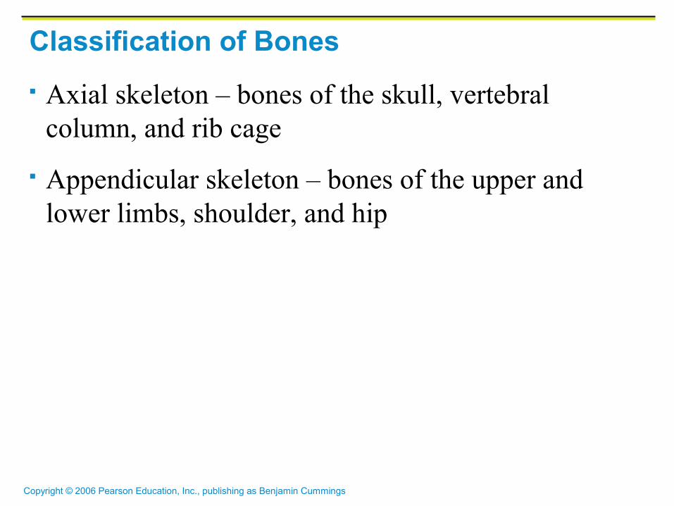
Classification of Bones
Axial skeleton – bones of the skull, vertebral column, and rib cage
Appendicular skeleton – bones of the upper and lower limbs, shoulder, and hip
Copyright © 2006 Pearson Education, Inc., publishing as Benjamin Cummings
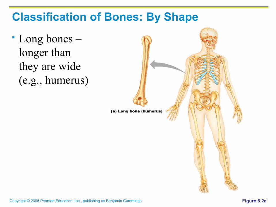
Classification of Bones: By Shape
Long bones – longer than they are wide (e.g., humerus)
Figure 6.2a
Copyright © 2006 Pearson Education, Inc., publishing as Benjamin Cummings
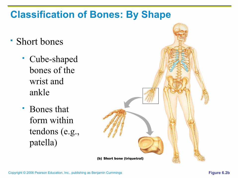
Classification of Bones: By Shape
Short bones
Cube-shaped bones of the wrist and ankle
Bones that form within tendons (e.g., patella)
Figure 6.2b

Copyright © 2006 Pearson Education, Inc., publishing as Benjamin Cummings
Classification of Bones: By Shape
Flat bones – thin, flattened, and a bit curved (e.g., sternum, and most skull bones)
Figure 6.2c

Copyright © 2006 Pearson Education, Inc., publishing as Benjamin Cummings
Classification of Bones: By Shape
Irregular bones – bones with complicated shapes (e.g., vertebrae and hip bones)
Figure 6.2d

Copyright © 2006 Pearson Education, Inc., publishing as Benjamin Cummings
Function of Bones
Support – form the framework that supports the body and cradles soft organs
Protection – provide a protective case for the brain, spinal cord, and vital organs
Movement – provide levers for muscles

Copyright © 2006 Pearson Education, Inc., publishing as Benjamin Cummings
Function of Bones
Mineral storage – reservoir for minerals, especially calcium and phosphorus
Blood cell formation – hematopoiesis occurs within the marrow cavities of bones
Copyright © 2006 Pearson Education, Inc., publishing as Benjamin Cummings
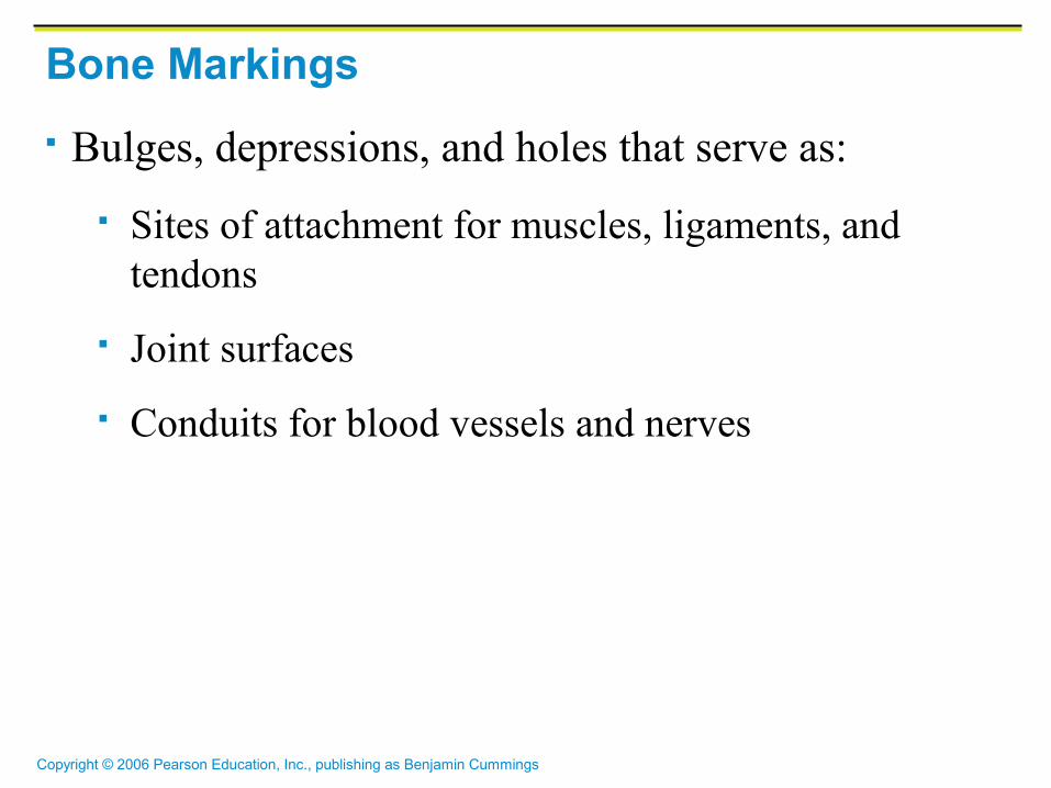
Bone Markings
Bulges, depressions, and holes that serve as:
Sites of attachment for muscles, ligaments, and tendons
Joint surfaces
Conduits for blood vessels and nerves

Copyright © 2006 Pearson Education, Inc., publishing as Benjamin Cummings
Bone Markings: Projections – Sites of Muscle and Ligament Attachment Tuberosity – rounded projection
Crest – narrow, prominent ridge of bone
Trochanter – large, blunt, irregular surface
Line – narrow ridge of bone

Copyright © 2006 Pearson Education, Inc., publishing as Benjamin Cummings
Tubercle – small rounded projection
Epicondyle – raised area above a condyle
Spine – sharp, slender projection
Process – any bony prominence
Bone Markings: Projections – Sites of Muscle and Ligament Attachment

Copyright © 2006 Pearson Education, Inc., publishing as Benjamin Cummings
Bone Markings: Projections – Projections That Help to Form Joints
Head – bony expansion carried on a narrow neck
Facet – smooth, nearly flat articular surface
Condyle – rounded articular projection
Ramus – armlike bar of bone

Copyright © 2006 Pearson Education, Inc., publishing as Benjamin Cummings
Bone Markings: Depressions and Openings
Meatus – canal-like passageway
Sinus – cavity within a bone
Fossa – shallow, basin-like depression
Groove – furrow
Fissure – narrow, slit-like opening
Foramen – round or oval opening through a bone

Copyright © 2006 Pearson Education, Inc., publishing as Benjamin Cummings
Gross Anatomy of Bones: Bone Textures
Compact bone – dense outer layer
Spongy bone – honeycomb of trabeculae filled with yellow bone marrow

Copyright © 2006 Pearson Education, Inc., publishing as Benjamin Cummings
Bone Markings
Table 6.1
Copyright © 2006 Pearson Education, Inc., publishing as Benjamin Cummings
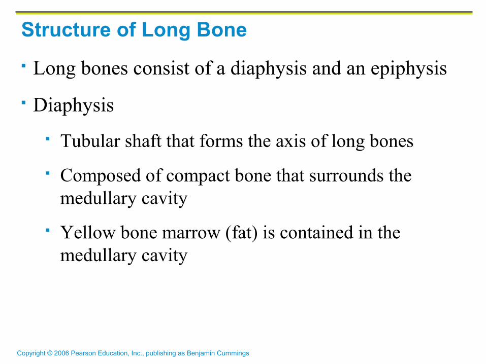
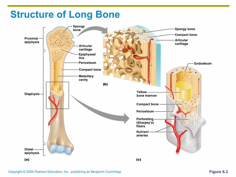
Structure of Long Bone
Long bones consist of a diaphysis and an epiphysis
Diaphysis
Tubular shaft that forms the axis of long bones
Composed of compact bone that surrounds the medullary cavity
Yellow bone marrow (fat) is contained in the medullary cavity

Copyright © 2006 Pearson Education, Inc., publishing as Benjamin Cummings
Structure of Long Bone
Epiphyses
Expanded ends of long bones
Exterior is compact bone, and the interior is spongy bone
Joint surface is covered with articular (hyaline) cartilage
Epiphyseal line separates the diaphysis from the epiphyses
Copyright © 2006 Pearson Education, Inc., publishing as Benjamin Cummings
Structure of Long Bone
Figure 6.3

Copyright © 2006 Pearson Education, Inc., publishing as Benjamin Cummings
Structure of Long Bone
Figure 6.3a

Copyright © 2006 Pearson Education, Inc., publishing as Benjamin Cummings
Structure of Long Bone
Figure 6.3b

Copyright © 2006 Pearson Education, Inc., publishing as Benjamin Cummings
Structure of Long Bone
Figure 6.3c

Copyright © 2006 Pearson Education, Inc., publishing as Benjamin Cummings
Bone Membranes
Periosteum – double-layered protective membrane
Outer fibrous layer is dense regular connective tissue
Inner osteogenic layer is composed of osteoblasts and osteoclasts
Richly supplied with nerve fibers, blood, and lymphatic vessels, which enter the bone via nutrient foramina
Secured to underlying bone by Sharpey’s fibers

Copyright © 2006 Pearson Education, Inc., publishing as Benjamin Cummings
Bone Membranes
Endosteum – delicate membrane covering internal surfaces of bone

Copyright © 2006 Pearson Education, Inc., publishing as Benjamin Cummings
Structure of Short, Irregular, and Flat Bones
Thin plates of periosteum-covered compact bone on the outside with endosteum-covered spongy bone (diploë) on the inside
Have no diaphysis or epiphyses
Contain bone marrow between the trabeculae

Copyright © 2006 Pearson Education, Inc., publishing as Benjamin Cummings
Structure of a Flat Bone
Figure 6.4

Copyright © 2006 Pearson Education, Inc., publishing as Benjamin Cummings
Location of Hematopoietic Tissue (Red Marrow)
In infants
Found in the medullary cavity and all areas of spongy bone
In adults
Found in the diploë of flat bones, and the head of the femur and humerus
Copyright © 2006 Pearson Education, Inc., publishing as Benjamin Cummings
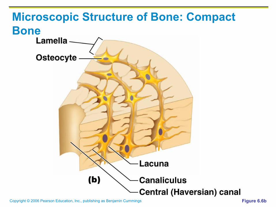
Microscopic Structure of Bone: Compact Bone Haversian system, or osteon – the structural unit of
compact bone
Lamella – weight-bearing, column-like matrix tubes composed mainly of collagen
Haversian, or central canal – central channel containing blood vessels and nerves
Volkmann’s canals – channels lying at right angles to the central canal, connecting blood and nerve supply of the periosteum to that of the Haversian canal
Copyright © 2006 Pearson Education, Inc., publishing as Benjamin Cummings
Microscopic Structure of Bone: Compact Bone Osteocytes – mature bone cells
Lacunae – small cavities in bone that contain osteocytes
Canaliculi – hairlike canals that connect lacunae to each other and the central canal

Copyright © 2006 Pearson Education, Inc., publishing as Benjamin Cummings
Microscopic Structure of Bone: Compact Bone
Figure 6.6a, b

Copyright © 2006 Pearson Education, Inc., publishing as Benjamin Cummings
Microscopic Structure of Bone: Compact Bone
Figure 6.6a
Copyright © 2006 Pearson Education, Inc., publishing as Benjamin Cummings
Microscopic Structure of Bone: Compact Bone
Figure 6.6b

Copyright © 2006 Pearson Education, Inc., publishing as Benjamin Cummings
Microscopic Structure of Bone: Compact Bone
Figure 6.6c

Copyright © 2006 Pearson Education, Inc., publishing as Benjamin Cummings
Chemical Composition of Bone: Organic
Osteoblasts – bone-forming cells
Osteocytes – mature bone cells
Osteoclasts – large cells that resorb or break down bone matrix
Osteoid – unmineralized bone matrix composed of proteoglycans, glycoproteins, and collagen

Copyright © 2006 Pearson Education, Inc., publishing as Benjamin Cummings
Chemical Composition of Bone: Inorganic
Hydroxyapatites, or mineral salts
Sixty-five percent of bone by mass
Mainly calcium phosphates
Responsible for bone hardness and its resistance to compression

Copyright © 2006 Pearson Education, Inc., publishing as Benjamin Cummings
Hydroxylapatite, also called hydroxyapatite (HA), is a naturally occurring mineral form of calcium apatite with the formula Ca5(PO4)3(OH), but is usually written Ca10(PO4)6(OH)2 to denote that the crystal unit cell comprises two entities. Hydroxylapatite is the hydroxyl endmember of the complex apatite group. The OH- ion can be replaced by fluoride, chloride or carbonate, producing fluorapatite or chlorapatite. fluorosis.

Copyright © 2006 Pearson Education, Inc., publishing as Benjamin Cummings
Bone Development
Osteogenesis and ossification – the process of bone tissue formation, which leads to:
The formation of the bony skeleton in embryos
Bone growth until early adulthood
Bone thickness, remodeling, and repair
Copyright © 2006 Pearson Education, Inc., publishing as Benjamin Cummings
Formation of the Bony Skeleton
Begins at week 8 of embryo development
Intramembranous ossification – bone develops from a fibrous membrane
Endochondral ossification – bone forms by replacing hyaline cartilage

Copyright © 2006 Pearson Education, Inc., publishing as Benjamin Cummings
Intramembranous Ossification
Formation of most of the flat bones of the skull and the clavicles
Fibrous connective tissue membranes are formed by mesenchymal cells
Recommended