Copyright © 2005 Pearson Education, Inc. publishing as Benjamin Cummings
Chapter 18:The Genetics of Viruses
and Bacteria
Copyright © 2005 Pearson Education, Inc. publishing as Benjamin Cummings
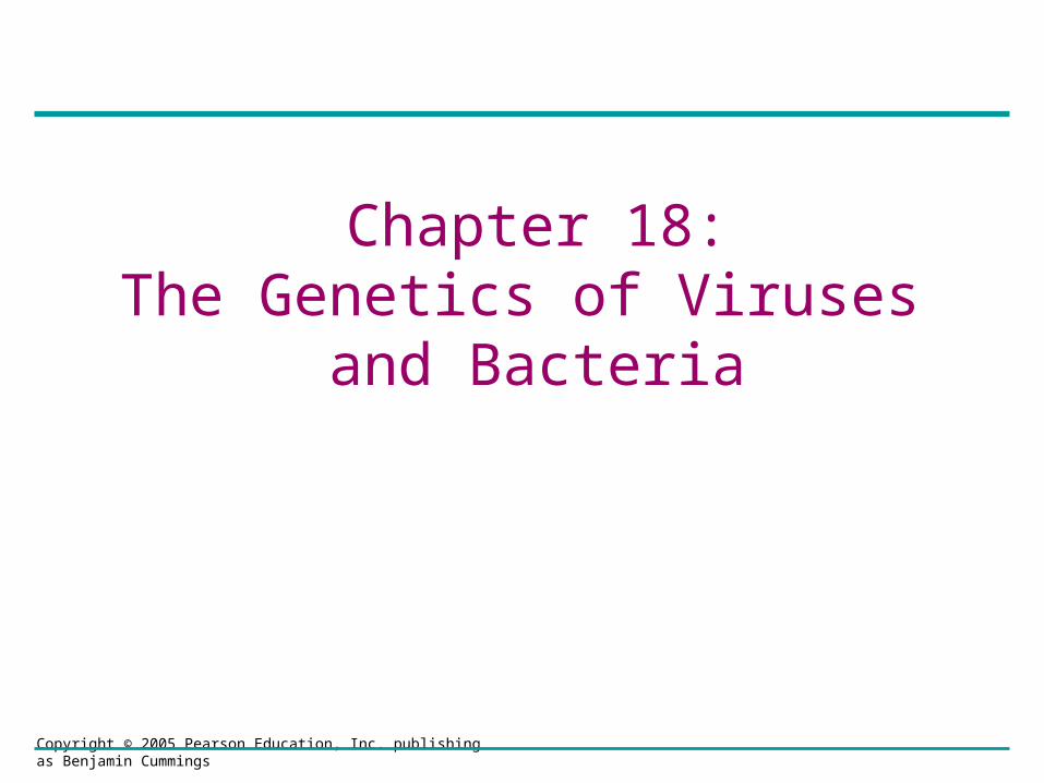
Figure 18.1 T4 bacteriophage infecting an E. coli cell
0.5 m
Copyright © 2005 Pearson Education, Inc. publishing as Benjamin Cummings
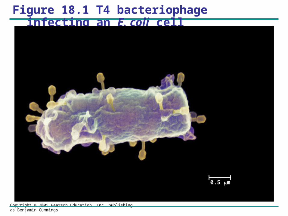
Figure 18.2 Comparing the size of a virus, a bacterium, and an animal cell
0.25 m
Virus
Animalcell
Bacterium
Animal cell nucleus
Copyright © 2005 Pearson Education, Inc. publishing as Benjamin Cummings
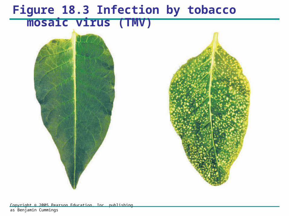
Figure 18.3 Infection by tobacco mosaic virus (TMV)
Copyright © 2005 Pearson Education, Inc. publishing as Benjamin Cummings
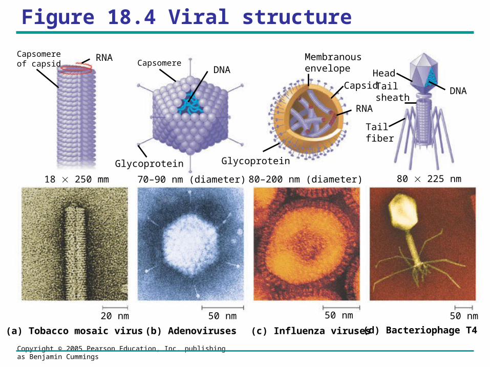
Figure 18.4 Viral structure
18 250 mm 70–90 nm (diameter) 80–200 nm (diameter) 80 225 nm
20 nm 50 nm 50 nm 50 nm
(a) Tobacco mosaic virus (b) Adenoviruses (c) Influenza viruses (d) Bacteriophage T4
RNA
RNACapsomereof capsid
DNACapsomere
Glycoprotein Glycoprotein
Membranousenvelope
CapsidDNA
Head
Tail fiber
Tail sheath
Copyright © 2005 Pearson Education, Inc. publishing as Benjamin Cummings
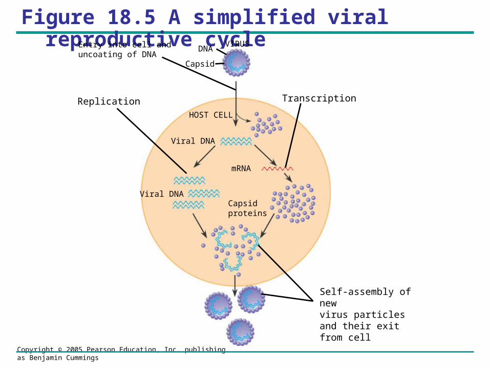
Figure 18.5 A simplified viral reproductive cycleVIRUS
Capsid proteins
mRNA
Viral DNA
HOST CELL
Viral DNA
Entry into cell anduncoating of DNA
Replication Transcription
DNA
Capsid
Self-assembly of new virus particles and their exit from cell
Copyright © 2005 Pearson Education, Inc. publishing as Benjamin Cummings
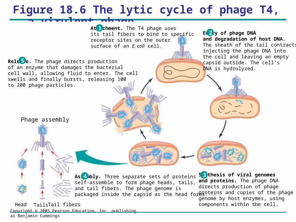
Figure 18.6 The lytic cycle of phage T4, a virulent phage
Attachment. The T4 phage usesits tail fibers to bind to specificreceptor sites on the outer surface of an E. coli cell.
Entry of phage DNA and degradation of host DNA.The sheath of the tail contracts,injecting the phage DNA intothe cell and leaving an emptycapsid outside. The cell’sDNA is hydrolyzed.
Synthesis of viral genomes and proteins. The phage DNAdirects production of phageproteins and copies of the phagegenome by host enzymes, usingcomponents within the cell.
Assembly. Three separate sets of proteinsself-assemble to form phage heads, tails,and tail fibers. The phage genome ispackaged inside the capsid as the head forms.
Release. The phage directs productionof an enzyme that damages the bacterialcell wall, allowing fluid to enter. The cellswells and finally bursts, releasing 100 to 200 phage particles.
12
4 3
5
Phage assembly
Head Tails Tail fibers
Copyright © 2005 Pearson Education, Inc. publishing as Benjamin Cummings
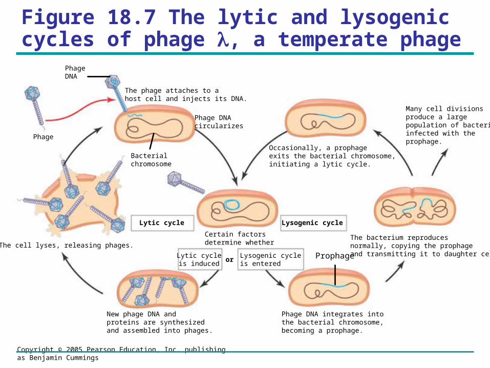
Figure 18.7 The lytic and lysogenic cycles of phage , a temperate phage
Many cell divisions produce a large population of bacteria infected with the prophage.
The bacterium reproducesnormally, copying the prophageand transmitting it to daughter cells.
Phage DNA integrates into the bacterial chromosome,becoming a prophage.
New phage DNA and proteins are synthesized and assembled into phages.
Occasionally, a prophage exits the bacterial chromosome, initiating a lytic cycle.
Certain factorsdetermine whether
The phage attaches to ahost cell and injects its DNA.
Phage DNAcircularizes
The cell lyses, releasing phages.
Lytic cycleis induced
Lysogenic cycleis entered
Lysogenic cycleLytic cycle
or Prophage
Bacterialchromosome
Phage
PhageDNA
Copyright © 2005 Pearson Education, Inc. publishing as Benjamin Cummings
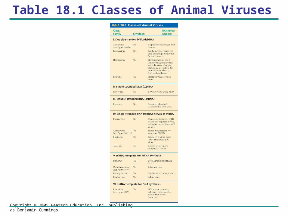
Table 18.1 Classes of Animal Viruses
Copyright © 2005 Pearson Education, Inc. publishing as Benjamin Cummings
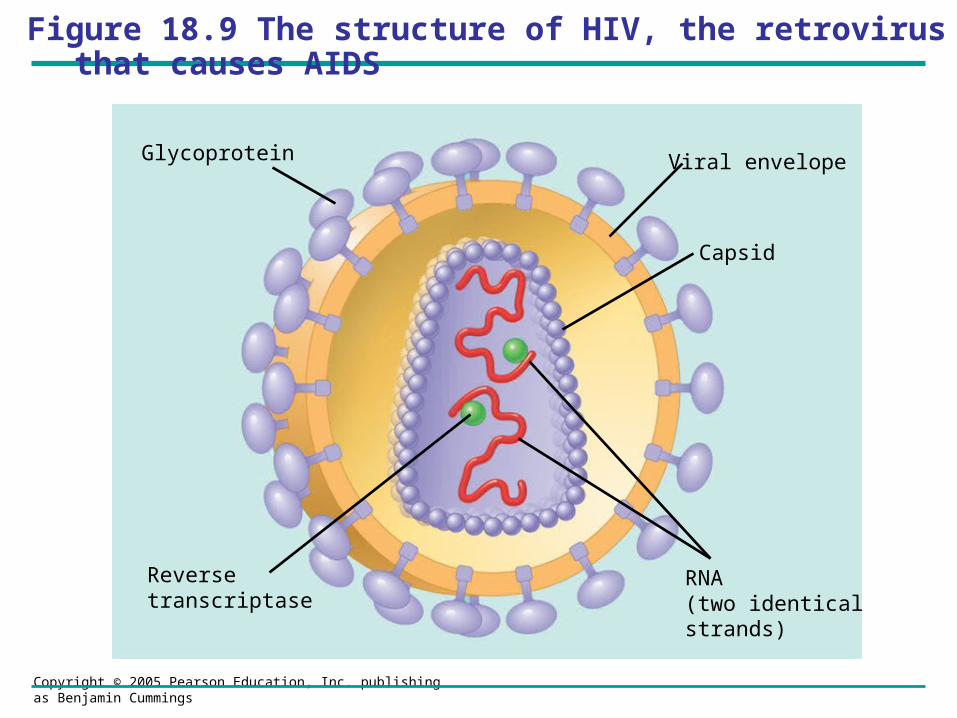
Figure 18.9 The structure of HIV, the retrovirus that causes AIDS
Reversetranscriptase
Viral envelope
Capsid
Glycoprotein
RNA(two identicalstrands)
Copyright © 2005 Pearson Education, Inc. publishing as Benjamin Cummings
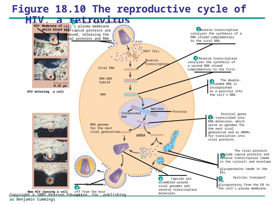
Figure 18.10 The reproductive cycle of HIV, a retrovirus
Vesicles transport theglycoproteins from the ER tothe cell’s plasma membrane.
7
The viral proteins include capsid proteins and reverse transcriptase (made in the cytosol) and envelope glycoproteins (made in the ER).
6
The double-stranded DNA is incorporatedas a provirus into the cell’s DNA.
4
Proviral genes are transcribed into RNA molecules, which serve as genomes for the next viral generation and as mRNAs for translation into viral proteins.
5
Reverse transcriptasecatalyzes the synthesis ofa second DNA strandcomplementary to the first.
3
Reverse transcriptasecatalyzes the synthesis of aDNA strand complementaryto the viral RNA.
2
New viruses budoff from the host cell.9
Capsids areassembled aroundviral genomes and reverse transcriptase molecules.
8
mRNA
RNA genomefor the nextviral generation
Viral RNA
RNA-DNAhybrid
DNA
ChromosomalDNA
NUCLEUSProvirus
HOST CELL
Reverse transcriptase
New HIV leaving a cell
HIV entering a cell
0.25 µm
HIV Membrane of white blood cell
The virus fuses with thecell’s plasma membrane.The capsid proteins areremoved, releasing the viral proteins and RNA.
1
Copyright © 2005 Pearson Education, Inc. publishing as Benjamin Cummings
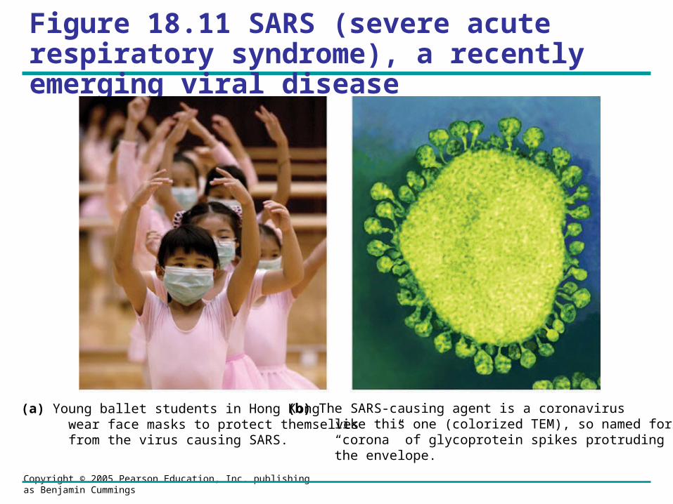
Figure 18.11 SARS (severe acute respiratory syndrome), a recently emerging viral disease
(a) Young ballet students in Hong Kong wear face masks to protect themselves from the virus causing SARS.
(b) The SARS-causing agent is a coronavirus like this one (colorized TEM), so named for the “corona” of glycoprotein spikes protruding from the envelope.
Copyright © 2005 Pearson Education, Inc. publishing as Benjamin Cummings
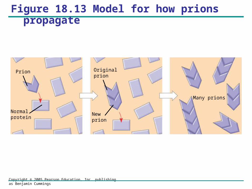
Figure 18.13 Model for how prions propagate
Prion
Normalprotein
Originalprion
Newprion
Many prions
Copyright © 2005 Pearson Education, Inc. publishing as Benjamin Cummings
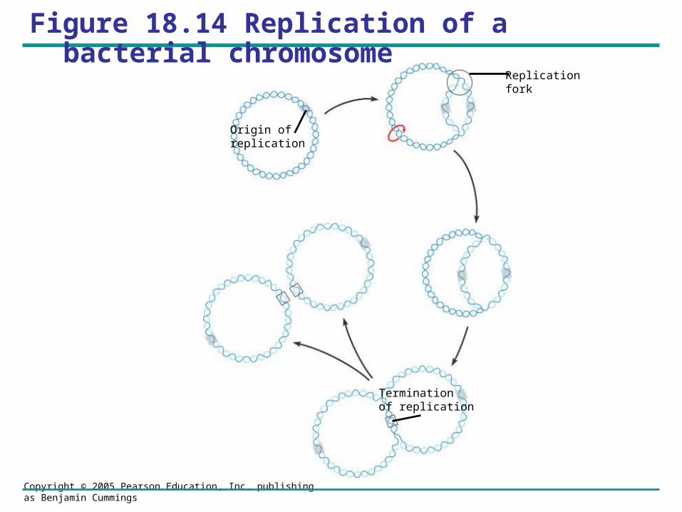
Figure 18.14 Replication of a bacterial chromosome
Replicationfork
Origin of replication
Termination of replication
Copyright © 2005 Pearson Education, Inc. publishing as Benjamin Cummings
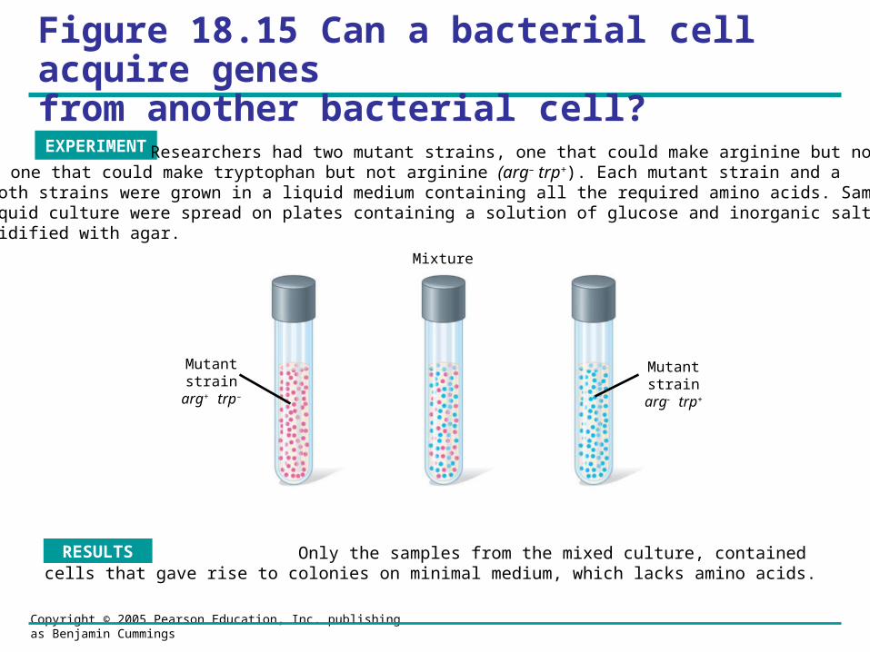
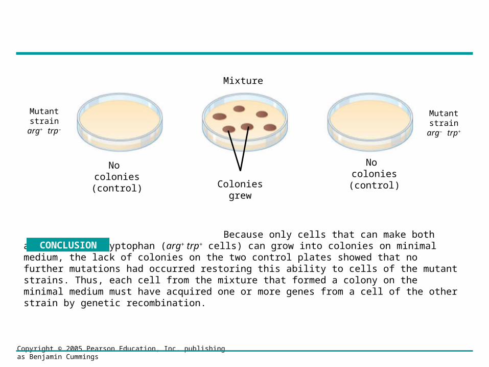
Figure 18.15 Can a bacterial cell acquire genes from another bacterial cell?
Only the samples from the mixed culture, contained cells that gave rise to colonies on minimal medium, which lacks amino acids.
RESULTS
EXPERIMENT Researchers had two mutant strains, one that could make arginine but not tryptophan (arg+ trp–) and one that could make tryptophan but not arginine (arg– trp+). Each mutant strain and a mixture of both strains were grown in a liquid medium containing all the required amino acids. Samples from each liquid culture were spread on plates containing a solution of glucose and inorganic salts (minimal medium), solidified with agar.
Mutantstrain
arg+ trp–
Mutantstrain
arg trp+
Mixture
Copyright © 2005 Pearson Education, Inc. publishing as Benjamin Cummings
Mutantstrain
arg+ trp–
Mutantstrain
arg– trp+
No colonies(control)
No colonies(control)
Mixture
Coloniesgrew
Because only cells that can make both arginine and tryptophan (arg+ trp+ cells) can grow into colonies on minimal medium, the lack of colonies on the two control plates showed that no further mutations had occurred restoring this ability to cells of the mutant strains. Thus, each cell from the mixture that formed a colony on the minimal medium must have acquired one or more genes from a cell of the other strain by genetic recombination.
CONCLUSION
Copyright © 2005 Pearson Education, Inc. publishing as Benjamin Cummings
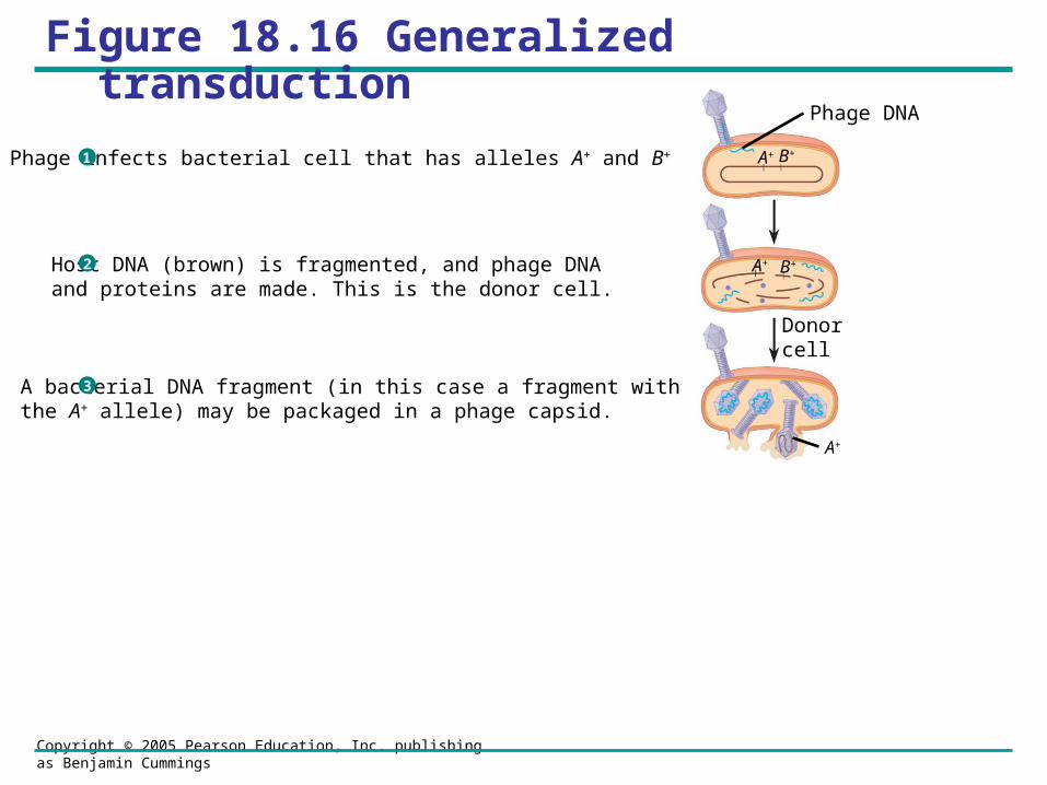
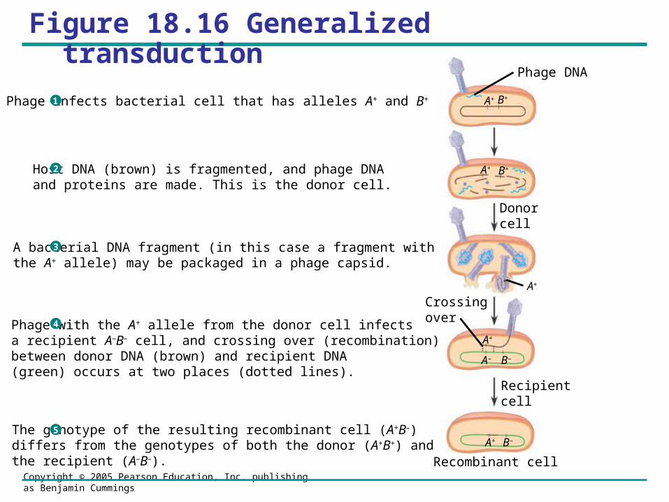
Figure 18.16 Generalized transduction
Phage DNA
Donorcell
A+ B+
A+ B+
A+
Phage infects bacterial cell that has alleles A+ and B+
Host DNA (brown) is fragmented, and phage DNA and proteins are made. This is the donor cell.
A bacterial DNA fragment (in this case a fragment withthe A+ allele) may be packaged in a phage capsid.
1
2
3
Copyright © 2005 Pearson Education, Inc. publishing as Benjamin Cummings
Figure 18.16 Generalized transduction
Phage DNA
Donorcell
Recipientcell
A+ B+
A+ B+
A+
A+ B–
A– B–
A+
Recombinant cell
Crossingover
Phage infects bacterial cell that has alleles A+ and B+
Host DNA (brown) is fragmented, and phage DNA and proteins are made. This is the donor cell.
A bacterial DNA fragment (in this case a fragment withthe A+ allele) may be packaged in a phage capsid.
Phage with the A+ allele from the donor cell infects a recipient A–B– cell, and crossing over (recombination)between donor DNA (brown) and recipient DNA(green) occurs at two places (dotted lines).
The genotype of the resulting recombinant cell (A+B–) differs from the genotypes of both the donor (A+B+) and the recipient (A–B–).
1
2
3
4
5
Copyright © 2005 Pearson Education, Inc. publishing as Benjamin Cummings
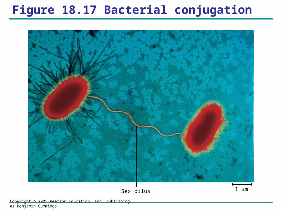
Figure 18.17 Bacterial conjugation
Sex pilus 1 m
Copyright © 2005 Pearson Education, Inc. publishing as Benjamin Cummings
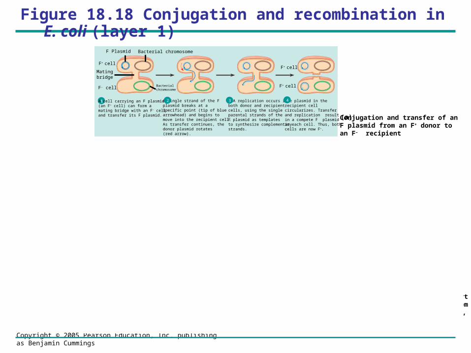
Figure 18.18 Conjugation and recombination in E. coli (layer 1)
1 A cell carrying an F plasmid(an F+ cell) can form amating bridge with an F– celland transfer its F plasmid.
A single strand of the F plasmid breaks at a specific point (tip of blue arrowhead) and begins tomove into the recipient cell. As transfer continues, the donor plasmid rotates(red arrow).
2 DNA replication occurs inboth donor and recipientcells, using the single parental strands of the F plasmid as templates to synthesize complementary strands.
3 The plasmid in the recipient cell circularizes. Transfer and replication result in a compete F plasmid in each cell. Thus, both cells are now F+.
4
F Plasmid Bacterial chromosome
Bacterial chromosomeF– cell
F+ cell
F+ cell
F+ cell Hfr cell
F factorThe circular F plasmid in an F+ cellcan be integrated into the circularchromosome by a single crossoverevent (dotted line).
1The resulting cell is called an Hfr cell (for High frequency of recombination).
2
Since an Hfr cell has all the F-factor genes, it can form a mating bridge with an F– cell and transfer DNA.
3 A single strand of the F factorbreaks and begins to move through the bridge. DNA replication occurs in both donor and recipient cells, resulting in double-stranded DNA
4 The location and orientation of the F factor in the donor chromosome determine the sequence of gene transfer during conjugation. In this example, the transfer sequence for four genes is A-B-C-D.
5 The mating bridgeusually breaks well before the entire chromosome and the rest of the F factor are transferred.
6
Two crossovers can result in the exchange of similar (homologous) genes between the transferred chromosome fragment (brown) and the recipient cell’s chromosome (green).
7 The piece of DNA ending up outside thebacterial chromosome will eventually be degraded by the cell’s enzymes. The recipient cell now contains a new combination of genes but no F factor; it is a recombinant F– cell.
8
Temporarypartialdiploid
Recombinant F–
bacterium
Conjugation and transfer of an F plasmid from an F+ donor to an F– recipient
(a)
Conjugation and transfer of part of the bacterial chromosome from an Hfr donor to an F– recipient, resulting in recombination
(b)
A+B+ C+
D+
F– cell A–B–
C–
D–
A–B–
C–
D– D–
A–
C–B– D–
A–
C–
B–
A+
B+C+D+A+
B+C+D+A+B+
D+C+
A+
A+
B+
A–B–
C–
D–
A–B+
C–
D–
A+
B+ B–
A+
A+
B+
F+ cell
Mating bridge
Copyright © 2005 Pearson Education, Inc. publishing as Benjamin Cummings
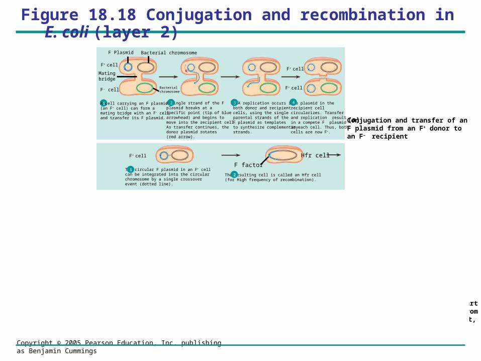
Figure 18.18 Conjugation and recombination in E. coli (layer 2)
1 A cell carrying an F plasmid(an F+ cell) can form amating bridge with an F– celland transfer its F plasmid.
A single strand of the F plasmid breaks at a specific point (tip of blue arrowhead) and begins tomove into the recipient cell. As transfer continues, the donor plasmid rotates(red arrow).
2 DNA replication occurs inboth donor and recipientcells, using the single parental strands of the F plasmid as templates to synthesize complementary strands.
3 The plasmid in the recipient cell circularizes. Transfer and replication result in a compete F plasmid in each cell. Thus, both cells are now F+.
4
F Plasmid Bacterial chromosome
Bacterial chromosomeF– cell
F+ cell
F+ cell
F+ cell Hfr cell
F factorThe circular F plasmid in an F+ cellcan be integrated into the circularchromosome by a single crossoverevent (dotted line).
1The resulting cell is called an Hfr cell (for High frequency of recombination).
2
Since an Hfr cell has all the F-factor genes, it can form a mating bridge with an F– cell and transfer DNA.
3 A single strand of the F factorbreaks and begins to move through the bridge. DNA replication occurs in both donor and recipient cells, resulting in double-stranded DNA
4 The location and orientation of the F factor in the donor chromosome determine the sequence of gene transfer during conjugation. In this example, the transfer sequence for four genes is A-B-C-D.
5 The mating bridgeusually breaks well before the entire chromosome and the rest of the F factor are transferred.
6
Two crossovers can result in the exchange of similar (homologous) genes between the transferred chromosome fragment (brown) and the recipient cell’s chromosome (green).
7 The piece of DNA ending up outside thebacterial chromosome will eventually be degraded by the cell’s enzymes. The recipient cell now contains a new combination of genes but no F factor; it is a recombinant F– cell.
8
Temporarypartialdiploid
Recombinant F–
bacterium
Conjugation and transfer of an F plasmid from an F+ donor to an F– recipient
(a)
Conjugation and transfer of part of the bacterial chromosome from an Hfr donor to an F– recipient, resulting in recombination
(b)
A+B+ C+
D+
F– cell A–B–
C–
D–
A–B–
C–
D– D–
A–
C–B– D–
A–
C–
B–
A+
B+C+D+A+
B+C+D+A+B+
D+C+
A+
A+
B+
A–B–
C–
D–
A–B+
C–
D–
A+
B+ B–
A+
A+
B+
F+ cell
Mating bridge
Copyright © 2005 Pearson Education, Inc. publishing as Benjamin Cummings
Figure 18.18 Conjugation and recombination in E. coli (layer 3)
1 A cell carrying an F plasmid(an F+ cell) can form amating bridge with an F– celland transfer its F plasmid.
A single strand of the F plasmid breaks at a specific point (tip of blue arrowhead) and begins tomove into the recipient cell. As transfer continues, the donor plasmid rotates(red arrow).
2 DNA replication occurs inboth donor and recipientcells, using the single parental strands of the F plasmid as templates to synthesize complementary strands.
3 The plasmid in the recipient cell circularizes. Transfer and replication result in a compete F plasmid in each cell. Thus, both cells are now F+.
4
F Plasmid Bacterial chromosome
Bacterial chromosomeF– cell
F+ cell
F+ cell
F+ cell Hfr cell
F factorThe circular F plasmid in an F+ cellcan be integrated into the circularchromosome by a single crossoverevent (dotted line).
1The resulting cell is called an Hfr cell (for High frequency of recombination).
2
Since an Hfr cell has all the F-factor genes, it can form a mating bridge with an F– cell and transfer DNA.
3 A single strand of the F factorbreaks and begins to move through the bridge. DNA replication occurs in both donor and recipient cells, resulting in double-stranded DNA
4 The location and orientation of the F factor in the donor chromosome determine the sequence of gene transfer during conjugation. In this example, the transfer sequence for four genes is A-B-C-D.
5 The mating bridgeusually breaks well before the entire chromosome and the rest of the F factor are transferred.
6
Two crossovers can result in the exchange of similar (homologous) genes between the transferred chromosome fragment (brown) and the recipient cell’s chromosome (green).
7 The piece of DNA ending up outside thebacterial chromosome will eventually be degraded by the cell’s enzymes. The recipient cell now contains a new combination of genes but no F factor; it is a recombinant F– cell.
8
Temporarypartialdiploid
Recombinant F–
bacterium
Conjugation and transfer of an F plasmid from an F+ donor to an F– recipient
(a)
Conjugation and transfer of part of the bacterial chromosome from an Hfr donor to an F– recipient, resulting in recombination
(b)
A+B+ C+
D+
F– cell A–B–
C–
D–
A–B–
C–
D– D–
A–
C–B– D–
A–
C–
B–
A+
B+C+D+A+
B+C+D+A+B+
D+C+
A+
A+
B+
A–B–
C–
D–
A–B+
C–
D–
A+
B+ B–
A+
A+
B+
F+ cell
Mating bridge
Hfr cell
Copyright © 2005 Pearson Education, Inc. publishing as Benjamin Cummings
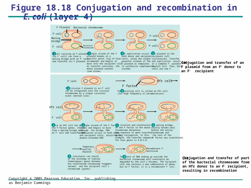
Figure 18.18 Conjugation and recombination in E. coli (layer 4)
1 A cell carrying an F plasmid(an F+ cell) can form amating bridge with an F– celland transfer its F plasmid.
A single strand of the F plasmid breaks at a specific point (tip of blue arrowhead) and begins tomove into the recipient cell. As transfer continues, the donor plasmid rotates(red arrow).
2 DNA replication occurs inboth donor and recipientcells, using the single parental strands of the F plasmid as templates to synthesize complementary strands.
3 The plasmid in the recipient cell circularizes. Transfer and replication result in a compete F plasmid in each cell. Thus, both cells are now F+.
4
F Plasmid Bacterial chromosome
Bacterial chromosomeF– cell
F+ cell
F+ cell
F+ cell Hfr cell
F factorThe circular F plasmid in an F+ cellcan be integrated into the circularchromosome by a single crossoverevent (dotted line).
1The resulting cell is called an Hfr cell (for High frequency of recombination).
2
Since an Hfr cell has all the F-factor genes, it can form a mating bridge with an F– cell and transfer DNA.
3 A single strand of the F factorbreaks and begins to move through the bridge. DNA replication occurs in both donor and recipient cells, resulting in double-stranded DNA
4 The location and orientation of the F factor in the donor chromosome determine the sequence of gene transfer during conjugation. In this example, the transfer sequence for four genes is A-B-C-D.
5 The mating bridgeusually breaks well before the entire chromosome and the rest of the F factor are transferred.
6
Two crossovers can result in the exchange of similar (homologous) genes between the transferred chromosome fragment (brown) and the recipient cell’s chromosome (green).
7 The piece of DNA ending up outside thebacterial chromosome will eventually be degraded by the cell’s enzymes. The recipient cell now contains a new combination of genes but no F factor; it is a recombinant F– cell.
8
Temporarypartialdiploid
Recombinant F–
bacterium
Conjugation and transfer of an F plasmid from an F+ donor to an F– recipient
(a)
Conjugation and transfer of part of the bacterial chromosome from an Hfr donor to an F– recipient, resulting in recombination
(b)
A+B+ C+
D+
F– cell A–B–
C–
D–
A–B–
C–
D– D–
A–
C–B– D–
A–
C–
B–
A+
B+C+D+A+
B+C+D+A+B+
D+C+
A+
A+
B+
A–B–
C–
D–
A–B+
C–
D–
A+
B+ B–
A+
A+
B+
F+ cell
Mating bridge
Hfr cell
Recommended