Research Article Open Access
J Clinic Experiment Ophthalmol Cataract ISSN:2155-9570 JCEO, an open access journal
Open AccessReview Article
Malik, J Clinic Experiment Ophthalmol 2013, S:1 DOI: 10.4172/2155-9570.S1-007
Keywords: Local anesthesia; Catract surgery; Sub-Tenon; Akinesia
IntroductionProgression in phacoemulsification techniques for cataract
surgery[1-4] has also led to inevitable changes in the delivery of accompanying anesthesia from general to local modalities [5-7] i.e. retrobulbar, peribulbar, Sub-Tenon and topical anesthesia. General anesthesia is preferred for complicated intracapsular cataract extraction (ICCE) surgery as this technique involves significant manipulation of the eye, and requires several corneal sutures to provide a watertight wound. Extracapsular cataract surgery [2,8-11] (ECCE) also requires a large corneal incision, but there was a trend towards local anesthesia such as retrobulbar [12-15], which allows quicker patient recovery, and thus facilitated day-case surgery.
With the introduction of the foldable intraocular lens [16] (IOL), phacoemulsification and small-incision surgery has developed [2,16-18]. Initially, this was performed via a scleral tunnel and required sutures, thus retrobulbar and peribulbar anesthesia were preferred [14,19]. With the advent of small, stepped, and self-sealing corneal incisions, very little manipulation is required, and this allowed the use of Sub-Tenon [20-23] and topical anesthesia [14,24-26].
An acceptable anesthesia modality for a cataract surgery implies the correct choice of anesthetic agent, combined with a compatible clinical technique, ensuring patient comfort both during and post surgery. Thus, the choice of an anesthesia modality must be assessed on an individual patient and surgeon basis.
This review discusses the efficacy and performance of the different available local anesthesia modalities for cataract surgery, including anesthesia requirements for patient safety and comfort. Clinical methodologies used for the implementation of varying local anesthesia modalities, with the associated complications, are also discussed with a view to identify an optimum local anesthesia strategy for cataract surgery.
Local Anesthesia RequirementsAkinesia
Many surgeons do not consider akinesia an important requirement for cataract surgery, but some prefer to operate under conditions in which eye movements are blunted if not completely paralyzed. Friedman et al. [27] report that both retrobulbar and peribulbar blocks
*Corresponding author: Adeela Malik, MBBS, BSc (Hons), Moorfield’s Eye Hospital NHS Foundation Trust, London, UK, E-mail: [email protected]
Received December 23, 2012; Accepted February 01, 2013; Published February 08, 2013
Citation: Malik A (2013) Efficacy and Performance of Various Local Anesthesia Modalities for Cataract Surgery. J Clinic Experiment Ophthalmol S1: 007. doi:10.4172/2155-9570.S1-007
Copyright: © 2013 Malik A. This is an open-access article distributed under the terms of the Creative Commons Attribution License, which permits unrestricted use, distribution, and reproduction in any medium, provided the original author and source are credited.
Efficacy and Performance of Various Local Anesthesia Modalities for Cataract SurgeryAdeela Malik*
Moorfield’s Eye Hospital NHS Foundation Trust, London, UK
produce the same degree of akinesia. Comparing retrobulbar and Sub-Tenon techniques, the same survey reports that akinesia was slightly less effective with Sub-Tenon; however, the statistical evidence for this was poor. Topical procedures do not lead to akinesia.
Patient preference (pain experience)
Significant pain during anesthetic administration, intraoperative surgery, or after the cataract procedure, is the major reasons for low patient satisfaction [28]. Perceived pain level thus determines patient’s preference for the anesthesia technique and is an important factor in the selection of an optimum strategy for anesthesia management during cataract surgery. During the anesthesia administration phase, topical anesthesia, unlike injection-type anesthesia modalities, is associated with minimal discomfort. However, with topical anesthesia, the agent blocks trigeminal nerve endings in the cornea and the conjunctiva only [29], leaving the intraocular structures in the anterior segment unanesthetized. Thus, manipulation of the iris or stretching of the ciliary and zonular tissues irritates the ciliary nerves, resulting in discomfort. Although injectable anesthesia provides a higher level of analgesia and akinesia than topical anesthesia, the degree of pain perceived by patients during anesthesia administration and intraoperative surgery varies for retrobulbar, peribulbar, and Sub-Tenon modalities. Patient-reported pain for various anesthesia techniques is discussed under “Comparative evaluation of performance of various local anesthesia techniques”.
Visual recovery
Rapid vision recovery is highly desirable in cataract surgery and is
AbstractThe efficacy and performance of various local anesthesia modalities for cataract surgery have been reviewed. A
comparative evaluation of these different modalities in terms of akinesia, patient-perceived pain during both anesthesia administration and intraoperative cataract surgery is presented.
Anesthesia requirements, anesthetic agents, and inherent complications of various anesthesia modalities are also discussed, with a description of the clinical procedural details for each modality.
The prevailing practice patterns of anesthesia techniques among refractive surgeons in the United Kingdom and United States are also illustrated.
Journal of Clinical & Experimental OphthalmologyJo
urna
l of C
linica
l & Experimental Ophthalmology
ISSN: 2155-9570
Citation: Malik A (2013) Efficacy and Performance of Various Local Anesthesia Modalities for Cataract Surgery. J Clinic Experiment Ophthalmol S1: 007. doi:10.4172/2155-9570.S1-007
Page 2 of 13
J Clinic Experiment Ophthalmol Cataract ISSN:2155-9570 JCEO, an open access journal
extremely useful, especially in monocular patients having surgery in the better eye. Modern ophthalmologic surgery is becoming faster and in a typical ophthalmology clinic, an uneventful cataract surgery may take 20 minutes or less; this allows the patient to be discharged with good vision and with an eye patch. Compared with a regional block, topical anesthesia permits early visual rehabilitation, primarily because in this modality only a limited block or no block of the optic nerve is involved.
The overall onset and duration times of the selected anesthetic agent can be adjusted depending on the method of administration, the type of anesthesia, the mixture, and the addition of hyaluronidase (Tables 1-3). Adjustment of the duration of the anesthesia will aid in postoperative recovery and assessment in most cases, but, as in other procedures, children or patients with hearing difficulties are treated under general anesthesia.
Sedation
Administration of an orbital block can result in a great deal of pain; consequently, some clinicians use deep sedation. However, many believe the patient should not be sedated deeply so that he or she remains cooperative. In the UK, the use of sedation decreased from 5.85% in 1996 to 3.9% in 2002/2003 [30,31]. The practice of sedation is,
however, not recommended by the Royal College of Anaesthetists [32] and thus should be used to cover only anxiety, not inadequate blocks. Complications of sedation include excessive restlessness, sudden movement, and airway obstruction, which can significantly increase the risk of the surgery [7,28,33]. If the patient is sedated, monitoring is required. The anesthetist should be present and only responsible for that list if the patients have intravenous (IV) sedation. It is considered safer to abandon surgery than to convert to general anesthesia and to continue with general anesthesia only when the patient is fully prepared. “Conscious” sedation, which allows full cooperation of the patient, is recommended [34]. The level of sedation desired, the route of administration, and the choice of drugs commonly used in ophthalmic anesthesia have been reviewed elsewhere [35].
Cost
Literature on the economic evaluation of different systems for cataract surgery and anesthesia is limited, and there is ongoing discussion of the most appropriate methods that can be used [36]. According to a survey reported by Mojon-Azzi and Mojon [37], outpatient surgery is more cost-effective, primarily because there are no costs for overnight stays and lower out-of-pocket expenses. In some European countries (e.g. Holland), the cost savings are estimated at about 20%. The survey also reports that the rate of outpatient cataract
Traditionalanaesthetic agents:
Type Onset Potency Toxicity Duration
Procaine2-chloroprocaine Tetracaine(Amethocaine)Cocaine
Ester Ester Ester Ester
Slow Rapid Slow Slow
LowIntermediateIntermediate
High
LowLowIntermediate
Very High
ShortShortIntermediate
LongBenoxinate(oxybuprocaine)
Ester It must be used in lowest possible dosage [36] to avoid:i) lethal reactions in low cholinesterase activitiesii) allergy to the para-aminobenzoic moitiesiii) absorption through nasal-lacrimal apparatus or mucous membrane
BupivacaineLidocaineRopivacaineEtidocaine
Amide AmideAmideAmide
SlowRapidSlowRapid
High IntermediateIntermediateHigh
HighLowIntermediateHigh
LongShortLongLong
Newly developedanaesthetic agentsArticaineLevobupivacaine 2-Choloroprocaine Proxymethacaine
Amide AmideEster Ester
Rapid SlowRapid Rapid
Low High Low Low
LongLong ShortIntermediate
Table 1: Properties of popular local anaesthetics used for ocular surgery [38,40,46,47].
Table 2: Typical anaesthetic agents for various conduction blocks, topical and intracameral anaesthetic techniques.
Technique Anaesthetic Agent Remarks
Regional/Conduction Blocks: [Retrobulbar, Peribulbar, Sub-Tenon’s]
i) Lidocaine 2% proceduresii) Lidocaine 2% + bupivacaine 0.755 [or ropivacaine 1%] iii) 50%-50% mixture of lidocaine 2% and bupivacaine 0.75%
For short duration
For procedures lasting one hour
For rapid onset and long duration procedures
Topical
Eye Drops: Propacaine Tetracaine Lidocaine Bupivacaine BenoxinateViscous lidocaine gel
IntracameralA mixture of preservative free 1% lidocaineand preservative free 0.5% bupivacaine eye drops
Citation: Malik A (2013) Efficacy and Performance of Various Local Anesthesia Modalities for Cataract Surgery. J Clinic Experiment Ophthalmol S1: 007. doi:10.4172/2155-9570.S1-007
Page 3 of 13
J Clinic Experiment Ophthalmol Cataract ISSN:2155-9570 JCEO, an open access journal
surgery in 10 European countries is influenced mainly by acute-bed density, density of practicing physicians, and public expenditure on health [37].
Anesthetic Agents for Local AnesthesiaThe choice of a local anesthetic agent is optimal only when the agent’s
inherent properties meet the characteristics of the surgical procedure and the requirements of the patient in terms of contraindications and perceived pain. The relative performance, efficacy, and underlying complications of local anesthetic agents, including toxicity, have been
discussed extensively in references [22,27,38-47]. The pharmacology and pathogenesis of toxicity of the various agents are also well documented [40,48]. Table 1 lists the properties of popular anesthetic agents. Lidocaine, bupivacaine, and ropivacaine are the most popular traditional agents used for conduction blockades. Levobupivacaine, articaine, and 2-chloroprocaine are recently developed agents. Table 2 lists the typical agents used for various anesthesia techniques and Table 3, the onset time to akinesia for the traditional and newer agents.
The use of hyaluronidase in regional anesthesia is controversial.
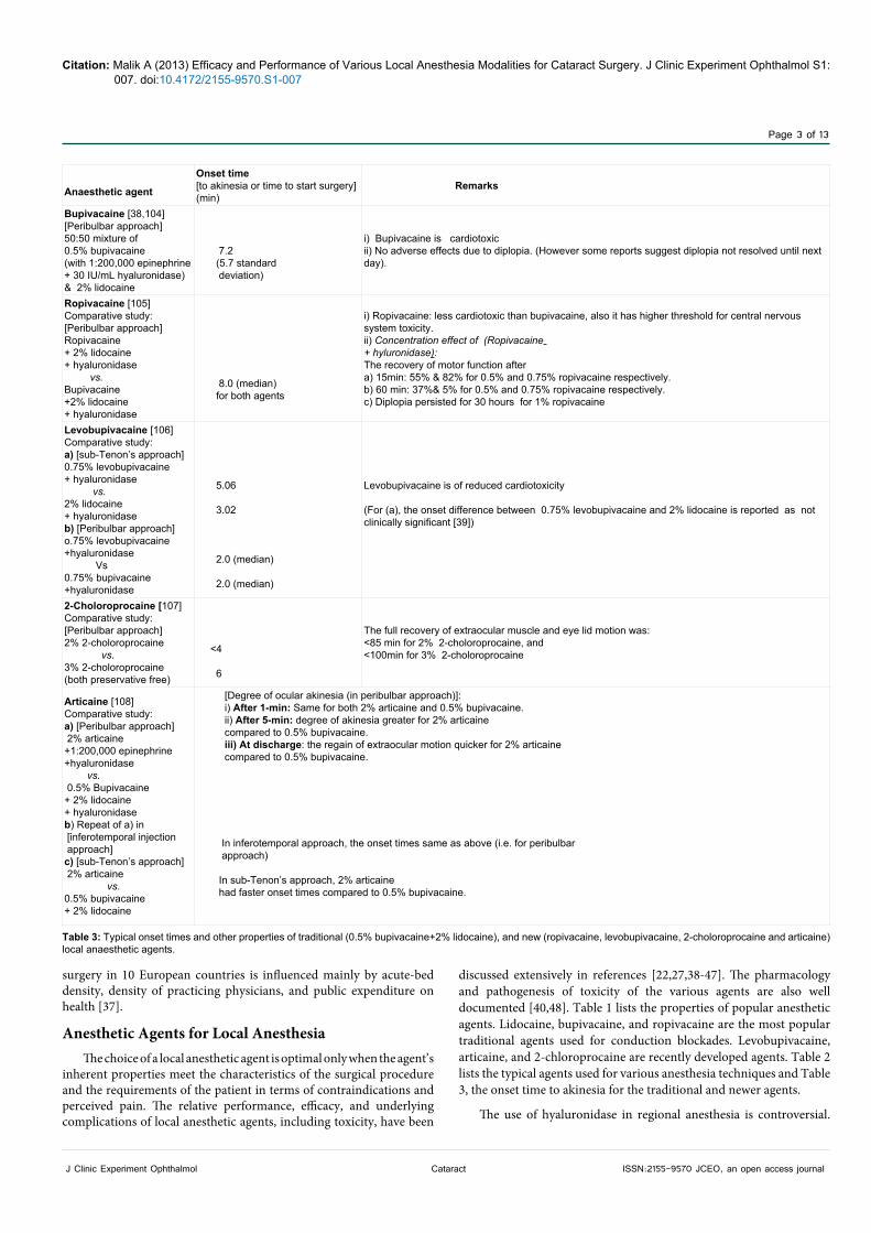
Table 3: Typical onset times and other properties of traditional (0.5% bupivacaine+2% lidocaine), and new (ropivacaine, levobupivacaine, 2-choloroprocaine and articaine) local anaesthetic agents.
Anaesthetic agent
Onset time[to akinesia or time to start surgery] (min)
Remarks
Bupivacaine [38,104] [Peribulbar approach]50:50 mixture of0.5% bupivacaine(with 1:200,000 epinephrine+ 30 IU/mL hyaluronidase)& 2% lidocaine
7.2 (5.7 standard deviation)
i) Bupivacaine is cardiotoxicii) No adverse effects due to diplopia. (However some reports suggest diplopia not resolved until next day).
Ropivacaine [105]Comparative study:[Peribulbar approach]Ropivacaine+ 2% lidocaine+ hyaluronidase vs.Bupivacaine+2% lidocaine+ hyaluronidase
8.0 (median) for both agents
i) Ropivacaine: less cardiotoxic than bupivacaine, also it has higher threshold for central nervous system toxicity.ii) Concentration effect of (Ropivacaine + hyluronidase):The recovery of motor function aftera) 15min: 55% & 82% for 0.5% and 0.75% ropivacaine respectively.b) 60 min: 37%& 5% for 0.5% and 0.75% ropivacaine respectively.c) Diplopia persisted for 30 hours for 1% ropivacaine
Levobupivacaine [106]Comparative study: a) [sub-Tenon’s approach]0.75% levobupivacaine+ hyaluronidase vs.2% lidocaine+ hyaluronidaseb) [Peribulbar approach]o.75% levobupivacaine+hyaluronidase Vs0.75% bupivacaine+hyaluronidase
5.06
3.02
2.0 (median)
2.0 (median)
Levobupivacaine is of reduced cardiotoxicity
(For (a), the onset difference between 0.75% levobupivacaine and 2% lidocaine is reported as not clinically significant [39])
2-Choloroprocaine [107]Comparative study:[Peribulbar approach]2% 2-choloroprocaine vs.3% 2-choloroprocaine(both preservative free)
<4
6
The full recovery of extraocular muscle and eye lid motion was:<85 min for 2% 2-choloroprocaine, and<100min for 3% 2-choloroprocaine
Articaine [108]Comparative study:a) [Peribulbar approach] 2% articaine+1:200,000 epinephrine+hyaluronidase vs. 0.5% Bupivacaine+ 2% lidocaine+ hyaluronidaseb) Repeat of a) in [inferotemporal injection approach]c) [sub-Tenon’s approach] 2% articaine vs.0.5% bupivacaine+ 2% lidocaine
[Degree of ocular akinesia (in peribulbar approach)]: i) After 1-min: Same for both 2% articaine and 0.5% bupivacaine. ii) After 5-min: degree of akinesia greater for 2% articaine compared to 0.5% bupivacaine. iii) At discharge: the regain of extraocular motion quicker for 2% articaine compared to 0.5% bupivacaine.
In inferotemporal approach, the onset times same as above (i.e. for peribulbar approach) In sub-Tenon’s approach, 2% articaine had faster onset times compared to 0.5% bupivacaine.
Citation: Malik A (2013) Efficacy and Performance of Various Local Anesthesia Modalities for Cataract Surgery. J Clinic Experiment Ophthalmol S1: 007. doi:10.4172/2155-9570.S1-007
Page 4 of 13
J Clinic Experiment Ophthalmol Cataract ISSN:2155-9570 JCEO, an open access journal
However, as an adjuvant in retrobulbar and peribulbar anesthesia [7,10,49], it enhances the onset of the block. It also improves the onset and quality of the block in the Sub-Tenon procedure by promoting diffusion to the periorbital and retro-orbital tissue [20,50-52] and alleviating the need for a facial nerve block, which can be painful [42,52]. Hyaluronidase [7] helps to avoid a decrease in retinal circulation and an increase in intraocular pressure (IOP) and to reduce the risk for muscle toxicity from local anesthetic agents.
Intracameral anesthesia (injection into the anterior chamber) is often used as an adjunct to topical anesthesia to enhance the anesthesia and thereby deal with incremental pain that may arise during topical anesthesia alone. Ester-linked benoxinate (oxybuprocaine), amide-linked lidocaine, and ester-linked tetracaine (amethocaine) are the most commonly used topical agents [7] for anesthesia of the cornea and conjunctiva.
Choice of Anesthetic Delivery Techniques in Local Anesthesia
Retrobulbar, Peribulbar, Sub-Tenon and Topical anesthesia are the popular modalities of local anesthesia (LA) [32,53-55]. The regional modalities i.e. retrobulbar, peribulbar, and Sub-Tenon anesthesia depend on injection-delivery of the anesthetic agent, and in some literature these modalities are referred to as intraconal, extraconal, and parabulbar blocks, respectively; but in this paper the former terminology is used exclusively.
Overall, local anesthesia for cataract surgery promises a better procedural safety profile [7,39] and has allowed development of day-surgery cases with quicker patient recovery generally. It is also more economical cost wise.
Substantial information [32] for safe and effective local anesthesia for ophthalmic surgery (intended for practice in the United Kingdom) is contained in joint guidelines issued by the Royal College of Anaesthetists and the Royal College of Ophthalmologists.
With increased popularity of day surgery, general anesthesia is reserved for cases that are unsuitable for local anesthesia: patients (such as a child or a younger adult) intolerant to the local anesthesia procedure, confused patients unable to comply, patients with marked uncontrolled tremor or jerky movements, patients with a history of allergic reaction to local anesthesia, and patients who refused consent for local anesthesia.
Local anesthesia is not free of complications. To reduce complications (discussed under Contraindications), it is essential for the clinician to be fully aware of the procedure (discussed under Local Anesthesia Requirements) involved in the implementation of the various delivery techniques.
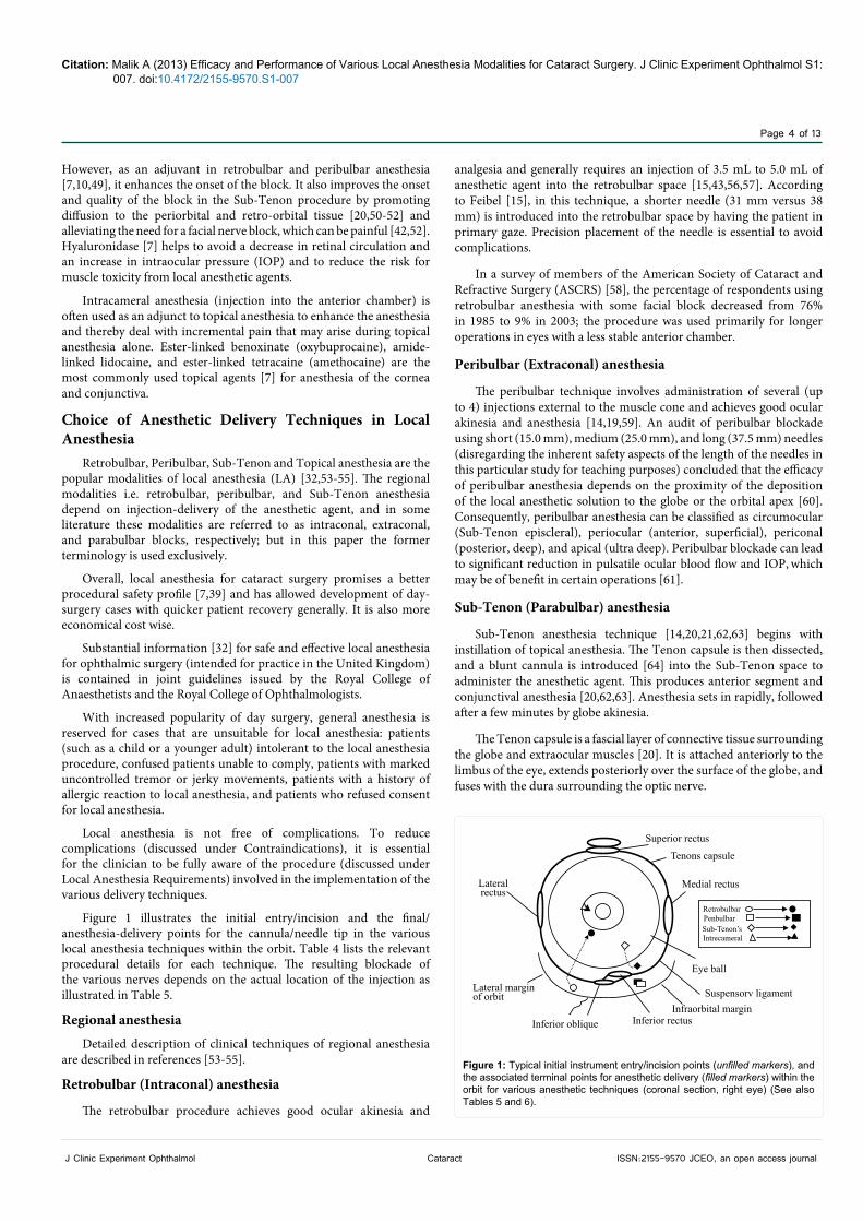
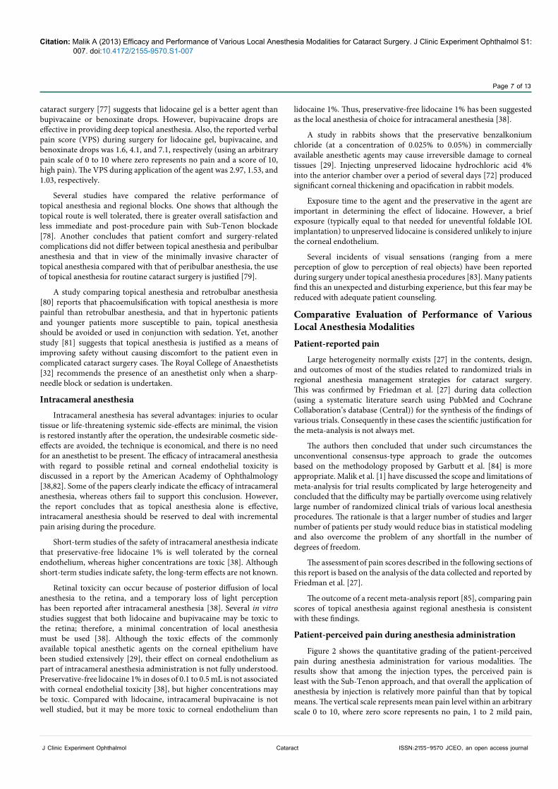
Figure 1 illustrates the initial entry/incision and the final/anesthesia-delivery points for the cannula/needle tip in the various local anesthesia techniques within the orbit. Table 4 lists the relevant procedural details for each technique. The resulting blockade of the various nerves depends on the actual location of the injection as illustrated in Table 5.
Regional anesthesiaDetailed description of clinical techniques of regional anesthesia
are described in references [53-55].
Retrobulbar (Intraconal) anesthesia
The retrobulbar procedure achieves good ocular akinesia and
analgesia and generally requires an injection of 3.5 mL to 5.0 mL of anesthetic agent into the retrobulbar space [15,43,56,57]. According to Feibel [15], in this technique, a shorter needle (31 mm versus 38 mm) is introduced into the retrobulbar space by having the patient in primary gaze. Precision placement of the needle is essential to avoid complications.
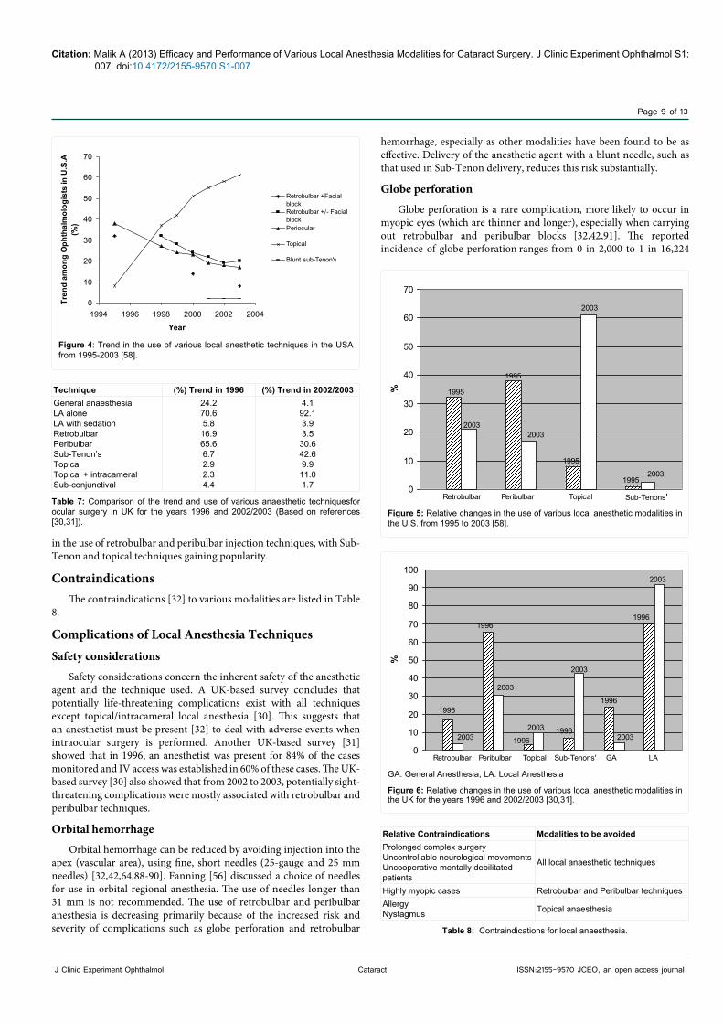
In a survey of members of the American Society of Cataract and Refractive Surgery (ASCRS) [58], the percentage of respondents using retrobulbar anesthesia with some facial block decreased from 76% in 1985 to 9% in 2003; the procedure was used primarily for longer operations in eyes with a less stable anterior chamber.
Peribulbar (Extraconal) anesthesia
The peribulbar technique involves administration of several (up to 4) injections external to the muscle cone and achieves good ocular akinesia and anesthesia [14,19,59]. An audit of peribulbar blockade using short (15.0 mm), medium (25.0 mm), and long (37.5 mm) needles (disregarding the inherent safety aspects of the length of the needles in this particular study for teaching purposes) concluded that the efficacy of peribulbar anesthesia depends on the proximity of the deposition of the local anesthetic solution to the globe or the orbital apex [60]. Consequently, peribulbar anesthesia can be classified as circumocular (Sub-Tenon episcleral), periocular (anterior, superficial), periconal (posterior, deep), and apical (ultra deep). Peribulbar blockade can lead to significant reduction in pulsatile ocular blood flow and IOP, which may be of benefit in certain operations [61].
Sub-Tenon (Parabulbar) anesthesia
Sub-Tenon anesthesia technique [14,20,21,62,63] begins with instillation of topical anesthesia. The Tenon capsule is then dissected, and a blunt cannula is introduced [64] into the Sub-Tenon space to administer the anesthetic agent. This produces anterior segment and conjunctival anesthesia [20,62,63]. Anesthesia sets in rapidly, followed after a few minutes by globe akinesia.
The Tenon capsule is a fascial layer of connective tissue surrounding the globe and extraocular muscles [20]. It is attached anteriorly to the limbus of the eye, extends posteriorly over the surface of the globe, and fuses with the dura surrounding the optic nerve.
Lateralrectus
Superior rectus
Lateral marginof orbit
Inferior oblique Inferior rectusInfraorbital margin
Suspensorv ligament
Eye ball
Tenons capsule
Medial rectus
RetrobulbarPenbulbarSub-Tenon’sIntrecameral
Figure 1: Typical initial instrument entry/incision points (unfilled markers), and the associated terminal points for anesthetic delivery (filled markers) within the orbit for various anesthetic techniques (coronal section, right eye) (See also Tables 5 and 6).
Citation: Malik A (2013) Efficacy and Performance of Various Local Anesthesia Modalities for Cataract Surgery. J Clinic Experiment Ophthalmol S1: 007. doi:10.4172/2155-9570.S1-007
Page 5 of 13
J Clinic Experiment Ophthalmol Cataract ISSN:2155-9570 JCEO, an open access journal
The Sub-Tenon technique has been used increasingly for posterior segment eye surgery such as retinal detachment surgery [65,66]. However, the technique requires a certain amount of skill to dissect into the Sub-Tenon space and correctly place the anesthetic agent as well as competence to deal with the increased risk for conjunctival bleeding and chemosis.
The incidence of conjunctival swelling associated with the Sub-Tenon block is around 39.4% [20,67]. Some of this is attributable to anterior leakage of the injectate, and chemosis is exaggerated if the solution is administered incorrectly into the anterior compartment of Sub-Tenon space or the subconjunctival space. The reported incidence of subconjunctival hemorrhage is 32% to 56% [20,68]. Cauterization of the conjunctival incision is often used to reduce hemorrhage.
Amin et al. [65] suggest the use of a standard IV cannula instead of the Sub-Tenon cannula to puncture the anesthetized and tented conjunctiva to reduce chemosis and subconjunctival hemorrhage, claiming that the “minimal risk” of puncturing the eye is assured because of the clear visibility of the needle tip at all times. Gray and Lucas [21] point out that the needle tip is not clearly visible once under the conjunctiva and in the event of hemorrhage, it would quickly be obscured. Thus, it has no additional safety compared with the peribulbar technique.
Topical anesthesia
For fast recovery and rehabilitation after surgery, cataract patients are often managed under topical anesthesia [69], which can be supplemented with intracameral anesthesia [7,14,25] or oral/IV sedation [24]. One bonus of topical anesthesia, especially in patients who have vision in the operated eye only, is that visual recovery is almost immediate.
Topical anesthesia has also been used with varying degrees of success for combined cataract and glaucoma surgery, trabeculectomy [70], viscocanalostomy, and secondary IOL transplantation.
Topical anesthetic agents [29] block trigeminal nerve endings in the cornea and the conjunctiva only, leaving the intraocular structures in the anterior segment unanesthetized. Thus, manipulation of the iris and stretching of the ciliary and zonular tissues during surgery can irritate the ciliary nerves, resulting in discomfort. For this reason, the addition of intracameral anesthesia as an adjunct is popular [6,14,38,71].
Several methods of topical anesthesia are available, but the use of eyedrops or viscous gels is the most common modalities. Considerations such as corneal epithelial toxicity [72], patient comfort, and patient history of allergies to local anesthetic agents determine the choice of suitable anesthetic eye drops for use in any particular situation.
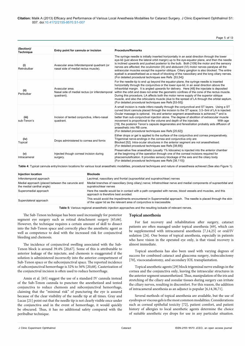
Table 4: Typical cannula entry/incision locations for various local anaesthetic modalities, procedural techniques and nature of anaesthesia achieved (See also Figure 1).
(Section)/Technique Entry point for cannula or incision Procedure/Remarks
(i)Retrobulbar
Avascular area Inferotemporal quadrant (or nasal side of medial rectus muscle).
The syringe needle is initially inserted horizontally in an axial direction through the lower eye-lid (just above the lateral orbit margin) up to the eye-equator plane, and then the needle is inclined upwards and pushed posterior to the bulb. Both [109] the motor and the sensory nerves are affected; the oculomotor (III) and abducent (VI) motor nerves paralyse all the extraocular muscles except the superior oblique. Ciliary ganglion is also blocked. The entire eyeball is anaesthetised as a result of blocking of the nasociliary and the long ciliary nerves.(For detailed procedural techniques see Refs [53,54])
(ii)Peribulbar
Avascular area.Nasal side of medial rectus (or inferotemporal quadrant).
For the needle-tip to end up beyond the equator-plane, the syringe needle is inserted horizontally through the conjunctiva or the lower eye-lid, in an axial direction above the infraorbital margin. It is angled upwards for delivery. Here [48] the injectate is deposited within the orbit and does not enter the geometric confines of the cone of the rectus muscle.
During this procedure, LA affects both the motor nerve supply of the superior oblique muscle, and also the orbicularis muscle (due to the spread of LA through the orbital septum.(For detailed procedural techniques see Refs [53-55])
(iii)sub-Tenon’s
Incision of tented conjunctiva, infero-nasal quadrant.
A small incision is made infero-nasally through the conjunctival and ST layers. Using a ST curved blunt cannula placed through the incision to the ST space; 3.5–5ml of LA is injected. Ocular massage is optional. Iris and anterior segment anaesthesia is achieved,48 and is better than sub-conjunctival injection alone. The degree of abolition of extraocular muscle movement is proportional to the volume and depth of the injectate. With age [19], the posterior Tenon’s capsule degenerates and fenestration probably aids diffusion of anaesthetic into RB cone.(For detailed procedural techniques see Refs [20,62])
(iv)Topical Drops administered to cornea and fornix
Either drops or gel is applied to the surface of the conjunctiva and cornea preoperatively. Trigeminal nerve-endings in the cornea and conjunctiva only are Blocked [52]. Intra-ocular structures in the anterior segment are not anaesthetised.(For detailed procedural techniques see Refs [38,69])
(v)Intracameral
Injected though corneal incision during surgery.
Preservative free anaesthetic (usually 1% lidocaine) is injected into the anterior chamber at the beginning of the operation through one of the corneal incisions required for phacoemulsification. It provides sensory blockage of the axis and the ciliary body(For detailed procedural techniques see Refs [38,110])
Table 5: Various regional anaesthetic injection approaches and the resulting lockade of relevant nerves.
Injection location BlockadeInferotemporal approach Lacrimal, nasociliary and frontal (supraorbital and supratrochlear) nerves Medial approach (placed between the caruncle and the medial canthal angle)
Medial branches of nasociliary (long ciliary) nerve; Infratrochlear nerve and medial components of supraorbital and supratrochlear nerves
Superomedial approach Here the needle would be in contact with a path congested with nerves, blood vessels and muscles, and this approach is therefore best avoided.
Superolateral approach This would avoid the impediments encountered in Superomedial approach. The needle is placed through the skin of the upper lid as the relevant area of conjunctiva is inaccessible.
Citation: Malik A (2013) Efficacy and Performance of Various Local Anesthesia Modalities for Cataract Surgery. J Clinic Experiment Ophthalmol S1: 007. doi:10.4172/2155-9570.S1-007
Page 6 of 13
J Clinic Experiment Ophthalmol Cataract ISSN:2155-9570 JCEO, an open access journal
Proparacaine, tetracaine, lidocaine, bupivacaine, or benoxinate anesthetic eye drops (Table 2) into the fornix of the operative eye are used primarily.
High or prolonged doses of local anesthetic agents are toxic to the corneal epithelium, and this prolongs wound healing and causes corneal erosion. Also, repeated administration of drops can cause clouding of the cornea, rendering surgery more difficult. Tetracaine (an ester-type anesthetic agent) is the most irritating of the eye drops listed above and should be avoided in patients allergic to this particular family of anesthetic agents [38]. Proparacaine, although an ester type, does not metabolize to p-aminobenzoate moiety, and thus may be used safely in patients allergic to other ester-type anesthetic agents.
An alternative to eye drops for topical application is the use of viscous lidocaine gel. The gel is often mixed with dilating medications and antibiotic and non-steroidal anti-inflammatory agents. It is reported that 5 mL of lidocaine gel 2% mixed with 4 drops tropicamide, 4 drops cyclopentolate 1%, 4 drops phenylephrine 10%, 10 drops moxifloxacin, and 4 drops ketorolac and applied to the operative eye twice before the surgery typically achieves excellent dilation and anesthesia [38]. However, drug absorption and corneal epithelial safety of this mixture have not been fully investigated.
Intracameral anesthesia
Intracameral anesthesia is a common adjunct to topical anesthesia in phacoemulsification [38]. It probably provides sensory blockage of the iris and ciliary body and thereby relieves discomfort experienced during IOL placement. Intracameral lidocaine alone dilates the pupil well [73], and this is believed to be because of the direct action of lidocaine on the iris, which in turn causes muscle relaxation. Preservative-free lidocaine 1% with epinephrine (0.3cc of 1: 1000) enhances pupillary dilation more than lidocaine 1% alone and thus obviates the need for preoperative dilating drops [74].
Efficacy and Performance of Various Local Anesthesia ModalitiesRegional (Injection type) anesthesia
The efficacy of anesthesia partly depends on the ease of irrigation
and the spread of the anesthetic agent after injection. A study using magnetic resonance imaging to examine the distribution of local anesthesia solution after combined peribulbar and retrobulbar, superomedial retrobulbar, and Sub-Tenon injection reports that for a combined peribulbar and retrobulbar block [75], a relatively large volume of local anesthetic solution spreads throughout the orbit and a reliable anesthesia is achieved. After superomedial retrobulbar and Sub-Tenon injection, the local anesthetic solution accumulates behind the globe and good analgesia and slight akinesia are achieved with a small volume of anesthetic solution. However, the superomedial approach is not favored because of the close proximity to the cluster of nerves and blood vessels.
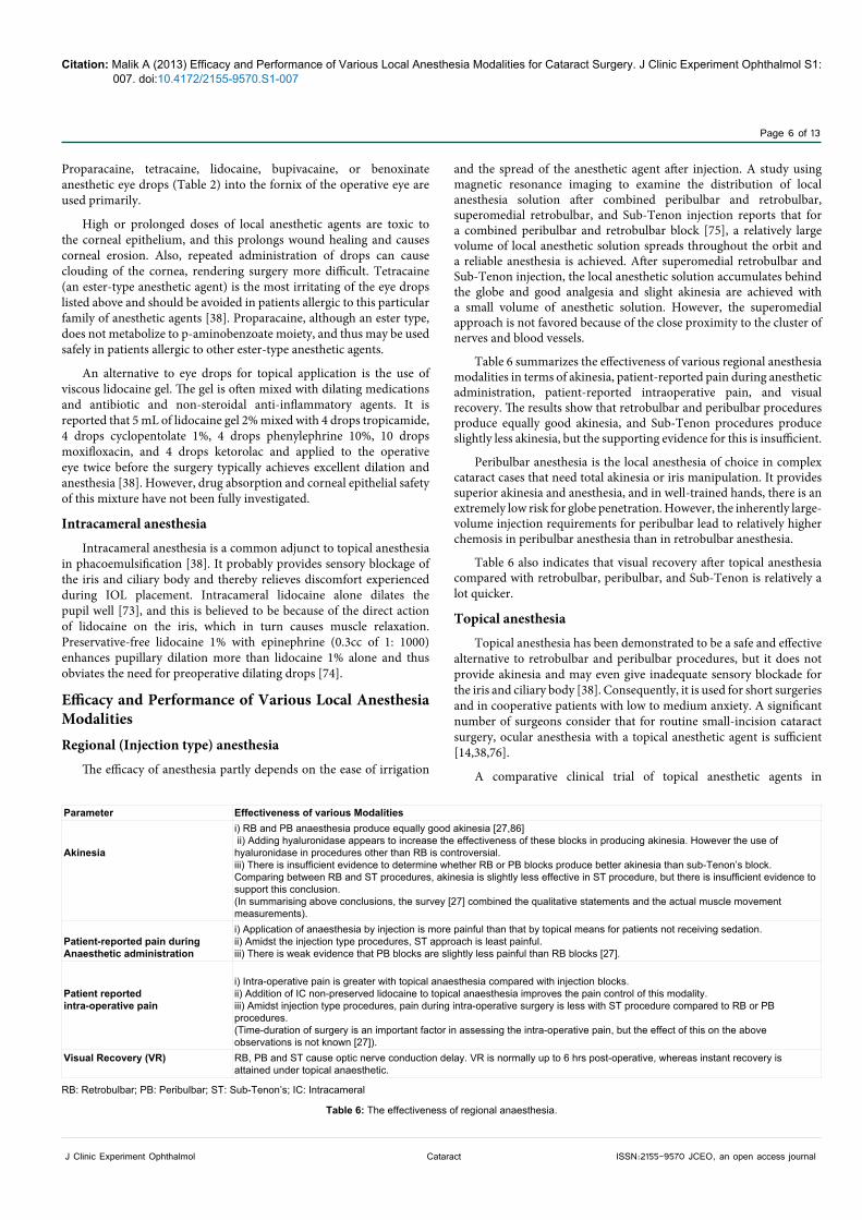
Table 6 summarizes the effectiveness of various regional anesthesia modalities in terms of akinesia, patient-reported pain during anesthetic administration, patient-reported intraoperative pain, and visual recovery. The results show that retrobulbar and peribulbar procedures produce equally good akinesia, and Sub-Tenon procedures produce slightly less akinesia, but the supporting evidence for this is insufficient.
Peribulbar anesthesia is the local anesthesia of choice in complex cataract cases that need total akinesia or iris manipulation. It provides superior akinesia and anesthesia, and in well-trained hands, there is an extremely low risk for globe penetration. However, the inherently large-volume injection requirements for peribulbar lead to relatively higher chemosis in peribulbar anesthesia than in retrobulbar anesthesia.
Table 6 also indicates that visual recovery after topical anesthesia compared with retrobulbar, peribulbar, and Sub-Tenon is relatively a lot quicker.
Topical anesthesia
Topical anesthesia has been demonstrated to be a safe and effective alternative to retrobulbar and peribulbar procedures, but it does not provide akinesia and may even give inadequate sensory blockade for the iris and ciliary body [38]. Consequently, it is used for short surgeries and in cooperative patients with low to medium anxiety. A significant number of surgeons consider that for routine small-incision cataract surgery, ocular anesthesia with a topical anesthetic agent is sufficient [14,38,76].
A comparative clinical trial of topical anesthetic agents in
RB: Retrobulbar; PB: Peribulbar; ST: Sub-Tenon’s; IC: Intracameral
Table 6: The effectiveness of regional anaesthesia.
Parameter Effectiveness of various Modalities
Akinesia
i) RB and PB anaesthesia produce equally good akinesia [27,86] ii) Adding hyaluronidase appears to increase the effectiveness of these blocks in producing akinesia. However the use of hyaluronidase in procedures other than RB is controversial. iii) There is insufficient evidence to determine whether RB or PB blocks produce better akinesia than sub-Tenon’s block. Comparing between RB and ST procedures, akinesia is slightly less effective in ST procedure, but there is insufficient evidence to support this conclusion.(In summarising above conclusions, the survey [27] combined the qualitative statements and the actual muscle movement measurements).
Patient-reported pain during Anaesthetic administration
i) Application of anaesthesia by injection is more painful than that by topical means for patients not receiving sedation.ii) Amidst the injection type procedures, ST approach is least painful.iii) There is weak evidence that PB blocks are slightly less painful than RB blocks [27].
Patient reported intra-operative pain
i) Intra-operative pain is greater with topical anaesthesia compared with injection blocks.ii) Addition of IC non-preserved lidocaine to topical anaesthesia improves the pain control of this modality. iii) Amidst injection type procedures, pain during intra-operative surgery is less with ST procedure compared to RB or PB procedures.(Time-duration of surgery is an important factor in assessing the intra-operative pain, but the effect of this on the above observations is not known [27]).
Visual Recovery (VR) RB, PB and ST cause optic nerve conduction delay. VR is normally up to 6 hrs post-operative, whereas instant recovery is attained under topical anaesthetic.
Citation: Malik A (2013) Efficacy and Performance of Various Local Anesthesia Modalities for Cataract Surgery. J Clinic Experiment Ophthalmol S1: 007. doi:10.4172/2155-9570.S1-007
Page 7 of 13
J Clinic Experiment Ophthalmol Cataract ISSN:2155-9570 JCEO, an open access journal
cataract surgery [77] suggests that lidocaine gel is a better agent than bupivacaine or benoxinate drops. However, bupivacaine drops are effective in providing deep topical anesthesia. Also, the reported verbal pain score (VPS) during surgery for lidocaine gel, bupivacaine, and benoxinate drops was 1.6, 4.1, and 7.1, respectively (using an arbitrary pain scale of 0 to 10 where zero represents no pain and a score of 10, high pain). The VPS during application of the agent was 2.97, 1.53, and 1.03, respectively.
Several studies have compared the relative performance of topical anesthesia and regional blocks. One shows that although the topical route is well tolerated, there is greater overall satisfaction and less immediate and post-procedure pain with Sub-Tenon blockade [78]. Another concludes that patient comfort and surgery-related complications did not differ between topical anesthesia and peribulbar anesthesia and that in view of the minimally invasive character of topical anesthesia compared with that of peribulbar anesthesia, the use of topical anesthesia for routine cataract surgery is justified [79].
A study comparing topical anesthesia and retrobulbar anesthesia [80] reports that phacoemulsification with topical anesthesia is more painful than retrobulbar anesthesia, and that in hypertonic patients and younger patients more susceptible to pain, topical anesthesia should be avoided or used in conjunction with sedation. Yet, another study [81] suggests that topical anesthesia is justified as a means of improving safety without causing discomfort to the patient even in complicated cataract surgery cases. The Royal College of Anaesthetists [32] recommends the presence of an anesthetist only when a sharp-needle block or sedation is undertaken.
Intracameral anesthesia
Intracameral anesthesia has several advantages: injuries to ocular tissue or life-threatening systemic side-effects are minimal, the vision is restored instantly after the operation, the undesirable cosmetic side-effects are avoided, the technique is economical, and there is no need for an anesthetist to be present. The efficacy of intracameral anesthesia with regard to possible retinal and corneal endothelial toxicity is discussed in a report by the American Academy of Ophthalmology [38,82]. Some of the papers clearly indicate the efficacy of intracameral anesthesia, whereas others fail to support this conclusion. However, the report concludes that as topical anesthesia alone is effective, intracameral anesthesia should be reserved to deal with incremental pain arising during the procedure.
Short-term studies of the safety of intracameral anesthesia indicate that preservative-free lidocaine 1% is well tolerated by the corneal endothelium, whereas higher concentrations are toxic [38]. Although short-term studies indicate safety, the long-term effects are not known.
Retinal toxicity can occur because of posterior diffusion of local anesthesia to the retina, and a temporary loss of light perception has been reported after intracameral anesthesia [38]. Several in vitro studies suggest that both lidocaine and bupivacaine may be toxic to the retina; therefore, a minimal concentration of local anesthesia must be used [38]. Although the toxic effects of the commonly available topical anesthetic agents on the corneal epithelium have been studied extensively [29], their effect on corneal endothelium as part of intracameral anesthesia administration is not fully understood. Preservative-free lidocaine 1% in doses of 0.1 to 0.5 mL is not associated with corneal endothelial toxicity [38], but higher concentrations may be toxic. Compared with lidocaine, intracameral bupivacaine is not well studied, but it may be more toxic to corneal endothelium than
lidocaine 1%. Thus, preservative-free lidocaine 1% has been suggested as the local anesthesia of choice for intracameral anesthesia [38].
A study in rabbits shows that the preservative benzalkonium chloride (at a concentration of 0.025% to 0.05%) in commercially available anesthetic agents may cause irreversible damage to corneal tissues [29]. Injecting unpreserved lidocaine hydrochloric acid 4% into the anterior chamber over a period of several days [72] produced significant corneal thickening and opacification in rabbit models.
Exposure time to the agent and the preservative in the agent are important in determining the effect of lidocaine. However, a brief exposure (typically equal to that needed for uneventful foldable IOL implantation) to unpreserved lidocaine is considered unlikely to injure the corneal endothelium.
Several incidents of visual sensations (ranging from a mere perception of glow to perception of real objects) have been reported during surgery under topical anesthesia procedures [83]. Many patients find this an unexpected and disturbing experience, but this fear may be reduced with adequate patient counseling.
Comparative Evaluation of Performance of Various Local Anesthesia ModalitiesPatient-reported pain
Large heterogeneity normally exists [27] in the contents, design, and outcomes of most of the studies related to randomized trials in regional anesthesia management strategies for cataract surgery. This was confirmed by Friedman et al. [27] during data collection (using a systematic literature search using PubMed and Cochrane Collaboration’s database (Central)) for the synthesis of the findings of various trials. Consequently in these cases the scientific justification for the meta-analysis is not always met.
The authors then concluded that under such circumstances the unconventional consensus-type approach to grade the outcomes based on the methodology proposed by Garbutt et al. [84] is more appropriate. Malik et al. [1] have discussed the scope and limitations of meta-analysis for trial results complicated by large heterogeneity and concluded that the difficulty may be partially overcome using relatively large number of randomized clinical trials of various local anesthesia procedures. The rationale is that a larger number of studies and larger number of patients per study would reduce bias in statistical modeling and also overcome the problem of any shortfall in the number of degrees of freedom.
The assessment of pain scores described in the following sections of this report is based on the analysis of the data collected and reported by Friedman et al. [27].
The outcome of a recent meta-analysis report [85], comparing pain scores of topical anesthesia against regional anesthesia is consistent with these findings.
Patient-perceived pain during anesthesia administration
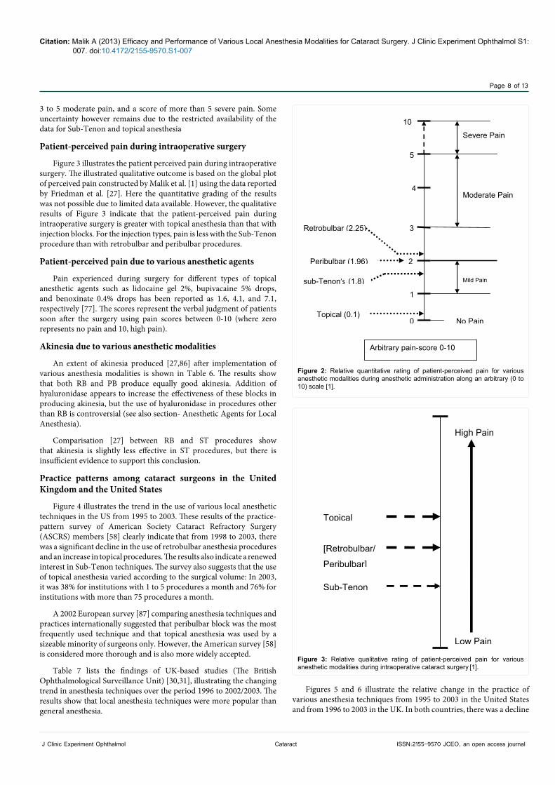
Figure 2 shows the quantitative grading of the patient-perceived pain during anesthesia administration for various modalities. The results show that among the injection types, the perceived pain is least with the Sub-Tenon approach, and that overall the application of anesthesia by injection is relatively more painful than that by topical means. The vertical scale represents mean pain level within an arbitrary scale 0 to 10, where zero score represents no pain, 1 to 2 mild pain,
Citation: Malik A (2013) Efficacy and Performance of Various Local Anesthesia Modalities for Cataract Surgery. J Clinic Experiment Ophthalmol S1: 007. doi:10.4172/2155-9570.S1-007
Page 8 of 13
J Clinic Experiment Ophthalmol Cataract ISSN:2155-9570 JCEO, an open access journal
3 to 5 moderate pain, and a score of more than 5 severe pain. Some uncertainty however remains due to the restricted availability of the data for Sub-Tenon and topical anesthesia
Patient-perceived pain during intraoperative surgery
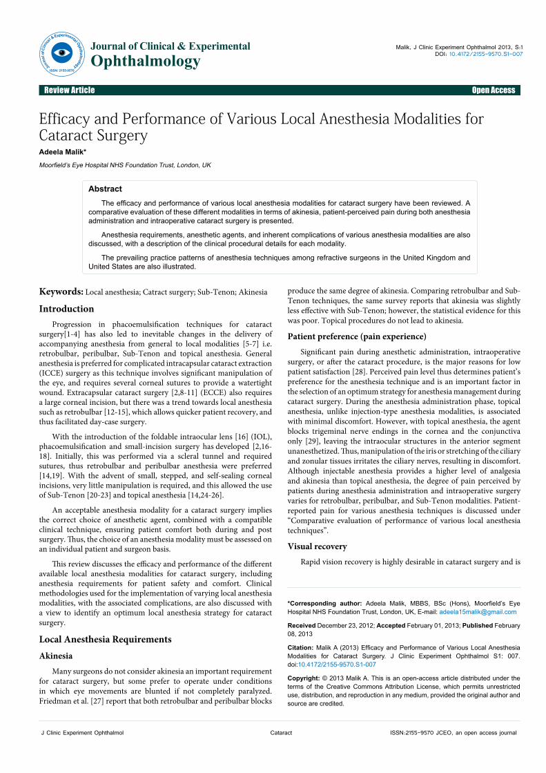
Figure 3 illustrates the patient perceived pain during intraoperative surgery. The illustrated qualitative outcome is based on the global plot of perceived pain constructed by Malik et al. [1] using the data reported by Friedman et al. [27]. Here the quantitative grading of the results was not possible due to limited data available. However, the qualitative results of Figure 3 indicate that the patient-perceived pain during intraoperative surgery is greater with topical anesthesia than that with injection blocks. For the injection types, pain is less with the Sub-Tenon procedure than with retrobulbar and peribulbar procedures.
Patient-perceived pain due to various anesthetic agents
Pain experienced during surgery for different types of topical anesthetic agents such as lidocaine gel 2%, bupivacaine 5% drops, and benoxinate 0.4% drops has been reported as 1.6, 4.1, and 7.1, respectively [77]. The scores represent the verbal judgment of patients soon after the surgery using pain scores between 0-10 (where zero represents no pain and 10, high pain).
Akinesia due to various anesthetic modalities
An extent of akinesia produced [27,86] after implementation of various anesthesia modalities is shown in Table 6. The results show that both RB and PB produce equally good akinesia. Addition of hyaluronidase appears to increase the effectiveness of these blocks in producing akinesia, but the use of hyaluronidase in procedures other than RB is controversial (see also section- Anesthetic Agents for Local Anesthesia).
Comparisation [27] between RB and ST procedures show that akinesia is slightly less effective in ST procedures, but there is insufficient evidence to support this conclusion.
Practice patterns among cataract surgeons in the United Kingdom and the United States
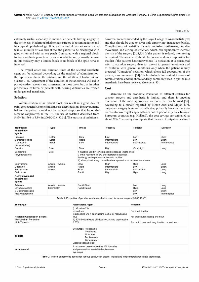
Figure 4 illustrates the trend in the use of various local anesthetic techniques in the US from 1995 to 2003. These results of the practice-pattern survey of American Society Cataract Refractory Surgery (ASCRS) members [58] clearly indicate that from 1998 to 2003, there was a significant decline in the use of retrobulbar anesthesia procedures and an increase in topical procedures. The results also indicate a renewed interest in Sub-Tenon techniques. The survey also suggests that the use of topical anesthesia varied according to the surgical volume: In 2003, it was 38% for institutions with 1 to 5 procedures a month and 76% for institutions with more than 75 procedures a month.
A 2002 European survey [87] comparing anesthesia techniques and practices internationally suggested that peribulbar block was the most frequently used technique and that topical anesthesia was used by a sizeable minority of surgeons only. However, the American survey [58] is considered more thorough and is also more widely accepted.
Table 7 lists the findings of UK-based studies (The British Ophthalmological Surveillance Unit) [30,31], illustrating the changing trend in anesthesia techniques over the period 1996 to 2002/2003. The results show that local anesthesia techniques were more popular than general anesthesia.
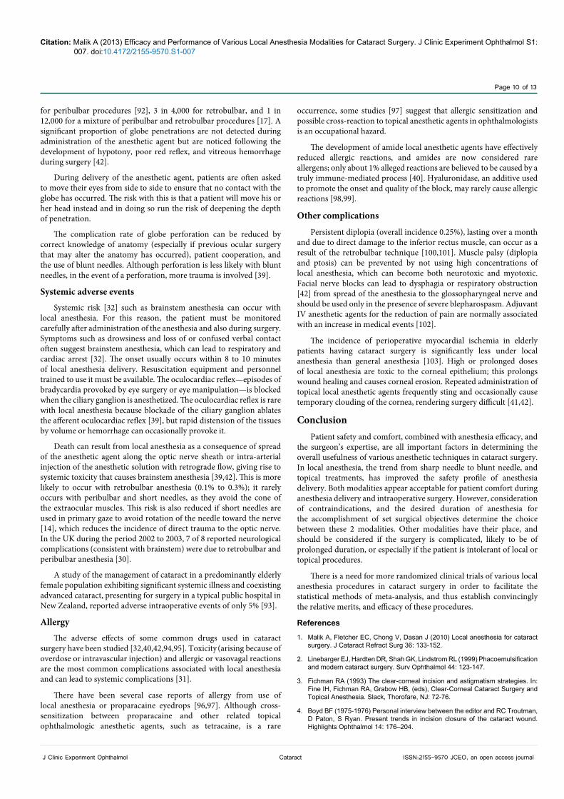
Figures 5 and 6 illustrate the relative change in the practice of various anesthesia techniques from 1995 to 2003 in the United States and from 1996 to 2003 in the UK. In both countries, there was a decline
Severe Pain
Moderate Pain
Mild Pain
No Pain
10
5
3
2
1
0
4
Retrobulbar (2.25)
Peribulbar (1.96)
Topical (0.1)
sub-Tenon’s (1.8)
Arbitrary pain-score 0-10
Figure 2: Relative quantitative rating of patient-perceived pain for various anesthetic modalities during anesthetic administration along an arbitrary (0 to 10) scale [1].
Low Pain
[Retrobulbar/
Peribulbar]
Sub-Tenon
Topical
High Pain
Figure 3: Relative qualitative rating of patient-perceived pain for various anesthetic modalities during intraoperative cataract surgery [1].
Citation: Malik A (2013) Efficacy and Performance of Various Local Anesthesia Modalities for Cataract Surgery. J Clinic Experiment Ophthalmol S1: 007. doi:10.4172/2155-9570.S1-007
Page 9 of 13
J Clinic Experiment Ophthalmol Cataract ISSN:2155-9570 JCEO, an open access journal
in the use of retrobulbar and peribulbar injection techniques, with Sub-Tenon and topical techniques gaining popularity.
ContraindicationsThe contraindications [32] to various modalities are listed in Table
8.
Complications of Local Anesthesia TechniquesSafety considerations
Safety considerations concern the inherent safety of the anesthetic agent and the technique used. A UK-based survey concludes that potentially life-threatening complications exist with all techniques except topical/intracameral local anesthesia [30]. This suggests that an anesthetist must be present [32] to deal with adverse events when intraocular surgery is performed. Another UK-based survey [31] showed that in 1996, an anesthetist was present for 84% of the cases monitored and IV access was established in 60% of these cases. The UK-based survey [30] also showed that from 2002 to 2003, potentially sight-threatening complications were mostly associated with retrobulbar and peribulbar techniques.
Orbital hemorrhage
Orbital hemorrhage can be reduced by avoiding injection into the apex (vascular area), using fine, short needles (25-gauge and 25 mm needles) [32,42,64,88-90]. Fanning [56] discussed a choice of needles for use in orbital regional anesthesia. The use of needles longer than 31 mm is not recommended. The use of retrobulbar and peribulbar anesthesia is decreasing primarily because of the increased risk and severity of complications such as globe perforation and retrobulbar
hemorrhage, especially as other modalities have been found to be as effective. Delivery of the anesthetic agent with a blunt needle, such as that used in Sub-Tenon delivery, reduces this risk substantially.
Globe perforation
Globe perforation is a rare complication, more likely to occur in myopic eyes (which are thinner and longer), especially when carrying out retrobulbar and peribulbar blocks [32,42,91]. The reported incidence of globe perforation ranges from 0 in 2,000 to 1 in 16,224
0
10
20
30
40
50
60
70
1994 1996 1998 2000 2002 2004
Year
Tren
d am
ong
Oph
thal
mol
ogis
ts in
U.S
.A
(%)
Retrobulbar +FacialblockRetrobulbar +/- FacialblockPeriocular
Topical
Blunt sub-Tenon's
Figure 4: Trend in the use of various local anesthetic techniques in the USA from 1995-2003 [58].
Table 7: Comparison of the trend and use of various anaesthetic techniquesfor ocular surgery in UK for the years 1996 and 2002/2003 (Based on references [30,31]).
Technique (%) Trend in 1996 (%) Trend in 2002/2003General anaesthesiaLA aloneLA with sedationRetrobulbarPeribulbarSub-Tenon’sTopicalTopical + intracameralSub-conjunctival
24.270.65.8
16.965.66.72.92.34.4
4.192.13.93.5
30.642.69.9
11.01.7 0
10
20
30
40
50
60
70
%
Retrobulbar Peribulbar Topical Sub-Tenons'
1995
2003
2003
2003
2003
1995
1995
1995
Figure 5: Relative changes in the use of various local anesthetic modalities in the U.S. from 1995 to 2003 [58].
0
10
20
30
40
50
60
70
80
90
100
%
Retrobulbar Peribulbar Topical Sub-Tenons' GA LA
1996
1996
19961996
1996
1996
2003
2003
2003
2003
2003
2003
GA: General Anesthesia; LA: Local Anesthesia
Figure 6: Relative changes in the use of various local anesthetic modalities in the UK for the years 1996 and 2002/2003 [30,31].
Relative Contraindications Modalities to be avoidedProlonged complex surgeryUncontrollable neurological movementsUncooperative mentally debilitated patients
All local anaesthetic techniques
Highly myopic cases Retrobulbar and Peribulbar techniquesAllergyNystagmus Topical anaesthesia
Table 8: Contraindications for local anaesthesia.
Citation: Malik A (2013) Efficacy and Performance of Various Local Anesthesia Modalities for Cataract Surgery. J Clinic Experiment Ophthalmol S1: 007. doi:10.4172/2155-9570.S1-007
Page 10 of 13
J Clinic Experiment Ophthalmol Cataract ISSN:2155-9570 JCEO, an open access journal
for peribulbar procedures [92], 3 in 4,000 for retrobulbar, and 1 in 12,000 for a mixture of peribulbar and retrobulbar procedures [17]. A significant proportion of globe penetrations are not detected during administration of the anesthetic agent but are noticed following the development of hypotony, poor red reflex, and vitreous hemorrhage during surgery [42].
During delivery of the anesthetic agent, patients are often asked to move their eyes from side to side to ensure that no contact with the globe has occurred. The risk with this is that a patient will move his or her head instead and in doing so run the risk of deepening the depth of penetration.
The complication rate of globe perforation can be reduced by correct knowledge of anatomy (especially if previous ocular surgery that may alter the anatomy has occurred), patient cooperation, and the use of blunt needles. Although perforation is less likely with blunt needles, in the event of a perforation, more trauma is involved [39].
Systemic adverse events
Systemic risk [32] such as brainstem anesthesia can occur with local anesthesia. For this reason, the patient must be monitored carefully after administration of the anesthesia and also during surgery. Symptoms such as drowsiness and loss of or confused verbal contact often suggest brainstem anesthesia, which can lead to respiratory and cardiac arrest [32]. The onset usually occurs within 8 to 10 minutes of local anesthesia delivery. Resuscitation equipment and personnel trained to use it must be available. The oculocardiac reflex—episodes of bradycardia provoked by eye surgery or eye manipulation—is blocked when the ciliary ganglion is anesthetized. The oculocardiac reflex is rare with local anesthesia because blockade of the ciliary ganglion ablates the afferent oculocardiac reflex [39], but rapid distension of the tissues by volume or hemorrhage can occasionally provoke it.
Death can result from local anesthesia as a consequence of spread of the anesthetic agent along the optic nerve sheath or intra-arterial injection of the anesthetic solution with retrograde flow, giving rise to systemic toxicity that causes brainstem anesthesia [39,42]. This is more likely to occur with retrobulbar anesthesia (0.1% to 0.3%); it rarely occurs with peribulbar and short needles, as they avoid the cone of the extraocular muscles. This risk is also reduced if short needles are used in primary gaze to avoid rotation of the needle toward the nerve [14], which reduces the incidence of direct trauma to the optic nerve. In the UK during the period 2002 to 2003, 7 of 8 reported neurological complications (consistent with brainstem) were due to retrobulbar and peribulbar anesthesia [30].
A study of the management of cataract in a predominantly elderly female population exhibiting significant systemic illness and coexisting advanced cataract, presenting for surgery in a typical public hospital in New Zealand, reported adverse intraoperative events of only 5% [93].
Allergy
The adverse effects of some common drugs used in cataract surgery have been studied [32,40,42,94,95]. Toxicity (arising because of overdose or intravascular injection) and allergic or vasovagal reactions are the most common complications associated with local anesthesia and can lead to systemic complications [31].
There have been several case reports of allergy from use of local anesthesia or proparacaine eyedrops [96,97]. Although cross-sensitization between proparacaine and other related topical ophthalmologic anesthetic agents, such as tetracaine, is a rare
occurrence, some studies [97] suggest that allergic sensitization and possible cross-reaction to topical anesthetic agents in ophthalmologists is an occupational hazard.
The development of amide local anesthetic agents have effectively reduced allergic reactions, and amides are now considered rare allergens; only about 1% alleged reactions are believed to be caused by a truly immune-mediated process [40]. Hyaluronidase, an additive used to promote the onset and quality of the block, may rarely cause allergic reactions [98,99].
Other complications
Persistent diplopia (overall incidence 0.25%), lasting over a month and due to direct damage to the inferior rectus muscle, can occur as a result of the retrobulbar technique [100,101]. Muscle palsy (diplopia and ptosis) can be prevented by not using high concentrations of local anesthesia, which can become both neurotoxic and myotoxic. Facial nerve blocks can lead to dysphagia or respiratory obstruction [42] from spread of the anesthesia to the glossopharyngeal nerve and should be used only in the presence of severe blepharospasm. Adjuvant IV anesthetic agents for the reduction of pain are normally associated with an increase in medical events [102].
The incidence of perioperative myocardial ischemia in elderly patients having cataract surgery is significantly less under local anesthesia than general anesthesia [103]. High or prolonged doses of local anesthesia are toxic to the corneal epithelium; this prolongs wound healing and causes corneal erosion. Repeated administration of topical local anesthetic agents frequently sting and occasionally cause temporary clouding of the cornea, rendering surgery difficult [41,42].
ConclusionPatient safety and comfort, combined with anesthesia efficacy, and
the surgeon’s expertise, are all important factors in determining the overall usefulness of various anesthetic techniques in cataract surgery. In local anesthesia, the trend from sharp needle to blunt needle, and topical treatments, has improved the safety profile of anesthesia delivery. Both modalities appear acceptable for patient comfort during anesthesia delivery and intraoperative surgery. However, consideration of contraindications, and the desired duration of anesthesia for the accomplishment of set surgical objectives determine the choice between these 2 modalities. Other modalities have their place, and should be considered if the surgery is complicated, likely to be of prolonged duration, or especially if the patient is intolerant of local or topical procedures.
There is a need for more randomized clinical trials of various local anesthesia procedures in cataract surgery in order to facilitate the statistical methods of meta-analysis, and thus establish convincingly the relative merits, and efficacy of these procedures.
References1. Malik A, Fletcher EC, Chong V, Dasan J (2010) Local anesthesia for cataract
surgery. J Cataract Refract Surg 36: 133-152.
2. Linebarger EJ, Hardten DR, Shah GK, Lindstrom RL (1999) Phacoemulsification and modern cataract surgery. Surv Ophthalmol 44: 123-147.
3. Fichman RA (1993) The clear-corneal incision and astigmatism strategies. In: Fine IH, Fichman RA, Grabow HB, (eds), Clear-Corneal Cataract Surgery and Topical Anesthesia. Slack, Thorofare, NJ: 72-76.
4. Boyd BF (1975-1976) Personal interview between the editor and RC Troutman, D Paton, S Ryan. Present trends in incision closure of the cataract wound. Highlights Ophthalmol 14: 176–204.
Citation: Malik A (2013) Efficacy and Performance of Various Local Anesthesia Modalities for Cataract Surgery. J Clinic Experiment Ophthalmol S1: 007. doi:10.4172/2155-9570.S1-007
Page 11 of 13
J Clinic Experiment Ophthalmol Cataract ISSN:2155-9570 JCEO, an open access journal
5. Fichman RA (1996) Use of topical anesthesia alone in cataract surgery. J Cataract Refract Surg 22: 612-614.
6. Gills JP, Cherchio M, Raanan MG (1997) Unpreserved lidocaine to control discomfort during cataract surgery using topical anesthesia. J Cataract Refract Surg 23: 545-550.
7. Kallio H, Rosenberg PH (2005) Advances in ophthalmic regional anaesthesia. Best Pract Res Clin Anaesthesiol 19: 215-227.
8. Seibel BS (2005) Phacodynamics; Mastering the Tools and Techniques of Phacoemulsification Surgery. (4th edn) Slack, Thorofare, NJ.
9. Gimbel HV (1991) Continuous curvilinear capsulorhexis and nuclear fracturing; evolution, technique, and complications. Ophthalmol Clin North Am 4: 235-249.
10. Holmberg AS, Philipson BT (1984) Sodium hyaluronate in cataract surgery. II. Report on the use of Healon in extracapsular cataract surgery using phacoemulsification. Ophthalmology 91: 53-59.
11. Ernest PH (1994) Cataract incision architecture. Int Ophthalmol Clin 34: 31-57.
12. Schein OD, Bass EB, Sharkey P, Luthra R, Tielsch JM, et al. (1995) Cataract surgical techniques. Preferences and underlying beliefs. Arch Ophthalmol 113: 1108-1112.
13. Packer M, Fishkind WJ, Fine IH, Seibel BS, Hoffman RS (2005) The physics of phaco: a review. J Cataract Refract Surg 31: 424-431.
14. Crandall AS (2001) Anesthesia modalities for cataract surgery. Curr Opin Ophthalmol 12: 9-11.
15. Feibel RM (1985) Current concepts in retrobulbar anesthesia. Surv Ophthalmol 30: 102-110.
16. Vargas LG, Peng Q, Escobar-Gomez M, Schmidbauer JM, Apple DJ (2001) Overview of modern foldable intraocular lenses and clinically relevant anatomy and histology of the crystalline lens. Int Ophthalmol Clin 41: 1-15.
17. Hamilton RC, Gimbel HV, Strunin L (1988) Regional anaesthesia for 12,000 cataract extraction and intraocular lens implantation procedures. Can J Anaesth 35: 615-623.
18. Alió J, Rodriguez-Prats JL, Galal A (2006) Advances in microincision cataract surgery intraocular lenses. Curr Opin Ophthalmol 17: 80-93.
19. Davis DB 2nd, Mandel MR (1986) Posterior peribulbar anesthesia: an alternative to retrobulbar anesthesia. J Cataract Refract Surg 12: 182-184.
20. Canavan KS, Dark A, Garrioch MA (2003) Sub-Tenon’s administration of local anaesthetic: a review of the technique. Br J Anaesth 90: 787-793.
21. Gray R, Lucas J (2002) “No needle” sub-Tenon’s anesthesia [letter]. Br J Ophthalmol 86: 831.
22. Greenbaum S (1997) Anesthesia in cataract aurgery. In: Greenbaum S (ed), Ocular Anesthesia. Saunders, Philadelphia, PA: 1-55.
23. Hansen EA, Mein CE, Mazzoli R (1990) Ocular anesthesia for cataract surgery: a direct sub-Tenon’s approach. Ophthalmic Surg 21: 696-699.
24. Rocha G, Turner C (2007) Safety of cataract surgery under topical anesthesia with oral sedation without anesthetic monitoring. Can J Ophthalmol 42: 288-294.
25. Ezra DG, Allan BD (2007) Topical anaesthesia alone versus topical anaesthesia with intracameral lidocaine for phacoemulsification. Cochrane Database Syst Rev : CD005276.
26. Kershner RM (1993) Topical anesthesia for small incision self-sealing cataract surgery. A prospective evaluation of the first 100 patients. J Cataract Refract Surg 19: 290-292.
27. Friedman DS, Bass EB, Lubomski LH, Fleisher LA, Kempen JH, et al. (2001) Synthesis of the literature on the effectiveness of regional anesthesia for cataract surgery. Ophthalmology 108: 519-529.
28. Fung D, Cohen MM, Stewart S, Davies A (2005) What determines patient satisfaction with cataract care under topical local anesthesia and monitored sedation in community setting? Anesth Analg 100: 1644-1650.
29. Tseng SH, Chen FK (1998) A randomized clinical trial of combined topical-intracameral anesthesia in cataract surgery. Ophthalmology 105: 2007-2011.
30. Eke T, Thompson JR (2007) Serious complications of local anaesthesia for cataract surgery: a 1 year national survey in the United Kingdom. Br J Ophthalmol 91: 470-475.
31. Eke T, Thompson JR (1999) The National Survey of Local Anaesthesia for Ocular Surgery. I. Survey methodology and current practice. Eye (Lond) 13 : 189-195.
32. Royal College of Anaesthetists (2009) Guidelines on the Provision of Anesthesia Services. Chapter 10. Ophthalmic Anesthesia Services. London, UK, Royal College of Anaesthetists.
33. Hodgkins PR, Luff AJ, Morrell AJ, Botchway LT, Featherston TJ, et al. (1992) Current practice of cataract extraction and anaesthesia. Br J Ophthalmol 76: 323-326.
34. Muttu S, Liu EH, Ang SB, Chew PT, Lee TL, et al. (2005) Comparison of dexmedetomidine and midazolam sedation for cataract surgery under topical anesthesia. J Cataract Refract Surg 31: 1845-1846.
35. Ahmad S (2006) Sedation techniques in ophthalmic anesthesia. Ophthalmol Clin North Am 19: 193-202.
36. Frick KD (2006) Economic evaluation of different systems for cataract surgery and anesthesia. Ophthalmol Clin North Am 19: 309-315.
37. Mojon-Azzi SM, Mojon DS (2007) The rate of outpatient cataract surgery in ten European countries: an analysis using data from the SHARE survey. Graefes Arch Clin Exp Ophthalmol 245: 1041-1044.
38. Cass GD (2006) Choices of local anesthetics for ocular surgery. Ophthalmol Clin North Am 19: 203-207.
39. Hamilton RC (1998) Complications of ophthalmic regional anesthesia. Ophthalmol Clin North Am 11: 99-114.
40. Jackson T, McLure HA (2006) Pharmacology of local anesthetics. Ophthalmol Clin North Am 19: 155-161.
41. Rosenwasser GO (1989) Complications of topical ocular anesthetics. Int Ophthalmol Clin 29: 153-158.
42. Rubin AP (1995) Complications of local anesthesia for ophthalmic surgery. Br J Anaesth 1995; 75:93-96.
43. Weindler J, Weindler M, Ruprecht KW (2004) [Local anesthesia in ophthalmic surgery]. Ophthalmologe 101: 847-864.
44. Coelho RP, Weissheimer J, Romão E, Velasco e Cruz AA (2005) Pain induced by phacoemulsification without sedation using topical or peribulbar anesthesia. J Cataract Refract Surg 31: 385-388.
45. Navaleza JS, Pendse SJ, Blecher MH (2006) Choosing anesthesia for cataract surgery. Ophthalmol Clin North Am 19: 233-237.
46. Stoelting RK (1999) Pharmacology and Physiology in Anesthetic Practice. (3rdedn) Lippincott-Raven, Philadelphia, PA.
47. Hamilton RC, Grizzard WS (1993) Complications. In: Gills JP, Hustead RF, Sanders DR (eds) Ophthalmic Anesthesia. Slack, Thorofare, NJ: 187-202.
48. Malik A. Pharmacology of local anesthetics. Duanes: Chapter
49. Atkinson WS (1961) The development of ophthalmic anesthesia; the Sanford R. Gifford Lecture. Am J Ophthalmol 51: 1-14.
50. Mauger TF, Craig EL Havener WH (1994) Havener’s Ocular Pharmacology. (6th edn) Mosby, St Louis, MO: 211-212.
51. Guise P, Laurent S (1999) Sub-Tenon’s block: the effect of hyaluronidase on speed of onset and block quality. Anaesth Intensive Care 27: 179-181.
52. Rowley SA, Hale JE, Finlay RD (2000) Sub-Tenon’s local anesthesia: the effect of hyaluronidase. Br J Ophthalmol 84: 435-436.
53. Hamilton RC (1996) Techniques of orbital regional anesthesia. In: Smith GB, Hamilton RC, Carr CA (eds) Ophthalmic Anesthesia; a Practical Handbook. (2nd edn) Arnold, London, UK: 104-147.
54. Freeman JM, Freeman JFD (1998) Retrobulbar and posterior peribulbar anesthesia for ophthalmic surgery. Ophthalmol Clin North Am 11: 39-45.
55. Kumar CM, Fanning GL (2002) Orbital regional anesthesia. In Kumar CM, Dodds C, Fanning GL (eds.) Ophthalmic Anesthesia. Swets and Zeitlinger, Lisse, The Netherlands: 61-68.
56. Fanning GL (2006) Orbital regional anesthesia. Ophthalmol Clin North Am 19: 221-232.
57. Lopatka CW, Sharvelle DJ, Magnante DO (2002) Retrobulbar anesthesia. Ophthalmology 109: 4-5.
Citation: Malik A (2013) Efficacy and Performance of Various Local Anesthesia Modalities for Cataract Surgery. J Clinic Experiment Ophthalmol S1: 007. doi:10.4172/2155-9570.S1-007
Page 12 of 13
J Clinic Experiment Ophthalmol Cataract ISSN:2155-9570 JCEO, an open access journal
58. Leaming DV (2004) Practice styles and preferences of ASCRS members--2003 survey. J Cataract Refract Surg 30: 892-900.
59. Mawer RJ, Coombes AG (1998) Current practice of local anaesthesia for routine ocular surgery. Br J Anaesth 80: 241-242.
60. van den Berg AA (2004) An audit of peribulbar blockade using 15 mm, 25 mm and 37.5 mm needles, and sub-Tenon’s injection. Anaesthesia 59: 775-780.
61. Chang BY, Hee WC, Ling R, Broadway DC, Beigi B (2000) Local anaesthetic techniques and pulsatile ocular blood flow. Br J Ophthalmol 84: 1260-1263.
62. Kumar CM, Dodds C (2006) Sub-Tenon’s Anesthesia. Ophthalmol Clin North Am 19: 209-219.
63. Kumar CM, Williamson S, Manickam B (2005) A review of sub-Tenon’s block: current practice and recent development. Eur J Anaesthesiol 22: 567-577.
64. Hamilton RC (1995) Techniques of orbital regional anaesthesia. Br J Anaesth 75: 88-92.
65. Amin S, Minihan M, Lesnik-Oberstein S, Carr C (2002) A new technique for delivering sub-Tenon’s anaesthesia in ophthalmic surgery. Br J Ophthalmol 86: 119-120.
66. Li HK, Abouleish A, Grady J, Groeschel W, Gill KS (2000) Sub-Tenon’s injection for local anesthesia in posterior segment surgery. Ophthalmology 107: 41-46.
67. Roman SJ, Chong Sit DA, Boureau CM, Auclin FX, Ullern MM (1997) Sub-Tenon’s anaesthesia: an efficient and safe technique. Br J Ophthalmol 81: 673-676.
68. Stevens J (1992) A new local anesthesia technique for cataract extraction by one quadrant sub-Tenon’s infiltration. Br J Ophthalmol 76: 620-624.
69. Fichman RA (1998) Topical anesthesia. Ophthalmol Clin North Am 11: 57-63.
70. Zabriskie NA, Ahmed II, Crandall AS, Daines B, Burns TA, et al. (2002) A comparison of topical and retrobulbar anesthesia for trabeculectomy. J Glaucoma 11: 306-314.
71. Koch PS (1997) Anterior chamber irrigation with unpreserved lidocaine 1% for anesthesia during cataract surgery. J Cataract Refract Surg 23: 551-554.
72. Judge AJ, Najafi K, Lee DA, Miller KM (1997) Corneal endothelial toxicity of topical anesthesia. Ophthalmology 104: 1373-1379.
73. Lee JJ, Moster MR, Henderer JD, Membreno JH (2003) Pupil dilation with intracameral 1% lidocaine during glaucoma filtering surgery. Am J Ophthalmol 136: 201-203.
74. Cionni RJ, Barros MG, Kaufman AH, Osher RH (2003) Cataract surgery without preoperative eyedrops. J Cataract Refract Surg 29: 2281-2283.
75. Niemi-Murola L, Krootila K, Kivisaari R, Kangasmäki A, Kivisaari L, et al. (2004) Localization of local anesthetic solution by magnetic resonance imaging. Ophthalmology 111: 342-347.
76. Crandall AS, Zabriskie NA, Patel BC, Burns TA, Mamalis N, et al. (1999) A comparison of patient comfort during cataract surgery with topical anesthesia versus topical anesthesia and intracameral lidocaine. Ophthalmology 106: 60-66.
77. Soliman MM, Macky TA, Samir MK (2004) Comparative clinical trial of topical anesthetic agents in cataract surgery: lidocaine 2% gel, bupivacaine 0.5% drops, and benoxinate 0.4% drops. J Cataract Refract Surg 30: 1716-1720.
78. Rüschen H, Celaschi D, Bunce C, Carr C (2005) Randomised controlled trial of sub-Tenon’s block versus topical anaesthesia for cataract surgery: a comparison of patient satisfaction. Br J Ophthalmol 89: 291-293.
79. Sauder G, Jonas JB (2003) Topical versus peribulbar anaesthesia for cataract surgery. Acta Ophthalmol Scand 81: 596-599.
80. Gombos K, Jakubovits E, Kolos A, Salacz G, Németh J (2007) Cataract surgery anaesthesia: is topical anaesthesia really better than retrobulbar? Acta Ophthalmol Scand 85: 309-316.
81. Jacobi PC, Dietlein TS, Jacobi FK (2000) A comparative study of topical vs retrobulbar anesthesia in complicated cataract surgery. Arch Ophthalmol 118: 1037-1043.
82. Karp CL, Cox TA, Wagoner MD, Ariyasu RG, Jacobs DS (2001) Intracameral anesthesia: a report by the American Academy of Ophthalmology. Ophthalmology 108: 1704-1710.
83. Ang CL, Au Eong KG, Lee SS, Chan SP, Tan CS (2007) Patients’ expectation and experience of visual sensations during phacoemulsification under topical anaesthesia. Eye (Lond) 21: 1162-1167.
84. Garbutt JC, West SL, Carey TS, Lohr KN, Crews FT (1999) Pharmacological treatment of alcohol dependence: a review of the evidence. JAMA 281: 1318-1325.
85. Zhao LQ, Zhu H, Zhao PQ, Wu QR, Hu YQ (2012) Topical anesthesia versus regional anesthesia for cataract surgery: a meta-analysis of randomized controlled trials. Ophthalmology 119: 659-667.
86. Alhassan MB, Kyari F, Ejere HO (2008) Peribulbar versus retrobulbar anaesthesia for cataract surgery. Cochrane Database Syst Rev : CD004083.
87. Eichel R, Goldberg I (2005) Anaesthesia techniques for cataract surgery: a survey of delegates to the Congress of the International Council of Ophthalmology, 2002. Clin Experiment Ophthalmol 33: 469-472.
88. Cionni RJ, Osher RH (1991) Retrobulbar hemorrhage. Ophthalmology 98: 1153-1155.
89. Edge KR, Nicoll JM (1993) Retrobulbar hemorrhage after 12,500 retrobulbar blocks. Anesth Analg 76: 1019-1022.
90. Price SR, Miller CD (1999) Local anaesthesia for routine ocular surgery. Br J Anaesth 82: 153-154.
91. Duker JS, Belmont JB, Benson WE, Brooks HL Jr, Brown GC, et al. (1991) Inadvertent globe perforation during retrobulbar and peribulbar anesthesia. Patient characteristics, surgical management, and visual outcome. Ophthalmology 98: 519-526.
92. Davis DB 2nd, Mandel MR (1994) Efficacy and complication rate of 16,224 consecutive peribulbar blocks. A prospective multicenter study. J Cataract Refract Surg 20: 327-337.
93. Riley AF, Malik TY, Grupcheva CN, Fisk MJ, Craig JP, et al. (2002) The Auckland cataract study: co-morbidity, surgical techniques, and clinical outcomes in a public hospital service. Br J Ophthalmol 86: 185-190.
94. Fraunfelder FW, Rich LF (2003) Possible adverse effects of drugs used in refractive surgery. J Cataract Refract Surg 29: 170-175.
95. Li J, Tripathi RC, Tripathi BJ (2008) Drug-induced ocular disorders. Drug Saf 31: 127-141.
96. Dannaker CJ, Maibach HI, Austin E (2001) Allergic contact dermatitis to proparacaine with subsequent cross-sensitization to tetracaine from ophthalmic preparations. Am J Contact Dermat 12: 177-179.
97. Herbst RA, Maibach HI (1991) Contact dermatitis caused by allergy to ophthalmic drugs and contact lens solutions. Contact Dermatitis 25: 305-312.
98. Quhill F, Bowling B, Packard RB (2004) Hyaluronidase allergy after peribulbar anesthesia with orbital inflammation. J Cataract Refract Surg 30: 916-917.
99. Watson D (1993) Hyaluronidase. Br J Anaesth 71: 422-425.
100. Gómez-Arnau JI, Yangüela J, González A, Andrés Y, García del Valle S, et al. (2003) Anaesthesia-related diplopia after cataract surgery. Br J Anaesth 90: 189-193.
101. Liu DTL, Chan W-M, Lam DSC (2005) Persistent diplopia after retrobulbar anesthesia [letter]. J Cataract Refract Surg 31: 864; author reply 864–865.
102. Katz J, Feldman MA, Bass EB, Lubomski LH, Tielsch JM, et al. (2001) Adverse intraoperative medical events and their association with anesthesia management strategies in cataract surgery; the Study of Medical Testing for Cataract Surgery Study Team. Ophthalmology 108: 1721-1726.
103. Glantz L, Drenger B, Gozal Y (2000) Perioperative myocardial ischemia in cataract surgery patients: general versus local anesthesia. Anesth Analg 91: 1415-1419.
104. Allman KG, Barker LL, Werrett GC, Gouws P, Sturrock GD, et al. (2002) Comparison of articaine and bupivacaine/lidocaine for peribulbar anaesthesia by inferotemporal injection. Br J Anaesth 88: 676-678.
105. Nicholson G, Sutton B, Hall GM (1999) Ropivacaine for peribulbar anesthesia. Reg Anesth Pain Med 24: 337-340.
106. McLure HA, Kumar CM, Ahmed S, Patel A (2005) A comparison of lidocaine 2% with levobupivacaine 0.75% for sub-Tenon’s block. Eur J Anaesthesiol 22: 500-503.
Citation: Malik A (2013) Efficacy and Performance of Various Local Anesthesia Modalities for Cataract Surgery. J Clinic Experiment Ophthalmol S1: 007. doi:10.4172/2155-9570.S1-007
Page 13 of 13
J Clinic Experiment Ophthalmol Cataract ISSN:2155-9570 JCEO, an open access journal
107. Cass G, Reynolds W, Lorenzen T, Leach D, Matson D, et al. (1999) Randomized double-blind study of the clinical duration and efficacy of Nesacaine-MPF 2% and 3% in peribulbar anaesthesia. J Cataract Refract Surg 25: 1656-1661.
108. Allman KG, McFadyen JG, Armstrong J, Sturrock GD, Wilson IH (2001) Comparison of articaine and bupivacaine/lidocaine for single medial canthus peribulbar anaesthesia. Br J Anaesth 87: 584-587.
109. Snell RS Lemp MA (1997) Clinical Anatomy of the eye. (2nd edn) Blackwell Science, Oxford, UK.
110. Gills JP, Johnson DE, Cherchio M, Raanan MG (1998) Intraocular anaesthesia. Ophthalmol Clin North Am 11: 65-71.
Thisarticlewasoriginallypublishedinaspecialissue,Cataract handledbyEditor(s).Dr.LineKessel,UniversityofCopenhagen,Denmark
Recommended