1
Correction of Hallux Valgus with Osteotomies
AS Flemister Jr MD
Professor of Orthopaedic Surgery
Chief, Division of Foot and Ankle
University of Rochester
Disclosures
• None
Hallux Valgus Radiographic Angles
• Hallux Valgus Angle (HVA)– Normal < 15°
• Intermetatarsal Angle (IMA)– Normal < 9-10°

2
Hallux Valgus Deformity
MILD <30° <12° <50%
MODERATE 30°- 40° 12-15° <75%
SEVERE >40° >15° >75%
HV Angle IM AngleSesamoid
Subluxation
Distal Metatarsal Articular Angle• Definition:
– Angle between the longitudinal axis of the metatarsal ( <10 degrees)
• May be difficult to measure accurately
• Poor inter/intra observer reliability
• Think younger HV pts
Congruent vs Incongruent

3
Indications
• Pain over medial eminence
• Transfer lesions
• 2nd toe impingement
• Inability to wear reasonable shoes
Surgical Goals
• Correct Hallux valgus
• Correct IM angle
• Restore or Maintain joint congruencey
• Maintain 1st metatarsal length
Hallux Valgus Surgery
> 130 Surgical Procedures
• Exostectomy
• Soft Tissue Procedures
• Distal Metatarsal Osteotomy
• Proximal Metatarsal Osteotomy
• Proximal Phalangeal Osteotomy
• 1st TMTJ Arthrodesis
• 1st MTP Joint Resection Arthroplasty
• 1st MTPJ Arthrodesis
Mutiple osteotomies described

4
When is an osteotomynot indicated?
1st TMT instability
Failure to recognize TMT instability

5
Arthritis
Osteotomies:How to choose which one?
• Degree of correction needed
• Consistency of correction
• Ease of performing the surgery
• Stability/early weightbearing
• Surgeon’s comfort level with technique
Distal Osteotomies• Stepcut (Mitchell):
shortening and dorsal malunion
• Oblique (Wilson): avg8.5mm shortening
• Distal > type (Chevron/Austin)
• Z type (SCARF)

6
Distal osteotomies/ Distal Chevron Osteotomy
• Indications(mild/moderate)– HVA < 30-35°
– IMA < 13-15°
• Contraindications– HV Pronation > 15°
– Severe deformities (> 40°)
Distal Chevron most common osteotomy among ortho surgeons in USA
Technique• Incision– Center over medial
eminence
• Pitfalls– Avoid dorsal and plantar
nerves
Technique• Drill apex hole in center of MT head

7
Technique• V shaped osteotomy
in center of head
60 degree angles
Avoid lateral blade penetration
Technique• Shift fragment
laterally 3 –5 millimeters
8
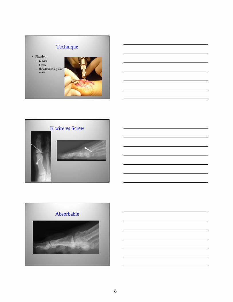
Technique
• Fixation– K-wire
– Screw
– Bioadsorbable pin or screw
K wire vs Screw
Absorbable

9
No evidence to support one type of fixation over another
Technique• Medial capsular
reefing
Technique
• Check intraoperative xray or fluoroscopy after medial closure
• Simulate WB

10
• Results– 80-94% patients satisfied– Average correction
• HVA 12-15°• IMA 4-7°
– Shortening of 1st MT avg. 2-3 mm– Limited correction of sesamoid
subluxation or hallux pronation– Higher satisfaction rate with IMA < 15°– Limited lateral release: No effect on
outcomes
Distal Chevron Osteotomy
Technique• Hallux Valgus dressing
Post OP
• Post Op, shoe or boot for 6 weeks
• Heel walking 4 weeks
• Change dressing week 1,3
• Toe spacer
• ROM at 4 weeks
• Toe spacer 8 weeks
11
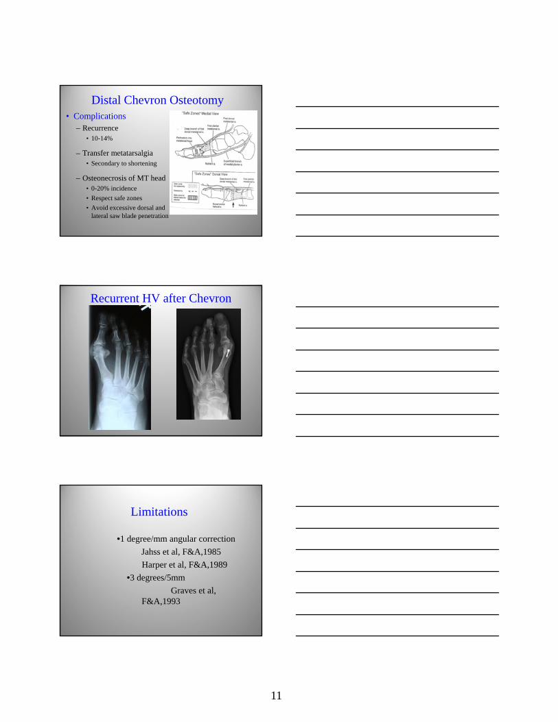
• Complications– Recurrence
• 10-14%
– Transfer metatarsalgia• Secondary to shortening
– Osteonecrosis of MT head• 0-20% incidence
• Respect safe zones
• Avoid excessive dorsal and lateral saw blade penetration
Distal Chevron Osteotomy
Recurrent HV after Chevron
Limitations
•1 degree/mm angular correction
Jahss et al, F&A,1985
Harper et al, F&A,1989
•3 degrees/5mm
Graves et al, F&A,1993

12
Limitations
• Average MT neck width is about 12-14mm
• Can only shift distal fragment 4-6mm and maintain adequate boney contact
Limitations
• Goal IMA <10 degrees
• 5 mm shift at most 5 degrees correction
• Pre-op IMA <14
Chevron Modifications
• Biplanar Chevron– Removes medial
wedge
– Allows medial rotation of capital fragment
– Useful in cases of high DMAA (15 degrees)

13
Modifications• Biplanar Chevron– Removes medial
wedge from metatarsal cut
– Allows rotation of the capital fragment
Double Osteotomy
• Post op– Biplanar Chevron
– Akin osteotomy
Distal Closing Wedge

14
Proximal Metatarsal Osteotomy(High DMAA )
Controversies
• Lateral Release/adductor tenotomy
• Is there sufficient evidence to support increased risk of AVN?
Chevron + DSTR

15
Complications
• AVN– Lateral release
– Lateral blade penetration
• Under Correction/Recurrence– Pushing the indications
Proximal Metatarsal Osteotomies (PMTO)
• Indicated for moderate to severe deformities
–HVA> 30 degrees
– IMA > 12-13 degrees
Distal Soft Tissue Procedure“Modified McBride” (1990)• Adjunct to bony procedure
– Medial capsulotomy and plication
– Lateral capsule release (multiple perforations)
– Adductor hallucis release (not always necessary)
– Deep transverse metatarsal ligament release (not always necessary)
– Varus stress
– Fibular sesamoid no longer excised

16
Proximal Metatarsal Osteotomy
• Proximal Metatarsal Osteotomies (PMTO)– Crescentic
– Proximal chevron
– Oblique (Ludloff and Mau)
– SCARF
– Medial opening wedge
How to decide?
Osteotomies:How to choose which one?
• Degree of correction needed
• Consistency of correction
• Ease of performing the surgery
• Stability/early weightbearing
• Surgeon’s comfort level with technique
• Complications– Recurrence
– Hallux Varus
– Arthrofibrosis
– Transfer metatarsalgia• Secondary to malunion,
shortening, or 1st MT dorsiflexion
– Delayed or nonunion– Incomplete correction
Proximal Osteotomies
17
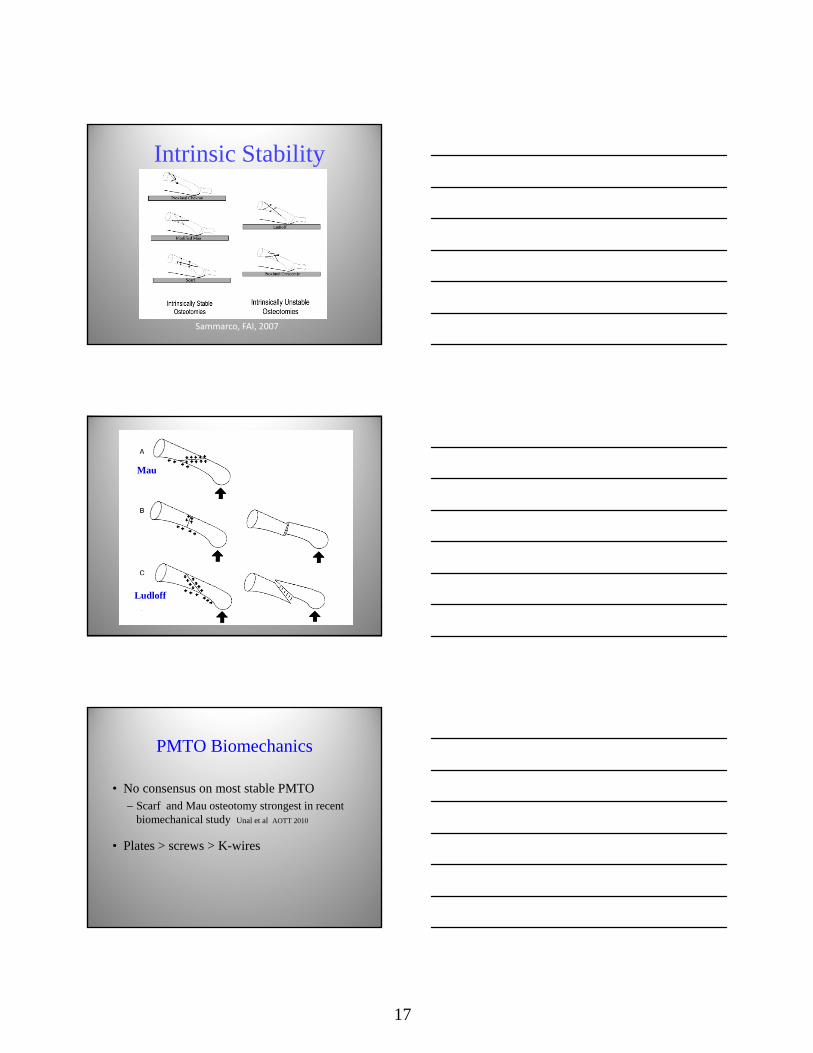
Sammarco, FAI, 2007
Intrinsic Stability
Mau
Ludloff
PMTO Biomechanics
• No consensus on most stable PMTO– Scarf and Mau osteotomy strongest in recent
biomechanical study Unal et al AOTT 2010
• Plates > screws > K-wires

18
Proximal Crescentic Osteotomy
• Rotational osteotomy
• 90-95% patient satisfaction
• Reliable correction• Zettl et al 2000 AOTS
• Thordarson 1992 FA
• Dreeban, Mann 1996 FAI
Proximal Crescentic Osteotomy
• Dorsiflexion malunions
• Nonunion
• Challenging to perform
• Difficulty w/fixation
– Plates may
help
Proximal Chevron Osteotomy
• Translational and Angular correction
• Outcomes similar to Cresentic but more stable
• Easley et al FAI 1996
• Markbreiter/Thompson FAI 1997
• Sammarco et al FAI 1998

19
Proximal Chevron Osteotomy
• Although better stability to prevent DF
• More tendency for loss of reduction
• Plate fixation may be required
• Deorio & Deorio, Tech in Ortho Surg 2011
Proximal Opening Wedge Osteotomy(POWO)
POWO

20
POWO
• Fairly reproducible
• Good results reported• Nery FA 2013
• Smith FASpec 2009
• Shurnas FAI 2009
• Wukich Oper Tech 2006
POWO
• Nonunion/delayed union
• Hardware removal
• Intraarticular hardware
Proximal Metatarsal Oblique Osteotomies
• Ludloff
• Mau
• SCARF

21
Ludloff Osteotomy
Ludloff osteotomy
• Fairly easy procedure
• Good clinical results• Chiodo FAI 2004
• Trinka JBJS 2008
Ludloff-? Stability - Loss of correction
-Shortening -Intraoperative fracture

22
SCARF• Widely used
• Translational
• Excellent stability
• Ease of internal fixation
• Mild to severe deformities
• Good reported clinical results
• Choi FAI 2013
• Adam CORR 2011
• Robinson 2009 FAI
• Weil 2000 FA clinic
SCARF
• Troughing
• IO fracture
• Wound healing
• Delayed union
• Learning Curve
Modified MAU

23
Modified Mau
• Translational & Rotational
• Excellent stability
• Ease of internal fixation
• Moderate to severe deformities
59 y/o female painful bunion
MAU Osteotomy

24
Distal soft tissue release
Draw out osteotomy
Mau Osteotomy

25
MAU
Mau
Mau

26
Mau
Mau
Mau

27
POST OP
• Splint x 7 days
• Fracture boot x 5 wks
• Heel WB
COMPLICATIONS
N=65
HALLUX VARUS: 9%
METATARSAL FRACTURE: 4.6%
LOSS OF CORRECTION: 4.6%
DORSIFLEXION MALUNION/TRANSFER LESION: 0%
NONUNION: 0%
LEARNING CURVE
Early Experience with the Modified Mau Osteotomy for Correction of Hallux Valgus: A
Retrospective AnalysisO’Dell, Walling, Clare
Other procedures
28
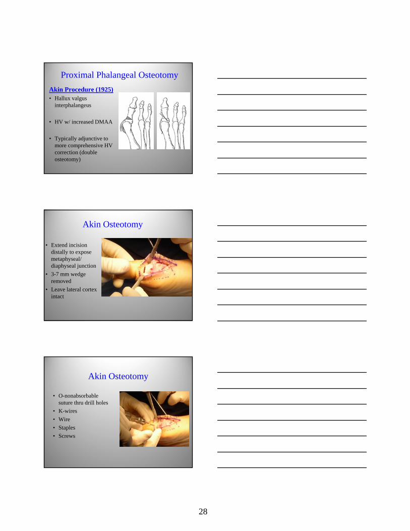
Proximal Phalangeal Osteotomy
Akin Procedure (1925)• Hallux valgus
interphalangeus
• HV w/ increased DMAA
• Typically adjunctive to more comprehensive HV correction (double osteotomy)
Akin Osteotomy
• Extend incision distally to expose metaphyseal/ diaphyseal junction
• 3-7 mm wedge removed
• Leave lateral cortex intact
Akin Osteotomy
• O-nonabsorbablesuture thru drill holes
• K-wires
• Wire
• Staples
• Screws

29
AKIN
Suture
30
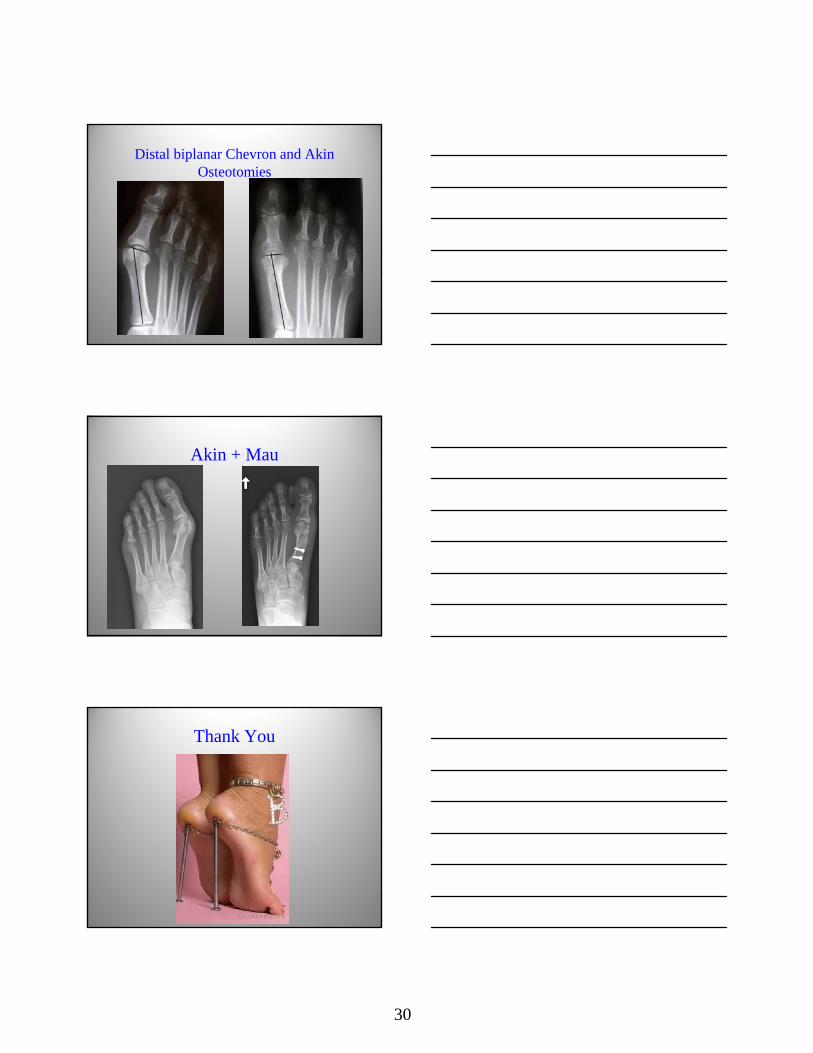
Distal biplanar Chevron and Akin Osteotomies
Akin + Mau
Thank You
Recommended