Criteria for Imaging
Effective: November 21, 2018
CLINICAL GUIDELINES
Prepared for Oxford Health Plans Provider Network. Clinical guidelines for medical necessity review of radiology services.
© 2018 eviCore healthcare. All rights reserved.
Please note the following:
CPT Copyright 2017 American Medical Association. All rights
reserved. CPT is a registered trademark of the American Medical
Association.
eviCore healthcare Cardiology Management Criteria V2.0.2018
© 2018 eviCore healthcare. All rights reserved. 400 Buckwalter Place Boulevard, Bluffton, SC 29910 • (800) 918-8924 www.eviCore.com
Page 2 of 49
Table of Contents CPT Code Page Cardiology Imaging Guidelines 75557 Cardiac MRI for Morphology and Function without Contrast 7 75561 Cardiac MRI for Morphology and Function without Contrast Followed by Contrast
Material and Further Sequences 7 I. Cardiac MRI – Coding II. Cardiac MRI – Indications (excluding Stress MRI) III. Cardiac MRI – Aortic Root and Proximal Ascending Aorta IV. Cardiac MRI – Evaluation of Pericardial Effusion or Diagnosis of Pericardial Tamponade
75559 Cardiac MRI for Morphology and Function without Contrast; with Stress
Imaging 10 75563 Cardiac MRI for Morphology and Function without Contrast Followed by Contrast
Material and Further Sequences; with Stress Imaging 10 78451 Myocardial Perfusion Imaging with SPECT – Single Study 10 78452 Myocardial Perfusion Imaging with SPECT – Multiple Studies 10 78453 Myocardial Perfusion Imaging, Planar Rest or Stress 10 78454 Myocardial Perfusion Imaging, Planar Rest and/or Stress 10
I. General Issues – Cardiac II. Stress Testing without Imaging – Procedures III. Stress Testing with Imaging – Procedures IV. Stress Testing with Imaging – Indications V. Stress Testing with Imaging – Preoperative VI. Transplant Patients VII. Non-imaging Heart Function and Cardiac Shunt Imaging VIII. Genetic lab testing in the evaluation of CAD
78459 PET Myocardial – Metabolic 19 78491 PET Myocardial Perfusion Imaging, Rest or Stress 19 78492 PET Myocardial Perfusion Imaging, Rest and Stress 19
I. Cardiac PET – Perfusion – Indications (CPT® 78491 and CPT® 78492) II. Cardiac PET – Absolute Quantitation of Myocardial Blood Flow (CPT®
0482T) III. Cardiac PET – Metabolic – Indications (CPT® 78459)
75571 Coronary Artery Calcium Scoring 21 75572 CT Heart Structure and Morphology with Contrast 21 75573 CT Heart Structure and Morphology in Congenital Heart Disease with Contrast 21 75574 CTA Coronary Arteries and Structure and Morphology with Function and with
Contrast 21
eviCore healthcare Cardiology Management Criteria V2.0.2018
© 2018 eviCore healthcare. All rights reserved. 400 Buckwalter Place Boulevard, Bluffton, SC 29910 • (800) 918-8924 www.eviCore.com
Page 3 of 49
0501T Noninvasive estimated coronary fractional flow reserve (FFR) derived from coronary computed tomography angiography data using computation fluid dynamics physiologic simulation software analysis of functional data to assess the severity of coronary artery disease; data preparation and transmission, analysis of fluid dynamics and simulated maximal coronary hyperemia, generation of estimated FFR model, with anatomical data review in comparison with estimated FFR model to reconcile discordant data, interpretation and report 21
0502T Noninvasive estimated coronary fractional flow reserve (FFR) derived from coronary computed tomography angiography data using computation fluid dynamics physiologic simulation software analysis of functional data to assess the severity of coronary artery disease; data preparation and transmission 21
0503T Noninvasive estimated coronary fractional flow reserve (FFR) derived from coronary computed tomography angiography data using computation fluid dynamics physiologic simulation software analysis of functional data to assess the severity of coronary artery disease; analysis of fluid dynamics and simulated maximal coronary hyperemia, and generation of estimated FFR model 21
0504T Noninvasive estimated coronary fractional flow reserve (FFR) derived from coronary computed tomography angiography data using computation fluid dynamics physiologic simulation software analysis of functional data to assess the severity of coronary artery disease; anatomical data review in comparison with estimated FFR model to reconcile discordant data, interpretation and report 22 I. CT for Coronary Calcium Scoring (CPT® 75571) II. CTA – Indications for CTA III. CTA – Additional Indications IV. Evaluation of left ventricular function following myocardial infarction or in
chronic heart failure V. Fractional Flow Reserve by Computed Tomography VI. CT Heart – Indications VII. Cardiac CT for congenital heart disease (CPT® 75573) VIII. Transcatheter Aortic Valve Replacement (TAVR)
Echocardiography 93303 Transthoracic Echocardiography for Congenital Cardiac Anomalies; Complete 28 93304 Transthoracic Echocardiography for Congenital Cardiac Anomalies; Follow-up or
Limited Study 28 93306 Echocardiography, Transthoracic, Real-time with Image Documentation (2D),
Includes M-mode Recording, when Performed, Complete, with Spectral Doppler Echocardiography, and with Color Flow Doppler Echocardiography 28
93307 Echocardiography, Transthoracic, Real-time with Image Documentation (2D) with or without M-mode Recording; Complete 28
93308 Echocardiography, Transthoracic, Real-time with Image Documentation (2D) with or without M-mode Recording; Follow-up or Limited Study 28
93320 Doppler Echocardiography, Pulsed Wave and/or Continuous Wave with Spectral Display; Complete 28
93321 Doppler Echocardiography, Pulsed Wave and/or Continuous Wave with Spectral Display; Follow- up or Limited Study 28
eviCore healthcare Cardiology Management Criteria V2.0.2018
© 2018 eviCore healthcare. All rights reserved. 400 Buckwalter Place Boulevard, Bluffton, SC 29910 • (800) 918-8924 www.eviCore.com
Page 4 of 49
93325 Doppler Echocardiography Color Flow Velocity Mapping 28 I. Transthoracic Echocardiography (TTE) II. 3D Echocardiography
Stress Echocardiography 93350 Echocardiography, Transthoracic, Real-Time with Image Documentation (2D),
Includes M-Mode Recording, when Performed, During Rest and Cardiovascular Stress Test Using Treadmill, Bicycle Exercise and/or Pharmacologically Induced Stress, with Interpretation and Report with or without M-Mode Recording, During Rest and Cardiovascular Stress Test, with Interpretation and Report 35
93351 Echocardiography, Transthoracic, Real-Time with Image Documentation (2D), Includes M-Mode Recording, when Performed, During Rest and Cardiovascular Stress Test Using Treadmill, Bicycle Exercise and/or Pharmacologically Induced Stress, with Interpretation and Report with or without M-Mode Recording, During Rest and Cardiovascular Stress Test, with Interpretation and Report; Including Performance of Continuous Electrocardiographic Monitoring, with Supervision by a Qualified Healthcare Professional 35 I. Stress Echocardiography (Stress Echo) II. General Issues – Cardiac III. Stress Testing without Imaging – Procedures IV. Stress Testing with Imaging-Procedures V. Stress Testing with Imaging - Indications VI. Stress Testing with Imaging – Preoperative VII. Transplant Patients VIII. Non-imaging Heart Function and Cardiac Shunt Imaging IX. Genetic lab testing in the evaluation of CAD
Diagnostic Heart Catheterization 93452 Left heart catheterization including intraprocedural injection(s) for left
ventriculography, imaging supervision and interpretation, when performed 44 93453 Combined right and left heart catheterization including intraprocedural injection(s)
for left ventriculography, imaging supervision and interpretation, when performed 44
93454 Catheter placement in coronary artery(s) for coronary angiography, including intraprocedural injection(s) for coronary angiography, imaging supervision and interpretation 44
93455 Catheter placement in coronary artery(s) for coronary angiography, including intraprocedural injection(s) for coronary angiography, imaging supervision and interpretation; with catheter placement(s) in bypass graft(s) (internal mammary, free arterial venous grafts) including intraprocedural injection(s) for bypass graft angiography 44
93456 Catheter placement in coronary artery(s) for coronary angiography, including intraprocedural injection(s) for coronary angiography, imaging supervision and interpretation; with right heart catheterization 44
93457 Catheter placement in coronary artery(s) for coronary angiography, including intraprocedural injection(s) for coronary angiography, imaging supervision and
eviCore healthcare Cardiology Management Criteria V2.0.2018
© 2018 eviCore healthcare. All rights reserved. 400 Buckwalter Place Boulevard, Bluffton, SC 29910 • (800) 918-8924 www.eviCore.com
Page 5 of 49
interpretation; with catheter placement(s) in bypass graft(s) (internal mammary, free arterial, venous grafts) including intraprocedural injection(s) for bypass graft angiography and right heart catheterization 44
93458 Catheter placement in coronary artery(s) for coronary angiography, including intraprocedural injection(s) for coronary angiography, imaging supervision and interpretation; with left heart catheterization including intraprocedural injection(s) for left ventriculography, when performed 44
93459 Catheter placement in coronary artery(s) for coronary angiography, including intraprocedural injection(s) for coronary angiography, imaging supervision and interpretation; with left heart catheterization including intraprocedural injection(s) for left ventriculography, when performed, catheter placement(s) in bypass graft(s) (internal mammary, free arterial, venous grafts) with bypass graft angiography 45
93460 Catheter placement in coronary artery(s) for coronary angiography, including intraprocedural injection(s) for coronary angiography, imaging supervision and interpretation; with right and left heart catheterization including intraprocedural injection(s) for left ventriculography, when performed 45
93461 Catheter placement in coronary artery(s) for coronary angiography, including intraprocedural injection(s) for coronary angiography, imaging supervision and interpretation; with right and left heart catheterization including intraprocedural injection(s) for left ventriculography, when performed, catheter placement(s) in bypass graft(s) (internal mammary, free arterial, venous grafts) with bypass graft angiography 45 I. Diagnostic Left Heart Catheterization (LHC) II. Right Heart Catheterization (RHC) III. Combined Right and Left Heart Catheterization Indications IV. Planned (Staged) Coronary Interventions
eviCore healthcare Cardiology Management Criteria V2.0.2018
© 2018 eviCore healthcare. All rights reserved. 400 Buckwalter Place Boulevard, Bluffton, SC 29910 • (800) 918-8924 www.eviCore.com
Page 6 of 49
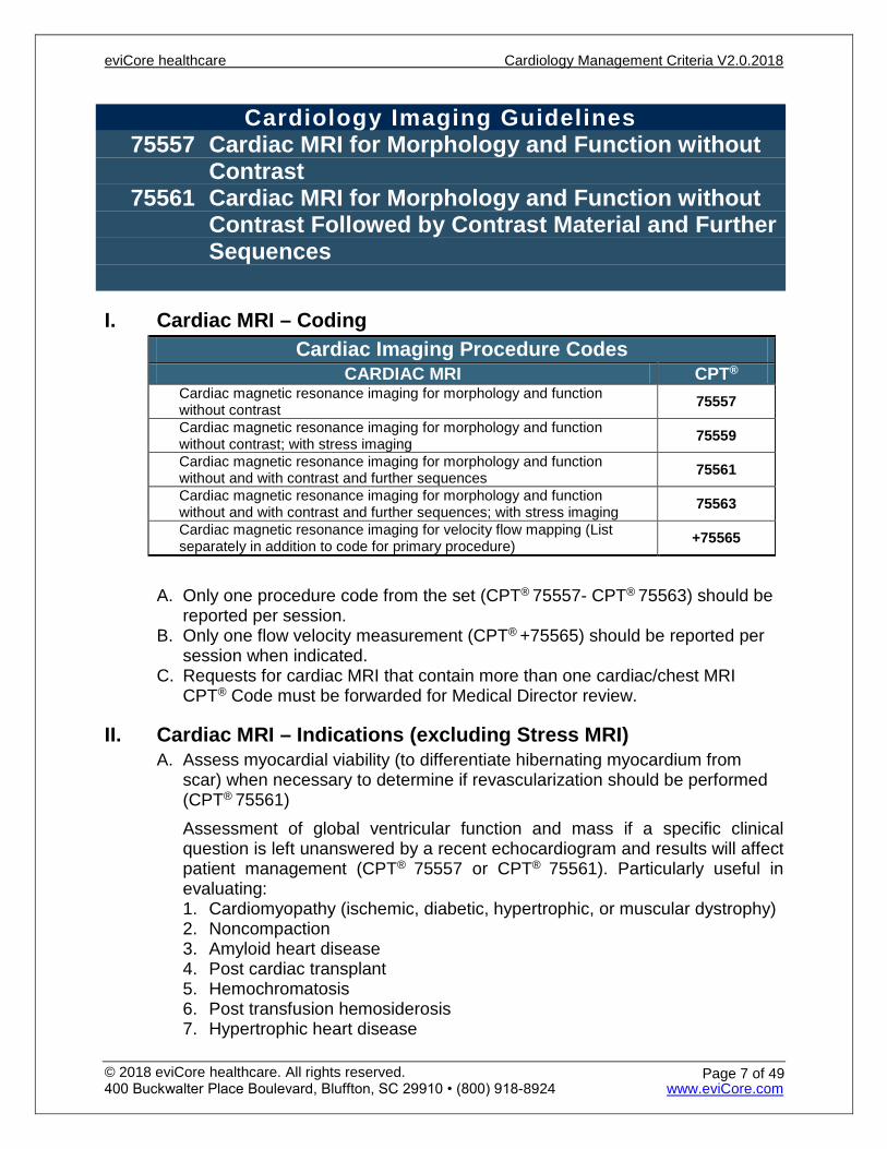
Cardiology Imaging Guidelines 75557 Cardiac MRI for Morphology and Function without
Contrast 75561 Cardiac MRI for Morphology and Function without
Contrast Followed by Contrast Material and Further Sequences
I. Cardiac MRI – Coding Cardiac Imaging Procedure Codes
CARDIAC MRI CPT® Cardiac magnetic resonance imaging for morphology and function without contrast 75557
Cardiac magnetic resonance imaging for morphology and function without contrast; with stress imaging 75559
Cardiac magnetic resonance imaging for morphology and function without and with contrast and further sequences 75561
Cardiac magnetic resonance imaging for morphology and function without and with contrast and further sequences; with stress imaging 75563
Cardiac magnetic resonance imaging for velocity flow mapping (List separately in addition to code for primary procedure) +75565
A. Only one procedure code from the set (CPT® 75557- CPT® 75563) should be
reported per session. B. Only one flow velocity measurement (CPT® +75565) should be reported per
session when indicated. C. Requests for cardiac MRI that contain more than one cardiac/chest MRI
CPT® Code must be forwarded for Medical Director review.
II. Cardiac MRI – Indications (excluding Stress MRI) A. Assess myocardial viability (to differentiate hibernating myocardium from
scar) when necessary to determine if revascularization should be performed (CPT® 75561) Assessment of global ventricular function and mass if a specific clinical question is left unanswered by a recent echocardiogram and results will affect patient management (CPT® 75557 or CPT® 75561). Particularly useful in evaluating: 1. Cardiomyopathy (ischemic, diabetic, hypertrophic, or muscular dystrophy) 2. Noncompaction 3. Amyloid heart disease 4. Post cardiac transplant 5. Hemochromatosis 6. Post transfusion hemosiderosis 7. Hypertrophic heart disease
eviCore healthcare Cardiology Management Criteria V2.0.2018
© 2018 eviCore healthcare. All rights reserved. 400 Buckwalter Place Boulevard, Bluffton, SC 29910 • (800) 918-8924 www.eviCore.com
Page 7 of 49
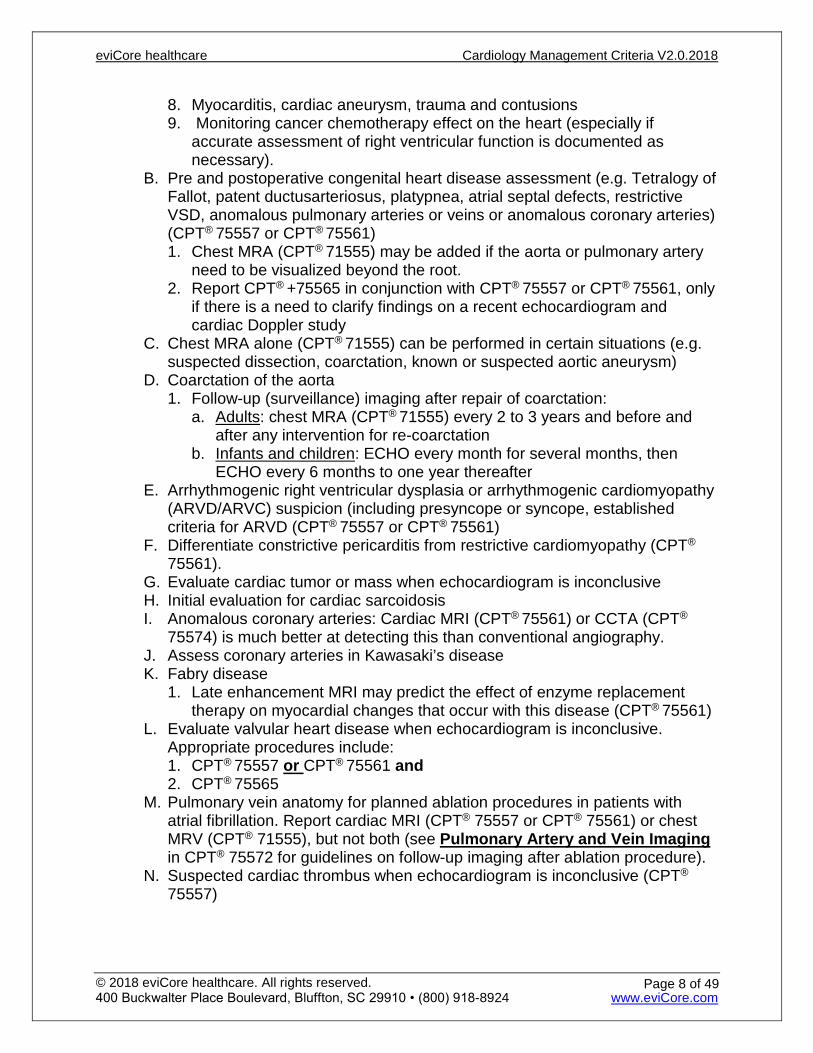
8. Myocarditis, cardiac aneurysm, trauma and contusions 9. Monitoring cancer chemotherapy effect on the heart (especially if
accurate assessment of right ventricular function is documented as necessary).
B. Pre and postoperative congenital heart disease assessment (e.g. Tetralogy of Fallot, patent ductusarteriosus, platypnea, atrial septal defects, restrictive VSD, anomalous pulmonary arteries or veins or anomalous coronary arteries) (CPT® 75557 or CPT® 75561) 1. Chest MRA (CPT® 71555) may be added if the aorta or pulmonary artery
need to be visualized beyond the root. 2. Report CPT® +75565 in conjunction with CPT® 75557 or CPT® 75561, only
if there is a need to clarify findings on a recent echocardiogram and cardiac Doppler study
C. Chest MRA alone (CPT® 71555) can be performed in certain situations (e.g. suspected dissection, coarctation, known or suspected aortic aneurysm)
D. Coarctation of the aorta 1. Follow-up (surveillance) imaging after repair of coarctation:
a. Adults: chest MRA (CPT® 71555) every 2 to 3 years and before and after any intervention for re-coarctation
b. Infants and children: ECHO every month for several months, then ECHO every 6 months to one year thereafter
E. Arrhythmogenic right ventricular dysplasia or arrhythmogenic cardiomyopathy (ARVD/ARVC) suspicion (including presyncope or syncope, established criteria for ARVD (CPT® 75557 or CPT® 75561)
F. Differentiate constrictive pericarditis from restrictive cardiomyopathy (CPT®
75561). G. Evaluate cardiac tumor or mass when echocardiogram is inconclusive H. Initial evaluation for cardiac sarcoidosis I. Anomalous coronary arteries: Cardiac MRI (CPT® 75561) or CCTA (CPT®
75574) is much better at detecting this than conventional angiography. J. Assess coronary arteries in Kawasaki’s disease K. Fabry disease
1. Late enhancement MRI may predict the effect of enzyme replacement therapy on myocardial changes that occur with this disease (CPT® 75561)
L. Evaluate valvular heart disease when echocardiogram is inconclusive. Appropriate procedures include: 1. CPT® 75557 or CPT® 75561 and 2. CPT® 75565
M. Pulmonary vein anatomy for planned ablation procedures in patients with atrial fibrillation. Report cardiac MRI (CPT® 75557 or CPT® 75561) or chest MRV (CPT® 71555), but not both (see Pulmonary Artery and Vein Imaging in CPT® 75572 for guidelines on follow-up imaging after ablation procedure).
N. Suspected cardiac thrombus when echocardiogram is inconclusive (CPT®
75557)
eviCore healthcare Cardiology Management Criteria V2.0.2018
© 2018 eviCore healthcare. All rights reserved. 400 Buckwalter Place Boulevard, Bluffton, SC 29910 • (800) 918-8924 www.eviCore.com
Page 8 of 49

O. Right ventricular function evaluation (CPT® 75557 in conjunction with CPT®
+75565) if a recent ECHO has been done, and there is documented need to perform cardiac MRI in order to resolve an unanswered question
P. Shunting through a VSD (CPT® 75557 in conjunction with CPT® +75565) if a recent ECHO has been done, including a bubble study, and there is documented need to perform cardiac MRI in order to resolve an unanswered question
Q. Evaluate for iron overload due to conditions requiring frequent blood transfusions (i.e. sickle cell, thalassemia, hemochromatosis, etc.) (CPT®
75557)
III. Cardiac MRI – Aortic Root and Proximal Ascending Aorta A. See Thoracic Aorta in the Chest Imaging Guidelines
IV. Cardiac MRI – Evaluation of Pericardial Effusion or Diagnosis of Pericardial Tamponade A. Contrast enhanced cardiac MRI (CPT® 75561) is useful for evaluating
pericarditis, neoplastic and other effusion, tamponade or myocardial infiltration if a specific clinical question is left unanswered by echocardiogram or another recent imaging study 1. Cardiac MRI – Indications for Stress MRI
B. If a nuclear perfusion (MPI) stress test was performed and was equivocal, a stress MRI is appropriate.
C. For indications for Stress MRI, see Stress Testing with Imaging – Indications
75557, 75561 Cardiac MRI
eviCore healthcare Cardiology Management Criteria V2.0.2018
© 2018 eviCore healthcare. All rights reserved. 400 Buckwalter Place Boulevard, Bluffton, SC 29910 • (800) 918-8924 www.eviCore.com
Page 9 of 49
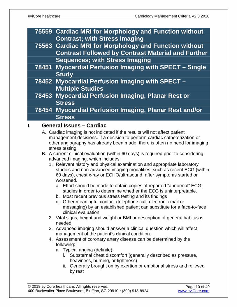
75559 Cardiac MRI for Morphology and Function without Contrast; with Stress Imaging
75563 Cardiac MRI for Morphology and Function without Contrast Followed by Contrast Material and Further Sequences; with Stress Imaging
78451 Myocardial Perfusion Imaging with SPECT – Single Study
78452 Myocardial Perfusion Imaging with SPECT – Multiple Studies
78453 Myocardial Perfusion Imaging, Planar Rest or Stress
78454 Myocardial Perfusion Imaging, Planar Rest and/or Stress
I. General Issues – Cardiac A. Cardiac imaging is not indicated if the results will not affect patient
management decisions. If a decision to perform cardiac catheterization or other angiography has already been made, there is often no need for imaging stress testing.
B. A current clinical evaluation (within 60 days) is required prior to considering advanced imaging, which includes: 1. Relevant history and physical examination and appropriate laboratory
studies and non-advanced imaging modalities, such as recent ECG (within 60 days), chest x-ray or ECHO/ultrasound, after symptoms started or worsened. a. Effort should be made to obtain copies of reported “abnormal” ECG
studies in order to determine whether the ECG is uninterpretable. b. Most recent previous stress testing and its findings c. Other meaningful contact (telephone call, electronic mail or
messaging) by an established patient can substitute for a face-to-face clinical evaluation.
2. Vital signs, height and weight or BMI or description of general habitus is needed.
3. Advanced imaging should answer a clinical question which will affect management of the patient’s clinical condition.
4. Assessment of coronary artery disease can be determined by the following: a. Typical angina (definite):
i. Substernal chest discomfort (generally described as pressure, heaviness, burning, or tightness)
ii. Generally brought on by exertion or emotional stress and relieved by rest
eviCore healthcare Cardiology Management Criteria V2.0.2018
© 2018 eviCore healthcare. All rights reserved. 400 Buckwalter Place Boulevard, Bluffton, SC 29910 • (800) 918-8924 www.eviCore.com
Page 10 of 49

iii. May radiate to the left arm or jaw iv. When clinical information is received indicating that a patient is
experiencing chest pain that is "exertional" or "due to emotional stress", this meets the typical angina definition under the Pre-Test Probability Grid. No further description of the chest pain is required (location within the chest is not required).
5. The Pre-Test Probability Grid (Table 1) is based on age, gender, and symptoms. All factors must be considered in order to approve for stress testing with imaging using the Pre-Test Probability Grid. a. Atypical angina (probable): Chest pain or discomfort (arm or jaw
pain) that lacks one of the characteristics of definite or typical angina. b. Non-anginal chest pain: Chest pain or discomfort that meets one or
none of the typical angina characteristics. c. Anginal variants or equivalents: a manifestation of myocardial
ischemia which is perceived by patients to be (otherwise unexplained) dyspnea, unusual fatigue, more often seen in women and may be unassociated with chest pain.
II. Stress Testing without Imaging – Procedures The Exercise Treadmill Test (ETT) is without imaging A. Necessary components of an ETT include:
1. ECG that can be interpreted for ischemia. 2. Patient capable of exercise on a treadmill or similar device (generally at
4METs or greater; see functional capacity below). B. An abnormal ETT (exercise treadmill test) includes any one of the following:
1. ST segment depression (usually described as horizontal or downsloping, greater or equal to 1.0 mm below baseline)
2. Development of chest pain 3. Significant arrhythmia (especially ventricular arrhythmia) 4. Hypotension
C. Functional capacity greater than or equal to 4METs equates to the following: 1. Can walk four blocks without stopping 2. Can walk up a hill 3. Can climb one flights of stairs without stopping 4. Can perform heavy work around the house
Practice Note: An observational study found that, compared with the Duke Activity Status Index, subjective assessment by clinicians generally underestimated exercise capacity(see reference 25).
III. Stress Testing with Imaging – Procedures A. Imaging Stress Tests include any one of the following:
1. Stress Echocardiography (see Stress Echocardiography (Stress Echo) – Coding)
2. MPI (see Myocardial Perfusion Imaging (MPI) – Coding) 3. Stress perfusion MRI (see Cardiac MRI – Indications for Stress MRI)
eviCore healthcare Cardiology Management Criteria V2.0.2018
© 2018 eviCore healthcare. All rights reserved. 400 Buckwalter Place Boulevard, Bluffton, SC 29910 • (800) 918-8924 www.eviCore.com
Page 11 of 49
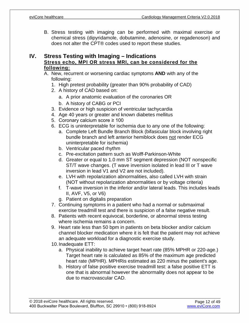
B. Stress testing with imaging can be performed with maximal exercise or chemical stress (dipyridamole, dobutamine, adenosine, or regadenoson) and does not alter the CPT® codes used to report these studies.
IV. Stress Testing with Imaging – Indications
Stress echo, MPI OR stress MRI, can be considered for the following: A. New, recurrent or worsening cardiac symptoms AND with any of the
following: 1. High pretest probability (greater than 90% probability of CAD) 2. A history of CAD based on:
a. A prior anatomic evaluation of the coronaries OR b. A history of CABG or PCI
3. Evidence or high suspicion of ventricular tachycardia 4. Age 40 years or greater and known diabetes mellitus 5. Coronary calcium score ≥ 100 6. ECG is uninterpretable for ischemia due to any one of the following:
a. Complete Left Bundle Branch Block (bifasicular block involving right bundle branch and left anterior hemiblock does not render ECG uninterpretable for ischemia)
b. Ventricular paced rhythm c. Pre-excitation pattern such as Wolff-Parkinson-White d. Greater or equal to 1.0 mm ST segment depression (NOT nonspecific
ST/T wave changes. (T wave inversion isolated in lead III or T wave inversion in lead V1 and V2 are not included).
e. LVH with repolarization abnormalities, also called LVH with strain (NOT without repolarization abnormalities or by voltage criteria)
f. T-wave inversion in the inferior and/or lateral leads. This includes leads II, AVF, V5, or V6)
g. Patient on digitalis preparation 7. Continuing symptoms in a patient who had a normal or submaximal
exercise treadmill test and there is suspicion of a false negative result. 8. Patients with recent equivocal, borderline, or abnormal stress testing
where ischemia remains a concern. 9. Heart rate less than 50 bpm in patients on beta blocker and/or calcium
channel blocker medication where it is felt that the patient may not achieve an adequate workload for a diagnostic exercise study.
10. Inadequate ETT: a. Physical inability to achieve target heart rate (85% MPHR or 220-age.)
Target heart rate is calculated as 85% of the maximum age predicted heart rate (MPHR). MPHRis estimated as 220 minus the patient's age.
b. History of false positive exercise treadmill test: a false positive ETT is one that is abnormal however the abnormality does not appear to be due to macrovascular CAD.
eviCore healthcare Cardiology Management Criteria V2.0.2018
© 2018 eviCore healthcare. All rights reserved. 400 Buckwalter Place Boulevard, Bluffton, SC 29910 • (800) 918-8924 www.eviCore.com
Page 12 of 49

B. Within 3 months of an acute coronary syndrome (e.g. ST segment elevation MI [STEMI], unstable angina, non-ST segment elevation MI [NSTEMI]), one MPIcan be performed to evaluate for inducible ischemia if all of the following related to the most recent acute coronary event apply: 1. Individual is hemodynamically stable 2. No recurrent chest pain symptoms and no signs of heart failure 3. No prior coronary angiography or imaging stress test in regards to the
current episode of symptoms C. Assessing myocardial viability in patients with significant ischemic ventricular
dysfunction (suspected hibernating myocardium) and persistent symptoms or heart failure such that revascularization would be considered. 1. NOTE: MRI, cardiac PET, MPI, or Dobutamine stress echo can be used to
assess myocardial viability depending on physician preference 2. PET and MPI perfusion studies are usually accompanied by PET
metabolic examinations (CPT® 78459). Tl-201 MPI perfusion studies may assess viability without accompanying PET metabolism information.
D. Unheralded syncope (not near syncope) E. Asymptomatic patient with an uninterpretable ECG that has never been
evaluated or is a new uninterpretable change. F. Patient with an elevated cardiac troponin. G. One routine study 2 years or more after a stent, except with a left main stent
where it can be done at 1 year. H. One routine study at 5 years or more after CABG, without cardiac symptoms. I. Every 2 years if there was documentation of previous “silent ischemia” on the
imaging portion of a stress test but not on the ECG portion. J. To assess for CAD prior to starting a taking Class IC antiarrhythmic agent
(flecainide or propafenone) and annually while taking the medication. K. Prior anatomic imaging study (coronary angiogram or CCTA) demonstrating
coronary stenosis in a major coronary branch which is of uncertain functional significance can have one stress test with imaging.
L. Evaluating new, recurrent or worsening left ventricular dysfunction/CHF.
V. Stress Testing with Imaging – Preoperative A. There are 2 steps that determine the need for imaging stress testing in
(stable) pre-operative patients: 1. Would the patient qualify for imaging stress testing independent of
planned surgery? a. If yes, proceed to stress testing guidelines above b. If no, go to step 2
2. Is the surgery considered high, moderate or low risk? (see Table 2) If high or moderate-risk, proceed below. If low-risk, there is no evidence to determine a need for preoperative cardiac testing. a. High Risk Surgery: All patients in this category should receive an
imaging stress test if there has not been an imaging stress test within 1 year*, unless the patient has developed new cardiac symptoms or a new change in the EKG since the last stress test.
eviCore healthcare Cardiology Management Criteria V2.0.2018
© 2018 eviCore healthcare. All rights reserved. 400 Buckwalter Place Boulevard, Bluffton, SC 29910 • (800) 918-8924 www.eviCore.com
Page 13 of 49

b. Intermediate Surgery: One or more risk factors and unable to perform an ETT per guidelines if there has not been an imaging stress test within 1 year* unless the patient has developed new cardiac symptoms or a new change in the EKG since the last stress test.
c. Low Risk: Preoperative imaging stress testing is not supported. 3. Clinical Risk Factors (for cardiac death & non-fatal MI at time of non-
cardiac surgery) a. Planned high risk surgery (open surgery on the aorta or open
peripheral vascular surgery) b. History of ischemic heart disease (previous MI, previous positive stress
test, use of nitroglycerin, typical angina, ECG Q waves, previous PCI or CABG)
c. History of compensated previous congestive heart failure (history of heart failure, previous pulmonary edema, third heart sound, bilateral rales, chest x-ray showing heart failure)
d. History of previous TIA or stroke e. Diabetes Mellitus f. Creatinine level > 2 mg/dL
*Time interval is based on consensus of eviCore executive cardiology panel.
Table 2 Cardiac Risk Stratification List
High Risk (> 5%) Intermediate Risk (1-5%) Low Risk (<1%) Open aortic and other
major open vascular surgery
Open peripheral vascular surgery
Open intraperitoneal and/or intrathoracic surgery
Open carotid endarterectomy
Head and neck surgery Open orthopedic surgery Open prostate surgery
Endoscopic procedures Superficial procedures Cataract surgery Breast surgery Ambulatory surgery Laparoscopic and
endovascular procedures that are unlikely to require further extensive surgical intervention
VI. Transplant Patients A. Stress Testing in patients for Non-Cardiac Transplant
1. Individuals who are candidates for any type of organ bone marrow or stem cell transplant can undergo imaging stress testing every year (usually stress echo or MPI) prior to transplant.
2. Individuals who have undergone organ transplant are at increased risk for ischemic heart disease secondary to their medication. Risk of vasculopathy is 7% at one year, 32% at five years and 53% at ten years. An imaging stress test can be repeated annually after transplant for at least two years or within one year of a prior cardiac imaging study if there is evidence of progressive vasculopathy.
eviCore healthcare Cardiology Management Criteria V2.0.2018
© 2018 eviCore healthcare. All rights reserved. 400 Buckwalter Place Boulevard, Bluffton, SC 29910 • (800) 918-8924 www.eviCore.com
Page 14 of 49
3. After two consecutive normal imaging stress tests, repeated testing is not supported more often than every other year without evidence for progressive vasculopathy or new symptoms.
4. Stress testing after five years may proceed according to normal patterns of consideration.
B. Post-Cardiac transplant assessment of transplant CAD: One of the following imaging studies may be performed annually: 1. MPI 2. Stress ECHO 3. Stress MRI 4. Cardiac PET perfusion with coronary flow quantitation (CPT® 78491 or
CPT® 78492)
VII. Non-imaging Heart Function and Cardiac Shunt Imaging A. Procedures reported with CPT® 78414 and CPT® 78428 are essentially
obsolete and should not be performed in lieu of other preferred modalities. B. Echocardiogram is the preferred method for cardiac shunt detection, rather
than the cardiac shunt imaging study described by CPT® 78428. C. Ejection fraction can be obtained by echocardiogram, MPI, MUGA study,
cardiac MRI, cardiac CT, or cardiac PET depending on the clinical situation, rather than by the non-imaging heart function study described by CPT®
78414.
VIII. Genetic lab testing in the evaluation of CAD A. Corus® CAD genetic expression score – refer to lab management program
guidelines
eviCore healthcare Cardiology Management Criteria V2.0.2018
© 2018 eviCore healthcare. All rights reserved. 400 Buckwalter Place Boulevard, Bluffton, SC 29910 • (800) 918-8924 www.eviCore.com
Page 15 of 49
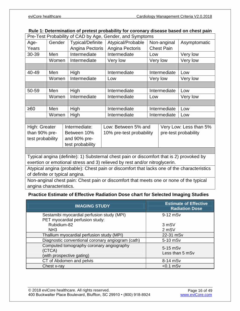
Rule 1: Determination of pretest probability for coronary disease based on chest pain Pre-Test Probability of CAD by Age, Gender, and Symptoms Age- Years
Gender Typical/Definite Angina Pectoris
Atypical/Probable Angina Pectoris
Non-anginal Chest Pain
Asymptomatic
30-39 Men Intermediate Intermediate Low Very low Women Intermediate Very low Very low Very low 40-49 Men High Intermediate Intermediate Low Women Intermediate Low Very low Very low 50-59 Men High Intermediate Intermediate Low Women Intermediate Intermediate Low Very low ≥60 Men High Intermediate Intermediate Low Women High Intermediate Intermediate Low High: Greater than 90% pre-test probability
Intermediate: Between 10% and 90% pre-test probability
Low: Between 5% and 10% pre-test probability
Very Low: Less than 5% pre-test probability
Typical angina (definite): 1) Substernal chest pain or discomfort that is 2) provoked by exertion or emotional stress and 3) relieved by rest and/or nitroglycerin. Atypical angina (probable): Chest pain or discomfort that lacks one of the characteristics of definite or typical angina. Non-anginal chest pain: Chest pain or discomfort that meets one or none of the typical angina characteristics.
Practice Estimate of Effective Radiation Dose chart for Selected Imaging Studies
IMAGING STUDY Estimate of Effective Radiation Dose
Sestamibi myocardial perfusion study (MPI) PET myocardial perfusion study: Rubidium-82 NH3
9-12 mSv 3 mSV 2 mSV
Thallium myocardial perfusion study (MPI) 22-31 mSv Diagnostic conventional coronary angiogram (cath) 5-10 mSv Computed tomography coronary angiography (CTCA) (with prospective gating)
5-15 mSv Less than 5 mSv
CT of Abdomen and pelvis 8-14 mSv Chest x-ray <0.1 mSv
eviCore healthcare Cardiology Management Criteria V2.0.2018
© 2018 eviCore healthcare. All rights reserved. 400 Buckwalter Place Boulevard, Bluffton, SC 29910 • (800) 918-8924 www.eviCore.com
Page 16 of 49
References:
1. Adabag AS, Grandits GA, Prineas RJ, et al. Relation of Heart Rate Parameters During Exercise Test to Sudden Death and All-Cause Mortality In Asymptomatic Men. Am J Cardiol 2008; 101:1437-1443. Accessed November 30, 2017. http://www.sciencedirect.com/science/article/pii/S0002914908001598.
2. Fihn SD, GardinJM, Abrams J, et al. 2012 ACCF/AHA/ACP/AATS/PCNA/SCAI/STS Guideline for the diagnosis and management of patients with stable ischemic heart disease: a report of the American College of Cardiology Foundation/American Heart Association Task Force on Practice Guidelines, and the American College of Physicians, American Association for Thoracic Surgery, Preventive Cardiovascular Nurses Association, Society for Cardiovascular Angiography and Interventions, and Society of Thoracic Surgeons. J Am CollCardiol 2012; 60:e44. Accessed November 30, 2017. http://circ.ahajournals.org/content/126/25/3097.
3. Qaseem A, Fihn SD, Williams S, et al. Diagnosis of stable ischemic heart disease: summary of a clinical practice guideline from the American College of Physicians/American College of Cardiology Foundation/American Heart Association/American Association for Thoracic Surgery/Preventive Cardiovascular Nurses Association/Society of Thoracic Surgeons. Ann Intern Med 2012; 157:729. Accessed November 30, 2017. http://annals.org/aim/fullarticle/1392193/diagnosis-stable-ischemic-heart-disease-summary-clinical-practice-guideline-from.
4. Rybicki FJ, Udelson JE, Peacock WF, et al. 2015 ACR/ACC/AHA/AATS/ACEP/ASNC/NASCI/SAEM/SCCT/SCMR/SCPC/SNMMI/STR/STS Appropriate Utilization of Cardiovascular Imaging in Emergency Department Patients With Chest Pain: A Joint Document of the American College of Radiology Appropriateness Criteria Committee and the American College of Cardiology Appropriate Use Criteria Task Force. J Am CollCardiol 2016; 67:853. Accessed November 30, 2017. http://www.onlinejacc.org/content/67/7/853?_ga=2.71380630.440582577.1512399151-2036160591.1507824385.
5. Fleisher LA, Fleischmann KE, Auerbach AD, et al. 2014 ACC/AHA Guideline on Perioperative Cardiovascular Evaluation and Management of Patients Undergoing Noncardiac Surgery: A Report of the American College of Cardiology/American Heart Association Task Force on Practice Guidelines. J Am CollCardiol 2014. Accessed November 30, 2017. http://www.sciencedirect.com/science/article/pii/S0735109714055363?via%3Dihub.
6. FriedewaldVE, King SB, Pepine CJ, et.al. The Editor’s Roundtable: Chronic stable angina pectoris. Am J Cardiol 2007 Dec; 100 (11):1635-1643. Accessed November 30, 2017. http://www.ajconline.org/article/S0002-9149(07)01706-7/fulltext.
7. Gibbons RJ, BaladyGJ, Bricker JT, et al. ACC/AHA 2002 Guideline Update for Exercise Testing Summary Article. A report of the American College of Cardiology/American Heart Association Task Force on Practice Guidelines (Committee to Update the 1997 Exercise Testing Guidelines). J Am Coll Cardio l2002; 40: 1531-1540. Accessed November 30, 2017. http://circ.ahajournals.org/content/106/14/1883.long.
8. Ho PM, Rumsfeld JS, Peterson PN. Chest pain on exercise treadmill test predicts future cardiac hospitalizations. ClinCardiol 2007; 30:505-510. Accessed November 30, 2017. http://onlinelibrary.wiley.com/doi/10.1002/clc.20139/abstract.
9. Lauer MS, Pothier CE, Magid DJ, et al. An externally validated model for predicting long-term survival after exercise treadmill testing in patients with suspected coronary artery disease and a normal electrocardiogram. Ann Intern Med 2007; 147:821-828. Accessed November 30, 2017. http://annals.org/aim/article-abstract/738017/externally-validated-model-predicting-long-term-survival-after-exercise-treadmill?doi=10.7326%2f0003-4819-147-12-200712180-00001.
10. Marshall AJ, Hutchings F, James AJ, et al. Prognostic value of a nine minute treadmill test in patients undergoing myocardial perfusion scintigraphy. Am J Cardiol 2010 Nov: 106(10):1423-1428. Accessed November 30, 2017. http://www.ajconline.org/article/S0002-9149(10)01408-6/fulltext.
11. MieresJH and Blumenthal RS. Does the treadmill test work in women? Cardiosource Spotlight, 2008 Jul 1; CS2-CS4. Accessed November 30, 2017. https://www.medscape.com/viewarticle/578141_3.
12. Peterson PN, Magid DJ, Ross C, et al. Association of exercise capacity on treadmill with future cardiac events in patients referred for exercise testing. Arch Intern Med 2008; 168(2):174-179. Accessed November 30, 2017. https://jamanetwork.com/journals/jamainternalmedicine/fullarticle/413829.
13. Picano E, Pasanisi E, Brown J, et al. A gatekeeper for the gatekeeper: Inappropriate referrals to stress echocardiography. Am Heart J 2007; 154: 285-290. Accessed November 30, 2017. http://www.ahjonline.com/article/S0002-8703(07)00356-0/fulltext.
14. Poirier P, Alpert MA, Fleisher LA, et al. Cardiovascular evaluation and management of severely obese patients undergoing surgery: a science advisory from the American Heart Association. Circulation 2009; 120:86-95. Accessed November 30, 2017. http://circ.ahajournals.org/content/120/1/86.
15. Sechtem U. Do heart transplant recipients need annual coronary angiography? European Heart Journal 2001; 22:895–897. Accessed November 30, 2017. https://academic.oup.com/eurheartj/article/22/11/895/524959.
16. Southard J, Baker L, Schaefer S. In search of the false-negative exercise treadmill testing evidence-based use of exercise echocardiography. ClinCardiol 2008; 31:35-40. Accessed November 30, 2017. http://onlinelibrary.wiley.com/doi/10.1002/clc.20174/abstract.
eviCore healthcare Cardiology Management Criteria V2.0.2018
© 2018 eviCore healthcare. All rights reserved. 400 Buckwalter Place Boulevard, Bluffton, SC 29910 • (800) 918-8924 www.eviCore.com
Page 17 of 49
17. Tavel ME. Stress testing in cardiac evaluation: Current concepts with emphasis on the ECG. Chest 2001; 119:907-925. Accessed November 30, 2017. http://journal.chestnet.org/article/S0012-3692(15)51692-9/fulltext.
18. Taylor DO, Edwards LB, Boucek MM, et al. Registry of the International Society for Heart and Lung Transplantation: Twenty-fourth official adult heart transplant report—2007. J Heart Lung Transplant 2007 August; 26(8):769-781. Accessed November 30, 2017. http://www.jhltonline.org/article/S1053-2498(07)00506-2/fulltext.
19. Diamond GA. A clinically relevant classification of chest discomfort. J Am CollCardiol 1983; 1:574–5. Accessed November 30, 2017. http://www.sciencedirect.com/science/article/pii/S073510978380093X?via%3Dihub.
20. WolkMJ, Bailey SR, Doherty JU, Douglas PS, Hendel RC, Kramer CM, Min JK, Patel MR, Rosenbaum L, Shaw LJ, StainbackRF, Allen JM. ACCF/AHA/ASE/ASNC/HFSA/HRS/SCAI/SCCT/SCMR/STS. 2013 Multi-modality appropriate use criteria for the detection and risk assessment of stable ischemic heart disease: a report of the American College of Cardiology Foundation, Appropriate Use Criteria Task Force, American Heart Association, American Society of Echocardiography, American Society of Nuclear Cardiology, Heart Failure Society of America, Heart Rhythm Society, Society for Cardiovascular Angiography and Interventions, Society of Cardiovascular Computed Tomography, Society for Cardiovascular Magnetic Resonance, and Society of Thoracic Surgeons. J Am CollCardiol 2014; 63: forthcoming. Accessed November 30, 2017. http://www.onlinejcf.com/article/S1071-9164(13)01274-8/fulltext.
21. Blank P, ScheopfUJ, Leipsic JA. CT in transcatheter aortic valve replacement. Radiology, 2013; 269(3) Accessed November 30, 2017. http://pubs.rsna.org/doi/10.1148/radiol.13120696.
22. Dill KE, George E, Abbara S, et al. ACR Appropriateness Criteria Imaging for Transcatheter Aortic Valve Replacement, Journal of the American College of Radiology, 2013 Dec;10(12): 957-965. Accessed November 30, 2017. http://www.jacr.org/article/S1546-1440(13)00565-6/fulltext.
23. MieresJH, Gulati M, BaireyMerz N, et al. American Heart Association Cardiac Imaging Committee of the Council on Clinical Cardiology, Cardiovascular Imaging and Intervention Committee of the Council on Cardiovascular Radiology. Role of Noninvasive Testing in the Clinical Evaluation of Women With Suspected Ischemic Heart Disease A Consensus Statement From the American Heart Association Circulation. 2014; 130(4):350. Accessed November 30, 2017. http://circ.ahajournals.org/content/130/4/350.long.
24. American College of Cardiology Foundation Appropriate Use Criteria Task Force, American Society of Echocardiography, American Heart Association, et al. ACCF/ASE/AHA/ASNC/HFSA/HRS/SCAI/SCCM/SCCT/SCMR 2011 Appropriate Use Criteria for Echocardiography. A Report of the American College of Cardiology Foundation Appropriate Use Criteria Task Force, American Society of Echocardiography, American Heart Association, American Society of Nuclear Cardiology, Heart Failure Society of America, Heart Rhythm Society, Society for Cardiovascular Angiography and Interventions, Society of Critical Care Medicine, Society of Cardiovascular Computed Tomography, and Society for Cardiovascular Magnetic Resonance Endorsed by the American College of Chest Physicians. J Am CollCardiol 2011; 57:1126. Accessed November 30, 2017. http://www.onlinejase.com/article/S0894-7317(10)01046-1/fulltext.
25. Melon CC, Eshtiaghi P, LuksunWJ, et al. Validated questionnaire vs physicians' judgment to estimate preoperative exercise capacity. JAMA Intern Med 2014; 174:1507Accessed November 30, 2017. https://jamanetwork.com/journals/jamainternalmedicine/fullarticle/1885465.
26. Taqueti V, Dorbala S, Wolinsky D. Myocardial perfusion imaging in women for the evaluation of stable ischemic heart disease— state-of-the-evidence and clinical recommendations. Journal of Nuclear Cardiology. June 2017. Accessed on October 25, 2017. https://link.springer.com/content/pdf/10.1007%2Fs12350-017-0926-8.pdf.
75559, 75563 Cardiac MRI for Morphology 78451, 78452, 78453, 78454 Myocardial Perfusion Imaging
eviCore healthcare Cardiology Management Criteria V2.0.2018
© 2018 eviCore healthcare. All rights reserved. 400 Buckwalter Place Boulevard, Bluffton, SC 29910 • (800) 918-8924 www.eviCore.com
Page 18 of 49
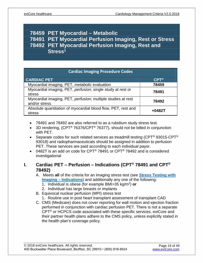
78459 PET Myocardial – Metabolic 78491 PET Myocardial Perfusion Imaging, Rest or Stress 78492 PET Myocardial Perfusion Imaging, Rest and
Stress1
Cardiac Imaging Procedure Codes
CARDIAC PET CPT® Myocardial imaging, PET, metabolic evaluation 78459 Myocardial imaging, PET, perfusion; single study at rest or stress 78491
Myocardial imaging, PET, perfusion; multiple studies at rest and/or stress 78492
Absolute quantitation of myocardial blood flow, PET, rest and stress +0482T
• 78491 and 78492 are also referred to as a rubidium study stress test. • 3D rendering, (CPT® 76376/CPT® 76377), should not be billed in conjunction
with PET. • Separate codes for such related services as treadmill testing (CPT® 93015-CPT®
93018) and radiopharmaceuticals should be assigned in addition to perfusion PET. These services are paid according to each individual payor.
• 0482T is an add on code for CPT® 78491 or CPT® 78492 and is considered investigational
I. Cardiac PET – Perfusion – Indications (CPT® 78491 and CPT®
78492) A. Meets all of the criteria for an imaging stress test (see Stress Testing with
Imaging – Indications) and additionally any one of the following: 1. Individual is obese (for example BMI>35 kg/m2) or 2. Individual has large breasts or implants
B. Equivocal nuclear perfusion (MPI) stress test 1. Routine use in post heart transplant assessment of transplant CAD
C. CMS (Medicare) does not cover reporting for wall motion and ejection fraction performed in conjunction with cardiac perfusion PET. There is not a separate CPT® or HCPCS code associated with these specific services. eviCore and their partner health plans adhere to the CMS policy, unless explicitly stated in the health plan’s coverage policy.
eviCore healthcare Cardiology Management Criteria V2.0.2018
© 2018 eviCore healthcare. All rights reserved. 400 Buckwalter Place Boulevard, Bluffton, SC 29910 • (800) 918-8924 www.eviCore.com
Page 19 of 49
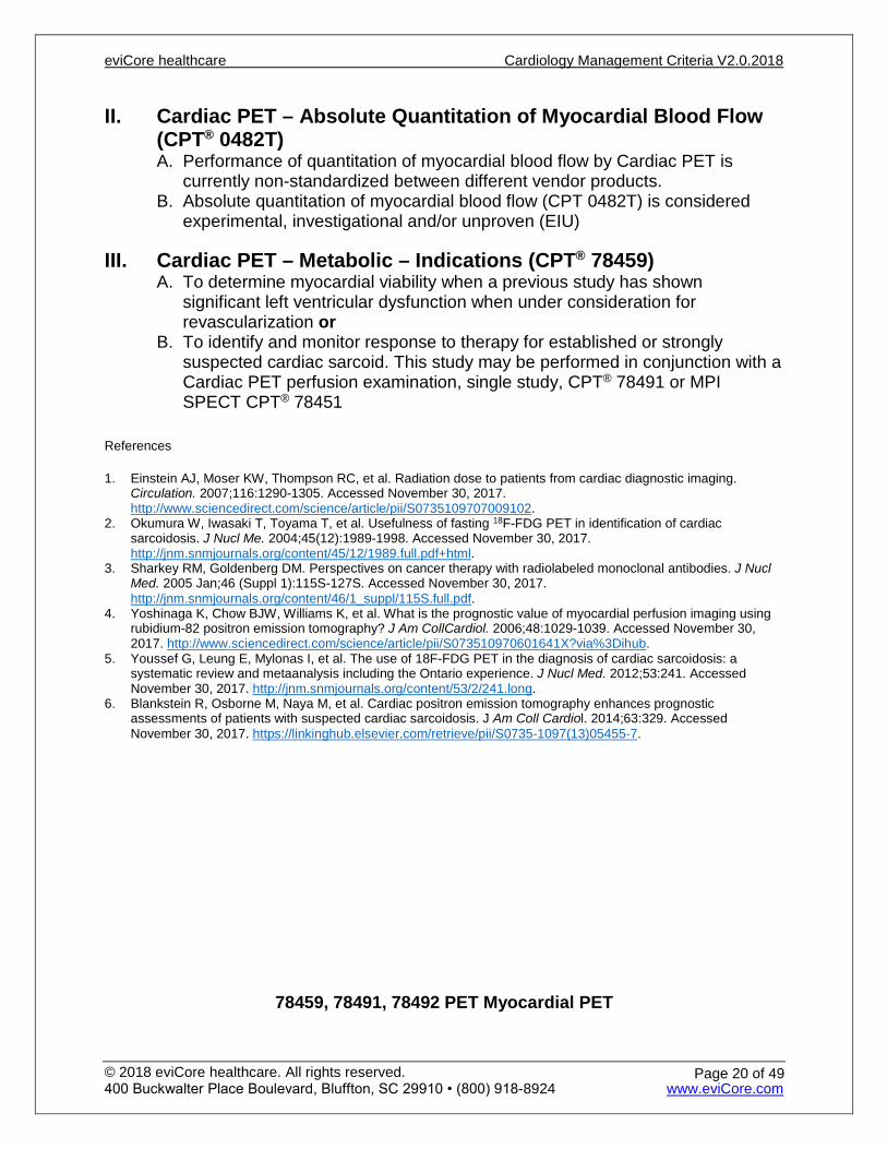
II. Cardiac PET – Absolute Quantitation of Myocardial Blood Flow (CPT® 0482T) A. Performance of quantitation of myocardial blood flow by Cardiac PET is
currently non-standardized between different vendor products. B. Absolute quantitation of myocardial blood flow (CPT 0482T) is considered
experimental, investigational and/or unproven (EIU)
III. Cardiac PET – Metabolic – Indications (CPT® 78459) A. To determine myocardial viability when a previous study has shown
significant left ventricular dysfunction when under consideration for revascularization or
B. To identify and monitor response to therapy for established or strongly suspected cardiac sarcoid. This study may be performed in conjunction with a Cardiac PET perfusion examination, single study, CPT® 78491 or MPI SPECT CPT® 78451
References
1. Einstein AJ, Moser KW, Thompson RC, et al. Radiation dose to patients from cardiac diagnostic imaging. Circulation. 2007;116:1290-1305. Accessed November 30, 2017. http://www.sciencedirect.com/science/article/pii/S0735109707009102.
2. Okumura W, Iwasaki T, Toyama T, et al. Usefulness of fasting 18F-FDG PET in identification of cardiac sarcoidosis. J Nucl Me. 2004;45(12):1989-1998. Accessed November 30, 2017. http://jnm.snmjournals.org/content/45/12/1989.full.pdf+html.
3. Sharkey RM, Goldenberg DM. Perspectives on cancer therapy with radiolabeled monoclonal antibodies. J Nucl Med. 2005 Jan;46 (Suppl 1):115S-127S. Accessed November 30, 2017. http://jnm.snmjournals.org/content/46/1_suppl/115S.full.pdf.
4. Yoshinaga K, Chow BJW, Williams K, et al. What is the prognostic value of myocardial perfusion imaging using rubidium-82 positron emission tomography? J Am CollCardiol. 2006;48:1029-1039. Accessed November 30, 2017. http://www.sciencedirect.com/science/article/pii/S073510970601641X?via%3Dihub.
5. Youssef G, Leung E, Mylonas I, et al. The use of 18F-FDG PET in the diagnosis of cardiac sarcoidosis: a systematic review and metaanalysis including the Ontario experience. J Nucl Med. 2012;53:241. Accessed November 30, 2017. http://jnm.snmjournals.org/content/53/2/241.long.
6. Blankstein R, Osborne M, Naya M, et al. Cardiac positron emission tomography enhances prognostic assessments of patients with suspected cardiac sarcoidosis. J Am Coll Cardiol. 2014;63:329. Accessed November 30, 2017. https://linkinghub.elsevier.com/retrieve/pii/S0735-1097(13)05455-7.
78459, 78491, 78492 PET Myocardial PET
eviCore healthcare Cardiology Management Criteria V2.0.2018
© 2018 eviCore healthcare. All rights reserved. 400 Buckwalter Place Boulevard, Bluffton, SC 29910 • (800) 918-8924 www.eviCore.com
Page 20 of 49
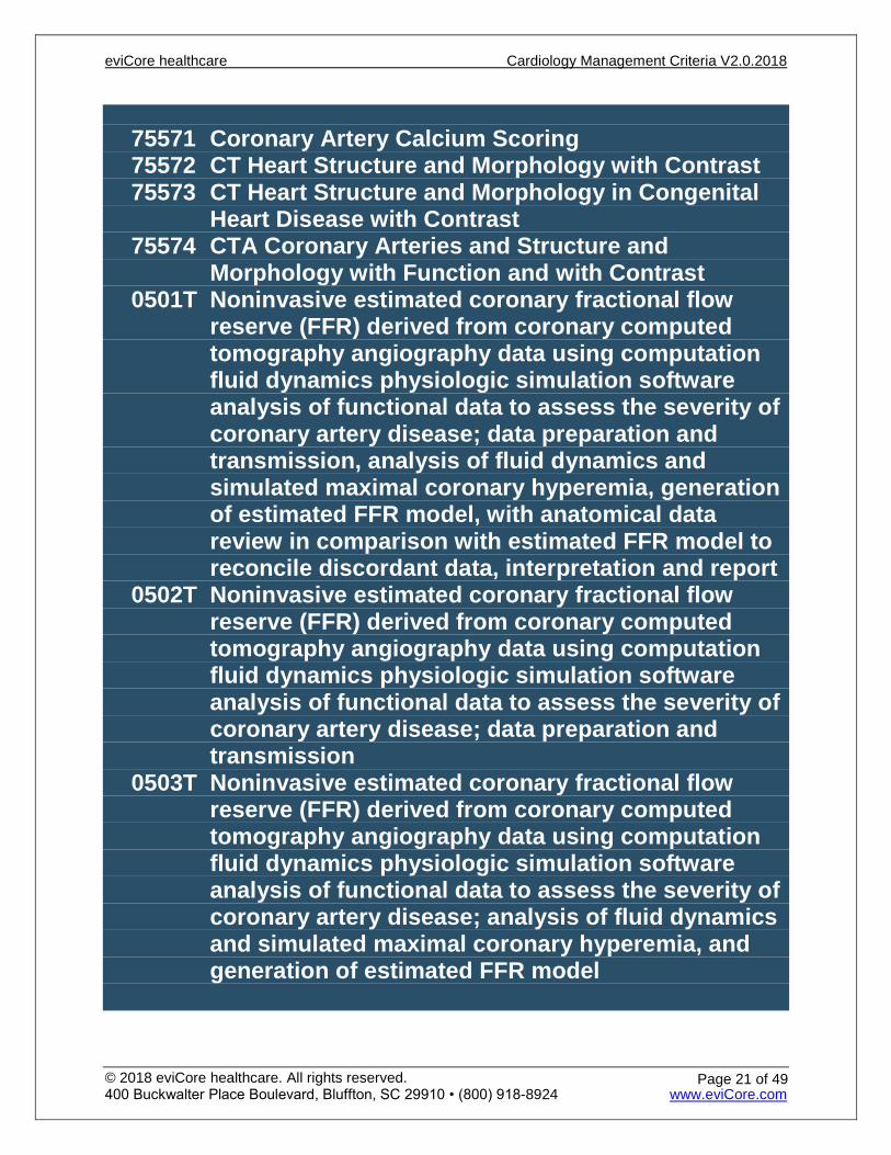
75571 Coronary Artery Calcium Scoring 75572 CT Heart Structure and Morphology with Contrast 75573 CT Heart Structure and Morphology in Congenital
Heart Disease with Contrast 75574 CTA Coronary Arteries and Structure and
Morphology with Function and with Contrast 0501T Noninvasive estimated coronary fractional flow
reserve (FFR) derived from coronary computed tomography angiography data using computation fluid dynamics physiologic simulation software analysis of functional data to assess the severity of coronary artery disease; data preparation and transmission, analysis of fluid dynamics and simulated maximal coronary hyperemia, generation of estimated FFR model, with anatomical data review in comparison with estimated FFR model to reconcile discordant data, interpretation and report
0502T Noninvasive estimated coronary fractional flow reserve (FFR) derived from coronary computed tomography angiography data using computation fluid dynamics physiologic simulation software analysis of functional data to assess the severity of coronary artery disease; data preparation and transmission
0503T Noninvasive estimated coronary fractional flow reserve (FFR) derived from coronary computed tomography angiography data using computation fluid dynamics physiologic simulation software analysis of functional data to assess the severity of coronary artery disease; analysis of fluid dynamics and simulated maximal coronary hyperemia, and generation of estimated FFR model
eviCore healthcare Cardiology Management Criteria V2.0.2018
© 2018 eviCore healthcare. All rights reserved. 400 Buckwalter Place Boulevard, Bluffton, SC 29910 • (800) 918-8924 www.eviCore.com
Page 21 of 49
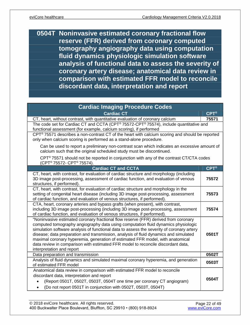
0504T Noninvasive estimated coronary fractional flow reserve (FFR) derived from coronary computed tomography angiography data using computation fluid dynamics physiologic simulation software analysis of functional data to assess the severity of coronary artery disease; anatomical data review in comparison with estimated FFR model to reconcile discordant data, interpretation and report
Cardiac Imaging Procedure Codes Cardiac CT CPT®
CT, heart, without contrast, with quantitative evaluation of coronary calcium 75571 The code set for Cardiac CT and CCTA (CPT® 75572-CPT® 75574), include quantitative and functional assessment (for example, calcium scoring), if performed CPT® 75571 describes a non-contrast CT of the heart with calcium scoring and should be reported only when calcium scoring is performed as a stand-alone procedure.
Can be used to report a preliminary non-contrast scan which indicates an excessive amount of calcium such that the original scheduled study must be discontinued. CPT® 75571 should not be reported in conjunction with any of the contrast CT/CTA codes (CPT® 75572- CPT® 75574).
Cardiac CT and CCTA CPT® CT, heart, with contrast, for evaluation of cardiac structure and morphology (including 3D image post-processing, assessment of cardiac function, and evaluation of venous structures, if performed).
75572
CT, heart, with contrast, for evaluation of cardiac structure and morphology in the setting of congenital heart disease (including 3D image post-processing, assessment of cardiac function, and evaluation of venous structures, if performed).
75573
CTA, heart, coronary arteries and bypass grafts (when present), with contrast, including 3D image post-processing (including 3D image post-processing, assessment of cardiac function, and evaluation of venous structures, if performed).
75574
"Noninvasive estimated coronary fractional flow reserve (FFR) derived from coronary computed tomography angiography data using computation fluid dynamics physiologic simulation software analysis of functional data to assess the severity of coronary artery disease; data preparation and transmission, analysis of fluid dynamics and simulated maximal coronary hyperemia, generation of estimated FFR model, with anatomical data review in comparison with estimated FFR model to reconcile discordant data, interpretation and report
0501T
Data preparation and transmission 0502T Analysis of fluid dynamics and simulated maximal coronary hyperemia, and generation of estimated FFR model 0503T
Anatomical data review in comparison with estimated FFR model to reconcile discordant data, interpretation and report • (Report 0501T, 0502T, 0503T, 0504T one time per coronary CT angiogram) • (Do not report 0501T in conjunction with 0502T, 0503T, 0504T)
0504T
eviCore healthcare Cardiology Management Criteria V2.0.2018
© 2018 eviCore healthcare. All rights reserved. 400 Buckwalter Place Boulevard, Bluffton, SC 29910 • (800) 918-8924 www.eviCore.com
Page 22 of 49
3D rendering, (CPT® 76376/CPT® 76377), should not be billed in conjunction with Cardiac CT and CCTA.
Only one code from the set: CPT® 75572 - CPT® 75574 can be reported per encounter.
CPT® 75574 includes evaluation of cardiac structure and morphology, when performed; therefore, additional code/s should not be assigned.
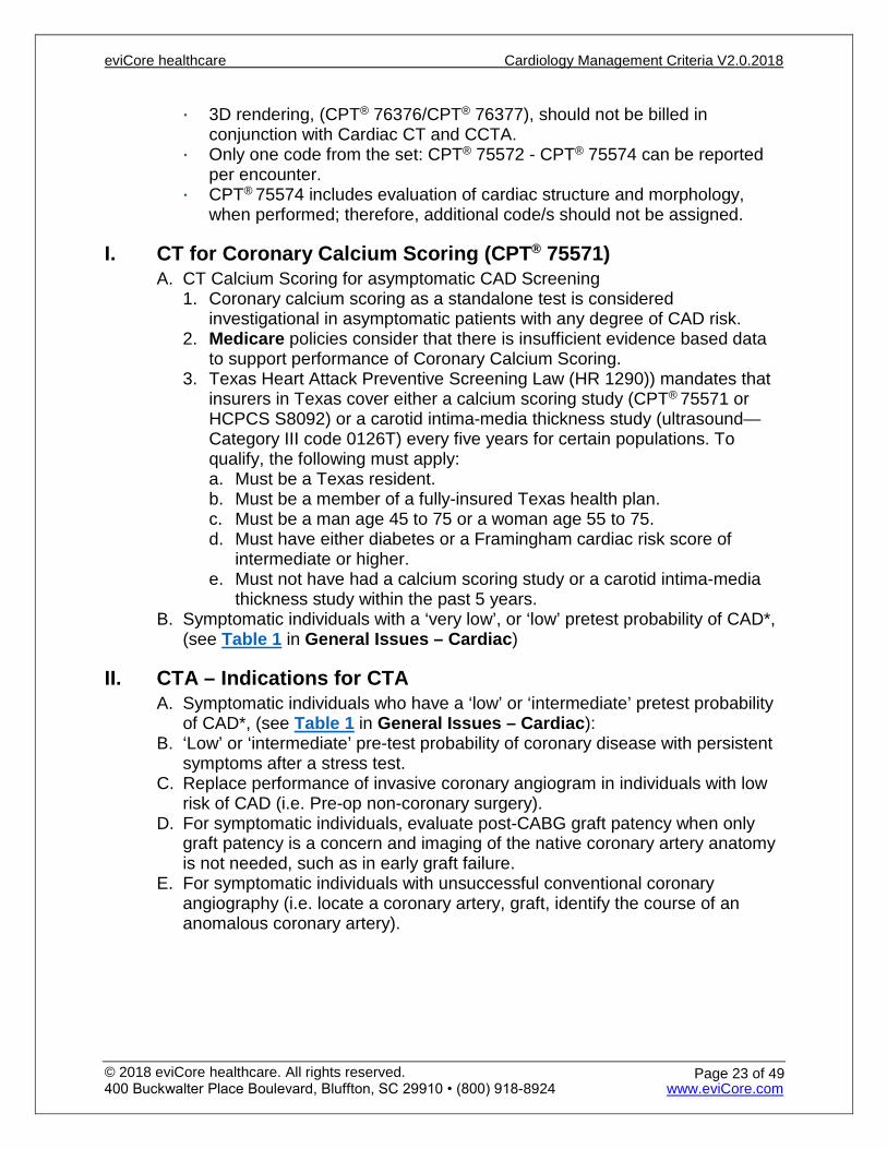
I. CT for Coronary Calcium Scoring (CPT® 75571) A. CT Calcium Scoring for asymptomatic CAD Screening
1. Coronary calcium scoring as a standalone test is considered investigational in asymptomatic patients with any degree of CAD risk.
2. Medicare policies consider that there is insufficient evidence based data to support performance of Coronary Calcium Scoring.
3. Texas Heart Attack Preventive Screening Law (HR 1290)) mandates that insurers in Texas cover either a calcium scoring study (CPT® 75571 or HCPCS S8092) or a carotid intima-media thickness study (ultrasound—Category III code 0126T) every five years for certain populations. To qualify, the following must apply: a. Must be a Texas resident. b. Must be a member of a fully-insured Texas health plan. c. Must be a man age 45 to 75 or a woman age 55 to 75. d. Must have either diabetes or a Framingham cardiac risk score of
intermediate or higher. e. Must not have had a calcium scoring study or a carotid intima-media
thickness study within the past 5 years. B. Symptomatic individuals with a ‘very low’, or ‘low’ pretest probability of CAD*,
(see Table 1 in General Issues – Cardiac)
II. CTA – Indications for CTA A. Symptomatic individuals who have a ‘low’ or ‘intermediate’ pretest probability
of CAD*, (see Table 1 in General Issues – Cardiac): B. ‘Low’ or ‘intermediate’ pre-test probability of coronary disease with persistent
symptoms after a stress test. C. Replace performance of invasive coronary angiogram in individuals with low
risk of CAD (i.e. Pre-op non-coronary surgery). D. For symptomatic individuals, evaluate post-CABG graft patency when only
graft patency is a concern and imaging of the native coronary artery anatomy is not needed, such as in early graft failure.
E. For symptomatic individuals with unsuccessful conventional coronary angiography (i.e. locate a coronary artery, graft, identify the course of an anomalous coronary artery).
eviCore healthcare Cardiology Management Criteria V2.0.2018
© 2018 eviCore healthcare. All rights reserved. 400 Buckwalter Place Boulevard, Bluffton, SC 29910 • (800) 918-8924 www.eviCore.com
Page 23 of 49
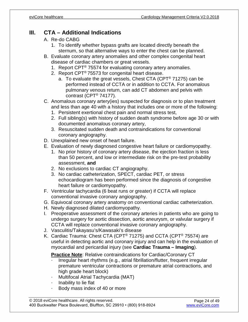
III. CTA – Additional Indications A. Re-do CABG
1. To identify whether bypass grafts are located directly beneath the sternum, so that alternative ways to enter the chest can be planned.
B. Evaluate coronary artery anomalies and other complex congenital heart disease of cardiac chambers or great vessels. 1. Report CPT® 75574 for evaluating coronary artery anomalies. 2. Report CPT® 75573 for congenital heart disease.
a. To evaluate the great vessels, Chest CTA (CPT® 71275) can be performed instead of CCTA or in addition to CCTA. For anomalous pulmonary venous return, can add CT abdomen and pelvis with contrast (CPT® 74177).
C. Anomalous coronary artery(ies) suspected for diagnosis or to plan treatment and less than age 40 with a history that includes one or more of the following: 1. Persistent exertional chest pain and normal stress test, 2. Full sibling(s) with history of sudden death syndrome before age 30 or with
documented anomalous coronary artery, 3. Resuscitated sudden death and contraindications for conventional
coronary angiography. D. Unexplained new onset of heart failure. E. Evaluation of newly diagnosed congestive heart failure or cardiomyopathy.
1. No prior history of coronary artery disease, the ejection fraction is less than 50 percent, and low or intermediate risk on the pre-test probability assessment, and
2. No exclusions to cardiac CT angiography. 3. No cardiac catheterization, SPECT, cardiac PET, or stress
echocardiogram has been performed since the diagnosis of congestive heart failure or cardiomyopathy.
F. Ventricular tachycardia (6 beat runs or greater) if CCTA will replace conventional invasive coronary angiography.
G. Equivocal coronary artery anatomy on conventional cardiac catheterization. H. Newly diagnosed dilated cardiomyopathy. I. Preoperative assessment of the coronary arteries in patients who are going to
undergo surgery for aortic dissection, aortic aneurysm, or valvular surgery if CCTA will replace conventional invasive coronary angiography.
J. Vasculitis/Takayasu’s/Kawasaki’s disease K. Cardiac Trauma: Chest CTA (CPT® 71275) and CCTA (CPT® 75574) are
useful in detecting aortic and coronary injury and can help in the evaluation of myocardial and pericardial injury (see Cardiac Trauma – Imaging). Practice Note: Relative contraindications for Cardiac/Coronary CT Irregular heart rhythms (e.g., atrial fibrillation/flutter, frequent irregular
premature ventricular contractions or premature atrial contractions, and high grade heart block)
Multifocal Atrial Tachycardia (MAT) Inability to lie flat Body mass index of 40 or more
eviCore healthcare Cardiology Management Criteria V2.0.2018
© 2018 eviCore healthcare. All rights reserved. 400 Buckwalter Place Boulevard, Bluffton, SC 29910 • (800) 918-8924 www.eviCore.com
Page 24 of 49

Inability to obtain a heart rate less than 65 beats per minute after beta-blockers
Inability to hold breath for at least 8 seconds Renal Insufficiency Asymptomatic patients and routine use in the evaluation of the coronary
arteries following heart transplantation CCTA should not be performed if there is extensive coronary calcification
(calcium score >1000). Evaluation of coronary stent patency (metal artifact limits accuracy) - <3.0
mm
IV. Evaluation of left ventricular function following myocardial infarction or in chronic heart failure
V. Fractional Flow Reserve by Computed Tomography A. Fractional flow reserve (FFR) is typically measured using invasive techniques.
FFR can be obtained noninvasively from coronary computed tomography angiography data (FFR-CT).
B. Indications for FFR-CT 1. To further assess CAD seen on a recent CCTA that is of uncertain
physiologic significance
VI. CT Heart – Indications A. If echocardiogram is inconclusive for:
1. Cardiac or pericardial tumor or mass 2. Cardiac thrombus 3. Pericarditis/constrictive pericarditis 4. Complications of cardiac surgery
B. Cardiac vein identification for lead placement in patients needing left ventricular pacing.
C. Pulmonary vein isolation procedure (ablation) for atrial fibrillation D. Cardiac MRI (CPT® 75557 or CPT® 75561), chest MRV (CPT® 71555), chest
CTV (CPT® 71275), or cardiac CT (CPT® 75572) can be performed to evaluate anatomy of the pulmonary veins prior to an ablation procedure performed for atrial fibrillation 1. Repeated post-procedure between 3-6 months after ablation because of a
1%-2% incidence of asymptomatic pulmonary vein stenosis. 2. If pulmonary vein stenosis is present on imaging following ablation and
symptoms of pulmonary vein stenosis (usually shortness of breath) are present, can be imaged at 1, 3, 6, and 12 months.
E. Recurrent laryngeal nerve palsy due to cardiac chamber enlargement. F. Clinical suspicion of arrhythmogenic right ventricular dysplasia` or
arrhythmogenic cardiomyopathy (ARVD/ARVC), especially if patient has presyncope or syncope if the clinical suspicion is supported by established criteria for ARVD.
eviCore healthcare Cardiology Management Criteria V2.0.2018
© 2018 eviCore healthcare. All rights reserved. 400 Buckwalter Place Boulevard, Bluffton, SC 29910 • (800) 918-8924 www.eviCore.com
Page 25 of 49
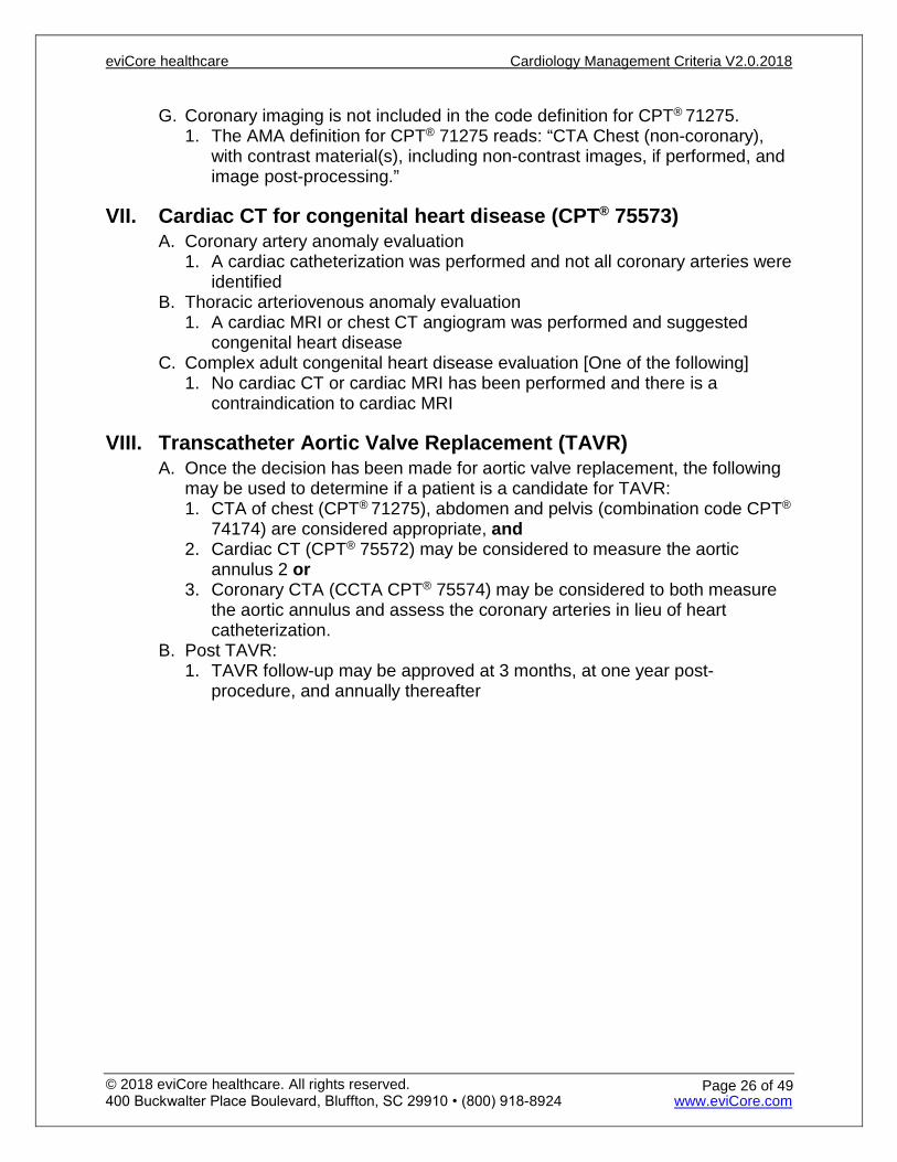
G. Coronary imaging is not included in the code definition for CPT® 71275. 1. The AMA definition for CPT® 71275 reads: “CTA Chest (non-coronary),
with contrast material(s), including non-contrast images, if performed, and image post-processing.”
VII. Cardiac CT for congenital heart disease (CPT® 75573) A. Coronary artery anomaly evaluation
1. A cardiac catheterization was performed and not all coronary arteries were identified
B. Thoracic arteriovenous anomaly evaluation 1. A cardiac MRI or chest CT angiogram was performed and suggested
congenital heart disease C. Complex adult congenital heart disease evaluation [One of the following]
1. No cardiac CT or cardiac MRI has been performed and there is a contraindication to cardiac MRI
VIII. Transcatheter Aortic Valve Replacement (TAVR) A. Once the decision has been made for aortic valve replacement, the following
may be used to determine if a patient is a candidate for TAVR: 1. CTA of chest (CPT® 71275), abdomen and pelvis (combination code CPT®
74174) are considered appropriate, and 2. Cardiac CT (CPT® 75572) may be considered to measure the aortic
annulus 2 or 3. Coronary CTA (CCTA CPT® 75574) may be considered to both measure
the aortic annulus and assess the coronary arteries in lieu of heart catheterization.
B. Post TAVR: 1. TAVR follow-up may be approved at 3 months, at one year post-
procedure, and annually thereafter
eviCore healthcare Cardiology Management Criteria V2.0.2018
© 2018 eviCore healthcare. All rights reserved. 400 Buckwalter Place Boulevard, Bluffton, SC 29910 • (800) 918-8924 www.eviCore.com
Page 26 of 49

Rule 1: Determination of pretest probability for coronary disease based on chest pain Pre-Test Probability of CAD by Age, Gender, and Symptoms
Age- Years Gender Typical/Definite
Angina Pectoris Atypical/Probable Angina Pectoris
Non-anginal Chest Pain Asymptomatic
30-39 Men Intermediate Intermediate Low Very low Women Intermediate Very low Very low Very low 40-49 Men High Intermediate Intermediate Low Women Intermediate Low Very low Very low 50-59 Men High Intermediate Intermediate Low Women Intermediate Intermediate Low Very low ≥60 Men High Intermediate Intermediate Low Women High Intermediate Intermediate Low
High: Greater than 90% pre-test probability
Intermediate: Between 10% and 90% pre-test probability
Low: Between 5% and 10% pre-test probability
Very Low: Less than 5% pre-test probability
Typical angina (definite): 1) Substernal chest pain or discomfort that is 2) provoked by exertion or emotional stress and 3) relieved by rest and/or nitroglycerin. Atypical angina (probable): Chest pain or discomfort that lacks one of the characteristics of definite or typical angina. Non-anginal chest pain: Chest pain or discomfort that meets one or none of the typical angina characteristics.
References: 1. Taylor AJ, Cequeira M, Hodgson JM, et al. ACCF/SCCT/ACR/AHA/ASE/ASNC/SCAI/SCMR 2010 appropriate
use criteria for cardiac computed tomography: a report of the American College of Cardiology Foundation Appropriate Use Criteria Task Force, the Society of Cardiovascular Computed Tomography, the American College of Radiology, the American Heart Association, the American Society of Echocardiography, the American Society of Nuclear Cardiology, the Society for Cardiovascular Angiography and Interventions, and the Society for Cardiovascular Magnetic Resonance. J Am CollCardiol. 210: 56; 1864-1894.
2. ACCF/ACR/SCCT/SCMR/ASNC/NASCI/SCAI/SIR Appropriateness Criteria for Cardiac Computed Tomography and Cardiac Magnetic Resonance Imaging. Journal of the American College of Cardiology Vol. 48, No. 7, 2006.
3. Andreini D, Pontone G, Pepi M, et al. Diagnostic accuracy of multidetector computed tomography coronary angiography in patients with dilated cardiomyopathy. J Am CollCardiol 2007 May;49:2044-2050.
4. BerbarieRF, Dockery WD, Johnson KB, et al. Use of multislice computed tomographic coronary angiography for the diagnosis of anomalous coronary arteries. Am J Cardiol 2006;98:402-406.
5. BudoffMJ, Achenbach S, Blumenthal RS, et al. Assessment of coronary artery disease by cardiac computed tomography. Circulation 2006;114:1761-1791. http://circ.ahajournals.org/cgi/content/full/114/16/1761. Accessed November 16, 2012.
6. Einstein AJ, HenzlovaMJ, and Rajagopalan S. Estimating risk of cancer associated with radiation exposure from 64-slice computed tomography coronary angiography. JAMA 2007;298:317-323.
7. Schlosser T, Konorza T, Hunold P, et al. Noninvasive visualization of coronary artery bypass grafts using 16-detector row computed tomography. J Am CollCardiol 2004;44:1224-1229.
8. Elie MC. Blunt cardiac injury. Mt Sinai J Med, 2006;73:542.75571, 75572, 75573, 75574 Coronary Artery Calcium Scoring, Heart Structure and Morphology;
0501T, 0502T, 0503T, 0504T Coronary Fractional Flow Reserve (FFR) Computed Tomography
eviCore healthcare Cardiology Management Criteria V2.0.2018
© 2018 eviCore healthcare. All rights reserved. 400 Buckwalter Place Boulevard, Bluffton, SC 29910 • (800) 918-8924 www.eviCore.com
Page 27 of 49

Echocardiography 93303 Transthoracic Echocardiography for Congenital
Cardiac Anomalies; Complete 93304 Transthoracic Echocardiography for Congenital
Cardiac Anomalies; Follow-up or Limited Study 93306 Echocardiography, Transthoracic, Real-time with
Image Documentation (2D), Includes M-mode Recording, when Performed, Complete, with Spectral Doppler Echocardiography, and with Color Flow Doppler Echocardiography
93307 Echocardiography, Transthoracic, Real-time with Image Documentation (2D) with or without M-mode Recording; Complete
93308 Echocardiography, Transthoracic, Real-time with Image Documentation (2D) with or without M-mode Recording; Follow-up or Limited Study
93320 Doppler Echocardiography, Pulsed Wave and/or Continuous Wave with Spectral Display; Complete
93321 Doppler Echocardiography, Pulsed Wave and/or Continuous Wave with Spectral Display; Follow- up or Limited Study
93325 Doppler Echocardiography Color Flow Velocity Mapping
eviCore healthcare Cardiology Management Criteria V2.0.2018
© 2018 eviCore healthcare. All rights reserved. 400 Buckwalter Place Boulevard, Bluffton, SC 29910 • (800) 918-8924 www.eviCore.com
Page 28 of 49
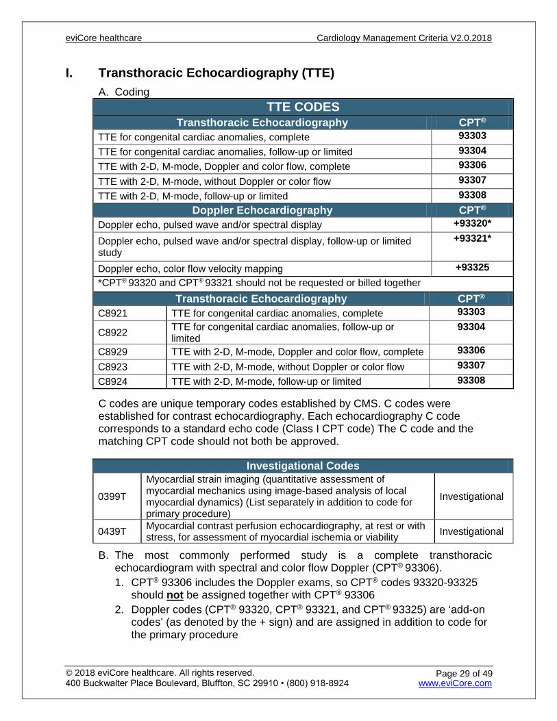
I. Transthoracic Echocardiography (TTE) A. Coding
TTE CODES Transthoracic Echocardiography CPT®
TTE for congenital cardiac anomalies, complete 93303 TTE for congenital cardiac anomalies, follow-up or limited 93304 TTE with 2-D, M-mode, Doppler and color flow, complete 93306 TTE with 2-D, M-mode, without Doppler or color flow 93307 TTE with 2-D, M-mode, follow-up or limited 93308
Doppler Echocardiography CPT® Doppler echo, pulsed wave and/or spectral display +93320*
Doppler echo, pulsed wave and/or spectral display, follow-up or limited study
+93321*
Doppler echo, color flow velocity mapping +93325 *CPT® 93320 and CPT® 93321 should not be requested or billed together
Transthoracic Echocardiography CPT® C8921 TTE for congenital cardiac anomalies, complete 93303
C8922 TTE for congenital cardiac anomalies, follow-up or limited
93304
C8929 TTE with 2-D, M-mode, Doppler and color flow, complete 93306 C8923 TTE with 2-D, M-mode, without Doppler or color flow 93307 C8924 TTE with 2-D, M-mode, follow-up or limited 93308
C codes are unique temporary codes established by CMS. C codes were established for contrast echocardiography. Each echocardiography C code corresponds to a standard echo code (Class I CPT code) The C code and the matching CPT code should not both be approved.
Investigational Codes
0399T
Myocardial strain imaging (quantitative assessment of myocardial mechanics using image-based analysis of local myocardial dynamics) (List separately in addition to code for primary procedure)
Investigational
0439T Myocardial contrast perfusion echocardiography, at rest or with stress, for assessment of myocardial ischemia or viability Investigational
B. The most commonly performed study is a complete transthoracic echocardiogram with spectral and color flow Doppler (CPT® 93306). 1. CPT® 93306 includes the Doppler exams, so CPT® codes 93320-93325
should not be assigned together with CPT® 93306 2. Doppler codes (CPT® 93320, CPT® 93321, and CPT® 93325) are ‘add-on
codes’ (as denoted by the + sign) and are assigned in addition to code for the primary procedure
eviCore healthcare Cardiology Management Criteria V2.0.2018
© 2018 eviCore healthcare. All rights reserved. 400 Buckwalter Place Boulevard, Bluffton, SC 29910 • (800) 918-8924 www.eviCore.com
Page 29 of 49
3. For a 2D transthoracic echocardiogram without Doppler, report CPT®
93307 4. Limited transthoracic echocardiogram should be billed if the report does
not “evaluate or document the attempt to evaluate” all of the required structures. a. A limited transthoracic echocardiogram is reported with CPT® 93308. b. CPT® 93321 (not CPT® 93320) should be reported with CPT® 93308 if
Doppler is included in the study. CPT® 93325 can be reported with CPT® 93308 if color flow Doppler is included in the study.
c. A limited congenital transthoracic echocardiogram is reported with CPT® 93304.
5. Providers performing echo on a patient, may not know what procedure codes they will be reporting until the initial study is completed. a. If a congenital issue is found on the initial echo, a complete echo is
reported with codes CPT® 93303, CPT® 93320, and CPT® 93325 because CPT® 93303 does NOT include Doppler and color flow mapping.
b. If no congenital issue is discovered, then CPT® 93306 is reported alone and includes 2-D, Doppler and color flow mapping.
c. Since providers may not know the appropriate code/s that will be reported at the time of the pre-authorization request, they may request all 4 codes (CPT® 93303, CPT® 93320, CPT® 93325, and CPT® 93306).
d. Depending upon individual health plan payer contracts, post-service audits may be completed to ensure proper claims submission.
e. CPT® 76376 and CPT® 76377 are not unique to 3D Echo. These codes also apply to 3D rendering of MRI and CT studies. (See Echocardiography – Coding)
f. CPT® 93325 may also be used with fetal echocardiography. 6. Doppler echo may be used for evaluation of the following:
a. Shortness of breath b. Known or suspected valvular disease c. Known or suspected hypertrophic obstructive cardiomyopathy d. Shunt detection
C. Transthoracic Echocardiography (TTE) – Indications 1. New or worsening cardiac signs or symptoms, such as:
a. Dyspnea b. Chest pain c. Palpitations d. Syncope e. Symptoms of heart failure f. Murmur
eviCore healthcare Cardiology Management Criteria V2.0.2018
© 2018 eviCore healthcare. All rights reserved. 400 Buckwalter Place Boulevard, Bluffton, SC 29910 • (800) 918-8924 www.eviCore.com
Page 30 of 49
2. Valve function and structure: a. Valvular stenosis or regurgitation b. Valvular structure c. Valve Surgery
i. If valve surgery is being considered can have TTE twice a year ii. One routine study (surveillance) 3 years or more after valve surgery
(repair or prosthetic valve implantation). iii. TAVR follow-up may be approved at, 3 months, and at one year
post-procedure and annually thereafter. 01. A baseline post-op TTE is usually performed within one week
after surgery. This baseline study may also be approved as an outpatient if not performed in the hospital prior to discharge.
3. Ventricular function including global and segmental wall motion for evaluating ejection fraction (EF) and coronary artery disease a. Dyspnea b. Symptoms of Heart Failure c. Cardiomyopathy d. Chemotherapy
i. See also: MUGA Study – Assessment of cardiac function for cardiotoxic chemotherapy
ii. Determine LV function in patients in patients on cardiotoxic chemotherapeutic drugs. 01. The time frame should be determined by the provider, but no
more often than baseline and at every 6 weeks. 02. May repeat every 4 weeks if cardiotoxic chemotherapeutic drug
is withheld for significant left ventricular cardiac dysfunction iii. If the LVEF is <50% on echocardiogram than follow up can be done
with MUGA at appropriate intervals. e. Arrhythmias
4. Ventricular structure including but not limited to: a. Infiltrative diseases (e.g. sarcoid, amyloid) b. Ventricular septal defect (VSD) c. Papillary muscle rupture/dysfunction d. Hypertrophy including:
i. asymmetric septal hypertrophy ii. spade heart iii. Hypertensive concentric hypertrophy iv. Infiltrative hypertrophy
5. Evaluation of right ventricular systolic pressure/pulmonary hypertension 6. Evaluation of atrial or ventricular chamber size (e.g. patients with atrial
fibrillation, tachyarrhythmias, or left ventricular dilatation) a. Yearly TTE may be indicated depending on the clinical circumstance.
7. Cardiac Defects or Masses a. Embolic source in patients with recent Transient Ischemic Attack (TIA),
stroke, or peripheral vascular emboli as an initial study before TEE b. ASD repair or VSD repair:
eviCore healthcare Cardiology Management Criteria V2.0.2018
© 2018 eviCore healthcare. All rights reserved. 400 Buckwalter Place Boulevard, Bluffton, SC 29910 • (800) 918-8924 www.eviCore.com
Page 31 of 49
i. Within the first year of surgery or ii. If newly symptomatic
c. Tumor evaluation including myxomas d. Clot detection e. Evaluation of congenital heart disease
8. Inflammatory a. Pericardial effusion/pericardial disease including pericardial cysts b. Congenital heart disease c. Endocarditis including:
i. Fever ii. Positive blood cultures indicating bacteremia or iii. A new murmur
9. Pacemaker insertion complication 10. Screening for first-degree relatives of patients with hypertrophic
cardiomyopathy (HCM) a. First-degree relatives who are 12 to 18 years old should be screened
yearly for HCM by 2D- echocardiography and ECG b. First-degree relatives who are older than age 18 should have 2D-echo
and ECG every five years to screen for delayed adult-onset LVH c. Systematic screening is usually not indicated for first-degree relatives
who are younger than age 12 unless there is a high-risk family history or the child is involved in particularly intense competitive sports
d. Affected individuals identified through family screening or otherwise should be evaluated every 12 to 18 months with 2D-echo, Holter monitor, and blood pressure response during maximal upright exercise
11. New abnormality on an EKG that has not been evaluated 12. Assess aortic root and proximal ascending aorta
D. Frequency of Echocardiography Testing 1. Repeat routine echocardiograms are not supported (annually or
otherwise) for evaluation of clinically stable syndromes 2. Once a year (when no change in clinical status), when there a history of:
a. Significant valve dysfunction b. Hypertrophic cardiomyopathy (see Stress Echocardiography –
Indications, other than ruling out CAD) c. Chronic pericardial effusions d. Left ventricular contractility/diastolic function prior to planned medical
therapy for heart failure or to evaluate the effectiveness of on-going therapy
e. Aortic root dilatation f. Pulmonary hypertension
3. Prior TAVR (see Transthoracic Echocardiography (TTE) – Indications) 4. Twice a year for the following assessments:
a. New or changing (not chronic stable) pericardial effusions b. New/changed medical therapy for congestive heart failure c. Hypertrophic cardiomyopathy when the results of the echo will
potentially change patient management
eviCore healthcare Cardiology Management Criteria V2.0.2018
© 2018 eviCore healthcare. All rights reserved. 400 Buckwalter Place Boulevard, Bluffton, SC 29910 • (800) 918-8924 www.eviCore.com
Page 32 of 49
d. Critical valvular heart disease when the results of the echo will potentially change patient management
5. Anytime, without regard for the number or timing of previous ECHO studies, if there are new signs or symptoms such as: a. Cardiac murmurs b. Myocardial infarction or acute coronary syndrome c. Congestive heart failure (new or worsening)
i. New symptoms of dyspnea ii. Orthopnea iii. Paroxysmal nocturnal dyspnea iv. Edema v. Elevated BNP
d. Pericardial disease e. Stroke/transient ischemic attack f. Decompression illness g. Prosthetic valve dysfunction or thrombosis
II. 3D Echocardiography A. Coding
1. The procedure codes used to report 3D rendering for echocardiography are not unique to echocardiography and are the same codes used to report the 3D post processing work for CT, MRI, ultrasound and other tomographic modalities a. CPT® 76376, not requiring image post-processing on an independent
workstation, is the most common code used for 3D rendering done with echocardiography
b. CPT® 76377 requires the use of an independent workstation B. 3D Echocardiography – Indications
1. 3D Echo Indications a. Echocardiography with 3-dimensional (3D) rendering is becoming
universally available, yet its utility remains limited based on the current literature. Current indications include: i. Left ventricular volume and ejection fraction assessment ii. Mitral valve anatomy specifically related to mitral valve stenosis iii. Guidance of transcatheter procedures
References
1. Bangalore S, Yao S, and Chaudhry F. Usefulness of stress echocardiography for risk stratification and prognosis of patients with left ventricular hypertrophy. American Journal of Cardiology 2007; 100: 536-543. Accessed on November 1, 2017. http://www.ajconline.org/article/S0002-9149(07)00852-1/fulltext.
2. Douglas P, Khandheria B, Stainback R, et al. ACCF/ASE/ACEP/AHA/ASNC/SCAI/SCCT/SCMR 2008 appropriateness criteria for stress echocardiography: a report of the American College of Cardiology Foundation Appropriateness Criteria Task Force, American Society of Echocardiography, American College of Emergency Physicians, American Heart Association, American Society of Nuclear Cardiology, Society for Cardiovascular Angiography and Interventions, Society of Cardiovascular Computed Tomography, and Society for Cardiovascular Magnetic Resonance endorsed by the Heart Rhythm Society and the Society of Critical Care Medicine. Journal of the American College of Cardiology. 2008; 51: 1127-1147. Accessed on November 1, 2017. http://www.sciencedirect.com/science/article/pii/S0735109707039629?via%3Dihub.
eviCore healthcare Cardiology Management Criteria V2.0.2018
© 2018 eviCore healthcare. All rights reserved. 400 Buckwalter Place Boulevard, Bluffton, SC 29910 • (800) 918-8924 www.eviCore.com
Page 33 of 49
3. Maron B, McKenna W, Danielson G, et al. American College of Cardiology/European Society of CardiologyClinical Expert Consensus Document on Hypertrophic Cardiomyopathy: A report of the American College ofCardiology Foundation Task Force on Clinical Expert Consensus Documents and the European Society ofCardiology Committee for Practice Guidelines. European Heart Journal 2003; 24:1965-1991. Accessed onNovember 1, 2017. https://academic.oup.com/eurheartj/article-lookup/doi/10.1016/S0195-668X(03)00479-2.
4. Metz LD, Beattie M, Hom R, et al. The prognostic value of normal exercise myocardial perfusion imaging andexercise echocardiography: A meta-analysis. J Am CollCardiol 2007;49 (2): 227-237. Accessed November 30,2017. http://www.sciencedirect.com/science/article/pii/S073510970602506X?via%3Dihub.
5. Pellikka P, Nagueh S, Elhendy A, et al. American Society of Echocardiography recommendations forperformance, interpretation, and application of stress echocardiography. Journal of the American Society ofEchocardiography 2007; 20 (9):1021-1041. Accessed on November 1, 2017.http://www.asecho.org/wordpress/wp-content/uploads/2013/05/Performance-Interpretation-and-App-of-Stress-Echo.pdf.
6. Tandogan I, Yetkin E, Yanik A, et al. Comparison of thallium-201 exercise SPECT and dobutamine stressechocardiography for diagnosis of coronary artery disease in patients with left bundle branch block. InternationalJournal of Cardiovascular Imaging 2001; 17:339-345 Accessed November 30, 2017.https://link.springer.com/article/10.1023/A:1011973530231.
7. Nishimura RA, Otto CM, Bonow RO, et al. 2014 AHA/ACC guideline for the management of patients with valvularheart disease: a report of the American College of Cardiology/American Heart Association Task Force onPractice Guidelines. J Am CollCardiol 2014; 63:e57. Accessed November 30, 2017.http://circ.ahajournals.org/content/129/23/2440.long.
8. Rudski L, Lai W, Afilalo J, et al. Guidelines for the echocardiographic assessment of the right heart in adults: areport from the American Society of Echocardiography endorsed by the European Association ofEchocardiography, a registered branch of the European Society of Cardiology, and the Canadian Society ofEchocardiography. Journal of the American Society of Echocardiography 2010; 23:685. Accessed on November1, 2017. http://www.onlinejase.com/article/S0894-7317(10)00434-7/fulltext.
9. Holmes DR Jr, Mack MJ, Kaul S, et al. 2012 ACCF/AATS/SCAI/STS expert consensus document ontranscatheter aortic valve replacement: developed in collaboration with the American Heart Association,American Society of Echocardiography, European Association for Cardio-Thoracic Surgery, Heart FailureSociety of America, Mended Hearts, Society of Cardiovascular Anesthesiologists, Society of CardiovascularComputed Tomography, and Society for Cardiovascular Magnetic Resonance. Journal of the American Collegeof Cardiology. 2012; 59:1200. Accessed on November 1, 2017.http://www.annalsthoracicsurgery.org/article/S0003-4975(12)00196-8/fulltext.
10. Zoghbi WA, Chambers JB, Dumesnil JG, et al. Recommendations for evaluation of prosthetic valves withechocardiography and doppler ultrasound: a report From the American Society of Echocardiography's Guidelinesand Standards Committee and the Task Force on Prosthetic Valves, developed in conjunction with the AmericanCollege of Cardiology Cardiovascular Imaging Committee, Cardiac Imaging Committee of the American HeartAssociation, the European Association of Echocardiography, a registered branch of the European Society ofCardiology, the Japanese Society of Echocardiography and the Canadian Society of Echocardiography,endorsed by the American College of Cardiology Foundation, American Heart Association, European Associationof Echocardiography, a registered branch of the European Society of Cardiology, the Japanese Society ofEchocardiography, and Canadian Society of Echocardiography.J AmSocEchocardiogr 2009; 22:975. AccessedNovember 30, 2017. http://www.onlinejase.com/article/S0894-7317(09)00676-2/abstract.
11. WolkMJ, Bailey SR, Doherty JU, et al. ACCF/AHA/ASE/ASNC/HFSA/HRS/SCAI/SCCT/SCMR/STS 2013multimodality appropriate use criteria for the detection and risk assessment of stable ischemic heart disease: areport of the American College of Cardiology Foundation Appropriate Use Criteria Task Force, American HeartAssociation, American Society of Echocardiography, American Society of Nuclear Cardiology, Heart FailureSociety of America, Heart Rhythm Society, Society for Cardiovascular Angiography and Interventions, Society ofCardiovascular Computed Tomography, Society for Cardiovascular Magnetic Resonance, and Society ofThoracic Surgeons. J Am CollCardiol 2014; 63:380. Accessed November 30, 2017.http://www.sciencedirect.com/science/article/pii/S0735109713061470?via%3Dihub.
12. Doherty JU, Kort S, Mehran R, et al ACC/AATS/AHA/ASE/ASNC/HRS/SCAI/SCCT/SCMR/STS 2017Appropriate Use Criteria for Multimodality Imaging in Valvular Heart Disease. A Report of the American Collegeof Cardiology Appropriate Use Criteria Task Force, American Association for Thoracic Surgery, American HeartAssociation, American Society of Echocardiography, American Society of Nuclear Cardiology, Heart RhythmSociety, Society for Cardiovascular Angiography and Interventions, Society of Cardiovascular ComputedTomography, Society for Cardiovascular Magnetic Resonance, and Society of Thoracic Surgeons. Journal of theAmerican College of Cardiology. September 2017; 70(13). Accessed on November 1, 2017.http://www.onlinejacc.org/content/70/13/1647.
93303, 93304, 93306, 93307, 93308, 93320, 93321, 93325 Echocardiography
eviCore healthcare Cardiology Management Criteria V2.0.2018
© 2018 eviCore healthcare. All rights reserved. 400 Buckwalter Place Boulevard, Bluffton, SC 29910 • (800) 918-8924 www.eviCore.com
Page 34 of 49
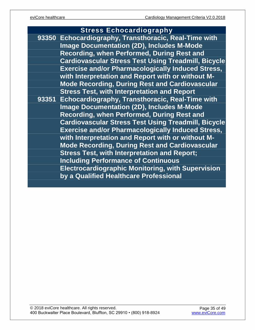
Stress Echocardiography 93350 Echocardiography, Transthoracic, Real-Time with
Image Documentation (2D), Includes M-Mode Recording, when Performed, During Rest and Cardiovascular Stress Test Using Treadmill, Bicycle Exercise and/or Pharmacologically Induced Stress, with Interpretation and Report with or without M-Mode Recording, During Rest and Cardiovascular Stress Test, with Interpretation and Report
93351 Echocardiography, Transthoracic, Real-Time with Image Documentation (2D), Includes M-Mode Recording, when Performed, During Rest and Cardiovascular Stress Test Using Treadmill, Bicycle Exercise and/or Pharmacologically Induced Stress, with Interpretation and Report with or without M-Mode Recording, During Rest and Cardiovascular Stress Test, with Interpretation and Report; Including Performance of Continuous Electrocardiographic Monitoring, with Supervision by a Qualified Healthcare Professional
eviCore healthcare Cardiology Management Criteria V2.0.2018
© 2018 eviCore healthcare. All rights reserved. 400 Buckwalter Place Boulevard, Bluffton, SC 29910 • (800) 918-8924 www.eviCore.com
Page 35 of 49
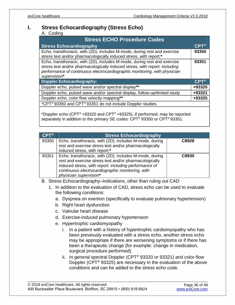
I. Stress Echocardiography (Stress Echo) A. Coding
Stress ECHO Procedure Codes Stress Echocardiography CPT® Echo, transthoracic, with (2D), includes M-mode, during rest and exercise stress test and/or pharmacologically induced stress, with report;*
93350
Echo, transthoracic, with (2D), includes M-mode, during rest and exercise stress test and/or pharmacologically induced stress, with report: including performance of continuous electrocardiographic monitoring, with physician supervision*
93351
Doppler Echocardiography: CPT® Doppler echo, pulsed wave and/or spectral display** +93320 Doppler echo, pulsed wave and/or spectral display, follow-up/limited study +93321 Doppler echo, color flow velocity mapping** +93325 *CPT® 93350 and CPT® 93351 do not include Doppler studies
*Doppler echo (CPT® +93320 and CPT® +93325), if performed, may be reportedseparately in addition to the primary SE codes: CPT® 93350 or CPT® 93351.
B. Stress Echocardiography–Indications, other than ruling out CAD 1. In addition to the evaluation of CAD, stress echo can be used to evaluate
the following conditions:a. Dyspnea on exertion (specifically to evaluate pulmonary hypertension)b. Right heart dysfunctionc. Valvular heart diseased. Exercise-induced pulmonary hypertensione. Hypertrophic cardiomyopathy
i. In a patient with a history of hypertrophic cardiomyopathy who hasbeen previously evaluated with a stress echo, another stress echomay be appropriate if there are worsening symptoms or if there hasbeen a therapeutic change (for example: change in medication,surgical procedure performed).
ii. In general spectral Doppler (CPT® 93320 or 93321) and color-flowDoppler (CPT® 93325) are necessary in the evaluation of the aboveconditions and can be added to the stress echo code.
CPT® Stress Echocardiography 93350 Echo, transthoracic, with (2D), includes M-mode, during
rest and exercise stress test and/or pharmacologically induced stress, with report;*
C8928
93351 Echo, transthoracic, with (2D), includes M-mode, during rest and exercise stress test and/or pharmacologically induced stress, with report: including performance of continuous electrocardiographic monitoring, with physician supervision*
C8930
eviCore healthcare Cardiology Management Criteria V2.0.2018
© 2018 eviCore healthcare. All rights reserved. 400 Buckwalter Place Boulevard, Bluffton, SC 29910 • (800) 918-8924 www.eviCore.com
Page 36 of 49
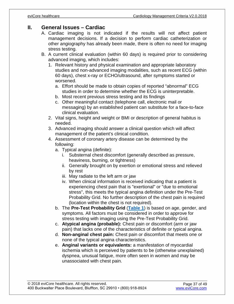
II. General Issues – CardiacA. Cardiac imaging is not indicated if the results will not affect patient
management decisions. If a decision to perform cardiac catheterization or other angiography has already been made, there is often no need for imaging stress testing.
B. A current clinical evaluation (within 60 days) is required prior to considering advanced imaging, which includes: 1. Relevant history and physical examination and appropriate laboratory
studies and non-advanced imaging modalities, such as recent ECG (within60 days), chest x-ray or ECHO/ultrasound, after symptoms started orworsened.a. Effort should be made to obtain copies of reported “abnormal” ECG
studies in order to determine whether the ECG is uninterpretable.b. Most recent previous stress testing and its findingsc. Other meaningful contact (telephone call, electronic mail or
messaging) by an established patient can substitute for a face-to-faceclinical evaluation.
2. Vital signs, height and weight or BMI or description of general habitus isneeded.
3. Advanced imaging should answer a clinical question which will affectmanagement of the patient’s clinical condition.
4. Assessment of coronary artery disease can be determined by thefollowing:a. Typical angina (definite):
i. Substernal chest discomfort (generally described as pressure,heaviness, burning, or tightness)
ii. Generally brought on by exertion or emotional stress and relievedby rest
iii. May radiate to the left arm or jawiv. When clinical information is received indicating that a patient is
experiencing chest pain that is "exertional" or "due to emotionalstress", this meets the typical angina definition under the Pre-TestProbability Grid. No further description of the chest pain is required(location within the chest is not required).
b. The Pre-Test Probability Grid (Table 1) is based on age, gender, andsymptoms. All factors must be considered in order to approve forstress testing with imaging using the Pre-Test Probability Grid.
c. Atypical angina (probable): Chest pain or discomfort (arm or jawpain) that lacks one of the characteristics of definite or typical angina.
d. Non-anginal chest pain: Chest pain or discomfort that meets one ornone of the typical angina characteristics.
e. Anginal variants or equivalents: a manifestation of myocardialischemia which is perceived by patients to be (otherwise unexplained)dyspnea, unusual fatigue, more often seen in women and may beunassociated with chest pain.
eviCore healthcare Cardiology Management Criteria V2.0.2018
© 2018 eviCore healthcare. All rights reserved. 400 Buckwalter Place Boulevard, Bluffton, SC 29910 • (800) 918-8924 www.eviCore.com
Page 37 of 49
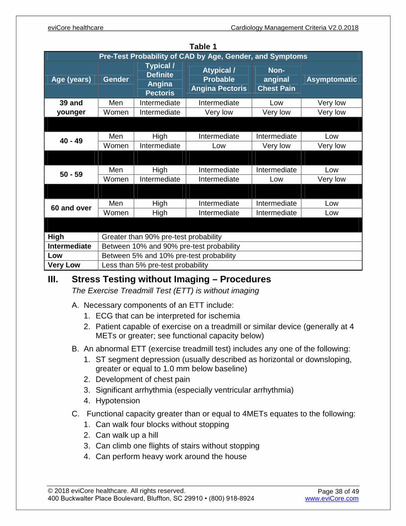
Table 1 Pre-Test Probability of CAD by Age, Gender, and Symptoms
Age (years) Gender
Typical / Definite Angina
Pectoris
Atypical / Probable
Angina Pectoris
Non-anginal
Chest Pain Asymptomatic
39 and younger
Men Intermediate Intermediate Low Very low Women Intermediate Very low Very low Very low
40 - 49 Men High Intermediate Intermediate Low Women Intermediate Low Very low Very low
50 - 59 Men High Intermediate Intermediate Low Women Intermediate Intermediate Low Very low
60 and over Men High Intermediate Intermediate Low Women High Intermediate Intermediate Low
High Greater than 90% pre-test probability Intermediate Between 10% and 90% pre-test probability Low Between 5% and 10% pre-test probability Very Low Less than 5% pre-test probability
III. Stress Testing without Imaging – ProceduresThe Exercise Treadmill Test (ETT) is without imaging
A. Necessary components of an ETT include:1. ECG that can be interpreted for ischemia2. Patient capable of exercise on a treadmill or similar device (generally at 4
METs or greater; see functional capacity below)B. An abnormal ETT (exercise treadmill test) includes any one of the following:
1. ST segment depression (usually described as horizontal or downsloping,greater or equal to 1.0 mm below baseline)
2. Development of chest pain3. Significant arrhythmia (especially ventricular arrhythmia)4. Hypotension
C. Functional capacity greater than or equal to 4METs equates to the following: 1. Can walk four blocks without stopping2. Can walk up a hill3. Can climb one flights of stairs without stopping4. Can perform heavy work around the house
eviCore healthcare Cardiology Management Criteria V2.0.2018
© 2018 eviCore healthcare. All rights reserved. 400 Buckwalter Place Boulevard, Bluffton, SC 29910 • (800) 918-8924 www.eviCore.com
Page 38 of 49
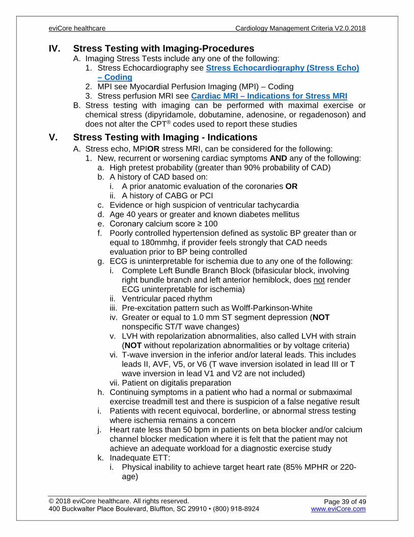
IV. Stress Testing with Imaging-ProceduresA. Imaging Stress Tests include any one of the following:
1. Stress Echocardiography see Stress Echocardiography (Stress Echo)– Coding
2. MPI see Myocardial Perfusion Imaging (MPI) – Coding3. Stress perfusion MRI see Cardiac MRI – Indications for Stress MRI
B. Stress testing with imaging can be performed with maximal exercise or chemical stress (dipyridamole, dobutamine, adenosine, or regadenoson) and does not alter the CPT® codes used to report these studies
V. Stress Testing with Imaging - Indications A. Stress echo, MPIOR stress MRI, can be considered for the following:
1. New, recurrent or worsening cardiac symptoms AND any of the following:a. High pretest probability (greater than 90% probability of CAD)b. A history of CAD based on:
i. A prior anatomic evaluation of the coronaries ORii. A history of CABG or PCI
c. Evidence or high suspicion of ventricular tachycardiad. Age 40 years or greater and known diabetes mellituse. Coronary calcium score ≥ 100f. Poorly controlled hypertension defined as systolic BP greater than or
equal to 180mmhg, if provider feels strongly that CAD needsevaluation prior to BP being controlled
g. ECG is uninterpretable for ischemia due to any one of the following:i. Complete Left Bundle Branch Block (bifasicular block, involving
right bundle branch and left anterior hemiblock, does not renderECG uninterpretable for ischemia)
ii. Ventricular paced rhythmiii. Pre-excitation pattern such as Wolff-Parkinson-Whiteiv. Greater or equal to 1.0 mm ST segment depression (NOT
nonspecific ST/T wave changes)v. LVH with repolarization abnormalities, also called LVH with strain
(NOT without repolarization abnormalities or by voltage criteria)vi. T-wave inversion in the inferior and/or lateral leads. This includes
leads II, AVF, V5, or V6 (T wave inversion isolated in lead III or Twave inversion in lead V1 and V2 are not included)
vii. Patient on digitalis preparationh. Continuing symptoms in a patient who had a normal or submaximal
exercise treadmill test and there is suspicion of a false negative resulti. Patients with recent equivocal, borderline, or abnormal stress testing
where ischemia remains a concernj. Heart rate less than 50 bpm in patients on beta blocker and/or calcium
channel blocker medication where it is felt that the patient may notachieve an adequate workload for a diagnostic exercise study
k. Inadequate ETT:i. Physical inability to achieve target heart rate (85% MPHR or 220-
age)
eviCore healthcare Cardiology Management Criteria V2.0.2018
© 2018 eviCore healthcare. All rights reserved. 400 Buckwalter Place Boulevard, Bluffton, SC 29910 • (800) 918-8924 www.eviCore.com
Page 39 of 49
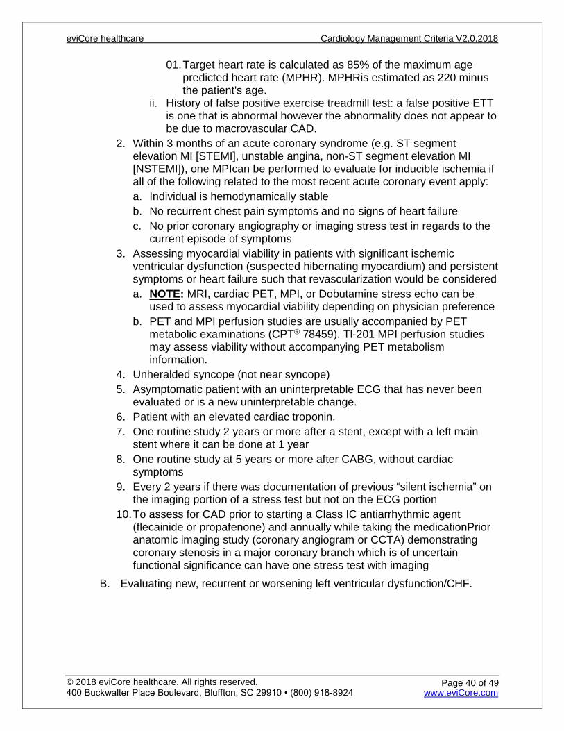
01. Target heart rate is calculated as 85% of the maximum agepredicted heart rate (MPHR). MPHRis estimated as 220 minusthe patient's age.
ii. History of false positive exercise treadmill test: a false positive ETTis one that is abnormal however the abnormality does not appear tobe due to macrovascular CAD.
2. Within 3 months of an acute coronary syndrome (e.g. ST segmentelevation MI [STEMI], unstable angina, non-ST segment elevation MI[NSTEMI]), one MPIcan be performed to evaluate for inducible ischemia ifall of the following related to the most recent acute coronary event apply:a. Individual is hemodynamically stableb. No recurrent chest pain symptoms and no signs of heart failurec. No prior coronary angiography or imaging stress test in regards to the
current episode of symptoms3. Assessing myocardial viability in patients with significant ischemic
ventricular dysfunction (suspected hibernating myocardium) and persistentsymptoms or heart failure such that revascularization would be considereda. NOTE: MRI, cardiac PET, MPI, or Dobutamine stress echo can be
used to assess myocardial viability depending on physician preferenceb. PET and MPI perfusion studies are usually accompanied by PET
metabolic examinations (CPT® 78459). Tl-201 MPI perfusion studiesmay assess viability without accompanying PET metabolisminformation.
4. Unheralded syncope (not near syncope)5. Asymptomatic patient with an uninterpretable ECG that has never been
evaluated or is a new uninterpretable change.6. Patient with an elevated cardiac troponin.7. One routine study 2 years or more after a stent, except with a left main
stent where it can be done at 1 year8. One routine study at 5 years or more after CABG, without cardiac
symptoms9. Every 2 years if there was documentation of previous “silent ischemia” on
the imaging portion of a stress test but not on the ECG portion10. To assess for CAD prior to starting a Class IC antiarrhythmic agent
(flecainide or propafenone) and annually while taking the medicationPrioranatomic imaging study (coronary angiogram or CCTA) demonstratingcoronary stenosis in a major coronary branch which is of uncertainfunctional significance can have one stress test with imaging
B. Evaluating new, recurrent or worsening left ventricular dysfunction/CHF.
eviCore healthcare Cardiology Management Criteria V2.0.2018
© 2018 eviCore healthcare. All rights reserved. 400 Buckwalter Place Boulevard, Bluffton, SC 29910 • (800) 918-8924 www.eviCore.com
Page 40 of 49
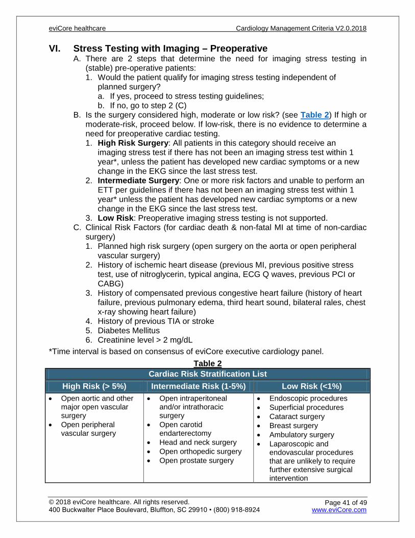
VI. Stress Testing with Imaging – PreoperativeA. There are 2 steps that determine the need for imaging stress testing in
(stable) pre-operative patients: 1. Would the patient qualify for imaging stress testing independent of
planned surgery?a. If yes, proceed to stress testing guidelines;b. If no, go to step 2 (C)
B. Is the surgery considered high, moderate or low risk? (see Table 2) If high or moderate-risk, proceed below. If low-risk, there is no evidence to determine a need for preoperative cardiac testing. 1. High Risk Surgery: All patients in this category should receive an
imaging stress test if there has not been an imaging stress test within 1year*, unless the patient has developed new cardiac symptoms or a newchange in the EKG since the last stress test.
2. Intermediate Surgery: One or more risk factors and unable to perform anETT per guidelines if there has not been an imaging stress test within 1year* unless the patient has developed new cardiac symptoms or a newchange in the EKG since the last stress test.
3. Low Risk: Preoperative imaging stress testing is not supported.C. Clinical Risk Factors (for cardiac death & non-fatal MI at time of non-cardiac
surgery) 1. Planned high risk surgery (open surgery on the aorta or open peripheral
vascular surgery)2. History of ischemic heart disease (previous MI, previous positive stress
test, use of nitroglycerin, typical angina, ECG Q waves, previous PCI orCABG)
3. History of compensated previous congestive heart failure (history of heartfailure, previous pulmonary edema, third heart sound, bilateral rales, chestx-ray showing heart failure)
4. History of previous TIA or stroke5. Diabetes Mellitus6. Creatinine level > 2 mg/dL
*Time interval is based on consensus of eviCore executive cardiology panel.Table 2
Cardiac Risk Stratification List High Risk (> 5%) Intermediate Risk (1-5%) Low Risk (<1%)
• Open aortic and othermajor open vascularsurgery
• Open peripheralvascular surgery
• Open intraperitonealand/or intrathoracicsurgery
• Open carotidendarterectomy
• Head and neck surgery• Open orthopedic surgery• Open prostate surgery
• Endoscopic procedures• Superficial procedures• Cataract surgery• Breast surgery• Ambulatory surgery• Laparoscopic and
endovascular proceduresthat are unlikely to requirefurther extensive surgicalintervention
eviCore healthcare Cardiology Management Criteria V2.0.2018
© 2018 eviCore healthcare. All rights reserved. 400 Buckwalter Place Boulevard, Bluffton, SC 29910 • (800) 918-8924 www.eviCore.com
Page 41 of 49

VII. Transplant PatientsA. Post-cardiac transplant assessment of transplant CAD:
1. One of the following imaging studies may be performed annuallya. MPIb. Stress Echocardiogramc. Stress MRId. Cardiac PET perfusion with coronary flow quantitation (CPT® 78491 or
CPT® 78492)B. Non-Cardiac Transplant Patients
1. Individuals who are awaiting an organ, bone marrow or stem celltransplant can undergo imaging stress testing every year (usually stressecho or MPI) prior to the transplant
2. Individuals who have undergone organ transplant are at increased risk forischemic heart disease secondary to their medication. An imaging stresstest can be repeated annually after transplant for at least two years orwithin one year of a prior cardiac imaging study if there is evidence ofprogressive vasculopathy
3. After two consecutive normal imaging stress tests, repeated testing issupported every two years unless there is evidence of progressivevasculopathy or new symptoms Stress testing after five years mayproceed according to normal patterns of consideration.
VIII. Non-imaging Heart Function and Cardiac Shunt ImagingA. Procedures reported with CPT® 78414 and CPT® 78428 are essentially
obsolete and should not be performed in lieu of other preferred modalities. B. Echocardiogram is the preferred method for cardiac shunt detection, rather
than the cardiac shunt imaging study described by CPT® 78428. C. Ejection fraction can be obtained by echocardiogram, MPI, MUGA study,
cardiac MRI, cardiac CT, or cardiac PET depending on the clinical situation, rather than by the non-imaging heart function study described by CPT® 78414.
IX. Genetic lab testing in the evaluation of CADA. Corus® CAD genetic expression score – refer to lab management program
guidelines
References
1. Bangalore S, Yao S, and Chaudhry F. Usefulness of stress echocardiography for risk stratification and prognosisof patients with left ventricular hypertrophy. American Journal of Cardiology 2007; 100: 536-543. Accessed onNovember 1, 2017. http://www.ajconline.org/article/S0002-9149(07)00852-1/fulltext.
2. Douglas P, Khandheria B, Stainback R, et al. ACCF/ASE/ACEP/AHA/ASNC/SCAI/SCCT/SCMR 2008appropriateness criteria for stress echocardiography: a report of the American College of Cardiology FoundationAppropriateness Criteria Task Force, American Society of Echocardiography, American College of EmergencyPhysicians, American Heart Association, American Society of Nuclear Cardiology, Society for CardiovascularAngiography and Interventions, Society of Cardiovascular Computed Tomography, and Society forCardiovascular Magnetic Resonance endorsed by the Heart Rhythm Society and the Society of Critical CareMedicine. Journal of the American College of Cardiology. 2008; 51: 1127-1147. Accessed on November 1, 2017.
eviCore healthcare Cardiology Management Criteria V2.0.2018
© 2018 eviCore healthcare. All rights reserved. 400 Buckwalter Place Boulevard, Bluffton, SC 29910 • (800) 918-8924 www.eviCore.com
Page 42 of 49

http://www.sciencedirect.com/science/article/pii/S0735109707039629?via%3Dihub. 3. Maron B, McKenna W, Danielson G, et al. American College of Cardiology/European Society of Cardiology
Clinical Expert Consensus Document on Hypertrophic Cardiomyopathy: A report of the American College ofCardiology Foundation Task Force on Clinical Expert Consensus Documents and the European Society ofCardiology Committee for Practice Guidelines. European Heart Journal 2003; 24:1965-1991. Accessed onNovember 1, 2017.https://academic.oup.com/eurheartj/article-lookup/doi/10.1016/S0195-668X(03)00479-2.
4. Metz LD, Beattie M, Hom R, et al. The prognostic value of normal exercise myocardial perfusion imaging andexercise echocardiography: A meta-analysis. J Am CollCardiol 2007;49 (2): 227-237. Accessed November 30,2017. http://www.sciencedirect.com/science/article/pii/S073510970602506X?via%3Dihub.
5. Pellikka P, Nagueh S, Elhendy A, et al. American Society of Echocardiography recommendations forperformance, interpretation, and application of stress echocardiography. Journal of the American Society ofEchocardiography 2007; 20 (9):1021-1041. Accessed on November 1, 2017.http://www.asecho.org/wordpress/wp-content/uploads/2013/05/Performance-Interpretation-and-App-of-Stress-Echo.pdf.
6. Tandogan I, Yetkin E, Yanik A, et al. Comparison of thallium-201 exercise SPECT and dobutamine stressechocardiography for diagnosis of coronary artery disease in patients with left bundle branch block. InternationalJournal of Cardiovascular Imaging 2001; 17:339-345 Accessed November 30, 2017.https://link.springer.com/article/10.1023/A:1011973530231.
7. Nishimura RA, Otto CM, Bonow RO, et al. 2014 AHA/ACC guideline for the management of patients with valvularheart disease: a report of the American College of Cardiology/American Heart Association Task Force onPractice Guidelines. J Am CollCardiol 2014; 63:e57. Accessed November 30, 2017.http://circ.ahajournals.org/content/129/23/2440.long.
8. Rudski L, Lai W, Afilalo J, et al. Guidelines for the echocardiographic assessment of the right heart in adults: areport from the American Society of Echocardiography endorsed by the European Association ofEchocardiography, a registered branch of the European Society of Cardiology, and the Canadian Society ofEchocardiography. Journal of the American Society of Echocardiography 2010; 23:685. Accessed on November1, 2017. http://www.onlinejase.com/article/S0894-7317(10)00434-7/fulltext.
9. Holmes DR Jr, Mack MJ, Kaul S, et al. 2012 ACCF/AATS/SCAI/STS expert consensus document ontranscatheter aortic valve replacement: developed in collaboration with the American Heart Association,American Society of Echocardiography, European Association for Cardio-Thoracic Surgery, Heart FailureSociety of America, Mended Hearts, Society of Cardiovascular Anesthesiologists, Society of CardiovascularComputed Tomography, and Society for Cardiovascular Magnetic Resonance. Journal of the American Collegeof Cardiology. 2012; 59:1200. Accessed on November 1, 2017.http://www.annalsthoracicsurgery.org/article/S0003-4975(12)00196-8/fulltext.
10. Zoghbi WA, Chambers JB, Dumesnil JG, et al. Recommendations for evaluation of prosthetic valves withechocardiography and doppler ultrasound: a report From the American Society of Echocardiography's Guidelinesand Standards Committee and the Task Force on Prosthetic Valves, developed in conjunction with the AmericanCollege of Cardiology Cardiovascular Imaging Committee, Cardiac Imaging Committee of the American HeartAssociation, the European Association of Echocardiography, a registered branch of the European Society ofCardiology, the Japanese Society of Echocardiography and the Canadian Society of Echocardiography,endorsed by the American College of Cardiology Foundation, American Heart Association, European Associationof Echocardiography, a registered branch of the European Society of Cardiology, the Japanese Society ofEchocardiography, and Canadian Society of Echocardiography. J Am SocEchocardiogr 2009; 22:975. AccessedNovember 30, 2017. http://www.onlinejase.com/article/S0894-7317(09)00676-2/abstract.
11. WolkMJ, Bailey SR, Doherty JU, et al. ACCF/AHA/ASE/ASNC/HFSA/HRS/SCAI/SCCT/SCMR/STS 2013multimodality appropriate use criteria for the detection and risk assessment of stable ischemic heart disease: areport of the American College of Cardiology Foundation Appropriate Use Criteria Task Force, American HeartAssociation, American Society of Echocardiography, American Society of Nuclear Cardiology, Heart FailureSociety of America, Heart Rhythm Society, Society for Cardiovascular Angiography and Interventions, Society ofCardiovascular Computed Tomography, Society for Cardiovascular Magnetic Resonance, and Society ofThoracic Surgeons. J Am CollCardiol 2014; 63:380. Accessed November 30, 2017.http://www.sciencedirect.com/science/article/pii/S0735109713061470?via%3Dihub.
12. Doherty JU, Kort S, Mehran R, et al ACC/AATS/AHA/ASE/ASNC/HRS/SCAI/SCCT/SCMR/STS 2017Appropriate Use Criteria for Multimodality Imaging in Valvular Heart Disease. A Report of the American Collegeof Cardiology Appropriate Use Criteria Task Force, American Association for Thoracic Surgery, American HeartAssociation, American Society of Echocardiography, American Society of Nuclear Cardiology, Heart RhythmSociety, Society for Cardiovascular Angiography and Interventions, Society of Cardiovascular ComputedTomography, Society for Cardiovascular Magnetic Resonance, and Society of Thoracic Surgeons. Journal of theAmerican College of Cardiology. September 2017; 70(13). Accessed on November 1, 2017.http://www.onlinejacc.org/content/70/13/1647. 93350, 93351 Echocardiography
eviCore healthcare Cardiology Management Criteria V2.0.2018
© 2018 eviCore healthcare. All rights reserved. 400 Buckwalter Place Boulevard, Bluffton, SC 29910 • (800) 918-8924 www.eviCore.com
Page 43 of 49
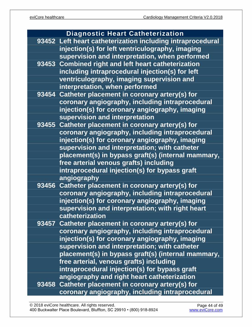
Diagnostic Heart Catheterization 93452 Left heart catheterization including intraprocedural
injection(s) for left ventriculography, imaging supervision and interpretation, when performed
93453 Combined right and left heart catheterization including intraprocedural injection(s) for left ventriculography, imaging supervision and interpretation, when performed
93454 Catheter placement in coronary artery(s) for coronary angiography, including intraprocedural injection(s) for coronary angiography, imaging supervision and interpretation
93455 Catheter placement in coronary artery(s) for coronary angiography, including intraprocedural injection(s) for coronary angiography, imaging supervision and interpretation; with catheter placement(s) in bypass graft(s) (internal mammary, free arterial venous grafts) including intraprocedural injection(s) for bypass graft angiography
93456 Catheter placement in coronary artery(s) for coronary angiography, including intraprocedural injection(s) for coronary angiography, imaging supervision and interpretation; with right heart catheterization
93457 Catheter placement in coronary artery(s) for coronary angiography, including intraprocedural injection(s) for coronary angiography, imaging supervision and interpretation; with catheter placement(s) in bypass graft(s) (internal mammary, free arterial, venous grafts) including intraprocedural injection(s) for bypass graft angiography and right heart catheterization
93458 Catheter placement in coronary artery(s) for coronary angiography, including intraprocedural
eviCore healthcare Cardiology Management Criteria V2.0.2018
© 2018 eviCore healthcare. All rights reserved. 400 Buckwalter Place Boulevard, Bluffton, SC 29910 • (800) 918-8924 www.eviCore.com
Page 44 of 49
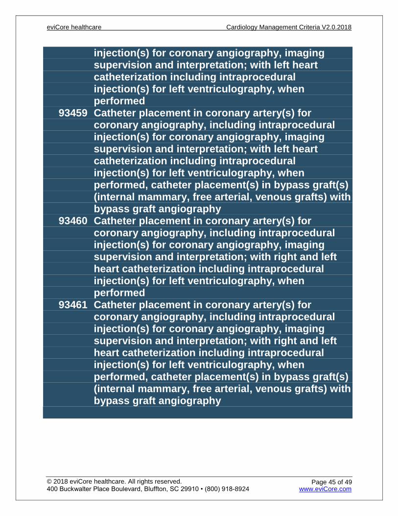
injection(s) for coronary angiography, imaging supervision and interpretation; with left heart catheterization including intraprocedural injection(s) for left ventriculography, when performed
93459 Catheter placement in coronary artery(s) for coronary angiography, including intraprocedural injection(s) for coronary angiography, imaging supervision and interpretation; with left heart catheterization including intraprocedural injection(s) for left ventriculography, when performed, catheter placement(s) in bypass graft(s) (internal mammary, free arterial, venous grafts) with bypass graft angiography
93460 Catheter placement in coronary artery(s) for coronary angiography, including intraprocedural injection(s) for coronary angiography, imaging supervision and interpretation; with right and left heart catheterization including intraprocedural injection(s) for left ventriculography, when performed
93461 Catheter placement in coronary artery(s) for coronary angiography, including intraprocedural injection(s) for coronary angiography, imaging supervision and interpretation; with right and left heart catheterization including intraprocedural injection(s) for left ventriculography, when performed, catheter placement(s) in bypass graft(s) (internal mammary, free arterial, venous grafts) with bypass graft angiography
eviCore healthcare Cardiology Management Criteria V2.0.2018
© 2018 eviCore healthcare. All rights reserved. 400 Buckwalter Place Boulevard, Bluffton, SC 29910 • (800) 918-8924 www.eviCore.com
Page 45 of 49
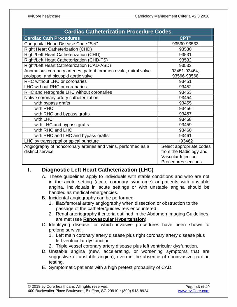
Cardiac Catheterization Procedure Codes Cardiac Cath Procedures CPT® Congenital Heart Disease Code “Set” 93530-93533 Right Heart Catheterization (CHD) 93530 Right/Left Heart Catheterization (CHD) 93531 Right/Left Heart Catheterization (CHD-TS) 93532 Right/Left Heart Catheterization (CAD-ASD) 93533 Anomalous coronary arteries, patent foramen ovale, mitral valve prolapse, and bicuspid aortic valve
93451-93464, 93566-93568
RHC without LHC or coronaries 93451 LHC without RHC or coronaries 93452 RHC and retrograde LHC without coronaries 93453 Native coronary artery catheterization; 93454
with bypass grafts 93455 with RHC 93456 with RHC and bypass grafts 93457 with LHC 93458 with LHC and bypass grafts 93459 with RHC and LHC 93460 with RHC and LHC and bypass grafts 93461
LHC by transseptal or apical puncture +93462 Angiography of noncoronary arteries and veins, performed as a distinct service
Select appropriate codes from the Radiology and Vascular Injection Procedures sections.
I. Diagnostic Left Heart Catheterization (LHC) A. These guidelines apply to individuals with stable conditions and who are not
in the acute setting (acute coronary syndrome) or patients with unstable angina. Individuals in acute settings or with unstable angina should be handled as medical emergencies.
B. Incidental angiography can be performed: 1. Iliac/femoral artery angiography when dissection or obstruction to the
passage of the catheter/guidewireis encountered.2. Renal arteriography if criteria outlined in the Abdomen Imaging Guidelines
are met (see Renovascular Hypertension).C. Identifying disease for which invasive procedures have been shown to
prolong survival: 1. Left main coronary artery disease plus right coronary artery disease plus
left ventricular dysfunction.2. Triple vessel coronary artery disease plus left ventricular dysfunction.
D. Unstable angina (new, accelerating, or worsening symptoms that are suggestive of unstable angina), even in the absence of noninvasive cardiac testing.
E. Symptomatic patients with a high pretest probability of CAD.
eviCore healthcare Cardiology Management Criteria V2.0.2018
© 2018 eviCore healthcare. All rights reserved. 400 Buckwalter Place Boulevard, Bluffton, SC 29910 • (800) 918-8924 www.eviCore.com
Page 46 of 49
F. Angina that is unresponsive to optimized medical therapy (see General Issues – Cardiac) and for which invasive procedures are needed to provide pain relief.
G. Left ventricular dysfunction (congestive heart failure) in patients suspected of having coronary artery disease.
H. Ventricular fibrillation or ventricular tachycardia where the etiology is unclear. I. Unheralded syncope (not near syncope). J. Recent noninvasive cardiac testing was equivocal, unsuccessful in
delineating the clinical problem, or led to a conclusion that intervention is indicated for the following conditions: 1. Suspicion of cardiomyopathy, endocarditis, or myocarditis2. Significant/serious ventricular arrhythmia3. Evaluating progression of known CAD when symptoms are persistent or
worsening4. An intermediate or large amount of myocardium (>5%) may be in jeopardy5. Evaluation of coronary grafts6. Evaluation of previously placed coronary artery stents7. Evaluation of structural disease
K. Ruling out coronary artery disease prior to planned non-coronary cardiac or great vessel surgery (i.e. cardiac valve surgery, aortic dissection, aortic aneurysm, congenital disease repair such as atrial septal defect, etc.).
L. Assessment for accelerated coronary artery disease associated with cardiac transplantation.
M. Pre-organ transplant (non-cardiac). Some institutions perform a heart cath as part of their initial evaluation protocol. Others use an imaging stress test for evaluation. Either is appropriate and can be approved but NOT both.
N. Valvular heart disease when there is a discrepancy between the clinical findings (history, physical exam, and non-invasive test results) or valvular surgery is being considered.
O. Suspected pericardial disease.
II. Right Heart Catheterization (RHC)A. General Information RHC
1. It is performed most commonly from the femoral vein, less often throughthe subclavian or internal jugular veins and interatrial septal punctureapproach.
2. It includes a full oximetry for detection and quantification of shunts.3. Pressure measurements are made and are done simultaneously with
aortic and left ventricular pressures.4. Cardiac outputs are calculated by several techniques including
thermodilution.B. Diagnostic Right Heart Catheterization – Indications
1. Atrial septal defect (ASD) including shunt detection and quantification2. Ventricular septal defect (VSD) including shunt detection and
quantification3. Patent foramen ovale (PFO)4. Anomalous pulmonary venous return
eviCore healthcare Cardiology Management Criteria V2.0.2018
© 2018 eviCore healthcare. All rights reserved. 400 Buckwalter Place Boulevard, Bluffton, SC 29910 • (800) 918-8924 www.eviCore.com
Page 47 of 49
5. Congenital defects including persistent left vena cava6. Pulmonary hypertension7. Pericardial diseases (constrictive or restrictive pericarditis)8. Valvular disease9. Right heart failure10. Left heart failure11. Preoperative evaluation for valve surgery12. Newly diagnosed or worsening cardiomyopathy13. During a left heart cath where the etiology of the symptoms remains
unclear.14. Pre-lung transplant to assess pulmonary pressures15. Uncertain intravascular volume status with an unclear etiology16. Assessment post cardiac transplant
a. For routine endomyocardial biopsyb. Assess for rejectionc. Assess pulmonary artery pressured. Can be done per the institution protocol or anytime organ rejection is
suspected and biopsy is needed for assessment17. Evaluation of right ventricular morphology18. Suspected arrhythmogenic right ventricular dysplasia
III. Combined Right and Left Heart Catheterization IndicationsA. Preoperative evaluation for valve surgeryB. Newly diagnosed or worsening cardiomyopathyC. If the major component of the patient symptoms is dyspnea, and the
indications for Diagnostic Left Heart Catheterization are also met D. If indications are met according to Diagnostic Left Heart Catheterization
(LHC) and Right Heart Catheterization, then a combination heart catheterization may be appropriate.
IV. Planned (Staged) Coronary InterventionsA. The CPT® codes for percutaneous coronary interventions (PCI) include the
following imaging services necessary for the procedure(s): 1. Contrast injection, angiography, ‘roadmapping’, and fluoroscopic guidance2. Vessel measurement3. Angiography following coronary angioplasty, stent placement, and
atherectomyB. Separate codes for these services should not be assigned in addition to the
PCI code/s because the services are already included. C. A repeat diagnostic left heart catheterization is not medically necessary when
the patient is undergoing a planned staged percutaneous coronary intervention.
D. CPT® 93530 to 93533 are appropriate for invasive evaluation of congenital heart disease.
eviCore healthcare Cardiology Management Criteria V2.0.2018
© 2018 eviCore healthcare. All rights reserved. 400 Buckwalter Place Boulevard, Bluffton, SC 29910 • (800) 918-8924 www.eviCore.com
Page 48 of 49

References
1. Boden WE, O’Rourke RA, TeoKK, et al. Impact of optimal medical therapy with or without percutaneous coronaryintervention on long-term cardiovascular end points in patients with stable coronary artery disease (from theCOURAGE trial). Am J Cardiol2009 July; 104(1):1-4. Accessed November 30, 2017.http://www.nejm.org/doi/full/10.1056/NEJMoa070829#t=article.
2. Friedewald V, King S, Pepine C, et.al. The Editor’s Roundtable: Chronic stable angina pectoris. AmericanJournal of Cardiology2007Dec, 100(11):1635-1643. Accessed on November 1, 2017.http://www.ajconline.org/article/S0002-9149(07)01706-7/fulltext.
3. Olade R, Cardiac Catheterization of Left Heart. Medscape Updated: Apr 13, 2016. Accessed November 1, 2017.https://emedicine.medscape.com/article/1819224-overview.
4. Amsterdam E, Wenger N, Brindis R, et al. 2014 AHA/ACC guideline for the management of patients with non-ST-elevation acute coronary syndromes: executive summary: a report of the American College ofCardiology/American Heart Association Task Force on Practice Guidelines. Society for CardiovascularAngiography and Interventions and the Society of Thoracic Surgeons Circulation. 2014; Accessed November 1,2017. http://circ.ahajournals.org/content/130/25/2354
93452, 93453, 93454, 93455, 93456, 93457, 93458, 93459, 93460, 93461: Diagnostic Hearth Catheterization
eviCore healthcare Cardiology Management Criteria V2.0.2018
© 2018 eviCore healthcare. All rights reserved. 400 Buckwalter Place Boulevard, Bluffton, SC 29910 • (800) 918-8924 www.eviCore.com
Page 49 of 49
Recommended