DEVELOPMENTAL STAGES OF EMBRYO
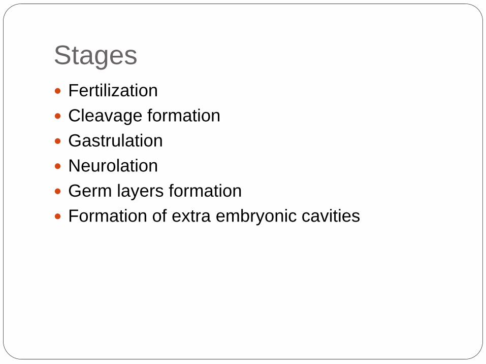
Stages
Fertilization
Cleavage formation
Gastrulation
Neurolation
Germ layers formation
Formation of extra embryonic cavities
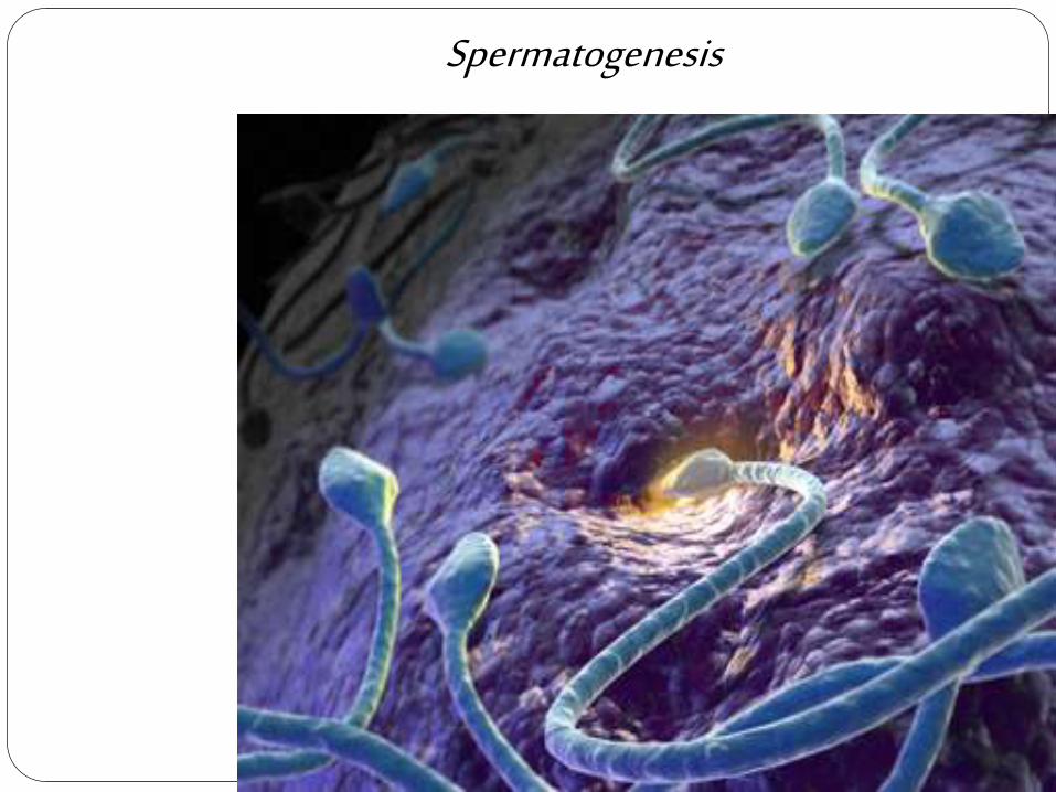
Spermatogenesis
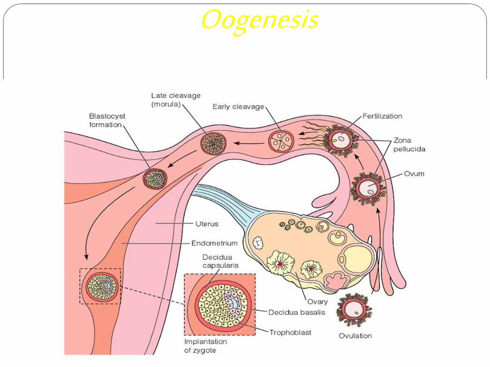
Oogenesis

FertilizationFertilization is
the process of
fusion of the
spermatozoon
with the mature
ovum.

Embryo
Stage of human
development
occurring between
the ovum and the
fetal stages, or from
2-8 weeks after
conception.

PREEMBRYONIC PERIOD (FIRST TO THIRD WEEK)
First week of life:
Day 1: Fertilization

Day 2 and 3: Cleavage
The original zygote divides about
30 hours after conception into two
daughter cells called
blastomeres.
Continued subdivisions of the
original cell result in increasing
numbers of blastomeres.
During cell division the dividing
cells decrease in size. This type
of cell division is called cleavage.
By the time the zygote is ready to
enter the uterus, it contains a
solid ball of 12 to 16 blastomeres
called the morula (from the latin
word for mulberry).

Day 4: Formation of the blastocyst
Fluid within the intercellular spaces
of the morula gradually increases,
and spaces on one side of the inner
cell mass come together, forming a
single cavity, the blastocele.
The outer layer organizes into the
trophoblasts, which give rise to the
placenta, and the inner layer of cells
form the embryo.
The cavity of the blastocele fills with
fluid, and the conceptus is now called
the blastocyst.

Preparation of the endometrium
Resting phase
Proliferative phase
Secretory phase

Attachment of the blastocyst The blastocyst attaches to the
uterine lining in the V-shaped.
When the trophoblast (the outer
cell layer) attaches to the
endometrium, it proliferates and
separates into an inner
cytotrophoblastic layer (fetal side)
and an outer syncytiotrophoblastic
(placental side).
The outer layer develops finger like
projections that proliferate and
superficially attach the blastocyst
to the endometrium within 6 days
after conception.

MAJOR EVENTS OF FIRST WEEK
NORMAL EVENTS POSSIBLE ABNORMAL
EVENTS
Fertilization and
formation of the
zygote (30hours).
Cleavage of the
zygote into 12 to 16
blastomeres- the
morula (day 2 and 3).
Formation of the
blastocyst ( day 5-8).
Abnormal
implantation
Maternal infection or
a genetic defect
Hydatidiform mole
Abortion
Ectopic implantation

Second week of life A slitlike amniotic cavity appears about
day 8, and the yolk sac appears as a
second cavity on day 12. Bilaminar
embryonic disc is formed in between
these two layers.
The endodermal disc becomes thicker
at it’s cephalad end, forming the
prochordal plate.
During early development of the
nervous system, the function of the
prochordal plate is to indicate the site of
the mouth and to form the membranes
of the mouth and throat.
The formation of the decidua, fetal
membranes, and placenta extends
beyond the second week, but their
development begins at this point.

THIRD WEEK OF LIFE
During the third week of life, the conceptus
develops rapidly. This period also coincides with
the first missed menstrual cycle of the mother.
The primitive streak is formed during the third
week, and three germ layers develop.
This periods from approximately day 15 to day
21, is called the “period of threes”; not only do
the three germ layers develop, but the primitive
streak, the notochord, and the neural tube are
formed.

GASTRULATION
Gastrulation is the process by
which the bilaminar embryo
becomes a trilaminar embryo.
On about day 15, the
cytotrophoblast cells proliferate
into the blastocyst to form the
extraembryonic mesoderm,
which later become the extra-
embryonic coelom.
The mesoderm lies between the
ectoderm and the endoderm,
completing the trilaminar disc of
the primitive streak. All tissues
and organs of the embryo are
developed from these three
layers.

Major derivatives of the embryonic germ layers
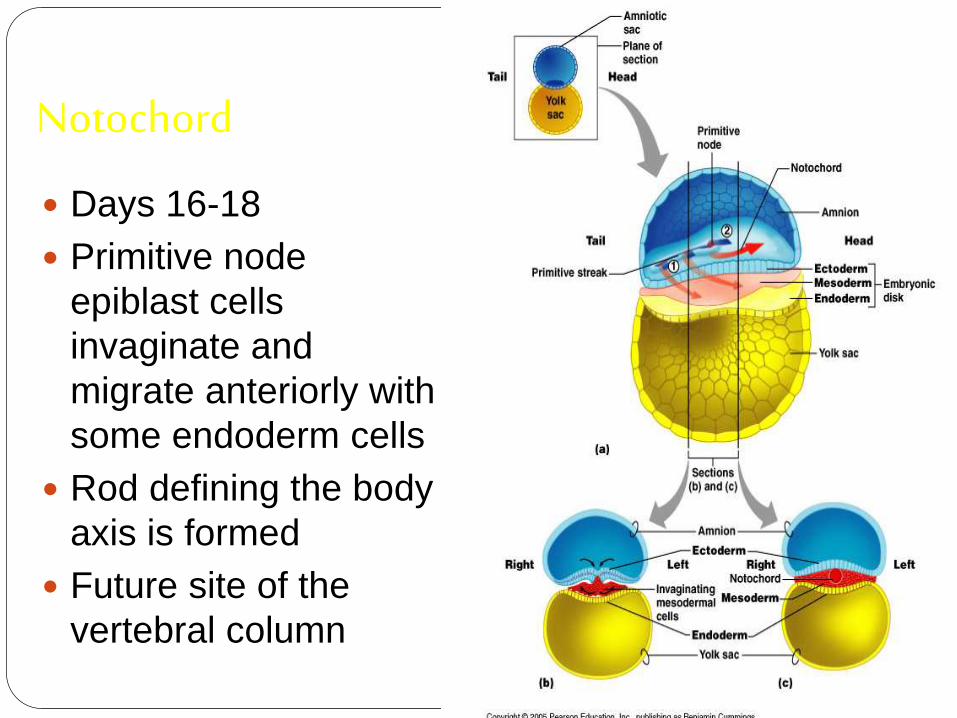
Notochord
Days 16-18
Primitive node
epiblast cells
invaginate and
migrate anteriorly with
some endoderm cells
Rod defining the body
axis is formed
Future site of the
vertebral column

Neurulation
Notochord signals overlying ectoderm
The neural tube is developed from the closure of the
neural plate and the neural fold- a process called
neurulation –at about 21 to 26 days.

Closure of neural tube: begins at end of week 3;
complete by end of week 4.
Extends cranially (eventually brain) and caudally
(spinal cord)
Neural crest, lateral ectodermal cells, pulled along and
form sensory nerve cells and other structures

DIVISON INTO SOMITES
About day 20, the mesoderm divides into paired bodies
called somites.
Located on either side of the developing neural tube, these
paired bodies give rise to the skeleton and muscle tissue.
During the somite period, day 20 to 30, 38 pairs of somites
develop.
Their total number eventually reaches 42 to 44 pairs, of
which 4 are occipital, 8 cervical, 12 thoracic, 5 lumbar, 5
sacral and 8 to 10 coccygeal.
Some of the somites – first occipital and the fifth to seventh
coccygeal –disappear, while the rest form the axial
skeleton.
By the end of the third week of life, the conceptus is about
1.0 mm in length.
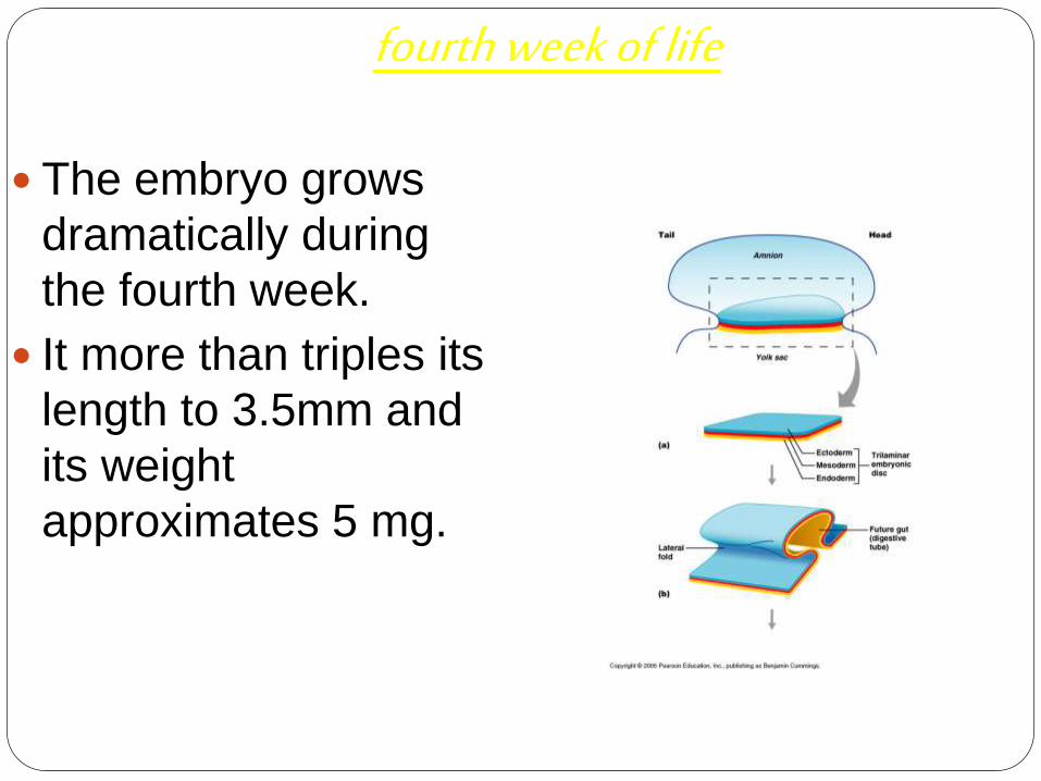
fourth week of life
The embryo grows
dramatically during
the fourth week.
It more than triples its
length to 3.5mm and
its weight
approximates 5 mg.

Elongation of the embryo has
occurred, and it has become
curved pon itself with the formation
of a head and tail fold.
Lateral body folds develop making
the embryo tubular rather than flat
and disc shaped.
Closure of the neural tube begins
in the area of the occiput and
proceeds upward and downward
from that point.
Somites formed in a craniocaudal
sequence as the neural tube
closes, can be observed through
the ectoderm.

The pericardial sac
around the heart
enlarges, causing the
head region to elevate.
The larygotracheal
groove and lung buds,
which will become the
respiratory system, are
present.
The mandible and
maxilla of the jaw
become distinct, and
rudimentary forms of the
eyes, ears and nose are

The intestinal system is
formed from the yolk sac,
and differentiation of the
buds, which will become the
oesophagus, stomach, liver
and pancreas, is progressing.
The thyroid and thymus
glands are also developing.
The primitive circulatory
system is established, and
the heart is beating.
The budlike projections on
the surface of the embryo are
the beginning of the limbs.

FIFTH WEEKS OF LIFE
As the embryo and then the fetus grow during the
first half of pregnancy, it is measured by its crown –
rump length (CRL).
The fetus is measured during the last half of
pregnancy by its crown heel length (CHL), or
standing height. The CRL grows from 4 to 8 mm in
this week, and exceeds the growth.
The growth of the head is rapid and exceeds the
growth of the body during this week. The embryo
lengthens and bends into a C shape, while an
additional 42 to 44 pairs of somites are added to its
caudal end.


The umbilical cord is
formed from the union of
the amnion, the yolk, and
the connecting stalk. It now
contains two umbilical
arteries and one umbilical
vein.
The doubling of the size of
the heart makes it
prominent, and its atria and
ventricles are visible
through the ectoderm. The
embryo’s four limb buds
are most vulnerable to
teratogens at this time.
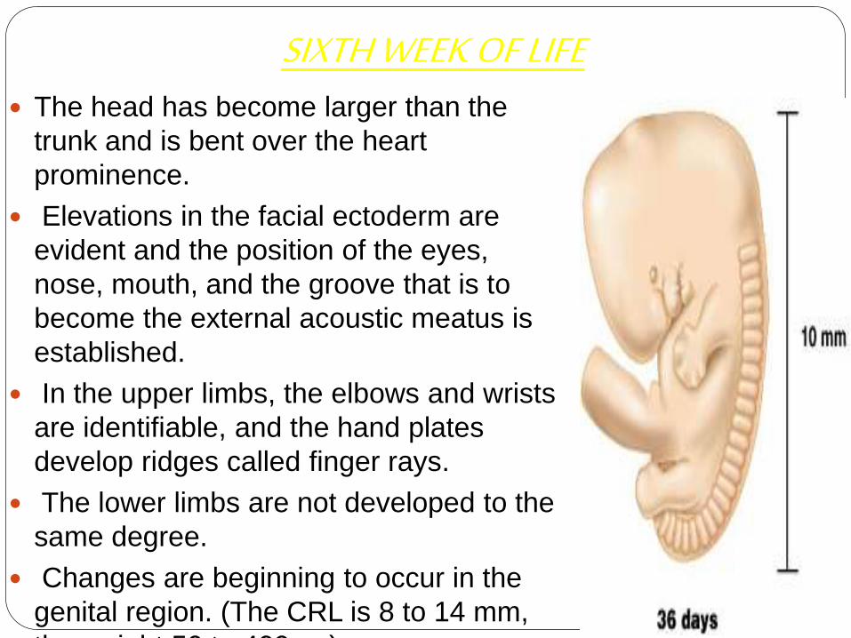
SIXTH WEEK OF LIFE The head has become larger than the
trunk and is bent over the heart
prominence.
Elevations in the facial ectoderm are
evident and the position of the eyes,
nose, mouth, and the groove that is to
become the external acoustic meatus is
established.
In the upper limbs, the elbows and wrists
are identifiable, and the hand plates
develop ridges called finger rays.
The lower limbs are not developed to the
same degree.
Changes are beginning to occur in the
genital region. (The CRL is 8 to 14 mm,
the weight 50 to 400mg).

SEVENTH WEEK OF LIFE
Cerebral hemispheres appear as the head enlarges
rapidly.
The eyes move from a lateral to a more frontal
position as the face elongates.
Prominences appear over the ventral body wall from
early because their function is vital to the maintenance
and survival of the embryo.
As the embryo continues to grow, the umbilical cord
shrinks.
The arm and hand of the upper limbs and the thigh,
leg, and foot segments of the lower limbs become
apparent.
The fingers develop, the their growth is critical at this
point (40 to 50 days).

EIGHTH WEEK OF LIFE
During this final week of the
embryonic period, the embryo
exhibits definite human
characteristics.
The cerebral hemispheres have
grown so rapidly that the head now
makes up 50% of the mass of the
embryo.
The face occupies the lower half of
the head, and the eyes continue to
move to a more frontal plane.
Eyelids folds develop. These will
become fused during the ninth week
and remain so until the seventh

The fingers lengthen, and the toes
are distinct by the end of the
eighth week.
The external ears are set low and
are taking on their final shape.
Sexual differences in the external
genitalia can now be seen by the
trained eye.
(The CRL is 21 to 30mm; the
weight 1000 to 3000 mg).

MAJOR EVENTS OF
fourth to eighth WEEK
NORMAL EVENTS POSSIBLE ABNORMAL
EVENTS
Conversion of the flat trilaminar embryonic
disc into a c- shaped cylindrical embryo.
Formation of the head, tail, and lateral folds.
Formation of the lateral and ventral body
walls.
Acquisition of an epithelial covering by the
umbilicus through the expansion of the
amnion.
Establishment of ventral position of the heart
and development of the brain in the cranial
region of the embryo.
Differentiation of the three germ layers into
various tissues and layers that will become
established as the major organ systems.
Appearance of the brain, limbs, ears, eyes,
Abnormalities of
the genes and
chromosomes.
Alterations of
maternal health,
such as infection
from rubella or
herpes.
Ingestion of
teratogenic
substances.
The risk of mortality
is greater than at
any other time of
life.

Anyquestion?

THANKYOU
Recommended