6/10/2015
1
Rutgers, The State University of New Jersey
DIFFICULT TO DEAL COLON POLYPS
SUSHIL AHLAWAT, MD, FACP, FASGE
Associate Professor of Medicine
Director of Endoscopy
Rutgers NJMS
Difficult Colon Polyp
Disclosures
• I do not have any relevant financial relationships with any commercial interests.
Difficult Colon Polyp
Colonoscopic Polypectomy
• The role of colonoscopic polypectomy in the prevention
of colorectal cancer is now well-established
• Resection of adenomatous colon polyps reduces
colorectal cancer incidence & mortality
• Role of endoscopic resection has expanded
• Only polyps with overt evidence of cancer or submucosal invasion should not be resected via colonoscopy otherwise all polyps are amenable to endoscopic resection
NEJM 2012;366:924
Difficult Colon Polyp
Malignant Potential of Polyp• Visual impression
– Ulcerations, friability, induration, failure to rise with sub-mucosal injection
• Biopsy– Sampling error
• Size of the polyps– Incidence of invasive
cancer is 10% in endoscopically resected polyps 2 cm or > that met visual criteria of being benign
Ahlawat S, et al. J Clin Gastroenterol 2011;45:347
6/10/2015
2
Difficult Colon Polyp
“Difficult” or “Defiant” Polyp
• No “impossible” polyp
• Polyp factors: – location, size, morphology, configuration
• Endoscopist factors: – experience, level of training, familiar/availability of
ancillary devices for complex polypectomy
• Patient factors:– comorbid condition may affect recovery from
complication
– Expectations: may not be ready to experience significant complication
Difficult Colon Polyp
Polyp Factors
• Size – Size alone can cause some hesitation
• Morphology– Flat or slightly elevated above mucosal surface
• Location or configuration– Located on the wall of colon that is not accessible to the snare
– Polyp in a segment of severe diverticular disease
– Polyp wrapped around a fold in a clam-shell fashion
– Polyp located behind a fold – difficult to approach
– Polyp on or behind the IC valve
– Located on appendiceal orifice
• Bleeding risk– Stalk >5 mm, piecemeal resection
Difficult Colon Polyp
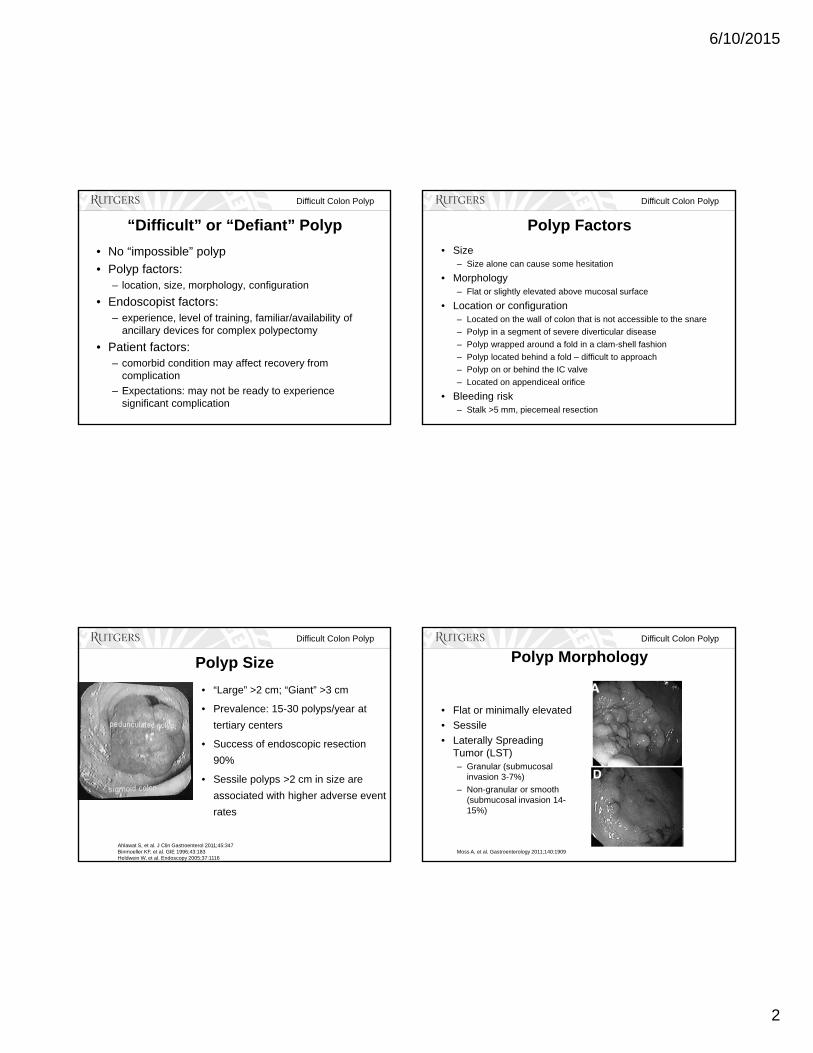
Polyp Size
• “Large” >2 cm; “Giant” >3 cm
• Prevalence: 15-30 polyps/year at
tertiary centers
• Success of endoscopic resection
90%
• Sessile polyps >2 cm in size are
associated with higher adverse event
rates
Ahlawat S, et al. J Clin Gastroenterol 2011;45:347Binmoeller KF, et al. GIE 1996;43:183Heldwein W, et al. Endoscopy 2005;37:1116
Difficult Colon Polyp
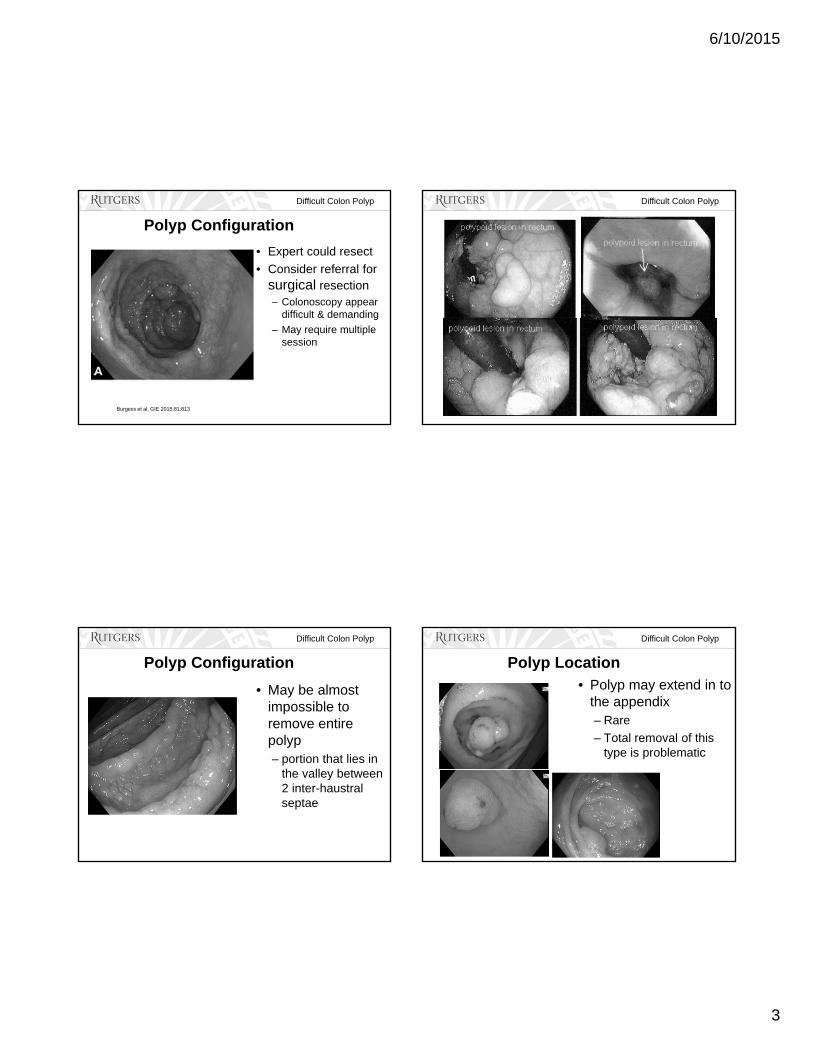
Polyp Morphology
• Flat or minimally elevated
• Sessile
• Laterally Spreading Tumor (LST)– Granular (submucosal
invasion 3-7%)
– Non-granular or smooth (submucosal invasion 14-15%)
Moss A, et al. Gastroenterology 2011;140:1909
6/10/2015
3
Difficult Colon Polyp
Polyp Configuration
• Expert could resect
• Consider referral for surgical resection – Colonoscopy appear
difficult & demanding
– May require multiple session
Burgess et al, GIE 2015;81:813
Difficult Colon Polyp
Difficult Colon Polyp
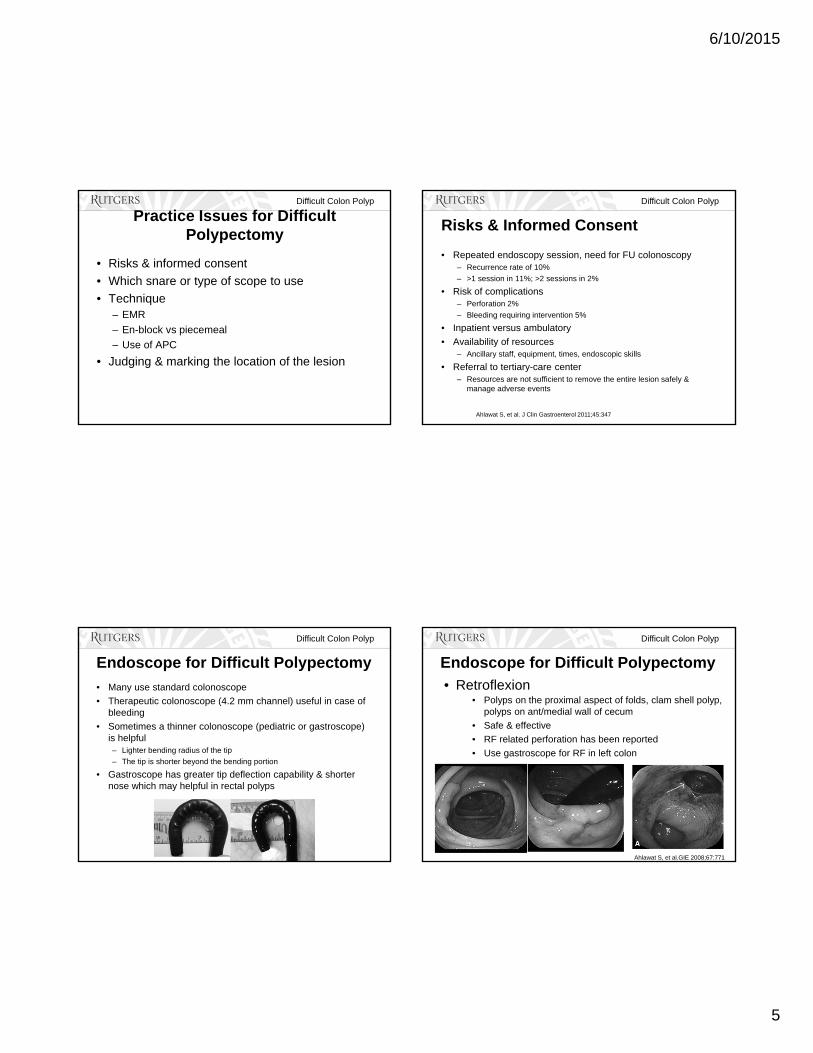
Polyp Configuration
• May be almost impossible to remove entire polyp– portion that lies in
the valley between 2 inter-haustral septae
Difficult Colon Polyp
Polyp Location• Polyp may extend in to
the appendix– Rare
– Total removal of this type is problematic

6/10/2015
4
Difficult Colon Polyp
Polyp Bleeding Risk
• Large pedunculated polyp(>2 cm) with broad stalk (>5 mm) may bleed during or after polypectomy– Large feeding vessels
• Endoloop with epinephrine injection or endoscopic clip may decrease risk of bleeding
Surg Endosc 2009;23:2732
Difficult Colon Polyp
Hogan et al. GIE 2007;66:1018
Difficult Colon Polyp
Polyp Bleeding Risk
• Laterally spreading tumors
• Sessile or flat lesion >2 cm
• Controlled by using thermal modalities or endoscopic clips
Difficult Colon Polyp
Presence of Sub-mucosal Fibrosis
• Previous attempts at resection or injudicious biopsy
• Fibrosis adheres mucosa & submucosa to MP resulting in incomplete separation of layers – Areas of non-lifting with submucosal injection
• Risk of submucosal invasive cancer can be determined accurately from gross appearance, biopsies are often not required
Moss A, et al. Gastroenterology 2011;140:1909
6/10/2015
5
Difficult Colon Polyp
Practice Issues for Difficult Polypectomy
• Risks & informed consent
• Which snare or type of scope to use
• Technique– EMR
– En-block vs piecemeal
– Use of APC
• Judging & marking the location of the lesion
Difficult Colon Polyp
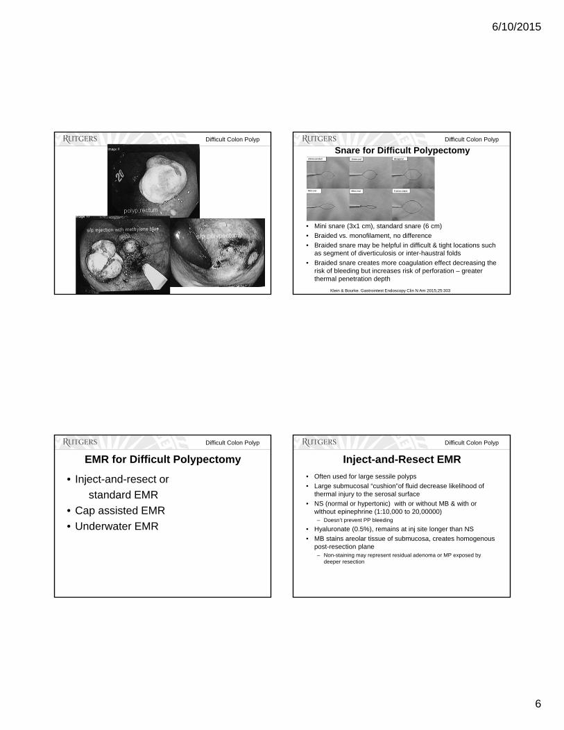
Risks & Informed Consent
• Repeated endoscopy session, need for FU colonoscopy– Recurrence rate of 10%
– >1 session in 11%; >2 sessions in 2%
• Risk of complications– Perforation 2%
– Bleeding requiring intervention 5%
• Inpatient versus ambulatory
• Availability of resources – Ancillary staff, equipment, times, endoscopic skills
• Referral to tertiary-care center – Resources are not sufficient to remove the entire lesion safely &
manage adverse events
Ahlawat S, et al. J Clin Gastroenterol 2011;45:347
Difficult Colon Polyp
Endoscope for Difficult Polypectomy
• Many use standard colonoscope
• Therapeutic colonoscope (4.2 mm channel) useful in case of bleeding
• Sometimes a thinner colonoscope (pediatric or gastroscope) is helpful– Lighter bending radius of the tip
– The tip is shorter beyond the bending portion
• Gastroscope has greater tip deflection capability & shorter nose which may helpful in rectal polyps
Difficult Colon Polyp
Endoscope for Difficult Polypectomy
• Polyps on the proximal aspect of folds, clam shell polyp, polyps on ant/medial wall of cecum
• Safe & effective
• RF related perforation has been reported
• Use gastroscope for RF in left colon
• Retroflexion
Ahlawat S, et al.GIE 2008;67:771
6/10/2015
6
Difficult Colon Polyp Difficult Colon Polyp
Snare for Difficult Polypectomy
• Mini snare (3x1 cm), standard snare (6 cm)
• Braided vs. monofilament, no difference
• Braided snare may be helpful in difficult & tight locations such as segment of diverticulosis or inter-haustral folds
• Braided snare creates more coagulation effect decreasing the risk of bleeding but increases risk of perforation – greater thermal penetration depth
Klein & Bourke. Gastrointest Endoscopy Clin N Am 2015;25:303
Difficult Colon Polyp
EMR for Difficult Polypectomy
• Inject-and-resect or
standard EMR
• Cap assisted EMR
• Underwater EMR
Difficult Colon Polyp
Inject-and-Resect EMR
• Often used for large sessile polyps
• Large submucosal “cushion”of fluid decrease likelihood of thermal injury to the serosal surface
• NS (normal or hypertonic) with or without MB & with or wIthout epinephrine (1:10,000 to 20,00000)– Doesn’t prevent PP bleeding
• Hyaluronate (0.5%), remains at inj site longer than NS
• MB stains areolar tissue of submucosa, creates homogenous post-resection plane– Non-staining may represent residual adenoma or MP exposed by
deeper resection
6/10/2015
7
Difficult Colon Polyp
Burgess et al. GIE;81:813
Difficult Colon Polyp
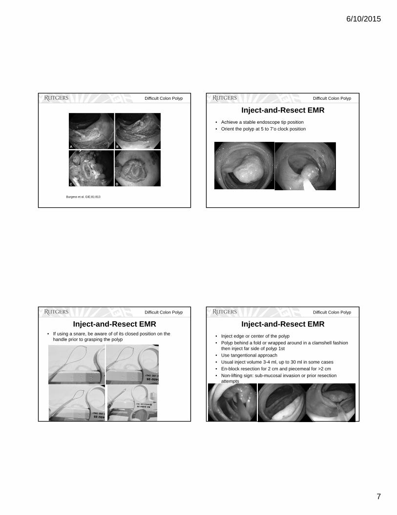
Inject-and-Resect EMR
• Achieve a stable endoscope tip position
• Orient the polyp at 5 to 7’o clock position
Difficult Colon Polyp
Inject-and-Resect EMR• If using a snare, be aware of of its closed position on the
handle prior to grasping the polyp
Difficult Colon Polyp
Inject-and-Resect EMR
• Inject edge or center of the polyp
• Polyp behind a fold or wrapped around in a clamshell fashion then inject far side of polyp 1st
• Use tangentional approach
• Usual inject volume 3-4 ml, up to 30 ml in some cases
• En-block resection for 2 cm and piecemeal for >2 cm
• Non-lifting sign: sub-mucosal invasion or prior resection attempts
6/10/2015
8
Difficult Colon Polyp
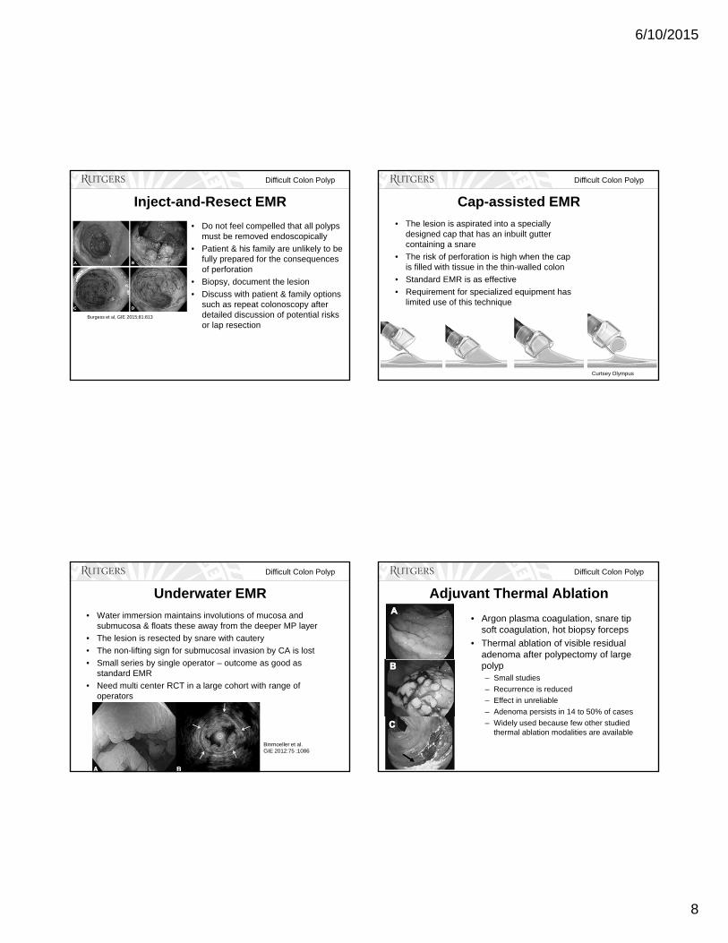
Inject-and-Resect EMR
• Do not feel compelled that all polyps must be removed endoscopically
• Patient & his family are unlikely to be fully prepared for the consequences of perforation
• Biopsy, document the lesion
• Discuss with patient & family options such as repeat colonoscopy after detailed discussion of potential risks or lap resection
Burgess et al, GIE 2015;81:813
Difficult Colon Polyp
Cap-assisted EMR
• The lesion is aspirated into a specially designed cap that has an inbuilt gutter containing a snare
• The risk of perforation is high when the cap is filled with tissue in the thin-walled colon
• Standard EMR is as effective
• Requirement for specialized equipment has limited use of this technique
Curtsey Olympus
Difficult Colon Polyp
Underwater EMR
• Water immersion maintains involutions of mucosa and submucosa & floats these away from the deeper MP layer
• The lesion is resected by snare with cautery
• The non-lifting sign for submucosal invasion by CA is lost
• Small series by single operator – outcome as good as standard EMR
• Need multi center RCT in a large cohort with range of operators
Binmoeller et al.GIE 2012:75 :1086
Difficult Colon Polyp
Adjuvant Thermal Ablation
• Argon plasma coagulation, snare tip soft coagulation, hot biopsy forceps
• Thermal ablation of visible residual adenoma after polypectomy of large polyp– Small studies
– Recurrence is reduced
– Effect in unreliable
– Adenoma persists in 14 to 50% of cases
– Widely used because few other studied thermal ablation modalities are available
6/10/2015
9
Difficult Colon Polyp Difficult Colon Polyp
Difficult Colon Polyp
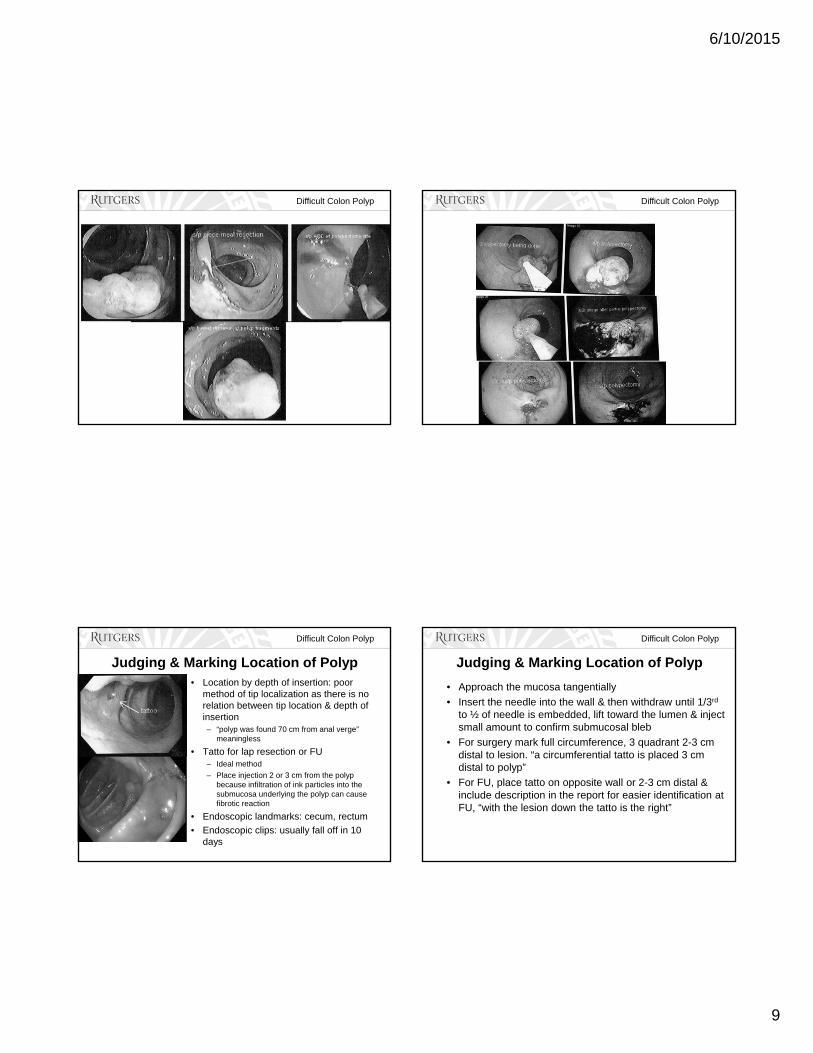
Judging & Marking Location of Polyp• Location by depth of insertion: poor
method of tip localization as there is no relation between tip location & depth of insertion – “polyp was found 70 cm from anal verge”
meaningless
• Tatto for lap resection or FU– Ideal method
– Place injection 2 or 3 cm from the polyp because infiltration of ink particles into the submucosa underlying the polyp can cause fibrotic reaction
• Endoscopic landmarks: cecum, rectum
• Endoscopic clips: usually fall off in 10 days
Difficult Colon Polyp
Judging & Marking Location of Polyp
• Approach the mucosa tangentially
• Insert the needle into the wall & then withdraw until 1/3rd
to ½ of needle is embedded, lift toward the lumen & inject small amount to confirm submucosal bleb
• For surgery mark full circumference, 3 quadrant 2-3 cm distal to lesion. “a circumferential tatto is placed 3 cm distal to polyp”
• For FU, place tatto on opposite wall or 2-3 cm distal & include description in the report for easier identification at FU, “with the lesion down the tatto is the right”
6/10/2015
10
Difficult Colon Polyp
Follow-up after Difficult Colon Polypectomy
• Recurrence or residual adenoma at 1st surveillance colonoscopy (3-6 months) is 10% to 30%, – Recurrence is usually diminutive & is managed endoscopically
• Recurrence rate at 2nd surveillance colonoscopy (12 months later) is 4% if no recurrence at 1st surveillance colonoscopy ; however, recurrence rate is 20% if recurrence occurs at 1st colonoscopy that has been treated– FU at 1 year is essential
• US Multi-Society Task Force on Colorectal Cancer recommends FU within 1 year for flat & sessile polyps >15 mm if there is any questions about incomplete resection
Moss et al. Gastroenterology 2011;140:1909Moss et al. Gut 2015;64:57
Ahlawat S, et al. J Clin Gastroenterol 2011;45:347
Difficult Colon Polyp
Complications• Bleeding
– 4 to 24 percent, our study 5%
– Definition of bleeding varies among studies
– Risk factors: size, large sessile, proximal location, anticoagulation use
– Pure cut vs pure coagulation vs endocut
– Endoscopic intervention successful
– Prophylactic clip ?
• Perforation– 0-2%
• Post polypectomy syndrome: transmural thermal injury– 1 to 4 percent
– Responds to conservative management
Difficult Colon Polyp Difficult Colon Polyp
6/10/2015
11
Difficult Colon Polyp
Conclusion
• The success or failure of colonoscopic polypectomy is determined by patient, polyp and endoscopist factors
• High rates of successful endoscopic resection of difficult colon polyps have been reported in tertiary-level advanced endoscopy units
• Only contraindication to endoscopic resection is a polyp that appear to have invasive cancer on visual inspection or fails to rise after submucosal injection
• DO NOT feel compelled that all polyps must be removed endoscopically; however, understand your “comfort level” & consider referral to an advanced endoscopy unit prior to surgery referral for laparoscopic resection
Recommended


![Forpath 2014 colon polyps Geboes.ppt [Mode de compatibilité] · Polyp – cancer sequence • Adenoma – cancer sequence (Morson et al 1984) Flat adenoma (Muto et al 1985) • Nonpolypoid](https://img.pdfslide.net/doc/110x75/5e0503d17d383847334e6909/forpath-2014-colon-polyps-mode-de-compatibilit-polyp-a-cancer-sequence-a.jpg)