Objective – Why AmpliSeq to Sanger CE ? Lessons From Initial Feasibility Experiments
• AmpliSeq primer design is readily transferable to Sanger CE sequencing
Abstract
• The introduction of defined Ion AmpliSeq™ panels for detection and characterization of actionable mutations occurring in tumor tissue has the potential to revolutionize translational oncology research.
• The Ion Ampliseq™ cancer hot spot panel version 2 (CHP v2) by Ion Torrent includes 207 actionable
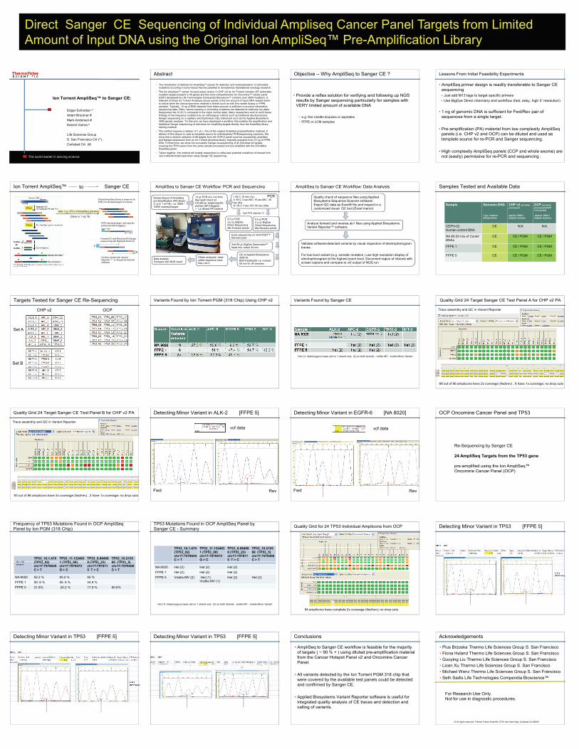
Direct Sanger CE Sequencing of Individual Ampliseq Cancer Panel Targets from Limited Amount of Input DNA using the Original Ion AmpliSeq™ Pre-Amplification Library
• Provide a reflex solution for verifying and following up NGS results by Sanger sequencing particularly for samples with VERY limited amount of available DNA
• e.g. fine needle biopsies or aspirates
• FFPE or LCM samples
• Just add M13 tags to target specific primers
• Use BigDye Direct chemistry and workflow (fast, easy, high 5’ resolution)
• 1 ng of genomic DNA is sufficient for Fwd/Rev pair of sequences from a single target.
• Pre-amplification (PA) material from low complexity AmpliSeq panels (i.e. CHP v2 and OCP) can be diluted and used as template source for re-PCR and Sanger sequencing.
• High complexity AmpliSeq panels (CCP and whole exome) are not (easily) permissive for re-PCR and sequencing .
mutation targets present in 50 genes and the more comprehensive Ion Oncomine™ cancer panel (OCP) developed by Life Technologies Compendia Bioscience™ contains over 2000 mutations. A hallmark of these Ion Torrent Ampliseq cancer panels is the low amount of input DNA needed which is critical when the clinical specimen material is limited such as with fine needle biopsy or FFPE samples. Typically, 10 ng of DNA obtained from these sources is sufficient to produce informative sequencing data. Often, cancer-causing or promoting mutations are detected at relatively low allele frequencies like 10-20 % compared to the major normal allele. Many researchers wish to verify these findings of low frequency mutations by an orthologous method such as traditional dye-fluorescent Sanger sequencing on a capillary electrophoresis (CE) instrument such as the Applied Biosystems 3500 genetic analyzer. To that end, we have developed a workflow that enables the amplification and traditional Sanger sequencing of individual Ion AmpliSeq targets directly from the AmpliSeq library starting material.
• The method requires a retainer of 1 µl (~ 5%) of the original AmpliSeq preamplification material. A dilution of this aliquot is used as template source for individualized PCR/sequencing reactions. We show that a random selection of 48 targets from the CHPv2 panel could be successfully amplified and Sanger-sequenced from an Ion Torrent Ampliseq library originally prepared from 10 ng of FFPE DNA. Furthermore, we show the successful Sanger-re-sequencing of all individual 24 targets covering the TP53 exons from the same sample processed and pre-amplified with the OncoMineAmpliSeq panel.
• Taken together, this method will enable researchers to reflex-test potential mutations of interest from very material-limited specimen using Sanger CE sequencing.
Ion Torrent AmpliSeq™ Sanger CEto AmpliSeq to Sanger CE Workflow: PCR and Sequencing AmpliSeq to Sanger CE Workflow: Data Analysis Samples Tested and Available Data
The world leader in serving science
Edgar Schreiber ^
Adam Broomer #
Mark Andersen #
Kamini Varma ^
Life Sciences Group
S. San Francisco CA (^) ,
Carlsbad CA (#)
Ion Torrent AmpliSeq™ to Sanger CE:
Ion Torrent AmpliSeq Sanger CE
take 1 µL (5%) of AmpliSeq pre-Amp
Dilute in 1 mL TE
Diluted AmpliSeq library is reservoir for PCR of individual targets of interest
PCR individual target with specific primer pair (M13-tagged )
Forward (F) and Reverse (R) Sanger sequencing with BigDye® Direct kit
Confirm variant with Variant Reporter ™ or Sequence Scanner software
20 µl
19 µl
F
R
to AmpliSeq to Sanger CE Workflow: PCR and Sequencing
Diluted aliquot of AmpliSeq pre-Amplification (PA) library (1 µl in 1 ml TE) : ca. 5000 -10000 copies/µl/target
13 µL PCR mix (1x) from Big Dye® Direct kit0.8 µM ea. target-specific primers (M13-tagged)+ 1 µl diluted PA material
I: 94 C 10 min (1x)II: 95 C 3 sec,60C 15 sec,68C 45 sec (8x)III: 95 C 3 sec,70C 50 sec (28x)
6.5 µl PCR2 µ uL BigDye Direct Sequencing Mix Forward primer
6.5 µl PCR2 µ uL BigDye Direct Sequencing Mix Reverse primer
PCR
Split PCR material 1:2
Add 55 µL BigDye Xterminator™ bead mix; vortex 30 min
CE on Applied Biosystems 3500 XLBDX FastSeq50 run module: 50 min for 24 samples
Obtain analyzed, base-called sequence trace files (.ab1)
Data analysisCompare with NGS result
Cycle sequencing on Veriti FAST™ thermal cycler
AmpliSeq to Sanger CE Workflow: Data Analysis
Quality check of sequence files using Applied Biosystems Sequence Scanner software: Export QC data as Excel® file and Inspect in a customized visual QC tool (Excel macro)
Validate software-detected variants by visual inspection of electropherogram traces.
For low level variant (e.g. somatic mutation ) use high resolution display of electropherogram at the highest zoom level. Document region of interest with screen capture and compare to vcf output of NGS run
Analyze forward and reverse ab1 files using Applied Biosystems Variant Reporter™ software.
Samples Tested and Available Data
Sample Genomic DNA
1 ng / reaction300 genomes
CHP v2 pre-Ampprocessed
approx. 5000 + targets/ reaction
OCP pre-Ampprocessed (OCP1+2 pooled)
approx. 5000 + targets/ reaction
CEPH-02Human control DNA
CE N/A N/A
NA 80:20 mix of Coriell DNAs
CE CE / PGM CE / PGM
FFPE 1 CE CE / PGM CE / PGM
FFPE 5 CE CE / PGM CE / PGM
Targets Tested for Sanger CE Re-Sequencing
Set B
CHP v2 OCP
Set A
Variants Found by Ion Torrent PGM (318 Chip) Using CHP v2 Variants Found by Sanger CE
Het (1): heterozygous base call on 1 strand only (2) on both strands ; visible MV : visible Minor Variant
Quality Grid 24 Target Sanger CE Test Panel A for CHP v2 PA
Trace assembly and QC in Variant Reporter
88 out of 96 amplicons have 2x coverage (fwd/rev) , 8 have 1x coverage; no drop outs
Quality Grid 24 Target Sanger CE Test Panel B for CHP v2 PA
Trace assembly and QC in Variant Reporter
Detecting Minor Variant in ALK-2 [FFPE 5]
vcf data
Detecting Minor Variant in EGFR-6 [NA 8020]
vcf data
OCP Oncomine Cancer Panel and TP53
Re-Sequencing by Sanger CE
24 AmpliSeq Targets from the TP53 gene
pre-amplified using the Ion AmpliSeq™
93 out of 96 amplicons have 2x coverage (fwd/rev) , 3 have 1x coverage; no drop outs
Fwd Rev Fwd Rev
p p g p qOncomine Cancer Panel (OCP)
Frequency of TP53 Mutations Found in OCP AmpliSeq Panel by Ion PGM (318 Chip)
TP53_10.1.470(TP53 02)
TP53_11.1324631 (TP53 08)
TP53_8.884088 (TP53 23)
TP53_10.215568 (TP53 5)(#) = CE
TP53 Mutations Found in OCP AmpliSeq Panel by Sanger CE - Summary
TP53_10.1.470(TP53_02)chr17:7578645 C > T
TP53_11.1324631 (TP53_08) chr17:7579472 G > C
TP53_8.884088 (TP53_23) chr17:7578115 T > C
TP53_10.215568 (TP53_5) chr17:7578406 C > T
Quality Grid for 24 TP53 Individual Amplicons from OCP Detecting Minor Variant in TP53 [FFPE 5]
(TP53_02)chr17:7578645 C > T
1 (TP53_08) chr17:7579472 G > C
8 (TP53_23) chr17:7578115 T > C
68 (TP53_5) chr17:7578406 C > T
NA 8020 52.2 % 50.2 % 52 %
FFPE 1 50. 6 % 55. 6 % 44.9 %
FFPE 5 21.8% 20.2 % 17.9 % 60.6%
(#) = CE assay #
NA 8020 Het (2) Het (2) Het (2)
FFPE 1 Het (2) Het (2) Het (2)
FFPE 5 Visible MV (2) Het (1)Visible MV (1)
Het (2) Het (2)
Het (1): heterozygous base call on 1 strand only (2) on both strands ; visible MV : visible Minor Variant
94 amplicons have complete 2x coverage (fwd/rev); no drop outs
Detecting Minor Variant in TP53 [FFPE 5]Detecting Minor Variant in TP53 [FFPE 5] Conclusions
• AmpliSeq to Sanger CE workflow is feasible for the majority of targets ( ~ 90 % + ) using diluted pre-amplification material
Acknowledgements
• Pius Brzoska Thermo Life Sciences Group S. San Francisco
• Fiona Hyland Thermo Life Sciences Group S. San Franciscofrom the Cancer Hotspot Panel v2 and Oncomine Cancer Panel.
• All variants detected by the Ion Torrent PGM 318 chip that were covered by the available test panels could be detected and confirmed by Sanger CE.
• Applied Biosystems Variant Reporter software is useful for integrated quality analysis of CE traces and detection and calling of variants.
y p
• Guoying Liu Thermo Life Sciences Group S. San Francisco
• Licen Xu Thermo Life Sciences Group S. San Francisco
• Michael Wenz Thermo Life Sciences Group S. San Francisco
• Seth Sadis Life Technologies Compendia Bioscience™
For Research Use Only.Not for use in diagnostic procedures.
© All rights reserved. Thermo Fisher Scientific, 5791 Van Allen Way, Carlsbad CA 92008
Recommended