DMD # 77586
1
Discovery and Validation of Pyridoxic Acid and Homovanillic Acid as Novel
Endogenous Plasma Biomarkers of Organic Anion Transporter (OAT) 1 and
OAT3 in Cynomolgus Monkeys
Hong Shen, David M. Nelson, Regina V. Oliveira, Yueping Zhang, Colleen A. Mcnaney,
Xiaomei Gu, Weiqi Chen, Ching Su, Michael D. Reily, Petia A. Shipkova,
Jinping Gan, Yurong Lai, Punit Marathe, and W. Griffith Humphreys
Departments of Metabolism and Pharmacokinetics (H.S., Y.Z., X.G., W.C., J.G., Y.L., P.M.,
W.G.H.), Discovery Toxicology (D.N., C.A.M., M.D.R.), Bioanalytical Research (R.O., P.A.S.),
and Discovery Pharmaceutics (C.S.), Pharmaceutical Candidate Optimization, Bristol-Myers
Squibb Research and Development, Princeton, New Jersey 08543, United States
This article has not been copyedited and formatted. The final version may differ from this version.DMD Fast Forward. Published on November 21, 2017 as DOI: 10.1124/dmd.117.077586
at ASPE
T Journals on M
arch 25, 2020dm
d.aspetjournals.orgD
ownloaded from

DMD # 77586
2
Running title: Identification of functional plasma markers of OAT1 and OAT3 in vivo
Address correspondence to:
Dr. Hong Shen
Department of Metabolism and Pharmacokinetics (MAP)
Pharmaceutical Candidate Optimization (PCO)
Bristol-Myers Squibb Company (BMS)
Route 206 & Province Line Road, Princeton, NJ 08543-4000
Telephone: (609) 252-4509
Facsimile: (609) 252-6802
E-mail: [email protected]
Number of text pages: 29
Number of tables: 3
Number of figures: 6
Number of references: 39
Number of words:
Abstract: 249
Introduction: 794
Discussion: 1832
Abbreviations: Ae, amount of unchanged drug recovered in the urine; AUC, area under the plasma
concentration-time curve; CCK-8, cholecystokinin octapeptide; CE, collision energy; CLNR,
This article has not been copyedited and formatted. The final version may differ from this version.DMD Fast Forward. Published on November 21, 2017 as DOI: 10.1124/dmd.117.077586
at ASPE
T Journals on M
arch 25, 2020dm
d.aspetjournals.orgD
ownloaded from

DMD # 77586
3
nonrenal plasma clearance; CLR, renal clearance; CLTOT, total plasma clearance; CsA, cyclosporin
A; DCA, dicarboxylic acid; DMEM, DDI, drug-drug interaction; Dulbecco’s modified Eagle’s
growth medium; DP, declustering potential; E3S, estrone-3-sulfate; E17βG, estradiol-17β-D-
glucuronide; fNR, the fraction excreted by nonrenal routes; fR, fraction excreted in the urine; FSM,
furosemide; GA, glucuronic acid; GFR, glomerular filtration rate; HBSS, Hanks’ balanced salt
solution; HEK, human embryonic kidney; HDA, hexadecanedioic acid; HVA, homovanillic acid;
IC50, concentration required to inhibit transport by 50%; IMC, indomethacin; Km, Michaelis-
Menten constant that corresponds to the substrate concentration at which the uptake rate is half of
Vmax; LC-MS/MS, liquid chromatography–tandem mass spectrometry; MATE, multidrug and
toxin extrusion protein; MFM, metformin; NTCP, sodium taurocholate cotransporting
polypeptide; OAT, organic anion transporter; OATP, organic anion transporting polypeptide;
OCT, organic cation transporter; PAH, para-aminohippuric acid; PBS, phosphate-buffered saline;
PCV, penciclovir; PDA, pyridoxic acid; PROB, probenecid; PYR, pyrimethanmine; RPTC, renal
proximal tubule cell; SRM, selective reaction monitoring; TCA, taurocholic acid; TDA,
tetradecanedioic acid, Vmax, maximum transport rate.
This article has not been copyedited and formatted. The final version may differ from this version.DMD Fast Forward. Published on November 21, 2017 as DOI: 10.1124/dmd.117.077586
at ASPE
T Journals on M
arch 25, 2020dm
d.aspetjournals.orgD
ownloaded from

DMD # 77586
4
ABSTRACT:
Perturbation of OAT1- and OAT3-mediated transport can alter the exposure, efficacy, and safety
of drugs. Although these have been reports of the endogenous biomarkers for OAT1/3, none of
these have all of the characteristics required for a clinical useful biomarker. Cynomolgus monkeys
were treated with intravenous probenecid (PROB) at a dose of 40 mg/kg in this study. As expected,
PROB increased the AUC of co-administered furosemide (FSM), a known substrate of OAT1 and
OAT3, by 4.1-fold, consistent with the values reported in humans (3.1- to 3.7-fold). Of 233 plasma
metabolites analyzed using a LC-MS/MS-based metabolomics method, 29 metabolites, including
pyridoxic acid (PDA) and homovanillic acid (HVA), were significantly increased at either 1 or 3
h in plasma from the monkeys pretreated with PROB compared with the treated animals. Plasma
of animals was then subjected to targeted LC-MS/MS analysis which confirmed that the PDA and
HVA AUCs increased by approximately 2- to 3-fold by PROB pretreatments. PROB also increased
plasma concentrations of hexadecanedioic acid (HDA) and tetradecanedioic acid (TDA) although
the increases were not statistically significant. Moreover, transporter profiling assessed using
stable cell lines constitutively expressing transporters, demonstrated that PDA and HVA are
substrates for human OAT1, OAT3, OAT2 (HVA) and OAT4 (PDA), but not OCT2, MATE1,
MATE2K, OATP1B1, OATP1B3, and NTCP. Collectively, these findings suggest that PDA and
HVA might serve as blood-based endogenous probes of cynomolgus monkey OAT1 and OAT3,
and investigation of PDA and HVA as circulating endogenous biomarkers of human OAT1 and
OAT3 function is warranted.
This article has not been copyedited and formatted. The final version may differ from this version.DMD Fast Forward. Published on November 21, 2017 as DOI: 10.1124/dmd.117.077586
at ASPE
T Journals on M
arch 25, 2020dm
d.aspetjournals.orgD
ownloaded from

DMD # 77586
5
INTRODUCTION
OAT1 (SLC22A6) and OAT3 (SLC22A8) function as influx-transporters that are mainly
expressed on the basolateral membrane of renal proximal tubular cells (RPTCs) and mediate
cellular uptake of substrates from blood into the cells (Motohashi et al., 2002). OAT1 and OAT3
can serve as the loci of drug-drug interactions (DDIs), and such DDIs can lead to undesired
changes in systemic and local exposures of victim drugs and toxins (Morrissey et al., 2013; Nigam
et al., 2015). For example, furosemide (FSM) is taken up from the blood in RPTCs via OAT1 and
OAT3 (Hasannejad et al., 2004), and probenecid (PROB), an inhibitor of OAT1 and OAT3, causes
significant alterations in the pharmacokinetic parameters of FSM in humans through increased
area under the plasma drug concentration-time curve (AUC), decreased total and renal clearance
(CLTOT and CLR), and decreased fraction excreted in the urine (fR) (Chennavasin et al., 1979; Smith
et al., 1980; Vree et al., 1995). Moreover, a few uremic toxins recently have been demonstrated to
be inhibitors of OAT1 and OAT3, and have the potential to inhibit renal secretion clearance of
drug substrates of OAT1 and OAT3 in patients with chronic kidney disease (Hsueh et al., 2016).
On the other hand, concomitant use of PROB is recommended by the United States Food and Drug
Administration (FDA) for reducing the pronounced nephrotoxicity of cidofovir by inhibiting
OAT1 and OAT3 to reduce the exposure of cidofovir in RPTCs to an extent that results in an
acceptable benefit-risk balance
(http://www.accessdata.fda.gov/drugsatfda_docs/label/1999/020638s003lbl.pdf).
Animal models have often been often used to assess pre-clinical drug-OAT interactions.
For example, the clinical famotidine-PROB interaction was reproduced in cynomolgus monkeys
recently as PROB caused an approximate 90% reduction in the tubular secretion clearance of
famotidine in monkeys (4.58 ± 1.25 versus 0.38 ± 0.36 mL/min/kg) (Tahara et al., 2006). Similarly,
This article has not been copyedited and formatted. The final version may differ from this version.DMD Fast Forward. Published on November 21, 2017 as DOI: 10.1124/dmd.117.077586
at ASPE
T Journals on M
arch 25, 2020dm
d.aspetjournals.orgD
ownloaded from

DMD # 77586
6
PROB significantly decreased the renal tubular secretion clearance of famotidine from 2.80 ± 0.36
to 0.37 ± 0.07 mL/min/kg in humans. Furthermore, the protective effect of PROB treatment on the
nephrotoxicity of cidofovir in humans was able to be recapitulated in monkeys (Lacy et al., 1998).
These results suggest that monkey is the more suitable animal model to predict the clinical DDIs
involving OAT1 and OAT3.
Recently endogenous biomarkers have been envisioned as a simple, fast, and cost-effective
tool to monitor transporter activity in a preclinical and clinical setting to facilitate development of
a drug candidate (Bergagnini-Kolev et al., 2017; Ito et al., 2012; Lai et al., 2016; Muller et al.,
2015; Yee et al., 2016). Therefore, the identification of a sensitive endogenous biomarker for
OAT1 and OAT3 would be of great value. In this regard, Imamura et al. reported that 6β-
hydroxycortisol could be an endogenous probe for OAT3 inhibition evaluation as PROB
significantly changed the AUC and CLR of 6β-hydroxycortisol in healthy subjects (Imamura et al.,
2014). However, 6β-hydroxycortisol is formed from cortisol by hepatic CYP3A4, and many drugs
are known to affect this drug-metabolizing enzyme can also change exposure of 6β-
hydroxycortisol (Peng et al., 2011). Very recently Tsuruya et al. reported that taurine and
glycochenodeoxycholate sulfate (GCDCA-S) were endogenous biomarkers of OAT1 and OAT3,
respectively (Tsuruya et al., 2016). However, the plasma levels of taurine and GCDCA-S were not
changed significantly by PROB treatment compared to control even though the renal secretion and
CLR were significantly decreased (Tsuruya et al., 2016). Reduced activities of blood-facing OAT1
and OAT3 by PROB are supposed to increase plasma concentration of a sensitive and selective
endogenous probe, mimicking systemic alterations of probe drugs such as FSM. To our
knowledge, all published “endogenous biomarkers” of OAT1 and OAT3 could not recapitulate
plasma drug concentration time profiles in the presence of OAT inhibitors. Several features
This article has not been copyedited and formatted. The final version may differ from this version.DMD Fast Forward. Published on November 21, 2017 as DOI: 10.1124/dmd.117.077586
at ASPE
T Journals on M
arch 25, 2020dm
d.aspetjournals.orgD
ownloaded from

DMD # 77586
7
including specificity, sensitivity, predictability, reproducibility, acute response and accessibility
have been considered in the identification and validation of ideal endogenous biomarkers for drug
transporters (Chu et al., 2017; Mariappan et al., 2017; Rodrigues et al., 2017). Given the
aforementioned reasons, there is a need for novel plasma biomarkers of renal organic anion
transporters.
In the present studies we provide direct experimental evidence that several organic anionic
compounds, including pyridoxic acid (PDA), homovanilic acid (HVA), hexadecanedioic acid
(HDA) and tetradecanedioic acid (TDA) are potential endogenous biomarkers of OAT1 and OAT3
in cynomolgus monkeys. Specifically, an untargeted metabolomics analysis was applied to plasma
samples to screen endogenous compounds that were associated with OAT1 and OAT3 inhibition
in monkeys. Follow-up quantitative LC/MS analysis further characterized the time-plasma
concentration profiles of selected endogenous compounds (i.e., PDA, HVA, HDA and TDA) after
PROB administration in cynomolgus monkeys. Moreover, transporter profiling assessed using
human embryonic kidney (HEK) 293 cells stably transfected with major human renal and hepatic
drug transporters demonstrated that PDA and HVA are OAT1 and OAT3 substrates and thus
potential novel plasma endogenous biomarkers of OAT1 and OAT3 inhibition.
This article has not been copyedited and formatted. The final version may differ from this version.DMD Fast Forward. Published on November 21, 2017 as DOI: 10.1124/dmd.117.077586
at ASPE
T Journals on M
arch 25, 2020dm
d.aspetjournals.orgD
ownloaded from
DMD # 77586
8
MATERIALS AND METHODS
Materials. [3H]Penciclovir (PCV) (1.3 Ci/mmol) and [14C]metformin ([14C]MFM) (98
mCi/mmol) were purchased from Moravek Biochemicals, Inc. (Brea, CA). [3H]para-
Aminohippuric acid (PAH) (4.5 Ci/mmol), [3H]estrone-3-sulfate ([3H]E3S; 44.0 Ci/mmol),
[3H]estradiol-17β-D-glucuronide ([3H]E17βG) (34.3 Ci/mmol), and [3H] cholecystokinin
octapeptide ([3H]CCK-8) (97.5 mCi/mmol) were purchased from PerkinElmer Life and Analytical
Sciences (Waltham, MA). Nonradiolabeled furosemide (FSM), probenecid (PROB), taurocholic
acid (TCA), and the corresponding stable isotope labeled internal standards were purchased from
Toronto Research Chemicals Inc. (North York, Ontario). 4-Pyridoxic Acid (PDA, ≥98%) was
purchased from Sigma Aldrich (St. Louis, MO), and Homovanilic acid (HVA, ≥98%) was
purchased from Acros Organics. Hexadecanedioic acid-d28, tetradecanedioic acid-d24, and
enalapril maleate-d5 (Enal-d5) were obtained from CDN Isotopes (Pointe-Claire, Quebec,
Canada). Other nonradiolabeled compounds were purchased from either Sigma-Aldrich (St. Louis,
MO) or Research Chemicals Inc. (North York, Ontario). and were of analytical grade. Cell culture
media and reagents were purchased from Invitrogen (Carlsbad, CA) or Mediatech, Inc., A Corning
Subsidiary (Manassas, VA).
Cynomolgus Monkeys Pharmacokinetic FSM-PROB Interaction Study Protocol. To
identify and verify the endogenous biomarkers of OAT1 and OAT3, we conducted a series of
experiments: cynomolgus monkey FSM-PROB interaction, metabolomics, targeted LC-/MS/MS,
and transporter profiling experiments. The experimental workflow is shown in Figure 1.
A single-dose, three-period crossover intravenous (IV) pharmacokinetic DDI study was
carried out at Bristol-Myers Squibb Company, and 3 male cynomolgus monkeys with body
weights ranging from 5.3 to 6.0 kg during study periods were included in this study. The
This article has not been copyedited and formatted. The final version may differ from this version.DMD Fast Forward. Published on November 21, 2017 as DOI: 10.1124/dmd.117.077586
at ASPE
T Journals on M
arch 25, 2020dm
d.aspetjournals.orgD
ownloaded from

DMD # 77586
9
experiment was performed in accordance with the National Institutes of Health guidelines and
approved by Bristol-Myers Squibb Animal Care and Use Committee. The animals were housed in
a temperature- and humidity-controlled room with a 12-h light/dark cycle.
In the first period, 40 mg/kg PROB dissolved in a sodium hydroxide solution (0.1 N) and
titrated to neutral pH with hydrochloric acid was intravenously infused via a femoral vein catheter
over 5 minutes (min) (5 mL/kg). Venous blood samples (2 mL) were collected before and 0.08,
0.17, 0.25, 0.5, 0.75, 1, 2, 3, 5, 7, and 24 hours (h) after administration in K2-EDTA-containing
tubes.
In the second period, after a washout period of 7 days, 2 mg/kg FSM dissolved in saline
was intravenously infused via a femoral vein over 5 min (5 mL/kg), and blood samples were
collected before and 0.08, 0.17, 0.25, 0.5, 0.75, 1, 2, 3, 5, 7, and 24 h after administration. In the
third period, 8 days after administrations of the previous dose of FSM, a FSM saline solution (2
mg/kg) was given by femoral vein infusion over 5 min to each monkey 30 min after PROB
administration (40 mg/kg, IV infusion for 5 min). Blood was sampled before and 0.08, 0.17, 0.25,
0.5, 0.75, 1, 2, 3, 5, 7, and 24 h after start of the FSM infusion. Blood samples were spun for 5 min
at 13,000 rpm within 1 h to obtain plasma. Urine samples were collected using metabolic cages
for the following intervals in all 3 periods: 0 to 3 h, 3 to 7 h and 7 to 24 h after administration, and
the volume of urine was recorded. The plasma and urine samples were stored at -80°C until liquid
chromatography–tandem mass spectrometry (LC-MS/MS) and metabolomics analysis was
conducted.
LC-MS/MS Analysis of Furosemide and Probenecid. The LC-MS/MS analysis was
performed on a Sciex Triple Quad API-4000 system (AB Sciex, Framingham, MA) coupled with
a Shimadzu Nexera LD-30AD ultra-performance liquid chromatography (UPLC) system
This article has not been copyedited and formatted. The final version may differ from this version.DMD Fast Forward. Published on November 21, 2017 as DOI: 10.1124/dmd.117.077586
at ASPE
T Journals on M
arch 25, 2020dm
d.aspetjournals.orgD
ownloaded from
DMD # 77586
10
(Shimadzu, Columbian, MD). The chromatographic separation was performed on an Agilent
Zorbax RRHD SB-C8 column (2.0 x 100 mm, 1.7 µm) from Agilent Technologies (Santa Clara,
CA) using mobile phases of 0.1% formic acid in water and 0.1% formic acid in acetonitrile. The
flow rate was 0.7 ml/min and total run time was 3.7 min. The LC column was maintained at 60 °C.
The analytes were monitored using selected reaction monitoring (SRM) in negative ionization
mode with the optimized nebulizing and desolvation gases. The source temperature was set at 400
ºC, and declustering potential (DP) and collision energy (CE) were optimized. Furosemide,
furosemide-d5 and probenecid were detected at the SRM transitions of m/z 329.1 → 285.0,
334.1→291.0 and 283.9 → 239.9, respectively.
Before the analysis, urine samples were diluted 100 fold into blank plasma and treated as
for plasma. The plasma and diluted urine samples were then extracted using protein precipitation.
Specifically, the 100 µL plasma and diluted urine samples were mixed with 100 µL of acetonitrile
containing 400 nM of furosemide-d5 (internal standard), followed by vortex mixing on a mixer
for 5 min at room temperature. The mixed solutions were then filtered using MultiScreen
hydrophilic flitration plate (Millipore, MA) by centrifugation at 4,000g for 10 min, and the filtered
solution was then injected (5 µL) on to LC-MS/MS. The assay was qualified over the analytical
range of 1 to 5,000 nM using a linear 1/x2 weighed regression.
Plasma Metabolomic Profiling. Frozen plasma samples were thawed and 50 µL aliquots were
subjected to protein precipitation by the addition of 600 µL of methanol containing 0.1% formic
acid and stable-labeled internal standards. The samples were mixed with a vortex mixer and
subjected to centrifugation for 10 min at 2700 x g (4,000 rpm). An aliquot (50 µL) of the resulting
supernatants were transferred to a 96-well plate for further hydrophilic interaction liquid
This article has not been copyedited and formatted. The final version may differ from this version.DMD Fast Forward. Published on November 21, 2017 as DOI: 10.1124/dmd.117.077586
at ASPE
T Journals on M
arch 25, 2020dm
d.aspetjournals.orgD
ownloaded from
DMD # 77586
11
chromatography (HILIC) LC-MS analysis and an additional 100 µL were transferred to a separate
96-well plate for reverse phase (RP) LC-MS analysis. Both plates were dried to completeness
under a nitrogen stream at room temperature. For RP LC-MS analysis, the dried samples were
reconstituted by the addition of 20 µL of methanol, followed by vigorous shaking and the addition
of 180 µL of water. The samples were mixed with a vortex mixer and subjected to centrifugation
for 10 min at 4,000 rpm. The resulting supernatants were transferred to a 96-well plate, from which
10 µL were directly injected for analysis. For HILIC LC-MS analysis, the dried samples were
reconstituted by the addition of 20 µL of water, followed by vigorous shaking and the addition of
180 µL of 50:50 MeOH:ACN. The samples were mixed with a vortex mixer and subjected to
centrifugation for 10 min at 4000 rpm. The resulting supernatants were transferred to a 96-well
plate, from which 10 µL were directly injected for analysis.
HILIC and RP LC- MS analyses were performed on a Nexera X2 LC-30AD (Shimadzu,
Somerset, NJ) UHPLC system connected to a Exactive Plus (Thermo Fisher Scientific, Waltham,
MA) mass spectrometer. The UHPLC column for HILIC analyses was an Acquity BEH-NH2,
2.1x150mm, 1.7 u (Waters Corporation, Milford, MA) with mobile phases A (95:5 water:ACN,
10 mM NH4OAc, 0.05% NH4OH) and B(ACN, 0.05% NH4OH) at a flow rate of 300 µL /min
with starting conditions of 95%B to 37%B at 3.5 min, hold for 4 min and down to starting
conditions at 7min, for a total run time of 11 min. The UHPLC column for RP analyses was an
Acquity BEH C18, 2.1x150mm, 1.7 u (Waters Corporation, Milford, MA) with mobile phases A
(water, 0.1% formic acid) and B (98:2ACN: water, 0.1% formic acid) at a flow rate of 600 µL
/min with starting conditions of 100%A, to 80%A at 3 min, 40%A at 4min and 100%B by 7min
and after a 2 min hold, down to starting conditions at 9 min, for a total run time of 11 min. Both
This article has not been copyedited and formatted. The final version may differ from this version.DMD Fast Forward. Published on November 21, 2017 as DOI: 10.1124/dmd.117.077586
at ASPE
T Journals on M
arch 25, 2020dm
d.aspetjournals.orgD
ownloaded from

DMD # 77586
12
HILIC and RP LC-MS data was collected in positive and negative polarities (separate injections)
at 35,000 resolution and expected mass accuracy of 5 ppm.
LCMS data analysis was performed using in-house developed software, Expedient Data
Mining (EDM) as described previously (Hnatyshyn et al., 2013). Direct data input from raw files
was performed using Thermo Fisher Scientific MSFileReader software. The resulting list of
components were matched with accurate mass and retention time values using an in-house
database of endogenous metabolites stored within EDM. The annotated table of component
integrals was exported to Microsoft Excel for further statistical analysis using in-house Visual
Basic scripts for Microsoft Excel. For these analyses, mean intensities for each treatment group
were compared to the relevant concurrent control. For each component, fold-change was
calculated by dividing the treatment group value by the control group value and p-values were
calculated as a pair-wise comparison for a two-tailed distribution, using Student’s t-test (Excel
statistics package, Microsoft).
Characterization of PDA and HVA Uptake in Stable Cell Lines Constitutively
Expressing Major Renal Drug Transporters. Uptake studies were performed as described
previously (Shen et al., 2016b). Uptake of PDA and HVA were first measured at a single
concentration for 5-min incubation with OAT1-, OAT2-, OAT3-, OAT4-, OCT2-, MATE1-,
MATE2K-, OATP1B1-, OATP1B3-, and NTCP-HEK cells, and then time- and concentration
dependent uptake was measured. The initial concentration used for PDA was 1 μM because the
physiological baseline level ranged from 0.67 to 2.5 µM in cynomolgus monkeys (Figure 3A). The
concentration studied for HVA was 5 μM because of limited bioanalytical sensitivity of this
compound in Mock-HEK cells. In addition, the transport Michaelis-Menten constant value (Km)
This article has not been copyedited and formatted. The final version may differ from this version.DMD Fast Forward. Published on November 21, 2017 as DOI: 10.1124/dmd.117.077586
at ASPE
T Journals on M
arch 25, 2020dm
d.aspetjournals.orgD
ownloaded from

DMD # 77586
13
value of 274 ± 100 μM has been reported in rat Oat2-mediated transport for HVA (Mori et al.,
2003), which is significantly greater than the selected testing concentrations (274 ± 100 versus 5
μM). No corresponding data were available for PDA. Kinetic transport experiments were
conducted under linear-uptake conditions or for shortest incubation duration with acceptable
analytical sensitivity (Supplemental Figure 1).
Cells were grown to confluence in 24-well poly-D-lysine-coated plates 2 to 3 days after
seeding at cell density of 500.000 cells per well (BD Biosciences, San Jose, CA). All experiments
were conducted at 37°C using a working solution containing Hanks balanced salt solution (HBSS)
supplemented with 10 mM HEPES (pH 7.4 for OAT1-, OAT2-, OAT3-, OAT4-, OCT2-,
OATP1B1, OATP1B3-, and NTCP-HEK cells, and pH 8.4 for MATE1- and MATE2K-HEK cells,
respectively), with a probe substrate, PDA, or HVA. The probe substrates used were [3H]PAH
(OAT1), [3H]PCV (OAT2), [3H]E3S (OAT3 and OAT4), [14C]MFM (OCT2, MATE1, and
MATE2K), [3H]E17βG (OATP1B1), [3H]CCK-8 (OATP1B3, and TCA (NTCP). Compounds
were dissolved in dimethyl sulfoxide and diluted in Hanks balanced salt solution (HBSS)
(maximum 0.2% dimethyl sulfoxide). Plating medium was removed, and cell monolayers were
rinsed twice with prewarmed HBSS. Incubations were started by the addition of 200 μL of
substrate prewarmed at 37°C. After incubation for desired time at 37°C, the cell monolayers were
rinsed three times with 500 μL of ice-cold HBSS. The cells were then lysed with 300 μL buffer
(0.1% Triton X-100 or methanol), and compound concentrations in the cell lysates were measured
by either liquid scintillation counting (Tri-Carb 2910 TR Liquid Scintillation Analyzer,
PerkinElmer Life and Analytical Sciences, Waltham, MA) or LC–MS/MS as described below.
LC-MS/MS Analysis of PDA and HVA. Stock PDA solution (390 µg/mL, 2.13 mM) was
prepared by dissolving PDA in 0.5% NH4OH solution. Stock HVA (1.8 mg/mL, 9.5mM) solution
This article has not been copyedited and formatted. The final version may differ from this version.DMD Fast Forward. Published on November 21, 2017 as DOI: 10.1124/dmd.117.077586
at ASPE
T Journals on M
arch 25, 2020dm
d.aspetjournals.orgD
ownloaded from

DMD # 77586
14
was prepared in water. Stock solutions were stored in darkness at -30°C and brought to room
temperature before use. The highest calibration standard (5000 ng/mL) was prepared by diluting
appropriate aliquots of PDA and HVA stock solutions to a final volume of 2 mL with 1% BSA in
PBS (pH 7.4). Additional standard solutions were obtained by serial dilution from the 5000 ng/mL
standard with 1% BSA in PBS (pH 7.4) to final concentrations of 2500, 1000, 500, 250, 100, 50,
25, 10 and 5 ng/mL. Quality control (QC) samples were also prepared with 1% BSA in PBS (pH
7.4) at three concentration levels: 7.5 ng/mL, 75 ng/mL and 750 ng/mL. The IS stock solution of
Enal-d5 was prepared at 1 mg/mL in methanol. The IS working solution containing 1000 ng/mL
of Enal-d5 was prepared by dilution of IS stock solution with methanol and stored at 4°C.
Calibration curves for PDA and HVA were fitted by 1/x weighted least squares quadratic.
The calibration curves for PDA and HVA range from 5 to 5000 ng/mL. All coefficients of
determination (R2) of the calibration lines were ≥0.98. The mean accuracy (% of true value) of
individual calibrators was ≥ 15%. The lower limit of quantitation (LLOQ), defined as the lowest
concentration which could be determined with precision and accuracy of ± 20%, was 5ng/mL for
PDA and HVA.
Samples stability was also determined for QCs samples (n = 5) that were extracted and
stored in the instrument autosampler under refrigerated conditions (5°C). Samples were stable for
24 hrs. Average recovery for QCs samples (n = 5) prepared with 1% BSA in PBS (pH 7.4) was
101% and 102% for PDA and HVA, respectively.
Urine and plasma samples were diluted by a factor of 1:1, 1:2, 1:10 or 1:100 with 1% BSA
in PBS (pH 7.4) to ensure levels within the range of the calibration curve. Aliquots (50 µL) of
diluted animal samples (urine and plasma), calibrators standards or QCs were transferred to a 2
This article has not been copyedited and formatted. The final version may differ from this version.DMD Fast Forward. Published on November 21, 2017 as DOI: 10.1124/dmd.117.077586
at ASPE
T Journals on M
arch 25, 2020dm
d.aspetjournals.orgD
ownloaded from

DMD # 77586
15
mL 96-well plate and mixed for 1 min at 2000 rpm. The samples were extracted by adding 200 µL
of Enal-d5 in methanol (1000 ng/mL) and vortex-mixed for 5 min at 2000 rpm, centrifuged at 4
°C for 10 min at 4000 ×g. The supernatant (170 µL) was transferred to a 500 µL 96-well plate and
dried under heated nitrogen (45°C). Samples were then reconstituted with 50 μL of water:methanol
(98:2) containing 0.1% formic acid, vortex-mixed for 1 min at 2000 rpm, followed by 10 min of
centrifugation at 4000g at 4 °C before MS analysis. To avoid PDA and HVA degradation, all
samples were protected from direct light exposure during sample preparation and analysis.
LC-MS/MS analyses were carried out on a Waters Acquity UPLC system, consisting of a
Acquity binary solvent manager and Acquity sample manager with sample organizer (Waters,
Milford, MA, USA) coupled to a SCIEX 6500 tandem quadrupole mass spectrometer (Applied
Biosystems/MDS SCIEX, Toronto, Canada) equipped with an ESI source. The analytes (10 µL)
were separated on an Acquity UPLC BEH130 C18 (2.1 x 100 mm; 1.7 µm particle size) column
and eluted by a gradient program as follows: held 2% B for 0.5 min, 2% B to 20% B in 2.5 min,
20% B to 98% B in 1 min, 98% B to 2% B in 0.01 min and retained 1 min for equilibration. The
column was heated at 45°C, and the flow rate was 500 µL/min. The mobile phase consisted of an
aqueous phase (A: 0.1% formic acid in water) and an organic phase (B: 0.1% formic acid in
acetonitrile).
The ESI source was operated in negative ion mode, and its main working parameters were
set as follows: ion spray voltage, -4.5 kV; ion source temperature, 550°C; declustering potential, -
40 V for PDA and HVA and -150 V for Enal-d5; collision energy, -35 V; entrance potential, -10
V; and collision cell exit potential, -10 V. Multiple reaction monitoring (MRM) measurements of
-4PA and HVA analytes were performed using individually optimized cone voltage and collision
energy. The MRM precursor/product ion transitions were as follows: m/z 182> 138.0 for PDA,
This article has not been copyedited and formatted. The final version may differ from this version.DMD Fast Forward. Published on November 21, 2017 as DOI: 10.1124/dmd.117.077586
at ASPE
T Journals on M
arch 25, 2020dm
d.aspetjournals.orgD
ownloaded from

DMD # 77586
16
181.0 > 122.0 for HVA, and 380.3 > 114.2 for the internal standard, Enal-d5.The dwell time
established for each transition was 50ms. All peak integration and data processing were performed
using SCIEX Analyst 1.6.2 (Applied Biosystems/MDS SCIEX).
Pharmacokinetic, Transport and Statistical Analysis. The area under the plasma
concentration-time curve from zero to 24 h (AUC0-24 h) was calculated using mixed trapezoidal
rule. The area under the plasma concentration-time curve from zero to infinity (AUC) includes
AUC0-24 h and one extrapolated to infinity from the last measured concentration. The volume of
distribution at steady-state (VdSS) was determined by noncompartmental method:
Equation 1
where AUMC is the area under the curve of the first moment of the concentration-time curve.
The total plasma clearance (CLTOT) was calculated from:
Equation 2
The pharmacokinetics parameters including AUC0-24h, AUC, VdSS, and CLTOT for FSM,
PROB, PDA and HVA, following single intravenous administration of PROB, FSM with or
without co-administration of PROB were analyzed with a mixed trapezoidal model using Kinetica
program (Thermo Electron; Philadelphia, PA). The renal clearance (CLR) was estimated from:
Equation 3
where Xe0-24 h is the cumulative amount of unchanged FSM excreted in urine over 24 h. The renal
extraction ratio (ERR) of FSM was calculated by the following equation:
( )( )2SS AUC
AUMCDoseVd •=
AUCDoseCL =TOT
h
h
AUCXeCL
240
240R
−
−=
This article has not been copyedited and formatted. The final version may differ from this version.DMD Fast Forward. Published on November 21, 2017 as DOI: 10.1124/dmd.117.077586
at ASPE
T Journals on M
arch 25, 2020dm
d.aspetjournals.orgD
ownloaded from
DMD # 77586
17
Equation 4
where fu is the fraction of unbound FSM in human plasma reported (i.e., 0.041) (Rane et
al., 1978), and GFR is the glomerular filtration rate in cynomolgus monkeys (i.e., 10.4 mL/min)
(Davies and Morris, 1993). The fraction of FSM excreted unchanged in the urine (fR) was
calculated by dividing Xe0-24 h by the dose. Nonrenal plasma clearance(CLNR) was estimated as the
difference between the total plasma and renal clearances. The fraction excreted by nonrenal routes
(fNR) was calculated by dividing the nonrenal clearance by the total plasma clearance. Paired
Student’s t-test was performed to compare pharmacokinetic parameters between groups using
GraphPad Prism version 7 (GraphPad Software, Inc., San Diego, CA). A P-value of less than 0.05
was considered to be statistically significant (* P < 0.05, ** P < 0.01, and *** P < 0.001). In order
to compare pharmacokinetic parameter between groups, the data are also reported as geometric
mean ratio with a two-sided 90% confidence interval (90% CI).
Transport data represent the results from a single study run in triplicate and a minimum of
two experiments on different days. The results were reported as mean ± SD (n = 3). To estimate
transport kinetics parameters of PDA and HVA into transporter-expressing HEK 293 cells, the
transporter-mediated uptake was calculated by subtracting the uptake in MOCK-HEK cells from
that in transporter-expressing HEK 293 cells. The following equation was used to estimate the
parameters:
Equation 5
GFRfCLER
u
R
•=R
[ ][ ]SKSVV
+=
m
max x
This article has not been copyedited and formatted. The final version may differ from this version.DMD Fast Forward. Published on November 21, 2017 as DOI: 10.1124/dmd.117.077586
at ASPE
T Journals on M
arch 25, 2020dm
d.aspetjournals.orgD
ownloaded from
DMD # 77586
18
where V is the rate of uptake measured at the given concentration; Vmax is the maximal rate of
uptake; Km represents the Michaelis-Menten constant at which the transport rate is half its
maximal value and [S] is the substrate concentration.
Statistical differences between cell lines or treatments were determined by an unpaired
two-tailed Student t-test. (GraphPad Prism version 7; GraphPad Software, Inc.; San Diego, CA).
A P-value of less than 0.05 was considered to be statistically significant (* P < 0.05, ** P < 0.01,
and *** P < 0.001).
This article has not been copyedited and formatted. The final version may differ from this version.DMD Fast Forward. Published on November 21, 2017 as DOI: 10.1124/dmd.117.077586
at ASPE
T Journals on M
arch 25, 2020dm
d.aspetjournals.orgD
ownloaded from

DMD # 77586
19
RESULTS
Effects of PROB on Pharmacokinetics of FSM in Cynomolgus Monkeys. Mean FSM
plasma concentration-time profiles after IV administration of 2 mg/kg of FSM in the absence and
presence of PROB (40 mg/kg, IV) are illustrated in Figure 2A. The plasma concentrations of FSM
were higher in the presence than in the absence of PROB at all time points and in all animals. This
statement was supported by the significant increase in AUC0-24 h (11.5 ± 0.7 and 47.7 ± 5.0 µM•h
after IV administration of 2 mg/kg FSM alone and with 40 mg/kg PROB, respectively; P < 0.01;
Table 1). The geometric mean and 90% confidence interval (CI) of FSM AUC0-24 h ratio were 4.1
(3.6 to 4.8). Consistently, the CLTOT of FSM was reduced significantly by PROB pretreatment (8.8
± 0.6 versus 2.1 ± 0.3 mL/min/kg; P < 0.001). However, the administration of PROB did not cause
a significant difference in VdSS and terminal elimination half-life (T1/2) of FSM although mean
values were decreased by PROB pretreatment (0.33 ± 0.11 versus 0.21 ± 0.03 L/kg and 5.9 ± 1.4
versus 4.7 ± 0.8 h, respectively; P > 0.05) (Table 1).
Mean urinary excretion rates of FSM in cynomolgus monkeys are shown in Figure 2C. A
2.1- to 2.4-fold reduction of FSM urinary excretion in the presence of probenecid was observed (P
< 0.01). The renal clearance (CLR) and extraction ratio (ER) of FSM were reduced significantly
with PROB pretreatment (3.7 ± 0.6 versus 0.44 ± 0.12 mL/min/kg and 49.5 ± 8.8 versus 5.9 ± 1.4,
respectively; P < 0.01). Furthermore, the non-renal clearance of (CLNR) of FSM in the presence of
PROB was also reduced markedly (5.0 ± 0.6 versus 1.7 ± 0.2 mL/min/kg, respectively; P < 0.01).
Therefore, the reduced CLTOT by PROB pretreatment was not solely due to either decreased CLR
or CLNR.
Figure 2B illustrates PROB concentration in plasma after IV PROB alone and concurrently
with FSM. The PROB plasma concentrations at 24 h after PROB administration (C24 h) were not
This article has not been copyedited and formatted. The final version may differ from this version.DMD Fast Forward. Published on November 21, 2017 as DOI: 10.1124/dmd.117.077586
at ASPE
T Journals on M
arch 25, 2020dm
d.aspetjournals.orgD
ownloaded from

DMD # 77586
20
significantly different (7.3 ± 5.7 and 8.9 ± 6.5 μM, P > 0.05). (Table 1). Comparing other PROB
pharmacokinetic parameters in the absence or presence of FSM indicates that the mean PROB
AUC0-24 h, C24 h, CLTOT and T1/2 values were almost identical (Table 1).
Identification of Potential Plasma Endogenous Biomarkers of OAT1 and OAT3 by
Metabolomics. To identify potential endogenous plasma probes of OAT1 and OAT3, LC-MS-
based metabolomics was used to determine the alterations in plasma concentrations of endogenous
compounds between treatments in cynomolgus monkeys. A total of 233 metabolites of known
structural identity by matching accurate mass and retention time values with in-house database of
endogenous metabolites were measured, and the concentration values used to determine statistical
significance using paired Student t-test (Supplementary Table 1). Of those metabolites monitored,
29 endogenous molecules were identified to be present at concentrations at least 3-fold higher at
1 h and/or 3 h in PROB pretreatment groups (administered alone or with FSM) (Table 2),
suggesting that the changes of endogenous metabolites in the plasma were associated with
inhibition of monkey OAT1 and OAT3, with associated reduction of OAT-mediated renal
clearance. However, only some metabolites were able to return to baseline at 24 h, which is
included as criterion for transporter biomarker candidate.
Based on the structure similarities, the identified metabolites can be categorized into three
subset groups. The first subset of metabolites are long-chain dicarboxylic acids and derivatives
(Table 2), which include tetradecanedioic acid (TDA) [HOOC(CH2)12COOH] and
hexadecanedioic acid (HDA) [HOOC(CH2)14COOH]. The second subset consists of small acids
that are produced by gut microflora. The metabolites include indole-3 acetic acid, cresol sulfate,
phenyl sulfate, phenyllactic acid, indoxyl sulfate, and phenylacetylglycine. The third are amino
acids and derivatives, which include hydroxy isovaleric acid, tyrosine, and aspartic acid. The
This article has not been copyedited and formatted. The final version may differ from this version.DMD Fast Forward. Published on November 21, 2017 as DOI: 10.1124/dmd.117.077586
at ASPE
T Journals on M
arch 25, 2020dm
d.aspetjournals.orgD
ownloaded from

DMD # 77586
21
metabolites whose plasma concentrations were significantly increased by PROB pretreatments
include PDA, HVA, glucuronic acid, pantothenic acid, xanthurenic acid, and kynurenic acid (Table
2). The administration of PROB caused increases in plasma PDA and HVA concentrations at 1
and 3 h (approximately 3- to 6-fold), and the concentrations returned to base line at 24 h (Table
2). PDA and HVA represent novel types of endogenous biomarkers not known previously to
interact with monkey and human OAT1 and OAT3. Therefore, we conducted follow-up
experiments to determine time-course of change in plasma PDA and HVA concentrations and
transporter-expressing cell uptake that comprehend the sensitivity and specificity of the probes as
described below.
Time-Dependent Effects on Plasma PDA, HVA, HDA and TDA Levels of PROB
Pretreatment. Administration of 40 mg/kg PROB intravenously caused increases in plasma PDA
and HVA concentrations in monkeys. The plasma PDA concentration increased by approximately
3-fold over the first 24 h (113.5 ± 23.3 and 120.0 ± 17.9 versus 40.5 ± 2.2 µM•h, P < 0.05), and
gradually declined to the basal level (1.5 ± 0.3 µM) at 24 h after PROB pretreatment (Figure 3A
and Table 3). The PDA concentrations were greater at any time points after PROB pretreatment
compared to those after furosemide administration. Similarly, the increase in plasma HVA
concentration was persistent. The plasma HVA concentration peaked at 3 to 4 h, returning to the
base line (85 ± 32 nM) within 24 h after PROB pretreatment with approximately 2-fold increase
in AUC (6.5 ± 1.0 and 8.7 ± 1.9 versus 4.1 ± 0.7 µM•h, P < 0.05) (Figure 3B and Table 3). As
shown in Table 3, PROB pretreatment significantly decreased CLR of PDA compared to FSM
treatment (P < 0.05). The pretreatment also decreased CLR of HVA although the reduction were
not statistically significant.
This article has not been copyedited and formatted. The final version may differ from this version.DMD Fast Forward. Published on November 21, 2017 as DOI: 10.1124/dmd.117.077586
at ASPE
T Journals on M
arch 25, 2020dm
d.aspetjournals.orgD
ownloaded from

DMD # 77586
22
We also detected the effects of PROB on plasma concentrations of TDA and HDA since
they are reported as substrates of OAT1 and OAT3. PROB pretreatment increased plasma
concentrations of TDA and other long chain dicarboxylic acids in monkeys (Table 2). Figures 3E
and 3F show that PROB pretreatments increase plasma concentrations, although the increases were
not statistically significant (Table 3).
Measurement of PDA and HVA Uptake in Stable Cell Lines Constitutively
Expressing Renal Transporter. In order to determine whether PDA and HVA are substrates for
major renal drug transporters, the cellular uptake of the molecules were measured at a single
concentration (1 and 5 µM for PDA and HVA, respectively) in human OAT1-, OAT2-, OAT3-,
OAT4-, OCT2-, MATE1-, MATE2K-, OATP1B1, OATP1B3-, and NTCP-HEK cells after a 5
min incubation. The cellular uptake of PDA and HVA into the HEK cells stably transfected with
the cynomolgus monkey renal organic anion transporters was not evaluated because the in vitro
models are not available in the United States (Tahara et al., 2005). The uptake of PDA in OAT1-
and OAT3-HEK cells was approximately 55- and 52-fold higher than that in the control cells (1757
± 703 vs. 30.6 ± 6.7 and 1548 ± 40.0 vs. 29.8 ± 5.9 pmol/mg/5 min, respectively) (Figure 4A). In
addition, the PDA uptake in OAT1- and OAT3-HEK cells is significantly inhibited by PROB, a
potent OAT1 and OAT3 inhibitor (Figure 4A). Furthermore, PDA is a substrate for human OAT4
because the uptake of PDA in OAT4-HEK cells is significantly greater than that in Mock-HEK
cells (1.8-fold; P < 0.01) and the uptake is reduced by 1 mM PROB (Figure 5A). On the other
hand, there was no significant OAT2-, OCT2-, MATE1-, and MATE2K-mediated uptake of PDA
compared with the control (P > 0.05) (Figures 4A and 5A). Similarly, the uptake of HVA in OAT1
and OAT3-expressing cells was approximately 45- and 6-fold higher than those in the control cells
(8.33 ± 0.40 vs. 0.18 ± 0.02 and 1.09 ± 0.09 vs. 0.18 ± 0.02 pmol/mg/5 min, respectively; P <
This article has not been copyedited and formatted. The final version may differ from this version.DMD Fast Forward. Published on November 21, 2017 as DOI: 10.1124/dmd.117.077586
at ASPE
T Journals on M
arch 25, 2020dm
d.aspetjournals.orgD
ownloaded from

DMD # 77586
23
0.001) (Figure 4B). Additionally, there was also significant OAT2-mediated uptake of HVA
compared with the control (5-fold; P < 0.001). However, there was no significant OCT2-, MATE1-
, and MATE2K-mediated uptake of HVA compared with the control (P > 0.05). Moreover, both
PDA and HVA are not substrates for human hepatic transporters OATP1B1, OATP1B3, and
NTCP because there are no significant difference in the uptake between the transporter-
overexpressing cells and mock cells (P > 0.05) (Supplemental Figure 2).
The affinities of OAT1- and OAT3 meditated transport for PDA and HVA were further
determined over a range of PDA and HVA concentrations (0.2-500 µM) in OAT1- and OAT3-
HEK cells after a 2.5-min incubation. The incubation time was set by the linear-uptake condition
and the lower limit of bioanalytical methods (Supplemental Figure 1). As shown in Figure 6, the
apparent Km values were 33.0 ± 5.3 and 52.1 ± 15.3 µM for OAT1- and OAT3-mediated uptake
of PDA, respectively, while the apparent Km values of OAT1-, OAT-2 and OAT3-mediated uptake
of HVA were 108 ± 6.2, 124 ± 10.8 and 438 ± 63 µM, respectively.
This article has not been copyedited and formatted. The final version may differ from this version.DMD Fast Forward. Published on November 21, 2017 as DOI: 10.1124/dmd.117.077586
at ASPE
T Journals on M
arch 25, 2020dm
d.aspetjournals.orgD
ownloaded from

DMD # 77586
24
DISCUSSION
OAT transporters are mainly expressed in renal proximal tubular cells and mediate active
renal secretion of their substrates. Inhibition of OAT transporter function could lead to decreased
renal elimination and increased plasma exposure of xenobiotics and endogenous metabolites that
are transported by these transporters. The general approach of using cynomolgus monkey as a
model for study of human transporter function has become routine and takes advantage of the
similarity and function of drug transporters between humans and monkeys (Shen et al., 2016b;
Shen et al., 2013; Tahara et al., 2006). It is recognized that such approaches can identify
endogenous biomarker candidates that are selective for specific transporter (Chu et al., 2015; Shen
et al., 2016a; Thakare et al., 2017). To demonstrate that cynomolgus monkey permits quantitative
prediction of human OAT-mediated DDIs, we measured the effects of the clinically relevant OAT
inhibitor PROB on the pharmacokinetics of the OAT1 and OAT3 probe substrate FSM in
monkeys, and compared these with the changes observed in human subjects. As shown in Figure
2, the intravenous administration of PROB generated a plasma total C24 h of 7.3 ± 5.7 µM (Figure
2 and Table 1). Based on the IC50 values for both monkey OAT1 and OAT3 (< 10 µM) (Tahara et
al., 2005), sustained inhibition of OAT1 and OAT3 transport function is anticipated. In agreement
with the in vitro inhibition, a pronounced increase in AUC of FSM, a known substrate of OAT1
and OAT3, resulting from PROB pretreatment was evident (Figure 2 and Table 1). At the relevant
dose (i.e., 40 mg/kg, IV), PROB decreased the FSM CLR by 8.3-fold in cynomolgus monkeys. The
decreases in the CLR of coadministered FSM observed in the clinic were 3.6- to 5.1-fold for PROB
(Chennavasin et al., 1979; Smith et al., 1980). Furthermore, PROB decreased FSM CLNR in
monkeys and humans by 2.9- and 1.5-fol, respectively, suggesting inhibition of FSM metabolism
in both species. In the current study, we demonstrate that intravenous pretreatment of cynomolgus
This article has not been copyedited and formatted. The final version may differ from this version.DMD Fast Forward. Published on November 21, 2017 as DOI: 10.1124/dmd.117.077586
at ASPE
T Journals on M
arch 25, 2020dm
d.aspetjournals.orgD
ownloaded from

DMD # 77586
25
monkeys with PROB (40 mg/kg) increased in the AUC of FSM by 4.1-fold consistent with the
values (3.1- to 3.7-fold) reported in humans, indicating that drug-drug interactions associated with
OAT transporter inhibitions could be reproduced in cynomoglous monkey. These results support
previous findings in cynomolgus monkeys where it was found that OAT1 and OAT3 exhibit very
similar transport kinetics compared to corresponding human orthologs, and cynomolgus monkeys
are recommended as more appropriate alternative systems for predicting DDIs involving renal
drug transporters in humans (Tahara et al., 2006; Tahara et al., 2005).
In the present study, we applied a combination of fit-for-purpose untargeted metabolomics
and quantitative multiple reaction monitoring LC-MS/MS methods (Figure 1) to determine if OAT
inhibition is associated with changes in plasma concentrations of endogenous metabolites that are
reflecting of OAT function. Of 233 plasma metabolites examined using metabolomics method, 29
metabolites including PDA and HVA were significantly increased at 1 or 3 h in plasma from
monkeys by PROB. Of those, a number of metabolites identified had not previously been reported
as substrates for OAT1 and OAT3 (Table 2). LC-MS/MS methods were further optimized for
determination of plasma PDA and HVA concentration-time profiles. We found that PDA and HVA
to be consistently elevated in plasma from cynomolgus monkeys pretreated with PROB alone and
with FSM (AUC0-24 h of 2.8- to 2.9-fold and 1.6- to 2.1-fold, respectively), similar to the increase
in plasma FSM concentration (4.1-fold) (Figures 2 and 3, and Tables 1 and 3). Furthermore, we
demonstrated that PDA and HVA are substrates for human OAT1, OAT3, OAT2 (HVA), and
OAT4 (PDA) but not OCT2, MATE1, MATE2K, OATP1B1, OATP1B3, and NTCP (Figures 4
and 5, and Supplemental Figure 2). However, we did not study the transport of PDA and HVA by
cynomolgus monkey OAT1 and OAT3 because the in vitro cell models are not available in the
United States. The species-dependent differences in OAT1- and OAT3-mediated transport of PDA
This article has not been copyedited and formatted. The final version may differ from this version.DMD Fast Forward. Published on November 21, 2017 as DOI: 10.1124/dmd.117.077586
at ASPE
T Journals on M
arch 25, 2020dm
d.aspetjournals.orgD
ownloaded from

DMD # 77586
26
and HVA cannot be excluded although monkey OAT1 and OAT3 exhibit similar transport kinetics
(i.e. Km and Vmax values) to human orthologs (Tahara et al., 2005). Collectively, the results from
the present study demonstrate that circulating PDA and HVA can be potentially used as
endogenous biomarkers of OAT1 and OAT3 inhibition.
A few years ago, an investigation was conducted examining the changes of plasma and
urine metabolites with murine Oat1 (Slc22a6) deficiency. Using untargeted metabolomics
analysis, the researchers identified several physiologically important metabolites, including PDA,
that were increased in the plasma from Oat1 knockout mice compared with wild-type animals,
suggesting they are substrates for mouse Oat1 (Wikoff et al., 2011). In agreement, PROB increased
the plasma HVA levels in rhesus monkey (Bacopoulos et al., 1978). PDA has been shown to
undergo active renal secretion in humans, and elevated plasma PDA was observed in patients with
renal insufficiency (Coburn et al., 2002). It is worth noting that the present study shows a
considerable variation in the urinary excretion and CLR of PDA and HVA (Figures 3B and 3D and
Table 3). In addition, the volumes of urine samples collected in PROB groups during normal
diuresis and forced diuresis (i.e., administration of PROB alone and with FSM) are significantly
different. Despite large variation, PROB pretreatments, either alone or with FSM, significantly
reduced the CLR values of PDA in monkeys (Ratios of 0.10 and 0.33-0.46, respectively), similar
to the decrease in FSM CLR [0.12 (0.11-0.13)] (Tables 1 and 3).
Although the plasma levels of HVA are significantly increased by PROB pretreatments
compared with FSM treatment, the CLR value was only slightly reduced by PROB pretreatments
(Ratios of 0.8 and 0.49-0.50, respectively) and the changes were not statistically significant. It is
unclear why PROB pretreatments decreased the CLR of HVA to a lesser extent compared to PDA.
This article has not been copyedited and formatted. The final version may differ from this version.DMD Fast Forward. Published on November 21, 2017 as DOI: 10.1124/dmd.117.077586
at ASPE
T Journals on M
arch 25, 2020dm
d.aspetjournals.orgD
ownloaded from

DMD # 77586
27
However, the following observations may shed some light. Based on the in vitro uptake
experiments, the renal basolateral transporters OAT1, OAT2 and OAT3 are involved into the
uptake of HVA while PDA is only transported by OAT1 and OAT3 (Figures 4 and 6). OAT2 is
less sensitive to the inhibition by PROB compared to OAT1 and OAT3 (IC50 of 393 to 766 µM
versus < 10 µM) (Enomoto et al., 2002; Jia et al., 2015). In contrast to our findings, previous
studies examining expression of human organic anion transporters in the choroid plexus and their
interactions with neurotransmitter metabolites have reported that HVA is not a substrate for human
organic anion transporters OAT1 and OAT3 in mouse cells (second segment of the proximal tubule
cells, S2 cells) stably expressing OAT1 or OAT3 (Alebouyeh et al., 2003). The difference in
transport profiling is probably due to the use of different host cells (HEK 293 cells vs. S2 cells),
and endogenous expression of mouse Oat1, oat2 and Oat3 in S2 host cells would mask HVA
uptake. Although we showed high-affinity uptake of PDA and HVA by basolateral transporters
OAT1, OAT3, and/or OAT2 into stably transfected HEK cells (Figures 4 and 6), the mechanism(s)
of the export of PDA and HVA out of the proximal tubule cells into the urine is not fully
understood. These molecules may be extruded out of the cell by efflux transporters multidrug
resistance protein 2 and 4 (MRP2 and MRP4) since most MRP2 and MRP4 substrates are organic
anions, which are localized in the apical membrane of renal proximal tubule cells. In addition, the
renal apical transporter OAT4 may play a role in the transport of PDA from RPTCs into the urine
since OAT4 can operate in efflux mode in addition to influx fashion (Hagos et al., 2007).
PROB is a selective inhibitor of OAT1 and OAT3 as its IC50 values towards other
transporters are greater than 25 µM (Supplemental Table 2), although for other monkey
transporters there is still paucity of complete data for PROB inhibition. Therefore, even though the
results presented herein showcase PDA and HVA as novel cynomolgus monkey plasma OAT
This article has not been copyedited and formatted. The final version may differ from this version.DMD Fast Forward. Published on November 21, 2017 as DOI: 10.1124/dmd.117.077586
at ASPE
T Journals on M
arch 25, 2020dm
d.aspetjournals.orgD
ownloaded from

DMD # 77586
28
biomarkers, it cannot be assumed that the results translate directly to human subjects. In addition,
we cannot exclude that the formation of the biomarkers studied may be affected by PROB.
Moreover, PDA is the dead-end catabolite of the B6 vitamins including pyridoxine, pyridoxamine,
and pyridoxal (Merrill and Henderson, 1990). Although the plasma PDA baseline level exhibits
acceptable inter-individual variability in monkeys and humans (1.6 ± 0.4 µM and 43 ± 17 nM)
(Coburn et al., 2002), the use of vitamin B6 supplements causes elevation of PDA levels far above
the normal physiological level (Coburn et al., 2002; Zempleni, 1995). Therefore, the use of vitamin
B6 supplements should be excluded in a clinical study with the goal of assessing a change in PDA
plasma level.
Emerging metabolomics and genome-wide association data revealed that HDA and TDA,
two fatty acid dicarboxylates, were potential endogenous biomarkers of human OATP1B1 (Yee et
al., 2016). Although HDA and TDA are substrates of OATP1B1, OAT1 and OAT3 were also
involved the disposition of HDA and TDA (Yee et al., 2016). Consistently we have observed that
administration of rifampin, a selective inhibitor of hepatic OATPs over renal OATs, increased the
AUCs of HDA and TDA by approximately 2-fold in human subjects (Shen et al., 2017). Although
these results indicated that HDA and TDA are endogenous biomarkers of OATP1B1, the in vitro
transport profiling suggested the involvement of OAT1 and OAT3 in the disposition of HDA and
TDA (Yee et al., 2016). Metabolomics analysis in this study demonstrated the significant
elevations in plasma levels of at least 20 dicarboxylic acids and fatty acids in the monkeys
pretreated with PROB (Table 2 and Supplemental Table 1). A similar trend was more evident for
TDA with significant increases of AUC in the LC-MS/MS dataset (Figure 3C and Table 3). A
comparable degree of increase has been observed with HDA (Figure 3D and Table 3). As a result,
HDA and TDA may serve as dual hepatic OATP and renal OAT biomarkers although further
This article has not been copyedited and formatted. The final version may differ from this version.DMD Fast Forward. Published on November 21, 2017 as DOI: 10.1124/dmd.117.077586
at ASPE
T Journals on M
arch 25, 2020dm
d.aspetjournals.orgD
ownloaded from

DMD # 77586
29
studies are needed for the relevance of clinical interactions. The concentration-time profiles for
TDA and HDA appear different in the PROB alone and with FSM groups although the
concentrations in both groups are greater than those in that of FSM alone (Figures 3C and 3D).
This is likely due to the additional inhibitory effect of FSM toward OAT1- and OAT3-mediated
transport of TDA and HDA (Hasannejad et al., 2004; Nieskens et al., 2016).
In conclusion, the use of monkey as a transporter-mediated DDI model along with
metabolomics has been demonstrated to be a useful approach for transporter biomarker
identification. Our investigations showed that PDA and HVA are novel blood biomarkers of
monkey renal OAT as the changes in plasma exposures of PDA and HVA are similar to that of a
probe substrate in monkeys. Although interspecies difference in the transport and disposition of
PDA and HVA need be considered, information obtained in this monkey study on the extent of
these endogenous compounds after pretreatment with PROB along with the results from stably
transporter-overexpressing cell lines suggest that PDA and HVA are candidate biomarkers of
OAT1 and OAT3 for use in clinical settings.
This article has not been copyedited and formatted. The final version may differ from this version.DMD Fast Forward. Published on November 21, 2017 as DOI: 10.1124/dmd.117.077586
at ASPE
T Journals on M
arch 25, 2020dm
d.aspetjournals.orgD
ownloaded from

DMD # 77586
30
Acknowledgments
The authors wish to thank Joseph Cantone and Dieter Drexler (Ph.D.) for supporting the
bioanalysis of PDA and HVA in OAT4, OATP1B1, OATP1B3 and NTCP uptake experiment.
This article has not been copyedited and formatted. The final version may differ from this version.DMD Fast Forward. Published on November 21, 2017 as DOI: 10.1124/dmd.117.077586
at ASPE
T Journals on M
arch 25, 2020dm
d.aspetjournals.orgD
ownloaded from

DMD # 77586
31
Authorship Contributions
Participated in research design: Shen, Nelson, Oliveira, Lai, and Humphreys.
Conducted experiments: Shen, Oliveira, Zhang, Mcnaney, Gu, Cheng, and Su.
Contributed new reagents or analytic tools: Shen, Nelson, and Oliveira.
Performed data analysis: Shen, Nelson, Oliveira, Zhang, Shipkova, Reily,
Gan, Marathe, and Humphreys.
Wrote or contributed to the writing of the manuscript: Shen, Nelson, Oliveira, Lai, Marathe, and
Humphreys.
.
This article has not been copyedited and formatted. The final version may differ from this version.DMD Fast Forward. Published on November 21, 2017 as DOI: 10.1124/dmd.117.077586
at ASPE
T Journals on M
arch 25, 2020dm
d.aspetjournals.orgD
ownloaded from

DMD # 77586
32
References
Bergagnini-Kolev, M.C., Hebert, M.F., Easterling, T.R., and Lin, Y.S. (2017). Pregnancy
Increases the Renal Secretion of N1-methylnicotinamide, an Endogenous Probe for Renal
Cation Transporters, in Patients Prescribed Metformin. Drug metabolism and disposition: the
biological fate of chemicals 45, 325-329.
Chennavasin, P., Seiwell, R., Brater, D.C., and Liang, W.M. (1979). Pharmacodynamic analysis
of the furosemide-probenecid interaction in man. Kidney international 16, 187-195.
Chu, X., Chan, G.H., and Evers, R. (2017). Identification of Endogenous Biomarkers to Predict
the Propensity of Drug Candidates to Cause Hepatic or Renal Transporter-Mediated Drug-
Drug Interactions. Journal of pharmaceutical sciences 106, 2357-2367.
Chu, X., Shih, S.J., Shaw, R., Hentze, H., Chan, G.H., Owens, K., Wang, S., Cai, X., Newton, D.,
Castro-Perez, J., et al. (2015). Evaluation of cynomolgus monkeys for the identification of
endogenous biomarkers for hepatic transporter inhibition and as a translatable model to
predict pharmacokinetic interactions with statins in humans. Drug metabolism and
disposition: the biological fate of chemicals 43, 851-863.
Coburn, S.P., Reynolds, R.D., Mahuren, J.D., Schaltenbrand, W.E., Wang, Y., Ericson, K.L.,
Whyte, M.P., Zubovic, Y.M., Ziegler, P.J., Costill, D.L., et al. (2002). Elevated plasma 4-
pyridoxic acid in renal insufficiency. Am J Clin Nutr 75, 57-64.
Davies, B., and Morris, T. (1993). Physiological parameters in laboratory animals and humans.
Pharmaceutical research 10, 1093-1095.
Enomoto, A., Takeda, M., Shimoda, M., Narikawa, S., Kobayashi, Y., Kobayashi, Y., Yamamoto,
T., Sekine, T., Cha, S.H., Niwa, T., et al. (2002). Interaction of human organic anion
transporters 2 and 4 with organic anion transport inhibitors. J Pharmacol Exp Ther 301, 797-
802.
Hagos, Y., Stein, D., Ugele, B., Burckhardt, G., and Bahn, A. (2007). Human renal organic anion
transporter 4 operates as an asymmetric urate transporter. Journal of the American Society
of Nephrology : JASN 18, 430-439.
Hasannejad, H., Takeda, M., Taki, K., Shin, H.J., Babu, E., Jutabha, P., Khamdang, S., Aleboyeh,
M., Onozato, M.L., Tojo, A., et al. (2004). Interactions of human organic anion transporters
with diuretics. The Journal of pharmacology and experimental therapeutics 308, 1021-1029.
This article has not been copyedited and formatted. The final version may differ from this version.DMD Fast Forward. Published on November 21, 2017 as DOI: 10.1124/dmd.117.077586
at ASPE
T Journals on M
arch 25, 2020dm
d.aspetjournals.orgD
ownloaded from

DMD # 77586
33
Hnatyshyn, S., Shipkova, P., and Sanders, M. (2013). Expedient data mining for nontargeted high-
resolution LC-MS profiles of biological samples. Bioanalysis 5, 1195-1210.
Hsueh, C.H., Yoshida, K., Zhao, P., Meyer, T.W., Zhang, L., Huang, S.M., and Giacomini, K.M.
(2016). Identification and Quantitative Assessment of Uremic Solutes as Inhibitors of Renal
Organic Anion Transporters, OAT1 and OAT3. Mol Pharm 13, 3130-3140.
Imamura, Y., Tsuruya, Y., Damme, K., Heer, D., Kumagai, Y., Maeda, K., Murayama, N.,
Okudaira, N., Kurihara, A., Izumi, T., et al. (2014). 6beta-Hydroxycortisol is an endogenous
probe for evaluation of drug-drug interactions involving a multispecific renal organic anion
transporter, OAT3/SLC22A8, in healthy subjects. Drug metabolism and disposition: the
biological fate of chemicals 42, 685-694.
Ito, S., Kusuhara, H., Kumagai, Y., Moriyama, Y., Inoue, K., Kondo, T., Nakayama, H., Horita,
S., Tanabe, K., Yuasa, H., et al. (2012). N-methylnicotinamide is an endogenous probe for
evaluation of drug-drug interactions involving multidrug and toxin extrusions (MATE1 and
MATE2-K). Clinical pharmacology and therapeutics 92, 635-641.
Jia, W., Du, F., Liu, X., Jiang, R., Xu, F., Yang, J., Li, L., Wang, F., Olaleye, O.E., Dong, J., et al.
(2015). Renal tubular secretion of tanshinol: molecular mechanisms, impact on its systemic
exposure, and propensity for dose-related nephrotoxicity and for renal herb-drug
interactions. Drug metabolism and disposition: the biological fate of chemicals 43, 669-678.
Lai, Y., Mandlekar, S., Shen, H., Holenarsipur, V.K., Langish, R., Rajanna, P., Murugesan, S.,
Gaud, N., Selvam, S., Date, O., et al. (2016). Coproporphyrins in Plasma and Urine Can Be
Appropriate Clinical Biomarkers to Recapitulate Drug-Drug Interactions Mediated by
Organic Anion Transporting Polypeptide Inhibition. The Journal of pharmacology and
experimental therapeutics 358, 397-404.
Mariappan, T.T., Shen, H., and Marathe, P. (2017). Endogenous Biomarkers to Assess Drug-Drug
Interactions by Drug Transporters and Enzymes. Curr Drug Metab.
Merrill, A.H., Jr., and Henderson, J.M. (1990). Vitamin B6 metabolism by human liver. Ann N Y
Acad Sci 585, 110-117.
Mori, S., Takanaga, H., Ohtsuki, S., Deguchi, T., Kang, Y.S., Hosoya, K., and Terasaki, T. (2003).
Rat organic anion transporter 3 (rOAT3) is responsible for brain-to-blood efflux of
homovanillic acid at the abluminal membrane of brain capillary endothelial cells. J Cereb
Blood Flow Metab 23, 432-440.
This article has not been copyedited and formatted. The final version may differ from this version.DMD Fast Forward. Published on November 21, 2017 as DOI: 10.1124/dmd.117.077586
at ASPE
T Journals on M
arch 25, 2020dm
d.aspetjournals.orgD
ownloaded from
DMD # 77586
34
Morrissey, K.M., Stocker, S.L., Wittwer, M.B., Xu, L., and Giacomini, K.M. (2013). Renal
transporters in drug development. Annu Rev Pharmacol Toxicol 53, 503-529.
Motohashi, H., Sakurai, Y., Saito, H., Masuda, S., Urakami, Y., Goto, M., Fukatsu, A., Ogawa,
O., and Inui, K. (2002). Gene expression levels and immunolocalization of organic ion
transporters in the human kidney. Journal of the American Society of Nephrology : JASN
13, 866-874.
Muller, F., Pontones, C.A., Renner, B., Mieth, M., Hoier, E., Auge, D., Maas, R., Zolk, O., and
Fromm, M.F. (2015). N(1)-methylnicotinamide as an endogenous probe for drug interactions
by renal cation transporters: studies on the metformin-trimethoprim interaction. Eur J Clin
Pharmacol 71, 85-94.
Nieskens, T.T., Peters, J.G., Schreurs, M.J., Smits, N., Woestenenk, R., Jansen, K., van der Made,
T.K., Roring, M., Hilgendorf, C., Wilmer, M.J., et al. (2016). A Human Renal Proximal
Tubule Cell Line with Stable Organic Anion Transporter 1 and 3 Expression Predictive for
Antiviral-Induced Toxicity. AAPS J 18, 465-475.
Nigam, S.K., Wu, W., Bush, K.T., Hoenig, M.P., Blantz, R.C., and Bhatnagar, V. (2015). Handling
of Drugs, Metabolites, and Uremic Toxins by Kidney Proximal Tubule Drug Transporters.
Clin J Am Soc Nephrol 10, 2039-2049.
Peng, C.C., Templeton, I., Thummel, K.E., Davis, C., Kunze, K.L., and Isoherranen, N. (2011).
Evaluation of 6beta-hydroxycortisol, 6beta-hydroxycortisone, and a combination of the two
as endogenous probes for inhibition of CYP3A4 in vivo. Clinical pharmacology and
therapeutics 89, 888-895.
Rane, A., Villeneuve, J.P., Stone, W.J., Nies, A.S., Wilkinson, G.R., and Branch, R.A. (1978).
Plasma binding and disposition of furosemide in the nephrotic syndrome and in uremia.
Clinical pharmacology and therapeutics 24, 199-207.
Rodrigues, A.D., Taskar, K.S., Kusuhara, H., and Sugiyama, Y. (2017). Endogenous Probes for
Drug Transporters: Balancing Vision With Reality. Clinical pharmacology and therapeutics.
Shen, H., Chen, W., Drexler, D.M., Mandlekar, S., Holenarsipur, V.K., Shields, E.E., Langish, R.,
Sidik, K., Gan, J., Humphreys, W.G., et al. (2017). Comparative Evaluation of Plasma Bile
Acids, Dehydroepiandrosterone Sulfate, Hexadecanedioate and Tetradecanedioate with
Coproporphyrins I and III as Markers of OATP Inhibition in Healthy Subjects. Drug
metabolism and disposition: the biological fate of chemicals.
This article has not been copyedited and formatted. The final version may differ from this version.DMD Fast Forward. Published on November 21, 2017 as DOI: 10.1124/dmd.117.077586
at ASPE
T Journals on M
arch 25, 2020dm
d.aspetjournals.orgD
ownloaded from
DMD # 77586
35
Shen, H., Dai, J., Liu, T., Cheng, Y., Chen, W., Freeden, C., Zhang, Y., Humphreys, W.G.,
Marathe, P., and Lai, Y. (2016a). Coproporphyrins I and III as Functional Markers of
OATP1B Activity: In Vitro and In Vivo Evaluation in Preclinical Species. The Journal of
pharmacology and experimental therapeutics 357, 382-393.
Shen, H., Liu, T., Jiang, H., Titsch, C., Taylor, K., Kandoussi, H., Qiu, X., Chen, C., Sukrutharaj,
S., Kuit, K., et al. (2016b). Cynomolgus Monkey as a Clinically Relevant Model to Study
Transport Involving Renal Organic Cation Transporters: In Vitro and In Vivo Evaluation.
Drug metabolism and disposition: the biological fate of chemicals 44, 238-249.
Shen, H., Yang, Z., Zhao, W., Zhang, Y., and Rodrigues, A.D. (2013). Assessment of vandetanib
as an inhibitor of various human renal transporters: inhibition of multidrug and toxin
extrusion as a possible mechanism leading to decreased cisplatin and creatinine clearance.
Drug metabolism and disposition: the biological fate of chemicals 41, 2095-2103.
Smith, D.E., Gee, W.L., Brater, D.C., Lin, E.T., and Benet, L.Z. (1980). Preliminary evaluation of
furosemide-probenecid interaction in humans. Journal of pharmaceutical sciences 69, 571-
575.
Tahara, H., Kusuhara, H., Chida, M., Fuse, E., and Sugiyama, Y. (2006). Is the monkey an
appropriate animal model to examine drug-drug interactions involving renal clearance?
Effect of probenecid on the renal elimination of H2 receptor antagonists. The Journal of
pharmacology and experimental therapeutics 316, 1187-1194.
Tahara, H., Shono, M., Kusuhara, H., Kinoshita, H., Fuse, E., Takadate, A., Otagiri, M., and
Sugiyama, Y. (2005). Molecular cloning and functional analyses of OAT1 and OAT3 from
cynomolgus monkey kidney. Pharmaceutical research 22, 647-660.
Thakare, R., Gao, H., Kosa, R.E., Bi, Y.A., Varma, M.V.S., Cerny, M.A., Sharma, R., Kuhn, M.,
Huang, B., Liu, Y., et al. (2017). Leveraging of Rifampicin-Dosed Cynomolgus Monkeys to
Identify Bile Acid 3-O-Sulfate Conjugates as Potential Novel Biomarkers for Organic
Anion-Transporting Polypeptides. Drug metabolism and disposition: the biological fate of
chemicals 45, 721-733.
Tsuruya, Y., Kato, K., Sano, Y., Imamura, Y., Maeda, K., Kumagai, Y., Sugiyama, Y., and
Kusuhara, H. (2016). Investigation of Endogenous Compounds Applicable to Drug-Drug
Interaction Studies Involving the Renal Organic Anion Transporters, OAT1 and OAT3, in
Humans. Drug metabolism and disposition: the biological fate of chemicals 44, 1925-1933.
This article has not been copyedited and formatted. The final version may differ from this version.DMD Fast Forward. Published on November 21, 2017 as DOI: 10.1124/dmd.117.077586
at ASPE
T Journals on M
arch 25, 2020dm
d.aspetjournals.orgD
ownloaded from
DMD # 77586
36
Vree, T.B., van den Biggelaar-Martea, M., and Verwey-van Wissen, C.P. (1995). Probenecid
inhibits the renal clearance of frusemide and its acyl glucuronide. Br J Clin Pharmacol 39,
692-695.
Wikoff, W.R., Nagle, M.A., Kouznetsova, V.L., Tsigelny, I.F., and Nigam, S.K. (2011).
Untargeted metabolomics identifies enterobiome metabolites and putative uremic toxins as
substrates of organic anion transporter 1 (Oat1). J Proteome Res 10, 2842-2851.
Yee, S.W., Giacomini, M.M., Hsueh, C.H., Weitz, D., Liang, X., Goswami, S., Kinchen, J.M.,
Coelho, A., Zur, A.A., Mertsch, K., et al. (2016). Metabolomic and Genome-wide
Association Studies Reveal Potential Endogenous Biomarkers for OATP1B1. Clinical
pharmacology and therapeutics 100, 524-536.
Zempleni, J. (1995). Pharmacokinetics of vitamin B6 supplements in humans. J Am Coll Nutr 14,
579-586.
This article has not been copyedited and formatted. The final version may differ from this version.DMD Fast Forward. Published on November 21, 2017 as DOI: 10.1124/dmd.117.077586
at ASPE
T Journals on M
arch 25, 2020dm
d.aspetjournals.orgD
ownloaded from

DMD # 77586
37
Footnotes
Reprint requests:
Dr. Hong Shen
Room F1.3802, Route 206 & Province Line Road, Princeton, NJ 08543
Bristol-Myers Squibb Company
Telephone: (609) 252-4509; Facsimile: (609) 252-6802
This study is supported by Bristol-Myers Squibb Company.
This article has not been copyedited and formatted. The final version may differ from this version.DMD Fast Forward. Published on November 21, 2017 as DOI: 10.1124/dmd.117.077586
at ASPE
T Journals on M
arch 25, 2020dm
d.aspetjournals.orgD
ownloaded from

DMD # 77586
38
Figure Legends
Figure 1. Schematic overview of experimental workflow for identification and validation of
endogenous biomarkers of OAT1 and OAT3. Plasma samples were collected from monkey FSM-
PROB interaction study, and metabolites were quantified by LC coupled with MS. Raw data were
extracted, and analyzed by various tools to identify associations between drug treatments and
concentrations, determine significant correlations, and integrate results with transporter
knowledge. Three selected potential endogenous biomarkers were further validated by LC-MS/MS
with deuterated internal standards to confirm metabolomics observations. Transporter uptake
studies were performed to determine whether the selected probes were selective substrates for
human OAT1 and OAT3.
Figure 2. (A), Plasma concentration-time curves of FSM administered intravenously alone (2
mg/kg; open circles) or with PROB (40 mg/kg, IV; closed circles). (B), Plasma concentration time
curves of PROB administered intravenously alone (40 mg/kg; open triangles) or with FSM (2
mg/kg, IV; closed triangles). (C), Effects of PROB on urinary exertion rate of FSM administered
intravenously alone (2 mg/kg; open bar) or with PROB (40 mg/kg, IV; closed bar). Data are shown
as mean ± SD (n = 3). **P < 0.01, significantly different from urinary excretion rate in the absence
of PROB.
Figure 3. Effect of PROB on plasma concentration-time curves of PDA (A), HVA (B), TDA (C)
and HDA (D) in 3 cynomolgus monkeys after intravenous administration of PROB alone (40
mg/kg; open squares), FSM alone (2 mg/kg, close triangles), and PROB with FSM (open circles).
This article has not been copyedited and formatted. The final version may differ from this version.DMD Fast Forward. Published on November 21, 2017 as DOI: 10.1124/dmd.117.077586
at ASPE
T Journals on M
arch 25, 2020dm
d.aspetjournals.orgD
ownloaded from

DMD # 77586
39
Figure 4. Profiling of the transport of PDA and HVA by major drug transporters expressed at the
basolateral membrane of RPTC. Uptake in the HEK cells stably transfected with the control vector
(Mock-HEK), OAT1, OAT2, OAT3, or OCT2 was measured after a 5-min incubation at 37°C
with PDA (1 µM) (A), HVA (5 µM) (B), and radio-labeled probe substrates [1 µM [3H]PAH
(OAT1), 1 µM [3H]PCV (OAT2), 1 µM [3H]E3S (OAT3), and 2 µM [14C]MFM (OAT2)] (C).
Incubations were conducted in the absence and presence of 1 mM PROB (OAT1 and OAT3), 100
µM IMC (OAT2), or 100 μM PYR (OCT2) to evaluate the effects of these inhibitors on PDA and
HVA uptake. Each value represents the mean ± SD (n = 3). *P < 0.05, **P < 0.01 and ***P <
0.001 statistically significantly different from uptake in Mock-HEK cells, and $P < 0.05, $$P <
0.01 and $$$P < 0.001 statistically significantly different from uptake in the absence of an inhibitor.
Figure 5. Profiling of the transport of PDA and HVA by major drug transporters expressed at the
apical membrane of RPTC. Uptake in the HEK cells stably transfected with the control vector
(Mock-HEK), OAT4, MATE1, or MATE2K was measured after a 5-min incubation at 37°C with
PDA (1 µM) (A), HVA (5 µM) (B), and [3H]E3S (1 µM) and [14C]MFM (2 µM) (C). Incubations
were conducted in the absence and presence of 1 mM PROB (OAT4) or 100 μM PYR (MATE1
and MATE2K) to evaluate the effects of these inhibitors on PDA and HVA uptake. Each value
represents the mean ± SD (n = 3). ***P < 0.001 statistically significantly different from uptake in
Mock-HEK cells, and $$$P < 0.001 statistically significantly different from uptake in the absence
of an inhibitor.
Figure 6. Concentration-dependent uptake of PDA (A) and HVA (B) by human OAT1 and OAT3.
This article has not been copyedited and formatted. The final version may differ from this version.DMD Fast Forward. Published on November 21, 2017 as DOI: 10.1124/dmd.117.077586
at ASPE
T Journals on M
arch 25, 2020dm
d.aspetjournals.orgD
ownloaded from
DMD # 77586
40
Cells were incubated with PDA or HVA (0.23 to 500 µM) for 2 min (linear range). The net OAT1-
(open triangle) or OAT3-mediated uptake (open circle) was calculated by subtracting that in mock
cells from that in OAT1- or OAT3-HEK cells. The curves represent the best fit of the Michaelis-
Menten equation. Each value represents the mean ± SD (n = 3).
This article has not been copyedited and formatted. The final version may differ from this version.DMD Fast Forward. Published on November 21, 2017 as DOI: 10.1124/dmd.117.077586
at ASPE
T Journals on M
arch 25, 2020dm
d.aspetjournals.orgD
ownloaded from

DMD # 77586
41
Table 1. Pharmacokinetic Parameters of FSM and PROB in Cynomolgus Monkeys (n = 3) Following Intravenous
Administration of PROB alone (40 mg/kg), FSM alone (2 mg/kg), and PROB with FSM
Analyte Parameter PROB alone FSM alone FSM with PROB Ratio (90% CI)
FSM
AUC0-24 h (µM•h) NA 11.5 ± 0.7 47.7 ± 5.9** 4.1 (3.6-4.8)
CLTOT (mL/min/kg) NA 8.8 ± 0.6 2.1 ± 0.3*** 0.24 (0.21-0.28)
VdSS (L/kg) NA 0.33 ± 0.11 0.21 ± 0.03 0.66 (0.31-1.4)
T1/2 (h) NA 5.9 ± 1.4 4.7 ± 0.8 0.82 (0.40-1.7)
CLR (mL/min/kg) NA 3.7 ± 0.6 0.44 ± 0.12** 0.12 (0.11-0.13)
CLNR (mL/min/kg) NA 5.0 ± 0.6 1.7 ± 0.2** 0.34 (0.27-0.42)
ERR NA 49.5 ± 8.8 5.9 ± 1.4** 0.12 (0.11-0.13)
PROB
AUC0-24 h (μM•h) 3,436 ± 655 NA 3,386 ± 860 0.98 (0.58-1.6)
C24 h (µM) 7.3 ± 5.7 NA 8.9 ± 6.5 1.1 (0.37-3.2)
CLTOT (mL/min/kg) 0.69 ± 0.12 NA 0.71 ± 0.18 1.0 (0.60-1.7)
VdSS (L/kg) 0.20 ± 0.01 NA 0.21 ± 0.02 1.0 (0.85-1.3)
T1/2 (h) 3.7 ± 0.8 NA 3.7 ± 0.9 1.0 (0.87-1.1)
This article has not been copyedited and formatted. The final version may differ from this version.DMD Fast Forward. Published on November 21, 2017 as DOI: 10.1124/dmd.117.077586
at ASPE
T Journals on M
arch 25, 2020dm
d.aspetjournals.orgD
ownloaded from
42
Pharmacokinetic parameters were determined as described in Materials and Methods. Data are shown as mean ± SD (n = 3). PROB was
intravenously dosed 30 min prior to FSM administration in the coadministration treatment group.
NA, not applicable. ** p < 0.01, and *** p < 0.001, when the parameter was compared with that administered with FSM or PROB alone.
This article has not been copyedited and formatted. The final version may differ from this version.DMD Fast Forward. Published on November 21, 2017 as DOI: 10.1124/dmd.117.077586
at ASPE
T Journals on M
arch 25, 2020dm
d.aspetjournals.orgD
ownloaded from

43
Table 2. Comparison of Plasma Concentrations of the 29 Selected Endogenous Metabolites in Cynomolgus Monkeys (n = 3)
Following Intravenous Administration of PROB alone (40 mg/kg), FSM alone (2 mg/kg), and PROB with FSM.
Metabolite Type Metabolite Name
Ratio of PROB alone to FSM alone Ratio of PROB with FSM to FSM alone
C1 h C3 h C24 h C1 h C3 h C24 h
Dicarboxylic acids and
derivatives
C18-di-OH dihydroxyoctadecanoic acid 5.1 7.6 2.1 11.7 9.5 2.5
C18-2 dicarboxylic acid 3.2 3.7 0.8 8.6 5.1 0.8
C20-4 dicarboxylic acid 3.0 3.6 0.7 8.8 5.5 0.5
C18-1 dicarboxylic acid 2.7 3.6 0.7 7.8 5.8 0.5
C16-1 dicarboxylic acid 2.4 2.0 0.6 4.6 2.8 0.8
Tetradecanedioic acid (TDA) 2.3 3.7 2.6 8.8 13.7 2.0
C12 dicarboxylic acid 2.1 3.7 1.2 1.8 2.4 2.0
C10-1 dicarboxylic acid 2.1 2.8 0.7 3.4 4.5 0.7
C12-1 dicarboxylic acid 2.1 1.2 0.9 3.1 1.7 0.8
C12-OH hydroxydidecanoic acid 2.0 1.3 1.3 3.5 2.6 1.4
C14-1 dicarboxylic acid 1.9 2.3 1.6 4.2 4.4 1.7
Unsaturated tetradecanoyl carnitine (C14-1) 3.9 1.0 0.5 2.8 1.5 0.5
Unsaturated lauryl carnitine (C12-1) 3.3 1.1 0.6 1.9 1.2 0.6
Lauryl carnitine (C12) 3.0 1.1 0.5 1.7 0.9 0.5
Small acids produced by
gut microflora
Indole-3 acetic acid 8.6 11.2 1.6 7.6 7.7 1.7
Cresol sulfate 3.9 5.6 2.3 4.9 7.1 2.6
This article has not been copyedited and formatted. The final version may differ from this version.DMD Fast Forward. Published on November 21, 2017 as DOI: 10.1124/dmd.117.077586
at ASPE
T Journals on M
arch 25, 2020dm
d.aspetjournals.orgD
ownloaded from

44
Phenyl sulfate 3.1 2.9 1.5 2.6 3.4 1.6
Phenyllactic acid 2.9 3.3 2.2 3.3 3.0 2.0
Indoxyl sulfate 2.8 2.8 1.0 6.4 7.5 1.2
Phenylacetylglycine 2.6 3.3 0.5 3.4 3.4 0.6
Amino acids and derivatives
Hydroxy-isovaleric acid 3.9 1.5 4.4 12.6 1.8 6.2
Tyrosine 3.1 2.6 1.5 2.5 1.6 0.4
Aspartic acid 2.2 3.0 2.2 1.6 2.5 0.5
Others
Pyridoxic acid (PDA) 4.5 4.8 1.4 5.6 6.0 1.5
Homovanillic acid (HVA) 4.1 3.2 0.8 4.6 4.0 1.3
Glucuronic acid 8.8 8.5 2.6 10.5 11.2 2.4
Pantothenic acid 2.8 1.1 0.8 3.0 1.7 0.9
Xanthurenic acid 3.8 5.2 0.4 7.4 8.1 0.7
Kynurenic acid 3.5 4.1 0.6 6.6 6.0 1.0
This article has not been copyedited and formatted. The final version may differ from this version.DMD Fast Forward. Published on November 21, 2017 as DOI: 10.1124/dmd.117.077586
at ASPE
T Journals on M
arch 25, 2020dm
d.aspetjournals.orgD
ownloaded from
45
Plasma concentrations of approximately 230 endogenous metabolites were determined by liquid chromatography-mass spectrometry
(Supplementary Table 1). Each determination was made in samples collected at 1, 3, and 24 h post-dose from 3 cynomolgus monkeys,
and values for compounds with ≥ 3.0-fold different plasma concentration at any of 3 time points in the monkeys treated with PROB and
FSM are presented. P values were determined by paired Student’s t test (Supplementary Table 1).
This article has not been copyedited and formatted. The final version may differ from this version.DMD Fast Forward. Published on November 21, 2017 as DOI: 10.1124/dmd.117.077586
at ASPE
T Journals on M
arch 25, 2020dm
d.aspetjournals.orgD
ownloaded from
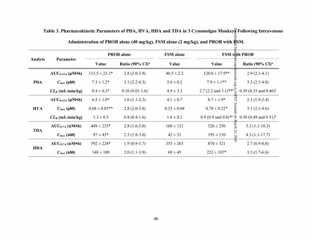
46
Table 3. Pharmacokinetic Parameters of PDA, HVA, HDA and TDA in 3 Cynomolgus Monkeys Following Intravenous
Administration of PROB alone (40 mg/kg), FSM alone (2 mg/kg), and PROB with FSM.
Analyte Parameter PROB alone FSM alone FSM with PROB
Value Ratio (90% CI)a Value Value Ratio (90% CI)a
PDA
AUC0-24 h (µM•h) 113.5 ± 23.3* 2.8 (2.0-3.9) 40.5 ± 2.2 120.0 ± 17.9** 2.9 (2.1-4.1)
Cmax (μM) 7.3 ± 1.2* 3.1 (2.2-4.3) 2.4 ± 0.2 7.9 ± 1.1** 3.3 (2.3-4.8)
CLR (mL/min/kg) 0.4 ± 0.2* 0.10 (0.01-1.6) 4.9 ± 3.3 2.7 (2.2 and 3.1)**b 0.39 (0.33 and 0.46)b
HVA
AUC0-24 h (µM•h) 6.5 ± 1.0* 1.6 (1.1-2.3) 4.1 ± 0.7 8.7 ± 1.9* 2.1 (1.9-2.4)
Cmax (μM) 0.68 ± 0.03** 2.8 (2.0-3.8) 0.25 ± 0.04 0.78 ± 0.22* 3.1 (2.1-4.6)
CLR (mL/min/kg) 1.3 ± 0.5 0.8 (0.4-1.6) 1.6 ± 0.2 0.9 (0.9 and 0.8)*b 0.50 (0.49 and 0.51)b
TDA AUC0-7 h (nM•h) 449 ± 233* 2.8 (1.6-5.0) 168 ± 121 526 ± 250 3.3 (1.1-10.3)
Cmax (nM) 97 ± 43* 2.3 (1.8-3.0) 42 ± 21 195 ± 110 4.3 (1.1-17.7)
HDA AUC0-7 h (nM•h) 592 ± 224* 1.9 (0.9-3.7) 355 ± 263 870 ± 321 2.7 (0.9-8.0)
Cmax (nM) 148 ± 109 2.0 (1.1-3.9) 69 ± 45 223 ± 107* 3.3 (1.7-6.6)
This article has not been copyedited and formatted. The final version may differ from this version.DMD Fast Forward. Published on November 21, 2017 as DOI: 10.1124/dmd.117.077586
at ASPE
T Journals on M
arch 25, 2020dm
d.aspetjournals.orgD
ownloaded from

47
Pharmacokinetic parameters were determined as described in Materials and Methods. Data are shown as mean ± SD (n = 3). PROB was
intravenously dosed 30 min prior to FSM administration in the coadministration treatment group.
a Ratio of pharmacokinetics parameter of PROB treatment to FSM treatment.
b Cage malfunction prevented urine collection from one monkey in the coadministration treatment group (n = 2).
* P < 0.05 and ** P < 0.01, when the parameter was compared with that administered with FSM alone.
This article has not been copyedited and formatted. The final version may differ from this version.DMD Fast Forward. Published on November 21, 2017 as DOI: 10.1124/dmd.117.077586
at ASPE
T Journals on M
arch 25, 2020dm
d.aspetjournals.orgD
ownloaded from

DMD # 77586
48
Figure 1
Plasma Sample Collection from
Monkey DDI Study
Data Alignment and Analysis Considering Experimental Factors
Metabolomics DataAcquisition
Period IPROB alone
Period IIFSM alone
Period IIIFSM-PROB
PotentialOAT1/3 EndogenousBiomarkers Identification
1 hr
2 4 hr
0
5 0
1 0 0
1 5 0
Inte
ns
ity
F S M a lo n e
P R O B a lo n e
F S M -P R O B
• Time-Course of Changes in Plasma Levels of PDA, HVA, HDA and TDA Validation
• PDA and HVA Transport in OAT1- and OAT3-HEK Cells Validation
This article has not been copyedited and formatted. The final version may differ from this version.DMD Fast Forward. Published on November 21, 2017 as DOI: 10.1124/dmd.117.077586
at ASPE
T Journals on M
arch 25, 2020dm
d.aspetjournals.orgD
ownloaded from

49
Figure 2
0.00
0.01
0.10
1.00
10.00
100.00
1,000.00
0 4 8 12 16 20 24
FSM
Con
cent
ratio
n [μ
M]
Time [h]
FSM aloneFSM with PROB
(A)
1
10
100
1,000
10,000
0 4 8 12 16 20 24
PRO
B C
once
ntra
tion
[μM
]
Time [h]
PROB alone
FSM with PROB(B)
0-3 h
0 -7 h
0 -24 h
0
2
4
6
8 F S M a lo n e
F S M w ith P R O BF
SM
Uri
na
ry E
xcr
etio
n R
ate
(µm
oL
/h)
(C )
* *
* *
* *
This article has not been copyedited and formatted. The final version may differ from this version.DMD Fast Forward. Published on November 21, 2017 as DOI: 10.1124/dmd.117.077586
at ASPE
T Journals on M
arch 25, 2020dm
d.aspetjournals.orgD
ownloaded from

50
Figure 3
0
3
6
9
12
0 4 8 12 16 20 24
PDA
Con
cent
ratio
n [μ
M]
Time [h]
PROB aloneFSM aloneFSM with PROB
(A)
0.0
0.3
0.6
0.9
1.2
0 4 8 12 16 20 24
HV
A C
once
ntra
tion
[μM
]
Time [h]
PROB aloneFSM aloneFSM with PROB
(B)
0
100
200
300
400
0 1 2 3 4 5 6 7
TDA
Con
cent
ratio
n [n
M]
Time [h]
PROB aloneFSM aloneFSM with PROB
(C)
0
100
200
300
400
0 1 2 3 4 5 6 7
HD
A C
once
ntra
tion
[nM
]
Time [h]
PROB aloneFSM aloneFSM with PROB
(D)
This article has not been copyedited and formatted. The final version may differ from this version.DMD Fast Forward. Published on November 21, 2017 as DOI: 10.1124/dmd.117.077586
at ASPE
T Journals on M
arch 25, 2020dm
d.aspetjournals.orgD
ownloaded from

51
Figure 4
Mock -H
E K
OA T 1 -H
E K
Mo ck -H
E K
OA T 2 -H
E K
Mo ck -H
E K
OA T 3 -H
E K
Mo ck -H
E K
OC T 2 -H
E K
02468
2 0
4 0
6 0
8 0
B ) . H V A U p ta k e b y B a so la te r a l T r a n sp o r te r s
Fol
d U
pta
ke
of H
VA
ove
r M
ock
Cel
ls T
reat
ed w
ith
ou I
nh
ibit
or
H V A (5 µ M ) + I n h ib i to r
O A T 1 O A T 2 O A T 3 O C T 2
* * *
$ $ $
* * *$ $
* * *
$ $ $
Mock -H
E K
OA
T 1 -HE K
Mo ck -H
E K
OA
T 2 -HE K
Mo ck -H
E K
OA
T 3 -HE K
Mo ck -H
E K
OC
T 2 -HE K
0
5
1 0
1 5
2 0
2 51 0 0
1 5 0
2 0 0
2 5 0
3 0 0
C ). P r o b e U p ta k e b y B a s o la te r a l T r a n sp o r te r s
Fo
ld U
pta
ke
of
Pro
be
ov
er M
ock
Cel
ls T
rea
ted
wit
ho
u I
nh
ibit
or
P r o b e + I n h ib i to r
O A T 1 O A T 2 O A T 3 O C T 2
* *
$ $
* * *$ $ $
* * *
$ $
* *
Mock -H
E K
OA
T 1 -HE K
Mo ck -H
E K
OA
T 2 -HE K
Mo ck -H
E K
OA
T 3 -HE K
Mo ck -H
E K
OC
T 2 -HE K
02468
1 0
5 0
1 0 0
1 5 0
A ) . P D A U p ta k e b y B a s o la te r a l T r a n s p o r te r s
Fo
ld U
pta
ke
of
PD
A o
ver
Mo
ckC
ells
Tre
ate
d w
ith
ou
In
hib
ito
r
P D A (1 µ M ) + I n h ib i to r
O A T 1 O A T 2 O A T 3 O C T 2
*
$
* * *
$ $ $
This article has not been copyedited and formatted. The final version may differ from this version.DMD Fast Forward. Published on November 21, 2017 as DOI: 10.1124/dmd.117.077586
at ASPE
T Journals on M
arch 25, 2020dm
d.aspetjournals.orgD
ownloaded from

52
Figure 5
Mock -H
E K
OA
T 4 -HE K
Mo ck -H
E K
MA
T E 1 -HE K
Mo ck -H
E K
MA
T E 2K-H
E K
0
1
2
3
4
5
A ). P D A U p ta k e b y A p ic a l T r a n sp o r te rs
Fo
ld U
pta
ke
of
PD
A o
ver
Mo
ckC
ells
Tre
ate
d w
ith
ou
In
hib
ito
r
P D A (1 µ M ) + I n h ib i to r
M A T E 1 M A T E 2 KO A T 4
* *$ $
Mock -H
E K
OA
T 4 -HE K
Mo ck -H
E K
MA
T E 1 -HE K
Mo ck -H
E K
MA
T E 2K-H
E K
0 .0
0 .5
1 .0
1 .5
2 .0
B ) . H V A U p ta k e b y A p ic a l T r a n sp o r te rs
Fo
ld U
pta
ke
of
HV
A o
ver
Mo
ckC
ells
Tre
ate
d w
ith
ou
In
hib
ito
r
H V A (5 µ M ) + I n h ib i to r
M A T E 1 M A T E 2 KO A T 4
Mock -H
E K
OA
T 4 -HE K
Mo ck -H
E K
MA
T E 1 -HE K
Mo ck -H
E K
MA
T E 2K-H
E K
0
1 0
2 0
3 0
4 0
C ). P r o b e U p ta k e b y A p ic a l T r a n sp o r te r s
Fo
ld U
pta
ke
of
Pro
be
ov
er M
ock
Cel
ls T
rea
ted
wit
ho
u I
nh
ibit
or
P r o b e + I n h ib i to r
M A T E 1 M A T E 2 K
* * *
* * *
$ $ $ $ $ $
O A T 4
* * *
$ $ $
This article has not been copyedited and formatted. The final version may differ from this version.DMD Fast Forward. Published on November 21, 2017 as DOI: 10.1124/dmd.117.077586
at ASPE
T Journals on M
arch 25, 2020dm
d.aspetjournals.orgD
ownloaded from
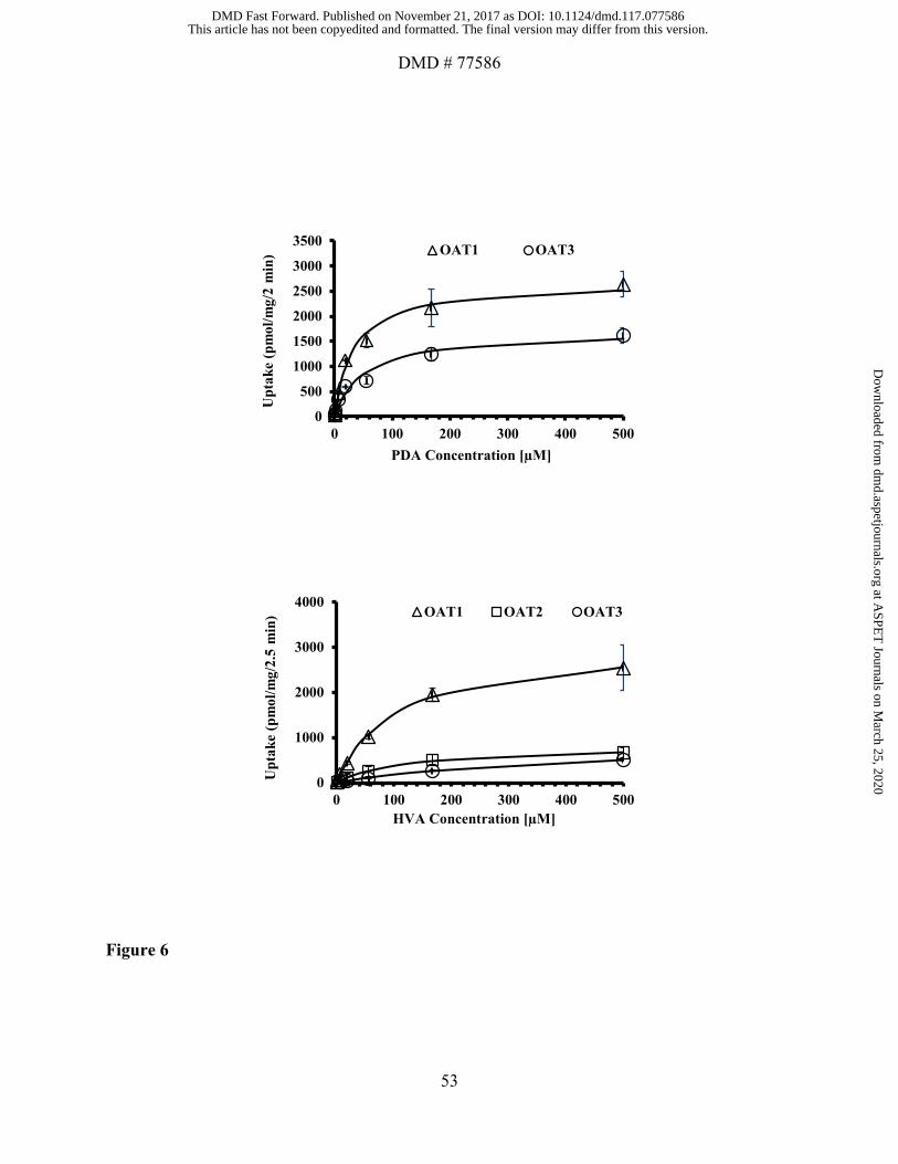
DMD # 77586
53
Figure 6
0
500
1000
1500
2000
2500
3000
3500
0 100 200 300 400 500
Upt
ake
(pm
ol/m
g/2
min
)
PDA Concentration [µM]
OAT1 OAT3
0
1000
2000
3000
4000
0 100 200 300 400 500
Upt
ake
(pm
ol/m
g/2.
5 m
in)
HVA Concentration [µM]
OAT1 OAT2 OAT3
This article has not been copyedited and formatted. The final version may differ from this version.DMD Fast Forward. Published on November 21, 2017 as DOI: 10.1124/dmd.117.077586
at ASPE
T Journals on M
arch 25, 2020dm
d.aspetjournals.orgD
ownloaded from
Recommended