Earth and Planetary Science Letters 392 (2014) 67–79
Contents lists available at ScienceDirect
Earth and Planetary Science Letters
www.elsevier.com/locate/epsl
Coupled Fe and S isotope variations in pyrite nodules from Archeanshale
Johanna Marin-Carbonne a,b,∗,Claire Rollion-Bard c, Andrey Bekker d, Olivier Rouxel e,Andrea Agangi f, Barbara Cavalazzi f,g, Cora C. Wohlgemuth-Ueberwasser f,h,Axel Hofmann f, Kevin D. McKeegan a
a Department of Earth, Planetary, and Space Sciences, University of California – 595 Charles Young Drive East, Los Angeles, CA 90095, USAb Institut de Physique du Globe – Sorbonne Paris Cité, Université Paris Diderot, CNRS, 1 rue Jussieu, 75238 Paris cedex 05, Francec CRPG-CNRS, Université de Lorraine, 15 rue Notre Dame des Pauvres, BP 20, 54501 Vandoeuvre lès Nancy, Franced Department of Earth Sciences, University of California, Riverside, CA, 92521, USAe Department of Marine Geosciences, IFREMER, Centre de Brest, 29280 Plouzané, Francef Department of Geology, University of Johannesburg, PO Box 524, Auckland Park 2006, Johannesburg, South Africag Dipartimento di Scienze Biologiche, Geologiche e Ambientali, Università di Bologna, Via Zamboni 67, I-40127 Bologna, Italyh PetroTectonics Centre, Department of Geological Sciences, Stockholm University, 106 91 Stockholm, Sweden
a r t i c l e i n f o a b s t r a c t
Article history:Received 4 October 2013Received in revised form 29 January 2014Accepted 3 February 2014Available online 25 February 2014Editor: B. Marty
Keywords:pyrite nodulesFe and S isotopesArcheanSIMS
Iron and sulfur isotope compositions recorded in ancient rocks and minerals such as pyrite (FeS2) havebeen widely used as a proxy for early microbial metabolisms and redox evolution of the oceans. However,most previous studies focused on only one of these isotopic systems. Herein, we illustrate the importanceof in-situ and coupled study of Fe and S isotopes on two pyrite nodules in a c. 2.7 Ga shale from theBubi Greenstone Belt (Zimbabwe). Fe and S isotope compositions were measured both by bulk-samplemass spectrometry techniques and by ion microprobe in-situ methods (Secondary Ion Mass Spectrometry,SIMS). Spatially-resolved analysis across the nodules shows a large range of variations at micrometer-scalefor both Fe and S isotope compositions, with δ56Fe and δ34S values from −2.1 to +0.7� and from−0.5 to +8.2�, respectively, and �33S values from −1.6 to +2.9�. The Fe and S isotope variationsin these nodules cannot be explained by tandem operation of Dissimilatory Iron Reduction (DIR) andBacterial Sulfate Reduction (BSR) as was previously proposed, but rather they reflect the contributions ofdifferent Fe and S sources during a complex diagenetic history. Pyrite formed from two different mineralprecursors: (1) mackinawite precipitated in the water column, and (2) greigite formed in the sedimentduring early diagenesis. The in-situ analytical approach reveals a complex history of the pyrite nodulegrowth and allows us to better constrain environmental conditions during the Archean.
© 2014 Elsevier B.V. All rights reserved.
1. Introduction
Variations in Fe and S isotope composition of sedimentarypyrites have placed important constraints on the chemistry andredox evolution of the Earth’s ocean and atmosphere over ge-ological time (e.g. Bekker et al., 2004; Farquhar et al., 2000;Johnson et al., 2008; Rouxel et al., 2003, 2005; Strauss, 2003).These variations record isotope fractionations during redox reac-tions, which in some cases might have been biologically medi-ated (Archer and Vance, 2006; Beard et al., 1999; Johnson et al.,2008). Sulfur isotopes have been used to document ancient mi-
* Corresponding author at: Institut de Physique du Globe – Sorbonne Paris Cité,Université Paris Diderot, CNRS, 1 rue Jussieu, 75238 Paris cedex 05, France.
E-mail address: [email protected] (J. Marin-Carbonne).
http://dx.doi.org/10.1016/j.epsl.2014.02.0090012-821X/© 2014 Elsevier B.V. All rights reserved.
crobial metabolisms, because the fractionations produced by livingorganisms can be large and reflect specific metabolic activity (e.g.Johnston, 2011). Indeed, in the process called Bacterial Sulfate Re-duction (BSR), dissolved sulfate is used by eukaryotes, bacteria, andcertain groups of Archea as an electron acceptor during organic Cremineralization or H2 oxidation. In other redox reactions, hydro-gen sulfide can act as an electron donor associated with O2, NO3 orCO2 reduction (Canfield, 2001). BSR preferentially metabolizes 32Srelative to 34S, thereby producing fractionation of the S isotopes upto 70� (Canfield, 2001; Sim et al., 2011).
The discovery of mass-independent fractionation (MIF) of S iso-topes in Archean sedimentary sulfides and sulfates (Farquhar et al.,2000) has deeply modified our understanding of the Precambriansulfur cycle. The prevailing hypothesis to explain S-MIF is basedon experimental studies and atmospheric models that invoke
68 J. Marin-Carbonne et al. / Earth and Planetary Science Letters 392 (2014) 67–79
photochemical reactions, and suggest an absence of atmosphericoxygen before 2.4 Ga (Farquhar et al., 2000; Ono et al., 2003;Pavlov and Kasting, 2002). Farquhar et al. (2001) suggested thatArchean S-MIF was created via photolysis of SO2 and/or SO byshort ultraviolet radiation (< 220 nm) that penetrated deeply intothe Archean atmosphere due to the lack of O2. SO2 photodissoci-ation in an oxygen-free atmosphere produces water-soluble SO2−
4with negative �33S values and elemental sulfur aerosols, mostlyS8, with positive �33S values (Farquhar et al., 2000). Although al-ternative views are still debated (see for alternative view Oduro etal., 2011; e.g., Ohmoto et al., 2006), O2 level below 10−5 PresentAtmospheric Level (PAL) is considered critical for the production ofmass-independent fractionation in S isotopes and its preservationin the sedimentary rock record (Thiemens, 2001). Based on pho-tochemical experiments, it was proposed that Archean seawatersulfate had negative �33S values (Farquhar et al., 2000; Ono et al.,2009, 2003), whereas Archean disseminated pyrites have mostlypositive �33S values (Farquhar and Wing, 2003). This is consis-tent with S isotope composition of hydrothermal barite and sulfidein base-metal barren, distal exhalite deposits (e.g., Farquhar andWing, 2003), which derived their S from seawater sulfate in distal,hydrothermally-influenced low-energy environments.
The Fe isotope composition of sedimentary pyrite is highly sen-sitive to the size of dissolved Fe(II) and S reservoirs and hencecan place important constrains on the redox state and chemistryof Precambrian oceans (Guilbaud et al., 2011; Rouxel et al., 2005).Iron isotopes fractionate through both redox and non-redox reac-tions (e.g., Johnson and Beard, 2005). Hence interpretation of ironisotope record of Fe-bearing marine deposits requires an under-standing of Fe sources and formation mechanisms of iron-bearingminerals, including oxides, sulfides, carbonates, and silicates, inmarine sediments. Each of these minerals can have various ori-gins, such as detrital, biochemical and hydrothermal, and thus canrecord different Fe isotope fractionations (Heimann et al., 2010;Johnson et al., 2008; Planavsky et al., 2009; Rouxel et al., 2005).
By coupling the S and Fe isotope systems, it is possible togain additional insights into the processes resulting in the for-mation of pyrite (Archer and Vance, 2006; Fabre et al., 2011;Hofmann et al., 2009; Rouxel et al., 2008). For example, it hasbeen proposed that coupled Fe and S isotope data can be usedas a proxy for microbial Fe(III) and sulfate reduction, especially forArchean sediments (Archer and Vance, 2006). Studies of Archeanrocks frequently use a bulk rock approach, although a growingnumber of studies focuses on individual crystals or crystal aggre-gates. A recent SIMS study of S isotopes of various Archean pyriteshas shown large intra-grain variability in δ34S values (Kamber andWhitehouse, 2007). Similarly, SIMS Fe isotope studies have alsoshown large ranges of δ56Fe values, from +0.9� to +5.2� ina single magnetite grain (Marin-Carbonne et al., 2011) and from−4.2� to +2.9� in pyrites from the 2.72 Ga Tumbiana Forma-tion (Yoshiya et al., 2012). Such variations have been interpretedas indicating multi-stage mineral formation and/or mineral alter-ation processes, and highlight the importance of spatially resolvedanalyses to better constrain processes in the water column, andduring diagenesis and, possibly, metamorphism. Pyrite nodules inArchean shales have been extensively investigated in the contextof the diagenetic history of ancient sedimentary rocks and havebeen used as a proxy for paleoenvironmental conditions on theearly Earth (Bekker et al., 2004; Kakegawa et al., 1998; Ono etal., 2009, 2003; Rouxel et al., 2005). Whether there exists anyisotopic variability in Fe within individual pyrite nodules is notknown.
Herein, we present bulk measurements along with in-situisotopic (Fe and S) and trace element analyses of pyrite nod-ules hosted in carbonaceous shale from the Late Archean (2.83–2.70 Ga) Bubi Greenstone Belt, Zimbabwe. We use these data to
constrain the origin and growth history of these pyrite nodulesand to explore the possibility of a microbially-induced fractiona-tion.
2. Samples and methods
2.1. Samples
We investigated pyrite nodules present in Late Archean car-bonaceous shales from core 690B92-02 (see supplementary mate-rial S1) drilled along the eastern margin of the Bubi GreenstoneBelt (Zimbabwe) north of the Damba nickel prospect (Hofmannet al., 2014; Prendergast, 2003). The drill core intersects basaltand underlying carbonaceous shale (see supplementary materialS2 for the detailed stratigraphic log), which are considered cor-relative with the 2.7 Ga Reliance Formation and the 2.83 to2.70 Ga Manjeri Formation of the Belingwe Greenstone Belt inZimbabwe (Hofmann and Kusky, 2004; Prendergast, 2003; Stoneet al., 1994). The metamorphic grade is not well-established, butrocks in the Bubi Greenstone Belt have been subjected to lowergreenschist grade metamorphism at most (Dziggel et al., 1998;Saggerson and Turner, 1976).
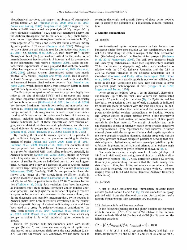
Pyrite occurs as nodules (up to 1 cm in diameter), discontinu-ous laminae (up to 0.5 cm thick), rare veinlets and disseminatedgrains (< 20 μm in size) in the shale (Fig. 1). Pyrite formed be-fore burial compaction at the stage of early diagenesis as indicatedby ellipsoidal shape of nodules with the long axis parallel to bed-ding, laminations in shale that bend around the nodules and rareptygmatically folded veinlets perpendicular to bedding. Nodulesand laminae consist of either massive pyrite, a fine intergrowthof pyrite with the host matrix, or concentrations of fine pyritecrystals in the host material. The margin of nodules and lami-nae frequently consist of relatively coarse pyrite crystals suggestiveof recrystallization. Pyrite represents the only observed Fe-sulfidemineral phase, with the exception of minor chalcopyrite crystals inthe more coarsely-crystalline domains. Nodules frequently containmarginal pressure shadows elongated parallel to bedding that con-sists of quartz, phyllosilicates, and rare coarsely-crystalline pyrite.A foliation is present in the shale and oriented at an oblique angleto bedding. A summary of pyrite textures is shown in Fig. 1.
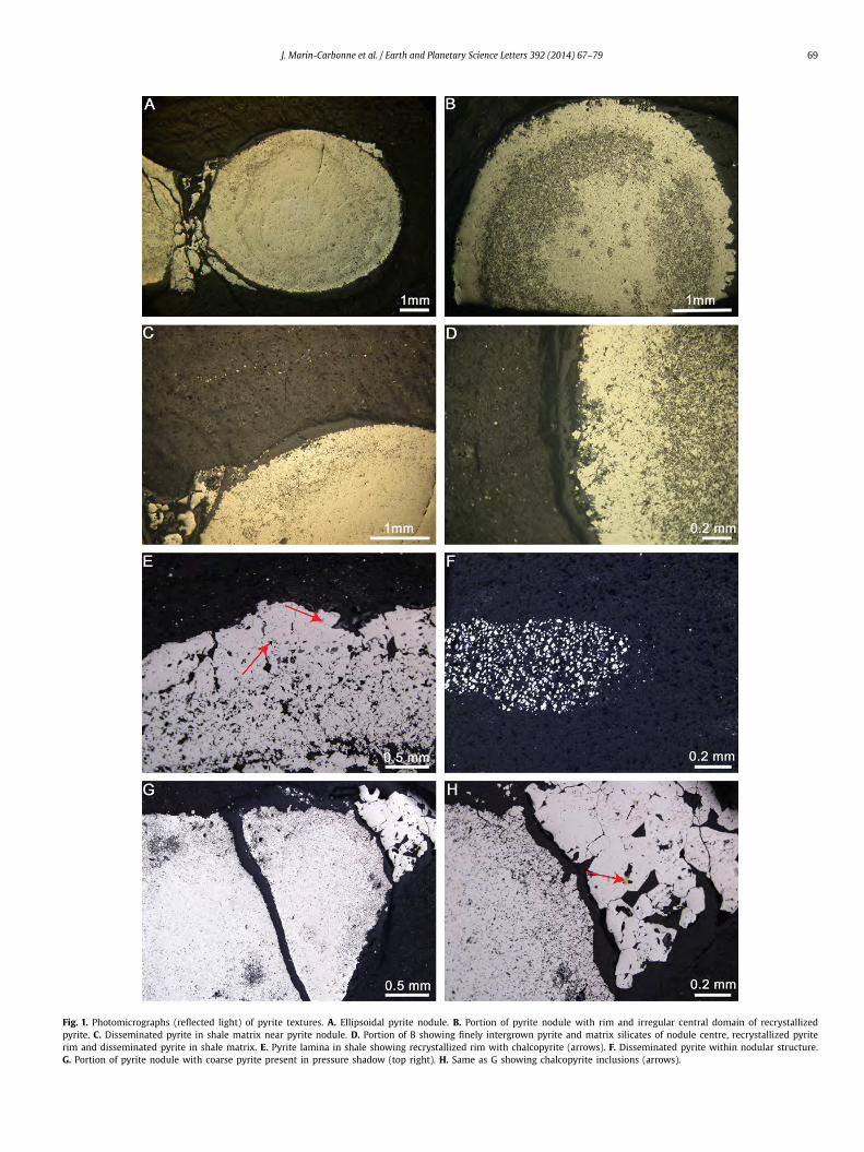
Our study focuses on a single sample of shale (at depth of144.5 m in drill core) containing several circular to slightly ellip-soidal pyrite nodules (Fig. 2). X-ray diffraction analysis (X-PertPro,University of Johannesburg) indicates that the shale mainly con-sists of quartz, illite, Fe-chlorite, and minor calcite mineral phases.The shale is relatively rich in organic carbon with Corg contentranging from 6.4 to 9.3 wt.% (Eltra Elemental Analyzer, Universityof Manitoba).
2.2. Methods
A slab of shale containing two, immediately adjacent pyritenodules (called nodule 1 and 2 in Fig. 2) was embedded in epoxy,polished with 1 μm size diamond paste and Au-coated for in-situisotopic measurements (see supplementary material S3).
2.2.1. Bulk sample Fe and S isotope analysesIn the following sections, iron and sulfur isotopes are expressed
in delta notation (δ56Fe, δ33S, and δ34S) relative to the interna-tional standards IRMM 14 (for Fe) and V-CDT (for S) based on thefollowing equation:
δ2A = [(2A/1Asample)/(2A/1Astandard
) − 1] × 1000
where A is Fe or S, 1 and 2 represent the heavy and light iso-topes, respectively (54 and 56 for Fe and 34 or 33 and 32 for S).
J. Marin-Carbonne et al. / Earth and Planetary Science Letters 392 (2014) 67–79 69
Fig. 1. Photomicrographs (reflected light) of pyrite textures. A. Ellipsoidal pyrite nodule. B. Portion of pyrite nodule with rim and irregular central domain of recrystallizedpyrite. C. Disseminated pyrite in shale matrix near pyrite nodule. D. Portion of B showing finely intergrown pyrite and matrix silicates of nodule centre, recrystallized pyriterim and disseminated pyrite in shale matrix. E. Pyrite lamina in shale showing recrystallized rim with chalcopyrite (arrows). F. Disseminated pyrite within nodular structure.G. Portion of pyrite nodule with coarse pyrite present in pressure shadow (top right). H. Same as G showing chalcopyrite inclusions (arrows).
70 J. Marin-Carbonne et al. / Earth and Planetary Science Letters 392 (2014) 67–79
Fig. 2. a. Scanning electron microscopy (SEM) images of the polished surface of the investigated pyrite nodules. The white dots indicate the in-situ Fe and S isotope and traceelement spot analyses. b. The Co and Ni concentration (wt.%, EPMA) profile of nodule 1 shows difference between the coarse-grained rim and the fine-grained core (see textfor the definition of core and rim). c. An SEM image of the core-rim transition zone showing textural difference.
Mass-independent fractionation has been calculated as the devia-tion from the Terrestrial Fractionation Line (TFL), using the mass-discrimination law (Farquhar et al., 2000):
�33 S =(
ln
(δ33 S
1000+ 1
)− 0.515 × ln
(δ34 S
1000
)+ 1
),
where the factor 0.515 defines the slope of the TFL.Pyrite nodules from the same sample of shale used for in-situ
work were drilled, crushed and millimetric-sized pyrite particleswere hand-picked. Sulfur isotope ratios of pyrite particles weredetermined at the Geophysical Laboratory using techniques de-scribed by Hu et al. (2003). Pyrite particles (0.5 to 1 mg) fromnodules were reacted with fluorine under a 25 W CO2 infraredlaser at 25–30 Torr in a vacuum chamber to produce SF6, whichwas then purified by dual gas chromatography. Multiple sulfurisotope ratios were measured with a Thermo Scientific MAT 253mass spectrometer in dual inlet mode (Hofmann et al., 2009;Ono et al., 2009). The precision for δ34S, δ33S, and �33S valueswas determined by the multiple analyses of CDT material and in-ternal reference materials (Maine and Alpha Aesar pyrite) and isbetter than 0.34�, 0.19�, and 0.03�, respectively (2σ ).
Fe isotope compositions were measured following the proce-dure described in Rouxel et al. (2005). Hand-picked particles frompyrite nodules were dissolved in concentrated HNO3–HCl acid mix-ture and Fe was purified on Bio-Rad AG1X8 anion resin. Fe isotoperatios were determined with a Thermo Scientific Neptune multi-collector inductively-coupled plasma mass-spectrometer operated
at IFREMER, Pole Spectrometry Ocean, Brest in France. Long-termreproducibility of δ56Fe measurements was determined on dupli-cate analysis of reference material and is about 0.08� (2σ ).
2.2.3. SIMS analysesIron isotope compositions were measured in-situ with a Cameca
ims 1270 ion microprobe at both CRPG (Nancy, France) and UCLA(Los Angeles, USA) following the procedure described in detail inMarin-Carbonne et al. (2011). Briefly, a 16O− primary beam ofabout 10 nA intensity was focused to a spot of about 15 μm. Themass resolution was set ∼7,000 and 54Fe+ and 56Fe+ were mea-sured in multicollection mode with two off-axis Faraday cups. Thegains of these Faraday cups were determined at the beginning ofthe analytical session and drift was monitored by frequent anal-yses of standards interspersed among analyses of the unknowns.The background of each detector was measured during the pre-sputtering for 1 min, i.e. at the beginning of each analysis. Ion cur-rents converted to count rates were typically ∼ 2.108 counts persecond (cps) for 56Fe. An analysis consisted of 30 cycles with 5 sacquisition time. Chromium was monitored on the masses 52 and53 by using electron multipliers, but chromium levels were neg-ligible in all samples. The internal precision for δ56Fe values wastypically better than 0.1� (2σ ), and the external reproducibilitybased on multiple measurements of our pyrite reference material(Balmat with δ56Fe = −0.399�; Whitehouse and Fedo, 2007) wasbetter than 0.2� (2σ ).
J. Marin-Carbonne et al. / Earth and Planetary Science Letters 392 (2014) 67–79 71
Sulfur isotope compositions were measured on the Cameca ims1280 HR2 (CRPG, Nancy, France) by simultaneous measurementsof 32S−, 33S−, and 34S− in multicollection mode with three off-axis Faraday cups. The relative gains of the Faraday cups wereintercalibrated at the beginning of each analytical session. The ana-lytical method is described in detail in Thomassot et al. (2009) andPhilippot et al. (2012) and is only summarized here. A Cs+ primarybeam of 5 nA intensity was focused to a spot of about 15–20 μm.Typical 32S− intensity was between 6 and 10.108 counts per sec-ond (cps) depending on the sulfide mineral analyzed. Several pyritestandards (Maine, Philippot et al., 2012 and Balmat pyrite, courtesyof M. Whitehouse) were used to determine (i) the instrumentalmass fractionation, and (ii) the reference mass discrimination line,from which �33S values were calculated. A typical analysis con-sists of 2 min of presputtering followed by data acquisition in 30cycles of 3 s each. The background of the detectors was measuredduring the presputtering and was then corrected for each analysis.The internal precision achieved under these conditions was betterthan 0.05� for δ34S and 0.03� for δ33S values (2σ ). The externalprecision, which is the standard deviation calculated from repeatedmeasurements on various reference materials, was 0.40� (2σ ) forδ34S and 0.06� (2σ ) for �33S values.
3. Results
Fe and S isotope compositions and EPMA trace element concen-trations have been obtained across each nodule (see supplemen-tary material S3 and S4), while LA-ICP-MS trace element concen-tration profiles were performed only for nodule 2 (see supplemen-tary material S4).
3.1. δ56 F e variations
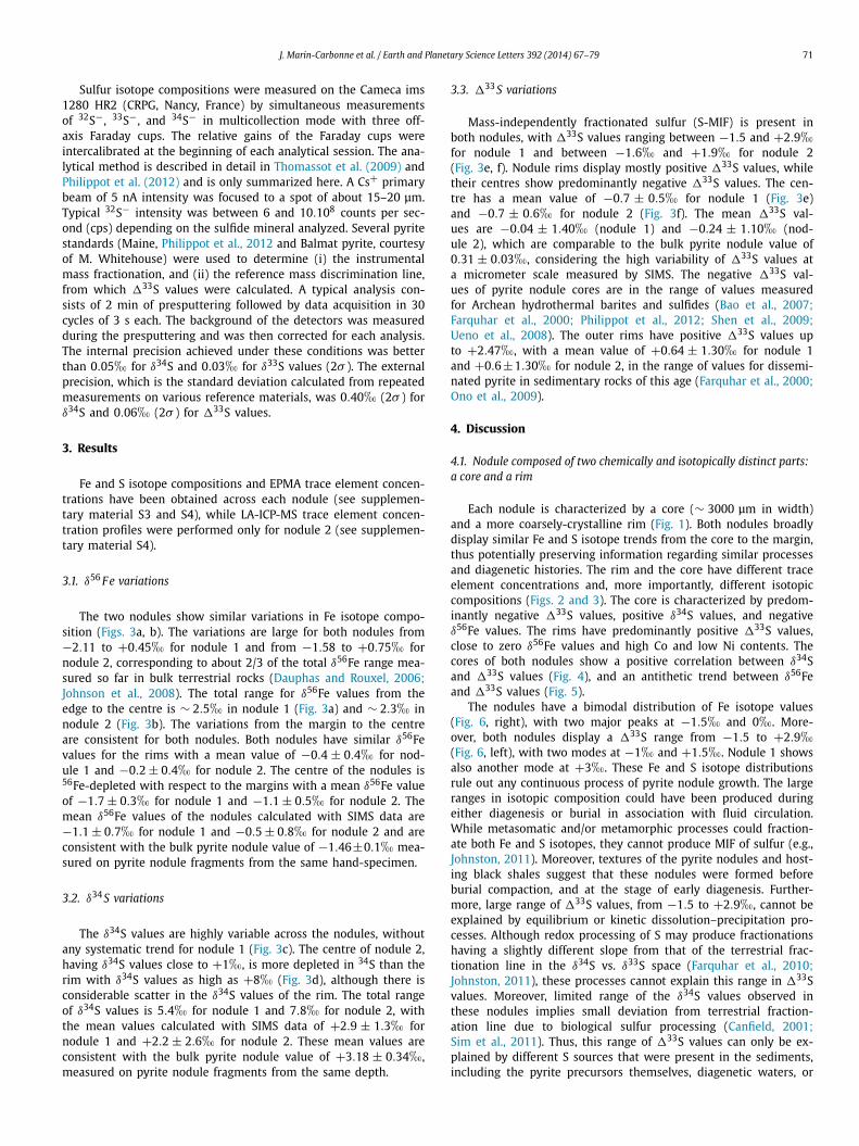
The two nodules show similar variations in Fe isotope compo-sition (Figs. 3a, b). The variations are large for both nodules from−2.11 to +0.45� for nodule 1 and from −1.58 to +0.75� fornodule 2, corresponding to about 2/3 of the total δ56Fe range mea-sured so far in bulk terrestrial rocks (Dauphas and Rouxel, 2006;Johnson et al., 2008). The total range for δ56Fe values from theedge to the centre is ∼ 2.5� in nodule 1 (Fig. 3a) and ∼ 2.3� innodule 2 (Fig. 3b). The variations from the margin to the centreare consistent for both nodules. Both nodules have similar δ56Fevalues for the rims with a mean value of −0.4 ± 0.4� for nod-ule 1 and −0.2 ± 0.4� for nodule 2. The centre of the nodules is56Fe-depleted with respect to the margins with a mean δ56Fe valueof −1.7 ± 0.3� for nodule 1 and −1.1 ± 0.5� for nodule 2. Themean δ56Fe values of the nodules calculated with SIMS data are−1.1 ± 0.7� for nodule 1 and −0.5 ± 0.8� for nodule 2 and areconsistent with the bulk pyrite nodule value of −1.46±0.1� mea-sured on pyrite nodule fragments from the same hand-specimen.
3.2. δ34 S variations
The δ34S values are highly variable across the nodules, withoutany systematic trend for nodule 1 (Fig. 3c). The centre of nodule 2,having δ34S values close to +1�, is more depleted in 34S than therim with δ34S values as high as +8� (Fig. 3d), although there isconsiderable scatter in the δ34S values of the rim. The total rangeof δ34S values is 5.4� for nodule 1 and 7.8� for nodule 2, withthe mean values calculated with SIMS data of +2.9 ± 1.3� fornodule 1 and +2.2 ± 2.6� for nodule 2. These mean values areconsistent with the bulk pyrite nodule value of +3.18 ± 0.34�,measured on pyrite nodule fragments from the same depth.
3.3. �33 S variations
Mass-independently fractionated sulfur (S-MIF) is present inboth nodules, with �33S values ranging between −1.5 and +2.9�for nodule 1 and between −1.6� and +1.9� for nodule 2(Fig. 3e, f). Nodule rims display mostly positive �33S values, whiletheir centres show predominantly negative �33S values. The cen-tre has a mean value of −0.7 ± 0.5� for nodule 1 (Fig. 3e)and −0.7 ± 0.6� for nodule 2 (Fig. 3f). The mean �33S val-ues are −0.04 ± 1.40� (nodule 1) and −0.24 ± 1.10� (nod-ule 2), which are comparable to the bulk pyrite nodule value of0.31 ± 0.03�, considering the high variability of �33S values ata micrometer scale measured by SIMS. The negative �33S val-ues of pyrite nodule cores are in the range of values measuredfor Archean hydrothermal barites and sulfides (Bao et al., 2007;Farquhar et al., 2000; Philippot et al., 2012; Shen et al., 2009;Ueno et al., 2008). The outer rims have positive �33S values upto +2.47�, with a mean value of +0.64 ± 1.30� for nodule 1and +0.6±1.30� for nodule 2, in the range of values for dissemi-nated pyrite in sedimentary rocks of this age (Farquhar et al., 2000;Ono et al., 2009).
4. Discussion
4.1. Nodule composed of two chemically and isotopically distinct parts:a core and a rim
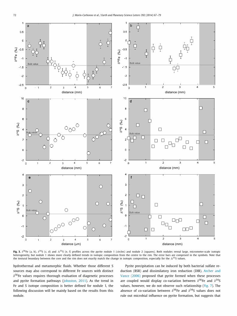
Each nodule is characterized by a core (∼ 3000 μm in width)and a more coarsely-crystalline rim (Fig. 1). Both nodules broadlydisplay similar Fe and S isotope trends from the core to the margin,thus potentially preserving information regarding similar processesand diagenetic histories. The rim and the core have different traceelement concentrations and, more importantly, different isotopiccompositions (Figs. 2 and 3). The core is characterized by predom-inantly negative �33S values, positive δ34S values, and negativeδ56Fe values. The rims have predominantly positive �33S values,close to zero δ56Fe values and high Co and low Ni contents. Thecores of both nodules show a positive correlation between δ34Sand �33S values (Fig. 4), and an antithetic trend between δ56Feand �33S values (Fig. 5).
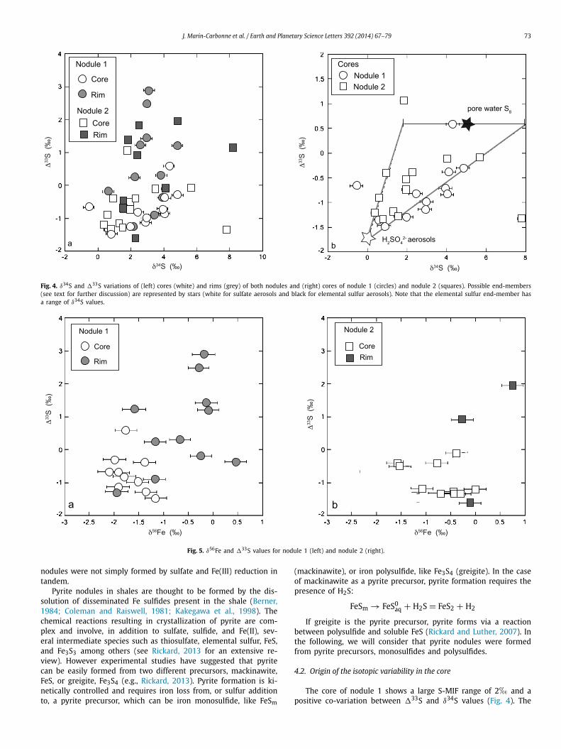
The nodules have a bimodal distribution of Fe isotope values(Fig. 6, right), with two major peaks at −1.5� and 0�. More-over, both nodules display a �33S range from −1.5 to +2.9�(Fig. 6, left), with two modes at −1� and +1.5�. Nodule 1 showsalso another mode at +3�. These Fe and S isotope distributionsrule out any continuous process of pyrite nodule growth. The largeranges in isotopic composition could have been produced duringeither diagenesis or burial in association with fluid circulation.While metasomatic and/or metamorphic processes could fraction-ate both Fe and S isotopes, they cannot produce MIF of sulfur (e.g.,Johnston, 2011). Moreover, textures of the pyrite nodules and host-ing black shales suggest that these nodules were formed beforeburial compaction, and at the stage of early diagenesis. Further-more, large range of �33S values, from −1.5 to +2.9�, cannot beexplained by equilibrium or kinetic dissolution–precipitation pro-cesses. Although redox processing of S may produce fractionationshaving a slightly different slope from that of the terrestrial frac-tionation line in the δ34S vs. δ33S space (Farquhar et al., 2010;Johnston, 2011), these processes cannot explain this range in �33Svalues. Moreover, limited range of the δ34S values observed inthese nodules implies small deviation from terrestrial fraction-ation line due to biological sulfur processing (Canfield, 2001;Sim et al., 2011). Thus, this range of �33S values can only be ex-plained by different S sources that were present in the sediments,including the pyrite precursors themselves, diagenetic waters, or
72 J. Marin-Carbonne et al. / Earth and Planetary Science Letters 392 (2014) 67–79
Fig. 3. δ56Fe (a, b), δ34S (c, d) and �33S (e, f) profiles across the pyrite nodule 1 (circles) and nodule 2 (squares). Both nodules reveal large, micrometer-scale isotopicheterogeneity, but nodule 1 shows more clearly defined trends in isotopic composition from the centre to the rim. The error bars are comprised in the symbols. Note thatthe textural boundary between the core and the rim does not exactly match the change in isotopic composition, especially for the �33S values.
hydrothermal and metamorphic fluids. Whether those different S
sources may also correspond to different Fe sources with distinct
δ56Fe values requires thorough evaluation of diagenetic processes
and pyrite formation pathways (Johnston, 2011). As the trend in
Fe and S isotope composition is better defined for nodule 1, the
following discussion will be mainly based on the results from this
nodule.
Pyrite precipitation can be induced by both bacterial sulfate re-
duction (BSR) and dissimilatory iron reduction (DIR). Archer and
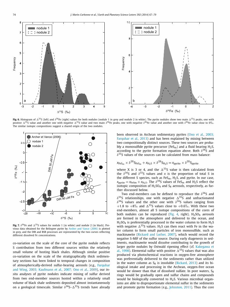
Vance (2006) proposed that pyrite formed when these processes
are coupled would display co-variation between δ56Fe and δ34S
values, however, we do not observe such relationship (Fig. 7). The
absence of co-variation between δ56Fe and δ34S values does not
rule out microbial influence on pyrite formation, but suggests that
J. Marin-Carbonne et al. / Earth and Planetary Science Letters 392 (2014) 67–79 73
Fig. 4. δ34S and �33S variations of (left) cores (white) and rims (grey) of both nodules and (right) cores of nodule 1 (circles) and nodule 2 (squares). Possible end-members(see text for further discussion) are represented by stars (white for sulfate aerosols and black for elemental sulfur aerosols). Note that the elemental sulfur end-member hasa range of δ34S values.
Fig. 5. δ56Fe and �33S values for nodule 1 (left) and nodule 2 (right).
nodules were not simply formed by sulfate and Fe(III) reduction intandem.
Pyrite nodules in shales are thought to be formed by the dis-solution of disseminated Fe sulfides present in the shale (Berner,1984; Coleman and Raiswell, 1981; Kakegawa et al., 1998). Thechemical reactions resulting in crystallization of pyrite are com-plex and involve, in addition to sulfate, sulfide, and Fe(II), sev-eral intermediate species such as thiosulfate, elemental sulfur, FeS,and Fe3S3 among others (see Rickard, 2013 for an extensive re-view). However experimental studies have suggested that pyritecan be easily formed from two different precursors, mackinawite,FeS, or greigite, Fe3S4 (e.g., Rickard, 2013). Pyrite formation is ki-netically controlled and requires iron loss from, or sulfur additionto, a pyrite precursor, which can be iron monosulfide, like FeSm
(mackinawite), or iron polysulfide, like Fe3S4 (greigite). In the caseof mackinawite as a pyrite precursor, pyrite formation requires thepresence of H2S:
FeSm → FeS0aq + H2S = FeS2 + H2
If greigite is the pyrite precursor, pyrite forms via a reactionbetween polysulfide and soluble FeS (Rickard and Luther, 2007). Inthe following, we will consider that pyrite nodules were formedfrom pyrite precursors, monosulfides and polysulfides.
4.2. Origin of the isotopic variability in the core
The core of nodule 1 shows a large S-MIF range of 2� and apositive co-variation between �33S and δ34S values (Fig. 4). The
74 J. Marin-Carbonne et al. / Earth and Planetary Science Letters 392 (2014) 67–79
Fig. 6. Histogram of �33S (left) and δ56Fe (right) values for both nodules (nodule 1 in grey and nodule 2 in white). The pyrite nodules show two main �33S peaks, one withpositive �33S value and another one with negative �33S value and two main δ56Fe peaks, one with negative δ56Fe value and another one with δ56Fe value close to 0�.The similar isotopic compositions suggest a shared origin of the two nodules.
Fig. 7. δ56Fe and �33S values for nodule 1 (in white) and nodule 2 (in black). Pre-vious data obtained for the Belingwe pyrite by Archer and Vance (2006) is plottedin grey, and the DIR and BSR processes are represented by the two curves reflectingdifferent dissolved Fe concentrations.
co-variation on the scale of the core of the pyrite nodule reflectsS contribution from two different sources within the relativelysmall volume of hosting black shales. Although similar positiveco-variation on the scale of the stratigraphically thick sedimen-tary sections has been linked to temporal changes in compositionof atmospherically-derived sulfur-bearing aerosols (e.g., Farquharand Wing, 2003; Kaufmann et al., 2007; Ono et al., 2009), our in-situ analyses of pyrite nodules indicate mixing of sulfur derivedfrom two end-member sources hosted within a relatively smallvolume of black shale sediments deposited almost instantaneouslyon a geological timescale. Similar δ34S–�33S trends have already
been observed in Archean sedimentary pyrites (Ono et al., 2003;Farquhar et al., 2013) and has been explained by mixing betweentwo compositionally distinct sources. These two sources are proba-bly a monosulfide pyrite precursor (FeSm) and a fluid bearing H2S,according to the pyrite formation equation above. Both δ34S andδ33S values of the sources can be calculated from mass balance:
xFeSm × δ3XSFeSm + xH2S × δ3XSH2S = xpyrite × δ3XSpyrite
where X is 3 or 4, and the �33S value is then calculated fromthe δ34S and δ33S values and x is the proportion of total S inthe different S species, such as FeSm, H2S, and pyrite. In our case,xpyrite = xFeSm + xH2S. The δ34S values of FeSm and H2S reflect theisotopic composition of H2SO4 and S8 aerosols, respectively, as fur-ther discussed below.
Two end-members can be defined to reproduce the δ34S andδ33S relationships, one with negative �33S and unfractionatedδ34S values and the other one with δ34S values ranging from+1.8 to +8� and �33S values close to +0.6�. With these twoend-members, almost all S isotope compositions of the cores ofboth nodules can be reproduced (Fig. 4, right). H2SO4 aerosolsare formed in the atmosphere and delivered to the ocean, andH2SO4 is preferentially processed in the water column to form H2Swith negative �33S values. H2S can then react with Fe in the wa-ter column to form small particles of iron monosulfide, such asmackinawite (Rickard and Luther, 2007), which would record thenegative S-MIF of the sulfur source. During early diagenesis in sed-iments, mackinawite would dissolve contributing to the growth oflarger pyrite nodules by Ostwald ripening effect (cf. Kakegawa etal., 1998). Elemental sulfur with positive �33S values that was alsoproduced via photochemical reactions in oxygen-free atmospherewas preferentially delivered to the sediments rather than utilizedin the water column as S8 is insoluble (Rickard, 2013) and its bi-ological intake and processing in the Archean, oxygen-free oceanwould be slower than that of dissolved sulfate. In pore waters, S8rings would be gradually open and sulfur chains and compoundswould be biologically converted to H2S. Various microbial organ-isms are able to disproportionate elemental sulfur in the sedimentsand promote pyrite formation (e.g., Johnston, 2011). Thus the core
J. Marin-Carbonne et al. / Earth and Planetary Science Letters 392 (2014) 67–79 75
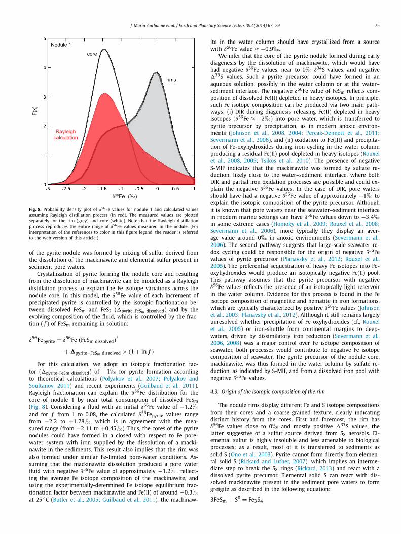
Fig. 8. Probability density plot of δ56Fe values for nodule 1 and calculated valuesassuming Rayleigh distillation process (in red). The measured values are plottedseparately for the rim (grey) and core (white). Note that the Rayleigh distillationprocess reproduces the entire range of δ56Fe values measured in the nodule. (Forinterpretation of the references to color in this figure legend, the reader is referredto the web version of this article.)
of the pyrite nodule was formed by mixing of sulfur derived fromthe dissolution of the mackinawite and elemental sulfur present insediment pore waters.
Crystallization of pyrite forming the nodule core and resultingfrom the dissolution of mackinawite can be modeled as a Rayleighdistillation process to explain the Fe isotope variations across thenodule core. In this model, the δ56Fe value of each increment ofprecipitated pyrite is controlled by the isotopic fractionation be-tween dissolved FeSm and FeS2 (�pyrite-FeSm dissolved) and by theevolving composition of the fluid, which is controlled by the frac-tion ( f ) of FeSm remaining in solution:
δ56Fepyrite = δ56Fe (FeSm dissolved)i
+ �pyrite–FeSm dissolved × (1 + ln f )
For this calculation, we adopt an isotopic fractionation fac-tor (�pyrite-FeSm dissolved) of −1� for pyrite formation accordingto theoretical calculations (Polyakov et al., 2007; Polyakov andSoultanov, 2011) and recent experiments (Guilbaud et al., 2011).Rayleigh fractionation can explain the δ56Fe distribution for thecore of nodule 1 by near total consumption of dissolved FeSm(Fig. 8). Considering a fluid with an initial δ56Fe value of −1.2�and for f from 1 to 0.08, the calculated δ56Fepyrite values rangefrom −2.2 to +1.78�, which is in agreement with the mea-sured range (from −2.11 to +0.45�). Thus, the cores of the pyritenodules could have formed in a closed with respect to Fe pore-water system with iron supplied by the dissolution of a macki-nawite in the sediments. This result also implies that the rim wasalso formed under similar Fe-limited pore-water conditions. As-suming that the mackinawite dissolution produced a pore waterfluid with negative δ56Fe value of approximately −1.2�, reflect-ing the average Fe isotope composition of the mackinawite, andusing the experimentally-determined Fe isotope equilibrium frac-tionation factor between mackinawite and Fe(II) of around −0.3�at 25 ◦C (Butler et al., 2005; Guilbaud et al., 2011), the mackinaw-
ite in the water column should have crystallized from a sourcewith δ56Fe value ≈ −0.9�.
We infer that the core of the pyrite nodule formed during earlydiagenesis by the dissolution of mackinawite, which would havehad negative δ56Fe values, near to 0� δ34S values, and negative�33S values. Such a pyrite precursor could have formed in anaqueous solution, possibly in the water column or at the water–sediment interface. The negative δ56Fe value of FeSm reflects com-position of dissolved Fe(II) depleted in heavy isotopes. In principle,such Fe isotope composition can be produced via two main path-ways: (i) DIR during diagenesis releasing Fe(II) depleted in heavyisotopes (δ56Fe ≈ −2�) into pore water, which is transferred topyrite precursor by precipitation, as in modern anoxic environ-ments (Johnson et al., 2008, 2004; Percak-Dennett et al., 2011;Severmann et al., 2006), and (ii) oxidation to Fe(III) and precipita-tion of Fe-oxyhydroxides during iron cycling in the water columnproducing a residual Fe(II) pool depleted in heavy isotopes (Rouxelet al., 2008, 2005; Tsikos et al., 2010). The presence of negativeS-MIF indicates that the mackinawite was formed by sulfate re-duction, likely close to the water–sediment interface, where bothDIR and partial iron oxidation processes are possible and could ex-plain the negative δ56Fe values. In the case of DIR, pore watersshould have had a negative δ56Fe value of approximately −1� toexplain the isotopic composition of the pyrite precursor. Althoughit is known that pore waters near the seawater–sediment interfacein modern marine settings can have δ56Fe values down to −3.4�in some extreme cases (Homoky et al., 2009; Rouxel et al., 2008;Severmann et al., 2006), more typically they display an aver-age value around 0� in anoxic environments (Severmann et al.,2006). The second pathway suggests that large-scale seawater re-dox cycling could be responsible for the origin of negative δ56Fevalues of pyrite precursor (Planavsky et al., 2012; Rouxel et al.,2005). The preferential sequestration of heavy Fe isotopes into Fe-oxyhydroxides would produce an isotopically negative Fe(II) pool.This pathway assumes that the pyrite precursor with negativeδ56Fe values reflects the presence of an isotopically light reservoirin the water column. Evidence for this process is found in the Feisotope composition of magnetite and hematite in iron formations,which are typically characterized by positive δ56Fe values (Johnsonet al., 2003; Planavsky et al., 2012). Although it still remains largelyunresolved whether precipitation of Fe oxyhydroxides (cf., Rouxelet al., 2005) or iron-shuttle from continental margins to deep-waters, driven by dissimilatory iron reduction (Severmann et al.,2006, 2008) was a major control over Fe isotope composition ofseawater, both processes would contribute to negative Fe isotopecomposition of seawater. The pyrite precursor of the nodule core,mackinawite, was thus formed in the water column by sulfate re-duction, as indicated by S-MIF, and from a dissolved iron pool withnegative δ56Fe values.
4.3. Origin of the isotopic composition of the rim
The nodule rims display different Fe and S isotope compositionsfrom their cores and a coarse-grained texture, clearly indicatingdistinct history from the cores. First and foremost, the rim hasδ56Fe values close to 0� and mostly positive �33S values, thelatter suggestive of a sulfur source derived from S8 aerosols. El-emental sulfur is highly insoluble and less amenable to biologicalprocesses; as a result, most of it is transferred to sediments assolid S (Ono et al., 2003). Pyrite cannot form directly from elemen-tal solid S (Rickard and Luther, 2007), which implies an interme-diate step to break the S8 rings (Rickard, 2013) and react with adissolved pyrite precursor. Elemental solid S can react with dis-solved mackinawite present in the sediment pore waters to formgreigite as described in the following equation:
3FeSm + S0 = Fe3S4
76 J. Marin-Carbonne et al. / Earth and Planetary Science Letters 392 (2014) 67–79
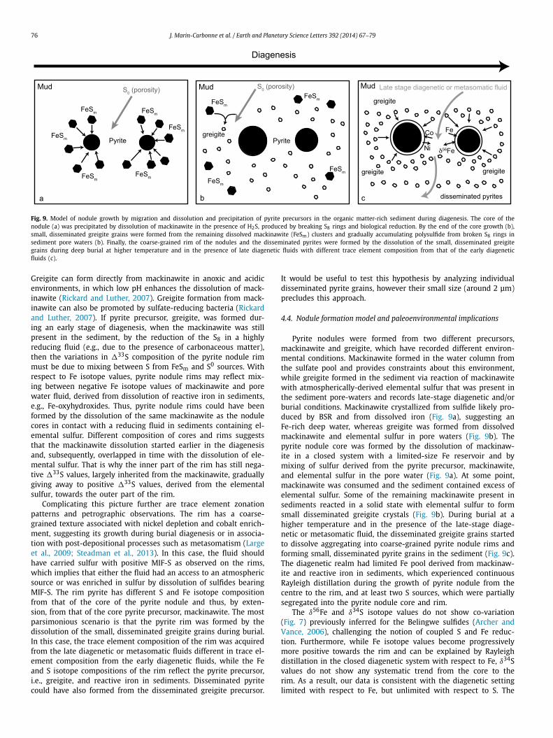
Fig. 9. Model of nodule growth by migration and dissolution and precipitation of pyrite precursors in the organic matter-rich sediment during diagenesis. The core of thenodule (a) was precipitated by dissolution of mackinawite in the presence of H2S, produced by breaking S8 rings and biological reduction. By the end of the core growth (b),small, disseminated greigite grains were formed from the remaining dissolved mackinawite (FeSm) clusters and gradually accumulating polysulfide from broken S8 rings insediment pore waters (b). Finally, the coarse-grained rim of the nodules and the disseminated pyrites were formed by the dissolution of the small, disseminated greigitegrains during deep burial at higher temperature and in the presence of late diagenetic fluids with different trace element composition from that of the early diageneticfluids (c).
Greigite can form directly from mackinawite in anoxic and acidicenvironments, in which low pH enhances the dissolution of mack-inawite (Rickard and Luther, 2007). Greigite formation from mack-inawite can also be promoted by sulfate-reducing bacteria (Rickardand Luther, 2007). If pyrite precursor, greigite, was formed dur-ing an early stage of diagenesis, when the mackinawite was stillpresent in the sediment, by the reduction of the S8 in a highlyreducing fluid (e.g., due to the presence of carbonaceous matter),then the variations in �33S composition of the pyrite nodule rimmust be due to mixing between S from FeSm and S0 sources. Withrespect to Fe isotope values, pyrite nodule rims may reflect mix-ing between negative Fe isotope values of mackinawite and porewater fluid, derived from dissolution of reactive iron in sediments,e.g., Fe-oxyhydroxides. Thus, pyrite nodule rims could have beenformed by the dissolution of the same mackinawite as the nodulecores in contact with a reducing fluid in sediments containing el-emental sulfur. Different composition of cores and rims suggeststhat the mackinawite dissolution started earlier in the diagenesisand, subsequently, overlapped in time with the dissolution of ele-mental sulfur. That is why the inner part of the rim has still nega-tive �33S values, largely inherited from the mackinawite, graduallygiving away to positive �33S values, derived from the elementalsulfur, towards the outer part of the rim.
Complicating this picture further are trace element zonationpatterns and petrographic observations. The rim has a coarse-grained texture associated with nickel depletion and cobalt enrich-ment, suggesting its growth during burial diagenesis or in associa-tion with post-depositional processes such as metasomatism (Largeet al., 2009; Steadman et al., 2013). In this case, the fluid shouldhave carried sulfur with positive MIF-S as observed on the rims,which implies that either the fluid had an access to an atmosphericsource or was enriched in sulfur by dissolution of sulfides bearingMIF-S. The rim pyrite has different S and Fe isotope compositionfrom that of the core of the pyrite nodule and thus, by exten-sion, from that of the core pyrite precursor, mackinawite. The mostparsimonious scenario is that the pyrite rim was formed by thedissolution of the small, disseminated greigite grains during burial.In this case, the trace element composition of the rim was acquiredfrom the late diagenetic or metasomatic fluids different in trace el-ement composition from the early diagenetic fluids, while the Feand S isotope compositions of the rim reflect the pyrite precursor,i.e., greigite, and reactive iron in sediments. Disseminated pyritecould have also formed from the disseminated greigite precursor.
It would be useful to test this hypothesis by analyzing individualdisseminated pyrite grains, however their small size (around 2 μm)precludes this approach.
4.4. Nodule formation model and paleoenvironmental implications
Pyrite nodules were formed from two different precursors,mackinawite and greigite, which have recorded different environ-mental conditions. Mackinawite formed in the water column fromthe sulfate pool and provides constraints about this environment,while greigite formed in the sediment via reaction of mackinawitewith atmospherically-derived elemental sulfur that was present inthe sediment pore-waters and records late-stage diagenetic and/orburial conditions. Mackinawite crystallized from sulfide likely pro-duced by BSR and from dissolved iron (Fig. 9a), suggesting anFe-rich deep water, whereas greigite was formed from dissolvedmackinawite and elemental sulfur in pore waters (Fig. 9b). Thepyrite nodule core was formed by the dissolution of mackinaw-ite in a closed system with a limited-size Fe reservoir and bymixing of sulfur derived from the pyrite precursor, mackinawite,and elemental sulfur in the pore water (Fig. 9a). At some point,mackinawite was consumed and the sediment contained excess ofelemental sulfur. Some of the remaining mackinawite present insediments reacted in a solid state with elemental sulfur to formsmall disseminated greigite crystals (Fig. 9b). During burial at ahigher temperature and in the presence of the late-stage diage-netic or metasomatic fluid, the disseminated greigite grains startedto dissolve aggregating into coarse-grained pyrite nodule rims andforming small, disseminated pyrite grains in the sediment (Fig. 9c).The diagenetic realm had limited Fe pool derived from mackinaw-ite and reactive iron in sediments, which experienced continuousRayleigh distillation during the growth of pyrite nodule from thecentre to the rim, and at least two S sources, which were partiallysegregated into the pyrite nodule core and rim.
The δ56Fe and δ34S isotope values do not show co-variation(Fig. 7) previously inferred for the Belingwe sulfides (Archer andVance, 2006), challenging the notion of coupled S and Fe reduc-tion. Furthermore, while Fe isotope values become progressivelymore positive towards the rim and can be explained by Rayleighdistillation in the closed diagenetic system with respect to Fe, δ34Svalues do not show any systematic trend from the core to therim. As a result, our data is consistent with the diagenetic settinglimited with respect to Fe, but unlimited with respect to S. The
J. Marin-Carbonne et al. / Earth and Planetary Science Letters 392 (2014) 67–79 77
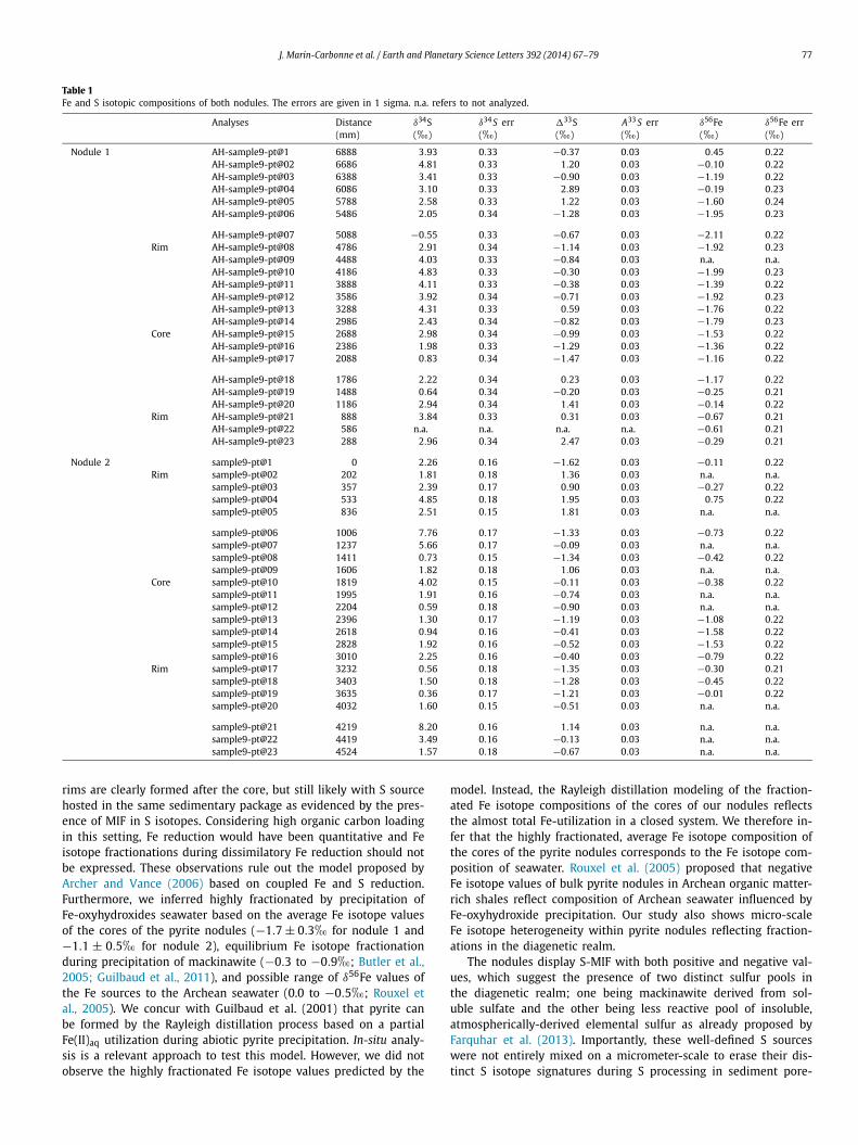
Table 1Fe and S isotopic compositions of both nodules. The errors are given in 1 sigma. n.a. refers to not analyzed.
Analyses Distance(mm)
δ34S(�)
δ34 S err(�)
�33S(�)
A33 S err(�)
δ56Fe(�)
δ56Fe err(�)
Nodule 1 AH-sample9-pt@1 6888 3.93 0.33 −0.37 0.03 0.45 0.22AH-sample9-pt@02 6686 4.81 0.33 1.20 0.03 −0.10 0.22AH-sample9-pt@03 6388 3.41 0.33 −0.90 0.03 −1.19 0.22AH-sample9-pt@04 6086 3.10 0.33 2.89 0.03 −0.19 0.23AH-sample9-pt@05 5788 2.58 0.33 1.22 0.03 −1.60 0.24AH-sample9-pt@06 5486 2.05 0.34 −1.28 0.03 −1.95 0.23
AH-sample9-pt@07 5088 −0.55 0.33 −0.67 0.03 −2.11 0.22Rim AH-sample9-pt@08 4786 2.91 0.34 −1.14 0.03 −1.92 0.23
AH-sample9-pt@09 4488 4.03 0.33 −0.84 0.03 n.a. n.a.AH-sample9-pt@10 4186 4.83 0.33 −0.30 0.03 −1.99 0.23AH-sample9-pt@11 3888 4.11 0.33 −0.38 0.03 −1.39 0.22AH-sample9-pt@12 3586 3.92 0.34 −0.71 0.03 −1.92 0.23AH-sample9-pt@13 3288 4.31 0.33 0.59 0.03 −1.76 0.22AH-sample9-pt@14 2986 2.43 0.34 −0.82 0.03 −1.79 0.23
Core AH-sample9-pt@15 2688 2.98 0.34 −0.99 0.03 −1.53 0.22AH-sample9-pt@16 2386 1.98 0.33 −1.29 0.03 −1.36 0.22AH-sample9-pt@17 2088 0.83 0.34 −1.47 0.03 −1.16 0.22
AH-sample9-pt@18 1786 2.22 0.34 0.23 0.03 −1.17 0.22AH-sample9-pt@19 1488 0.64 0.34 −0.20 0.03 −0.25 0.21AH-sample9-pt@20 1186 2.94 0.34 1.41 0.03 −0.14 0.22
Rim AH-sample9-pt@21 888 3.84 0.33 0.31 0.03 −0.67 0.21AH-sample9-pt@22 586 n.a. n.a. n.a. n.a. −0.61 0.21AH-sample9-pt@23 288 2.96 0.34 2.47 0.03 −0.29 0.21
Nodule 2 sample9-pt@1 0 2.26 0.16 −1.62 0.03 −0.11 0.22Rim sample9-pt@02 202 1.81 0.18 1.36 0.03 n.a. n.a.
sample9-pt@03 357 2.39 0.17 0.90 0.03 −0.27 0.22sample9-pt@04 533 4.85 0.18 1.95 0.03 0.75 0.22sample9-pt@05 836 2.51 0.15 1.81 0.03 n.a. n.a.
sample9-pt@06 1006 7.76 0.17 −1.33 0.03 −0.73 0.22sample9-pt@07 1237 5.66 0.17 −0.09 0.03 n.a. n.a.sample9-pt@08 1411 0.73 0.15 −1.34 0.03 −0.42 0.22sample9-pt@09 1606 1.82 0.18 1.06 0.03 n.a. n.a.
Core sample9-pt@10 1819 4.02 0.15 −0.11 0.03 −0.38 0.22sample9-pt@11 1995 1.91 0.16 −0.74 0.03 n.a. n.a.sample9-pt@12 2204 0.59 0.18 −0.90 0.03 n.a. n.a.sample9-pt@13 2396 1.30 0.17 −1.19 0.03 −1.08 0.22sample9-pt@14 2618 0.94 0.16 −0.41 0.03 −1.58 0.22sample9-pt@15 2828 1.92 0.16 −0.52 0.03 −1.53 0.22sample9-pt@16 3010 2.25 0.16 −0.40 0.03 −0.79 0.22
Rim sample9-pt@17 3232 0.56 0.18 −1.35 0.03 −0.30 0.21sample9-pt@18 3403 1.50 0.18 −1.28 0.03 −0.45 0.22sample9-pt@19 3635 0.36 0.17 −1.21 0.03 −0.01 0.22sample9-pt@20 4032 1.60 0.15 −0.51 0.03 n.a. n.a.
sample9-pt@21 4219 8.20 0.16 1.14 0.03 n.a. n.a.sample9-pt@22 4419 3.49 0.16 −0.13 0.03 n.a. n.a.sample9-pt@23 4524 1.57 0.18 −0.67 0.03 n.a. n.a.
rims are clearly formed after the core, but still likely with S sourcehosted in the same sedimentary package as evidenced by the pres-ence of MIF in S isotopes. Considering high organic carbon loadingin this setting, Fe reduction would have been quantitative and Feisotope fractionations during dissimilatory Fe reduction should notbe expressed. These observations rule out the model proposed byArcher and Vance (2006) based on coupled Fe and S reduction.Furthermore, we inferred highly fractionated by precipitation ofFe-oxyhydroxides seawater based on the average Fe isotope valuesof the cores of the pyrite nodules (−1.7 ± 0.3� for nodule 1 and−1.1 ± 0.5� for nodule 2), equilibrium Fe isotope fractionationduring precipitation of mackinawite (−0.3 to −0.9�; Butler et al.,2005; Guilbaud et al., 2011), and possible range of δ56Fe values ofthe Fe sources to the Archean seawater (0.0 to −0.5�; Rouxel etal., 2005). We concur with Guilbaud et al. (2001) that pyrite canbe formed by the Rayleigh distillation process based on a partialFe(II)aq utilization during abiotic pyrite precipitation. In-situ analy-sis is a relevant approach to test this model. However, we did notobserve the highly fractionated Fe isotope values predicted by the
model. Instead, the Rayleigh distillation modeling of the fraction-ated Fe isotope compositions of the cores of our nodules reflectsthe almost total Fe-utilization in a closed system. We therefore in-fer that the highly fractionated, average Fe isotope composition ofthe cores of the pyrite nodules corresponds to the Fe isotope com-position of seawater. Rouxel et al. (2005) proposed that negativeFe isotope values of bulk pyrite nodules in Archean organic matter-rich shales reflect composition of Archean seawater influenced byFe-oxyhydroxide precipitation. Our study also shows micro-scaleFe isotope heterogeneity within pyrite nodules reflecting fraction-ations in the diagenetic realm.
The nodules display S-MIF with both positive and negative val-ues, which suggest the presence of two distinct sulfur pools inthe diagenetic realm; one being mackinawite derived from sol-uble sulfate and the other being less reactive pool of insoluble,atmospherically-derived elemental sulfur as already proposed byFarquhar et al. (2013). Importantly, these well-defined S sourceswere not entirely mixed on a micrometer-scale to erase their dis-tinct S isotope signatures during S processing in sediment pore-
78 J. Marin-Carbonne et al. / Earth and Planetary Science Letters 392 (2014) 67–79
waters. We relate this unique preservation of atmospheric S sig-nature during diagenetic processing to the difference in reactivitybetween these two pools with the insoluble sulfur compounds de-rived from atmospheric elemental sulfur being less reactive withrespect to the seawater sulfate. Fe and S isotope variations ob-served in the Archean pyrite nodules reflect the contribution oftwo atmospherically-derived S sources and Fe pool fractionated byprecipitation of iron oxyhydroxides as well by diagenetic processesinvolved in the growth of pyrite nodules.
5. Conclusions
In-situ coupled Fe and S isotope study of the c. 2.7 Ga pyritenodules have revealed an extreme isotopic variability both in Feand S at the micrometer scale (see Table 1). It has been shownfor the first time that these nodules have both positive and nega-tive �33S values and highly variable Fe isotope composition. Theyreveal a complex crystallization history with, at least, two stepsof dissolution and precipitation of two different pyrite precur-sors involved in the Ostwald ripening process. The core of thepyrite nodules has grown by dissolution of fine-grained mackinaw-ite precipitated in the water column, while the rims were formedduring the late diagenesis by dissolution of disseminated greigitegrains crystallized during early diagenesis, when remaining dis-solved FeSm clusters reacted with broken S8 rings. The nodulesthus record S processing in two different environments, water col-umn and sediment pore-waters, at the time when the Earth’s at-mosphere was still anoxic. They also indicate the presence of twodistinct and yet contemporaneous seawater sulfur pools derivedfrom the atmosphere, soluble sulfate and insoluble elemental sul-fur, which, being different in their isotopic composition, are nowresolvable at a micrometer-scale within the pyrite nodules. It alsoimplies that temporal trends in �33S values earlier inferred frombulk-rock S isotope analyses showing positive co-variation between�33S and δ34S values (e.g., Kaufmann et al., 2007; Ono et al.,2009) represent local variations in relative contribution of thesetwo atmospherically-derived S sources and cannot be reliably usedfor global correlation of hosting sedimentary successions.
This study, based on two isotopic systems, reveals that path-ways for pyrite nodule formation are complex and involve severalpyrite precursors, such as mackinawite and greigite, and isotopi-cally distinct S and Fe sources. However, the isotopic and elementalrecords spatially preserved in these pyrite nodules do constrainthe conditions for the precipitation of pyrite precursors and thuscan be used as proxies for paleoenvironmental conditions in theArchaen oceans and atmosphere. The study also illustrates the im-portance of in-situ and coupled isotopic studies to reveal diagenetichistories and pyrite formation pathways.
Acknowledgements
Frantz Ossa Ossa is thanked for assistance with trace ele-ment analysis by x-ray diffraction. Vincent Busigny and JabraneLabidi (IPGP, Paris) are thanked for fruitful discussions. CarolineGuillemette (CRPG, Nancy) is thanked for the measurements ofS isotopic composition of Spain pyrite. Participation by A.B. wassupported by NSERC Discovery Grant. The UCLA ion microprobefacility is partially supported by a grant from the NSF Instrumen-tation and Facilities program. This is IPGP contribution no. 3491.
Appendix A. Supplementary material
Supplementary material related to this article can be found on-line at http://dx.doi.org/10.1016/j.epsl.2014.02.009.
References
Archer, C., Vance, D., 2006. Coupled Fe and S isotope evidence for Archean microbialFe(III) and sulfate reduction. Geology 34, 153–156.
Bao, H., Rumble III, D., Lowe, D.R., 2007. The five stable isotope compositions ofFig Tree barites: Implications on sulfur cycle in ca. 3.2 Ga oceans. Geochim.Cosmochim. Acta 71, 4868–4879.
Beard, B.L., Johnson, C.M., Cox, L., Sun, H., Nealson, K.H., Aguilar, C., 1999. Iron iso-tope biosignatures. Science 285, 1889–1892.
Bekker, A., Holland, H.D., Wang, P.L., Rumble, D., Stein, H.J., Hannah, J.L., Coetzee, L.L.,Beukes, N.J., 2004. Dating the rise of atmospheric oxygen. Nature 427, 117–120.
Berner, R.A., 1984. Sedimentary pyrite formation: An update. Geochim. Cosmochim.Acta 48, 605–615.
Butler, I.B., Archer, C., Vance, D., Oldroyd, A., Rickard, D., 2005. Fe isotope fractiona-tion on FeS formation in ambient aqueous solution. Earth Planet. Sci. Lett. 236,430–442.
Canfield, D.E., 2001. Isotope fractionation by natural populations of sulfate-reducingbacteria. Geochim. Cosmochim. Acta 65, 1117–1124.
Coleman, M.L., Raiswell, R., 1981. Carbon, oxygen and sulphur isotope variations inconcretions from the Upper Lias of N.E. England. Geochim. Cosmochim. Acta 45,329–340.
Dauphas, N., Rouxel, O., 2006. Mass spectrometry and natural variations of iron iso-topes. Mass Spectrom. Rev. 25, 515–550.
Dziggel, A., Klemd, R., Stevens, G., 1998. Gold mineralization at Turk Mine, Zim-babwe: A geochemical and statistical investigation of the hydrothermally over-printed host rock. Information Circular. Economic Geology Research Unit 327,University of the Witwatersrand, Johannesburg. 16 pp.
Fabre, S., Nedelec, A., Poitrasson, F., Strauss, H., Thomazo, C., Nogueira, A., 2011. Ironand sulphur isotopes from the Carajas mining province (Para, Brazil): Implica-tions for the oxidation of the ocean and the atmosphere across the Archaean,Proterozoic transition. Chem. Geol. 289, 124–139.
Farquhar, J., Wing, B., 2003. Multiple sulfur isotopes and the evolution of the atmo-sphere. Earth Planet. Sci. Lett. 213, 1–13.
Farquhar, J., Bao, H., Thiemens, M., 2000. Atmospheric influence of Earth’s earliestsulfur cycle. Science 289, 756–758.
Farquhar, J., Savarino, J., Airieau, S., Thiemens, M.H., 2001. Observation ofwavelength-sensitive mass-independent sulfur isotope effects during SO2
photolysis: Implications for the early atmosphere. J. Geophys. Res. 106,32829–32839.
Farquhar, J., Wu, N., Canfield, D.E., Oduro, H., 2010. Connections between sulfur cy-cle evolution, sulfur isotopes, sediments, and base metal sulfide deposits. Econ.Geol. 105, 509–533.
Farquhar, J., Cliff, J., Zerkle, A.L., Kamyshny, A., Poulton, S.W., Claire, M., Adams,D., Harms, B., 2013. Pathways for Neoarchean pyrite formation constrainedby mass-independent sulfur isotopes. Proc. Natl. Acad. Sci. USA 110 (44),17638–17643.
Guilbaud, R., Butler, I.B., Ellam, R.M., 2011. Abiotic pyrite formation produces a largeFe isotope fractionation. Science 332, 1548–1551.
Heimann, A., Johnson, C.M., Beard, B.L., Valley, J.W., Roden, E.E., Spicuzza, M.J.,Beukes, N.J., 2010. Fe, C, and O isotope compositions of banded iron forma-tion carbonates demonstrate a major role for dissimilatory iron reduction in∼ 2.5 Ga marine environments. Earth Planet. Sci. Lett. 294, 8–18.
Hofmann, A., Kusky, T., 2004. The Belingwe Greenstone Belt: Ensialic or Oceanic?.In: Timothy, M.K. (Ed.), Developments in Precambrian Geology. Elsevier,pp. 487–538.
Hofmann, A., Bekker, A., Rouxel, O., Rumble, D., Master, S., 2009. Multiple sulphurand iron isotope composition of detrital pyrite in Archaean sedimentary rocks:A new tool for provenance analysis. Earth Planet. Sci. Lett. 286, 436–445.
Hofmann, A., Bekker, A., Dirks, P., Gueguen, B., Rumble, D., Rouxel, O., 2014. Com-paring orthomagmatic and hydrothermal mineralization models for komatiite-hosted nickel deposits in Zimbabwe using multiple-sulfur, iron, and nickel iso-tope data. Miner. Depos. 49 (1), 75–100.
Homoky, W.B., Severmann, S., Mills, R.A., Statham, P.J., Fones, G.R., 2009. Pore-fluidFe isotopes reflect the extent of benthic Fe redox recycling: Evidence from con-tinental shelf and deep-sea sediments. Geology 37, 751–754.
Hu, G.X., Rumble III, D., Wang, P.L., 2003. An ultraviolet laser microprobe for the insitu analysis of multisulfur isotopes and its use in measuring Archean sulfur iso-tope mass-independent anomalies. Geochim. Cosmochim. Acta 67, 3101–3118.
Johnson, C., Beard, B., Beukes, N., Klein, C., O’Leary, J., 2003. Ancient geochemical cy-cling in the Earth as inferred from Fe isotope studies of banded iron formationsfrom the Transvaal Craton. Contrib. Mineral. Petrol. 144, 523–547.
Johnson, C.M., Beard, B.L., 2005. Biogeochemical cycling of iron isotopes. Sci-ence 309, 1025–1027.
Johnson, C.M., Beard, B.L., Roden, E.E., 2008. The iron isotope fingerprints of re-dox and biogeochemical cycling in modern and ancient Earth. Annu. Rev. EarthPlanet. Sci. 36, 457–493.
Johnson, C.M., Beard, B.L., Roden, E.E., Newman, D.K., Nealson, K.H., 2004. Iso-topic constraints on biogeochemical cycling of Fe. Rev. Mineral. Geochem. 55,359–408.
Johnston, D.T., 2011. Multiple sulfur isotopes and the evolution of Earth’s surfacesulfur cycle. Earth-Sci. Rev. 106, 161–183.
J. Marin-Carbonne et al. / Earth and Planetary Science Letters 392 (2014) 67–79 79
Kakegawa, T., Kawai, H., Ohmoto, H., 1998. Origins of pyrites in the 2.5 Ga Mt.McRae Shale, the Hamersley District, Western Australia. Geochim. Cosmochim.Acta 62, 3205–3220.
Kamber, B.S., Whitehouse, M.J., 2007. Micro-scale sulphur isotope evidence for sul-phur cycling in the late Archean shallow ocean. Geobiology 5, 5–17.
Kaufmann, A.J., Johnston, D.T., Farquhar, J., Masterson, A.L., Lyons, T.w., Bates, S.,Anbar, A.D., Arnold, G.L., Garvin, J., Buick, R., 2007. Late archean biospheric oxy-genation and atmospheric evolution. Science 317, 1900–1903.
Large, R.R., Danyushevsky, L., Hollit, C., Maslennikov, V., Meffre, S., Gilbert, S., Bull, S.,Scott, R., Emsbo, P., Thomas, H., Singh, B., Foster, J., 2009. Gold and trace elementzonation in Pyrite using a laser imaging technique: Implications for the timingof gold in Orogenic and Carlin-Style sediment-hosted deposits. Econ. Geol. 104,635–668.
Marin-Carbonne, J., Rollion-Bard, C., Luais, B., 2011. In-situ measurements of ironisotopes by SIMS: MC-ICP-MS intercalibration and application to a magnetitecrystal from the Gunflint chert. Chem. Geol. 285, 50–61.
Oduro, H., Harms, B., Sintim, H.O., Kaufman, A.J., Cody, G., Farquhar, J., 2011. Ev-idence of magnetic isotope effects during thermochemical sulfate reduction.Proc. Natl. Acad. Sci. USA 108, 17635–17638.
Ohmoto, H., Watanabe, Y., Ikemi, H., Poulson, S.R., Taylor, B.E., 2006. Sulphur isotopeevidence for an oxic Archaen atmosphere. Nature 442, 908–911.
Ono, S., Beukes, N.J., Rumble, D., 2009. Origin of two distinct multiple-sulfur isotopecompositions of pyrite in the 2.5 Ga Klein Naute Formation, Griqualand WestBasin, South Africa. Precambrian Res. 169, 48–57.
Ono, S., Eigenbrode, J.L., Pavlov, A.A., Kharecha, P., Rumble III, D., Kasting, J.F., Free-man, K.H., 2003. New insights into Archean sulfur cycle from mass-independentsulfur isotope records from the Hamersley Basin, Australia. Earth Planet. Sci.Lett. 213, 15–30.
Pavlov, A.A., Kasting, J.F., 2002. Mass-independent fractionation of sulfur isotopes inArchean sediments: Strong evidence for an anoxic Archean atmosphere. Astro-biology 2, 27–41.
Percak-Dennett, E.M., Beard, B.L., Xu, H., Konishi, H., Johnson, C.M., Roden, E.E., 2011.Iron isotope fractionation during microbial dissimilatory iron oxide reduction insimulated Archaean seawater. Geobiology 9, 205–220.
Philippot, P., Van Zuilen, M., Rollion-Bard, C., 2012. Variations in atmosphericsulphur chemistry on early Earth linked to volcanic activity. Nat. Geosci. 5,668–674.
Planavsky, N., Rouxel, O., Bekker, A., Shapiro, R., Fralick, P., Knudsen, A., 2009. Iron-oxidizing microbial ecosystems thrived in late Paleoproterozoic redox-stratifiedoceans. Earth Planet. Sci. Lett. 286, 230–242.
Planavsky, N., Rouxel, O.J., Bekker, A., Hofmann, A., Little, C.T.S., Lyons, T.W., 2012.Iron isotope composition of some Archean and Proterozoic iron formations.Geochim. Cosmochim. Acta 80, 158–169.
Polyakov, V.B., Soultanov, D.M., 2011. New data on equilibrium iron isotope frac-tionation among sulfides: Constraints on mechanisms of sulfide formation inhydrothermal and igneous systems. Geochim. Cosmochim. Acta 75, 1957–1974.
Polyakov, V.B., Clayton, R.N., Horita, J., Mineev, S.D., 2007. Equilibrium iron isotopefractionation factors of minerals: Reevaluation from the data of nuclear inelasticresonant X-ray scattering and Mossbauer spectroscopy. Geochim. Cosmochim.Acta 71, 3833–3846.
Prendergast, M.D., 2003. The nickeliferous late Archean Reliance komatiitic eventin the Zimbabwe Craton–Magmatic architecture, physical volcanology, and oregenesis. Econ. Geol. 98, 865–891.
Rickard, D., 2013. Sulfidic sediments and sedimentary rocks. Devel. Sedimentol-ogy 65, 1–801.
Rickard, D., Luther, G.W., 2007. Chemistry of iron sulfides. Chem. Rev. 107, 514–562.Rouxel, O.J., Bekker, A., Edwards, K.J., 2005. Iron isotope constraints on the Archean
and Paleoproterozoic Ocean redox state. Science 307, 1088–1091.Rouxel, O., Dobbek, N., Ludden, J., Fouquet, Y., 2003. Iron isotope fractionation during
oceanic crust alteration. Chem. Geol. 202, 155–182.Rouxel, O., Shanks III, W.C., Bach, W., Edwards, K.J., 2008. Integrated Fe- and S-
isotope study of seafloor hydrothermal vents at East Pacific Rise 9–10◦ N. Chem.Geol. 252, 214–227.
Saggerson, E.P., Turner, L.M., 1976. A review of the distribution of metamorphism inthe ancient Rhodesian craton. Precambrian Res. 3, 1–53.
Severmann, S., Johnson, C.M., Beard, B.L., McManus, J., 2006. The effect of early di-agenesis on the Fe isotope compositions of porewaters and authigenic mineralsin continental margin sediments. Geochim. Cosmochim. Acta 70, 2006–2022.
Severmann, S., Lyons, T.W., Anbar, A., McManus, J., Gordon, G., 2008. Modern ironisotope perspective on the benthic iron shuttle and the redox evolution of an-cient oceans. Geology 36, 487–490.
Shen, Y., Farquhar, J., Masterson, A., Kaufman, A.J., Buick, R., 2009. Evaluating therole of microbial sulfate reduction in the early Archean using quadruple isotopesystematics. Earth Planet. Sci. Lett. 279, 383–391.
Sim, M.S., Bosak, T., Ono, S., 2011. Large sulfur isotope fractionation does not requiredisproportionation. Science 333, 74–77.
Steadman, J.A., Large, R.R., Meffre, S., Bull, S.W., 2013. Age, origin and significance ofnodular sulfides in 2680 Ma carbonaceous black shale of the Eastern GoldfieldsSuperterrane, Yilgarn Craton, Western Australia. Precambrian Res. 230, 227–247.
Stone, A.G., Cumming, B., Foy, R.A., 1994. Final report on work done from 01/04/91to 31/03/94, EP0690-Gwampa Valley bulletin Z329, FalconBridge Exploration,Zimbabwe.
Strauss, H., 2003. Sulphur isotopes and the early Archaean sulphur cycle. Precam-brian Res. 126, 349–361.
Thiemens, M.H., 2001. The mass-independent ozone isotope effect. Science 293, 226.Thomassot, E., Cartigny, P., Harris, J.W., Lorand, J.P., Rollion-Bard, C., Chaussidon, M.,
2009. Metasomatic diamond growth: A multi-isotope study (13C, 15N, 33S, 34S)of sulphide inclusions and their host diamonds from Jwaneng (Botswana). EarthPlanet. Sci. Lett. 282, 79–90.
Tsikos, H., Matthews, A., Erel, Y., Moore, J.M., 2010. Iron isotopes constrain biogeo-chemical redox cycling of iron and manganese in a Palaeoproterozoic stratifiedbasin. Earth Planet. Sci. Lett. 298, 125–134.
Ueno, Y., Ono, S., Rumble, D., Maruyama, S., 2008. Quadruple sulfur isotope analysisof ca. 3.5 Ga Dresser Formation: New evidence for microbial sulfate reductionin the early Archean. Geochim. Cosmochim. Acta 72, 5675–5691.
Whitehouse, M.J., Fedo, C.M., 2007. Microscale heterogeneity of Fe isotopes in>3.71 Ga banded iron formation from the Isua Greenstone Belt southwestGreenland. Geology 35, 719–722.
Yoshiya, K., Nishizawa, M., Sawaki, Y., Ueno, Y., Komiya, T., Yamada, K., Yoshida, N.,Hirata, T., Wada, H., Maruyama, S., 2012. In situ iron isotope analyses of pyriteand organic carbon isotope ratios in the Fortescue Group: Metabolic variationsof a Late Archean ecosystem. Precambrian Res. 212–213, 169–193.
Recommended