Edinburgh Research Explorer
Transcriptomics reveal potential vaccine antigens and a drasticincrease of upregulated genes during Theileria parvadevelopment from arthropod to bovine infective stages
Citation for published version:Tonui, T, Corredor-Moreno, P, Kanduma, EG, Njuguna, J, Njahira, MN, Nyanjom, SG, Silva, JC, Djikeng, A& Pelle, R 2018, 'Transcriptomics reveal potential vaccine antigens and a drastic increase of upregulatedgenes during Theileria parva development from arthropod to bovine infective stages', PLoS ONE, vol. 13,no. 10, e0204047. https://doi.org/10.1371/journal.pone.0204047
Digital Object Identifier (DOI):10.1371/journal.pone.0204047
Link:Link to publication record in Edinburgh Research Explorer
Document Version:Publisher's PDF, also known as Version of record
Published In:PLoS ONE
General rightsCopyright for the publications made accessible via the Edinburgh Research Explorer is retained by the author(s)and / or other copyright owners and it is a condition of accessing these publications that users recognise andabide by the legal requirements associated with these rights.
Take down policyThe University of Edinburgh has made every reasonable effort to ensure that Edinburgh Research Explorercontent complies with UK legislation. If you believe that the public display of this file breaches copyright pleasecontact [email protected] providing details, and we will remove access to the work immediately andinvestigate your claim.
Download date: 03. Jun. 2020

RESEARCH ARTICLE
Transcriptomics reveal potential vaccine
antigens and a drastic increase of upregulated
genes during Theileria parva development
from arthropod to bovine infective stages
Triza Tonui1,2, Pilar Corredor-MorenoID1,3, Esther Kanduma4, Joyce Njuguna1, Moses
N. Njahira1,5, Steven G. Nyanjom2, Joana C. Silva6, Appolinaire Djikeng1,7, Roger PelleID1*
1 Biosciences eastern and central Africa—International Livestock Research Institute (BecA-ILRI) Hub,
Nairobi, Kenya, 2 Department of Biochemistry, Jomo Kenyatta University of Agriculture and Technology, City
Square, Nairobi, Kenya, 3 John Innes Centre, Norwich Research Park, Colney Lane, Norwich, United
Kingdom, 4 Department of Biochemistry, University of Nairobi, Nairobi, Kenya, 5 International Centre of
Insect Physiology and Ecology (ICIPE), Nairobi, Kenya, 6 Institute for Genome Sciences and Department of
Microbiology and Immunology, University of Maryland School of Medicine, Baltimore, United States of
America, 7 Centre for Tropical Livestock Genetics and Health, The Roslin Institute and Royal (Dick) School of
Veterinary Studies, The University of Edinburgh, Scotland, United Kingdom
Abstract
Theileria parva is a protozoan parasite transmitted by the brown ear tick Rhipicephalus
appendiculatus that causes East Coast fever (ECF) in cattle, resulting in substantial eco-
nomic losses in the regions of southern, eastern and central Africa. The schizont form of the
parasite transforms the bovine host lymphocytes into actively proliferating cancer-like cells.
However, how T. parva causes bovine host cells to proliferate and maintain a cancerous
phenotype following infection is still poorly understood. On the other hand, current efforts to
develop improved vaccines have identified only a few candidate antigens. In the present
paper, we report the first comparative transcriptomic analysis throughout the course of T.
parva infection. We observed that the development of sporoblast into sporozoite and then
the establishment in the host cells as schizont is accompanied by a drastic increase of upre-
gulated genes in the schizont stage of the parasite. In contrast, the ten highest gene expres-
sion values occurred in the arthropod vector stages. A comparative analysis showed that
2845 genes were upregulated in both sporozoite and schizont stages compared to the
sporoblast. In addition, 647 were upregulated only in the sporozoite whereas 310 were only
upregulated in the schizont. We detected low p67 expression in the schizont stage, an unex-
pected finding considering that p67 has been reported as a sporozoite stage-specific gene.
In contrast, we found that transcription of p67 was 20 times higher in the sporoblast than in
the sporozoite. Using the expression profiles of recently identified candidate vaccine anti-
gens as a benchmark for selection for novel potential vaccine candidates, we identified
three genes with expression similar to p67 and several other genes similar to Tp1—Tp10
schizont vaccine antigens. We propose that the antigenicity or chemotherapeutic potential
of this panel of new candidate antigens be further investigated. Structural comparisons of
the transcripts generated here with the existing gene models for the respective loci revealed
PLOS ONE | https://doi.org/10.1371/journal.pone.0204047 October 10, 2018 1 / 23
a1111111111
a1111111111
a1111111111
a1111111111
a1111111111
OPENACCESS
Citation: Tonui T, Corredor-Moreno P, Kanduma E,
Njuguna J, Njahira MN, Nyanjom SG, et al. (2018)
Transcriptomics reveal potential vaccine antigens
and a drastic increase of upregulated genes during
Theileria parva development from arthropod to
bovine infective stages. PLoS ONE 13(10):
e0204047. https://doi.org/10.1371/journal.
pone.0204047
Editor: Gordon Langsley, Institut national de la
sante et de la recherche medicale - Institut Cochin,
FRANCE
Received: April 27, 2018
Accepted: August 31, 2018
Published: October 10, 2018
Copyright: © 2018 Tonui et al. This is an open
access article distributed under the terms of the
Creative Commons Attribution License, which
permits unrestricted use, distribution, and
reproduction in any medium, provided the original
author and source are credited.
Data Availability Statement: All relevant data are
within the paper and its Supporting Information
files.
Funding: This work was supported by BecA-ILRI
Hub through the ABCF Program. The ABCF
Program is funded by the Australian Agency for
International Development (AusAID) through a
partnership between Australia’s Commonwealth
indels. Our findings can be used to improve the structural annotation of the T. parva
genome, and the identification of alternatively spliced transcripts.
Introduction
Theileria parva is an obligate intracellular protozoan parasite transmitted by the ixodid tick
Rhipicephalus appendiculatus, commonly known as ‘brown ear tick’ [1]. The parasite causes
East Coast fever (ECF), a lymphoproliferative disease of cattle endemic in the regions of east-
ern, southern and central Africa [2,3]. ECF accounts for over 1 million cattle deaths annually
and over USD 300 million in terms of economic losses per year [4]. The life cycle of Theileriaparva alternates between the cattle and the tick. The tick gut harbors the sexual stage of the
parasite life cycle following ingestion of piroplasms in the red blood cells of an infected bovine
host. Final parasite differentiation occurs in the immature stages of the tick once they have
detached from the host. A club-shaped motile kinete forms within the zygote, as the tick molts,
is then released into the body cavity and then migrates into the salivary glands via the hemo-
lymph. The kinetes then invade the epithelial cells of the tick salivary glands and develop into a
large syncytium, called sporoblast, within which several thousand minute, elongated sporozo-
ites originate, during the early part of a tick’s next feeding. As the tick feeds, sporozoites are
released into the saliva and inoculated into the skin of the mammalian host (feeding site). Spo-
rozoites then invade host lymphocytes and differentiate into multinucleate bodies, termed as
schizonts, in the lymphocyte cytoplasm after a period of three days. The schizonts cause trans-
formation of the infected host cells thereby inducing a cancer-like phenotype [5]. The parasite
establishes itself within the fundamental components of the host cell because it adequately
evades lysosomal digestion and controls the host cell’s proliferation machinery [6]. Some of
the schizonts undergo merogony, giving rise to merozoites. These merozoites mature into
piroplasms in the red blood cells and are infective to ticks. It is the tick larvae and nymph that
ingest the parasite by feeding on an infected bovine host, and transmit the parasites as the next
tick instar. Upon infection, T. parva-transformed cells depend neither on exogenous growth
factors nor on antigenic stimulation to proliferate. Rather, they express a broad range of cyto-
kines and lymphokines that play a pivotal role in contributing to cell proliferation and subse-
quent survival of parasitized cells [7]. What drives host cells to proliferate and maintain a
cancerous phenotype following infection by T. parva is unknown. However, the dependence
of the transformed phenotype on the presence of the parasite is well established, because when
the schizont is killed with drugs, such as parvaquone, buparvaquone and compound BW720c
[1], the host cell reverts to normal growth characteristics. This suggests that the parasite does
not cause permanent changes to the host genome but rather usurps pathways controlling the
cell cycle of the infected cell. In addition, the severity of ECF is generally dependent on the
level of parasitemia whereas the schizont stage of infection represents the main pathogenic
state of the disease [8]. Understanding the profile of gene expression in the parasite in the early
stages of sporozoite infection that lead to transformation of lymphocytes by schizonts could
shed light on the genes that are responsible for establishment of the disease and may represent
potential targets for vaccine and diagnostic development. Therefore, in the present study, we
have carried out comparative transcriptomic analyses of T. parva from the sporoblast and spo-
rozoite life cycle stages in the tick, as well as the schizont stage in the bovine host. This was
done using the Illumina MiSeq sequencing platform and sequences generated were processed
and analyzed using bioinformatic tools and expression profile of known ECF candidate vac-
cine antigens.
First comparative transcriptomics study of the protozoan Theileria parva in the tick vector and bovine host
PLOS ONE | https://doi.org/10.1371/journal.pone.0204047 October 10, 2018 2 / 23
Scientific and Industrial Research Organisation
(CSIRO) and the BecA-ILRI Hub; the Syngenta
Foundation for Sustainable Agriculture (SFSA); the
Bill & Melinda Gates Foundation (OPP1075938)
(BMGF); the UK Department for International
Development (DFID); and the Swedish Ministry of
Foreign Affairs through the Swedish International
Development Cooperation Agency (SIDA). TT was
a recipient of an Africa Biosciences Challenge Fund
(ABCF) Fellowship. The authors express their
gratitude to the UK Biotechnology and Biological
Sciences Research Council, Global Challenges
Research Fund for funding PCM’s participation in
the project.The funders had no role in study
design, data collection and analysis, decision to
publish, or preparation of the manuscript.
Competing interests: The authors have declared
that no competing interests exist.

Materials and methods
Sample collection and RNA purification
Rhipicephalus appendiculatus tick salivary glands infected with Theileria parva Muguga stabi-
late 3087 [9] for the purification of sporoblast and sporozoite forms of the parasite (Fig 1A)
were supplied by the tick vector unit of the International Livestock Research Institute (ILRI).
The stabilate 3087 was prepared as follows: the Muguga stock was isolated by feeding field
ticks from Muguga, in Kenya, on four cattle at the National Veterinary Research Centre
(NVRC), Muguga, and, after several tick-cattle passages, the stabilate number 10 was made in
1970. From it, NVRC made stabilate 147, which was used by ILRI (then ILRAD) to prepare
stabilate 3087 [10]. Fig 1B is a flow chart outlining the origin of the TpM 3087.
For the sporoblast form, dissected salivary glands from 2-day fed infected adult ticks were
used whereas for the sporozoite form, tick salivary grands from 4-day fed infected adult ticks
were used. So, about 350 tick salivary glands (2000 acini/salivary gland with averages of about
50 infected acini per tick and 30,000 sporozoites per acinus) were aseptically dissected from
infected adult ticks and collected in 2 ml RPMI 1640 (Flow Laboratories) containing 5% foetal
calf serum on ice then ground manually in a sterile chilled sintered glass tissue grinder by a
gentle up, down and circular motion of the pestle until a uniform cloudy suspension is formed.
The homogenate was centrifuged at 300 x g for 5 minutes in a pre-chilled centrifuge at 4˚C to
remove large tissue debris pelleted at the bottom of the tube. The supernatant containing the
sporozoites and sporoblast, respectively, were loaded onto a column of an 8 ml DEAE 52 cellu-
lose resin in the sporozoite buffer (per 1 litre: 9.4 g NaCl, 9 g D-glucose, 4.16 g Na2HPO4, 9 g
Tris base, pH 8.2) packed in a 10 ml polypropylene syringe containing a Whatman 4 qualita-
tive filter paper (Cat. # 1004–150) to hold the resin. Then 4 ml fractions of sporozoite buffer
were applied to the column and the sporozoites or sporoblasts were collected in 3.5 ml frac-
tions while ensuring that the DEAE-cellulose bed does not run dry at any one point before and
during collection. The sporozoites/sporoblasts fractions were pooled (~10 ml) into chilled fal-
con tubes and centrifuged at 12,000 x g for 10 minutes at 4˚C. The supernatant was carefully
pipetted off, and the remaining small yellowish/white pellet of parasites was stored at -80˚C for
RNA extraction. The schizont form of the parasite (Fig 1) was purified from approx. 2 x 10⁸
cells obtained from the in vitro established T. parva (Muguga) schizont-infected bovine periph-
eral blood mononuclear cell line TpM 3087 at the ILRI tissue culture unit as described [11].
Samples from the three parasite stages (i.e., sporoblast, sporozoite and schizont) were pro-
cessed for total RNA purification using the RNAzol1 RT isolation kit according to the manu-
facturers’ instructions (Sigma-Aldrich). Purified RNA was quantified by Nanodrop1-1000
spectrophotometer (Nanodrop technologies, Delaware, USA). The integrity of RNA was veri-
fied using 1.5% agarose RNA gel as described by Pelle and Murphy [12]. Poly(A)+ RNA was
purified from the total RNA isolated from the three stages of the parasite, using FastTrack1
MAG beads kit according to the manufacturer’s instructions. The integrity of RNA-poly (A+)
was checked on 1.5% agarose gel electrophoresis.
cDNA library preparation
Normalization. Normalization of the isolated mRNA from the sporoblast, sporozoite and
schizont stages of the parasite was done using Ambion1 ERCC Spike-In Control. Briefly, 2 μl
of ERCC spike in mix was added to 8 μl (50 ηg) of mRNA sample from each of the three stages
of the parasite. The importance of adding this internal control in gene expression profiling
study was to eliminate variations that occur due of such factors as quality of the starting mate-
rial, RNA yield, platform used, and human error during the experiment, among others.
First comparative transcriptomics study of the protozoan Theileria parva in the tick vector and bovine host
PLOS ONE | https://doi.org/10.1371/journal.pone.0204047 October 10, 2018 3 / 23

Fig 1. A) Theileria parva life cycle stages (not drawn to scale). The tick and cattle stages (sporoblast, sporozoite and
schizont) shadowed are the ones reported in the present study. The red line separates the bovine from the tick vector
stages. B) Flow chart of the origin of the stabilate T. parvaMuguga 3087 (stabilate 3087) used in this study
(shadowed box). The cow F100 was infected with TpM 3087 to generate F100 TpM. The stabilate 4138 is the Muguga
component of three stock components of the Muguga Cocktail ITM live vaccine. The date (day, month and year) and
place of preparation are indicated where known. NVRC; National Veterinary Research Centre, Muguga, Kenya. CVL;
Central Veterinary Laboratory, Lilongwe, Malawi.
https://doi.org/10.1371/journal.pone.0204047.g001
First comparative transcriptomics study of the protozoan Theileria parva in the tick vector and bovine host
PLOS ONE | https://doi.org/10.1371/journal.pone.0204047 October 10, 2018 4 / 23

Ambion1 ERCC Spike-In Control is composed of a set of unlabelled poly-adenylated tran-
scripts that mimic natural eukaryotic mRNAs, added to the RNA analysis to measure against
performance criteria of the platform used.
Truseq library construction. The 10 μl purified mRNA spiked with Ambion1 ERCC
RNA control were then used to prepare each library using the Truseq stranded Illumina protocol
according to the manufacturer’s instructions. The concentrations of the libraries were checked
using Qubit (Fisher Scientific) broad range and high sensitivity parameters. The integrity of the
library fragments was checked using Agilent Bioanalyzer and 1.2% agarose gel electrophoresis
with TAE buffer. The Qubit results>30.0 ηg/μl were considered of high concentrations and
diluted to bring the concentrations of the samples to approximately 15 ηg/μl to avoid over-clus-
tering errors on the sequencer. The libraries were then prepared for sequencing on an Illumina
MiSeq sequencer according to the manufacturer’s instructions. The reads obtained were then
exported to the ILRI high performance cluster (HPC) server for data analysis.
Sequence data filtering
MiSeq sequencing reads were cleaned using fastqc and fastx trimmer tool kit. Quality checks
were performed on the raw read using fastqc/0.11.3. Then the adapters were clipped from the
fasta sequences using fastx trimmer/0.0.13. Dynamic trimming of the end terminal reads was
performed using SolexaQA++/3.1.3 and the complementary reads were paired using python/
3.4.3.
Transcript quantification and differential expression analysis
The transcript abundances in the RNA-Seq samples were quantified using Kallisto version
0.43.0 [13] (https://pachterlab.github.io/kallisto/about.html). Kallisto performs a pseudo-align-
ment using the reference transcriptome to determine the compatibility of reads with target
transcripts. After indexing the Theileria parva reference transcriptome (Accession no:
GCF_000165365.1_ASM16536v1), the paired-end reads previously trimmed were mapped to
the indexed transcriptome. Comparison was done using both the original annotation released
with the original 2005 genome [14] and the updated genome annotation, a manual curation
based on current evidence, including schizont RNA (Tretina et al., in preparation; http://igs-
ilri.igs.umaryland.edu/eukaryotic.php). In case of discrepancies relative to our transcripts’
sequence information, the version of annotation used is stated. The locus tag identifiers for the
new genome annotation are very similar to those in the original annotation for genes with no
or minor structural changes. For instance, TP01_0001 simply becomes TpMuguga_01g00001.
However, in the case of genes with a fundamentally difference structure, the gene numbering
will be altered to start in the 2000’s. The Sleuth R package (http://pachterlab.github.io/sleuth/)
[15] was used to identify differentially expressed genes at the different infection stages. Sleuth
was used to explore previously calculated Kallisto quantifications and bootstraps; and read
counts were normalized to Transcripts Per Kilobase Million (TPM) thus accounting for gene
length and sequencing depth. Genes were considered differentially expressed when q.value cut
off (FDR adjusted p-value using Benjamini-Hochberg model [16]) was lower than 0.01. Paired
reads were also mapped to the T. parva genome using TopHat2 version 2.1.0 [17] and Bowtie2
version 2.2.8. Running options for TopHat2 included a maximum of 2 alignments for one read
(2 maximum multihits, -g 2), 10 mismatches (-N 10), 2 splice mismatches (-m 10), 10 bp gaps
(—read-gap-length 10) and an edit distance of 10 (—read-edit-dist 10). Cufflinks version 2.2.1
[18] was used to estimate transcript abundances and analyse differences in expression by iso-
form. The R package Cluster [19] was used to find genes with similar expression to the known
T. parva antigens.
First comparative transcriptomics study of the protozoan Theileria parva in the tick vector and bovine host
PLOS ONE | https://doi.org/10.1371/journal.pone.0204047 October 10, 2018 5 / 23

Unmapped Theileria parva reads
Kallisto was used to obtain the reads that did not map the reference transcriptome. These
reads for all the stages were subjected to sequence similarity searches against the National Cen-
ter for Biotechnology Information (NCBI) non-redundant (nr) database using the BLASTN
algorithm with an E-value of 10−10. Only the best hit with a 100% identity between query and
subject was retrieved for each unmapped read. Unmapped reads blasting the Theileria parvagenome were further considered for investigation. Genes that appeared repeatedly in the dif-
ferent stages were counted for each infection stage and subjected to a count-based differential
expression analysis using DESeq2 [20].
Functional enrichment of differentially expressed genes
Functional annotation of the significantly differentially expressed genes was assessed using
Database for Annotation, Visualization and Integrated Discovery (DAVID) (NIAID and
NIH). Gene ontology (GO) term enrichment was analysed individually for functional classifi-
cation of the upregulated genes in each of the three infection stages.
In silico search of N-terminal signal peptide, nuclear localization signal, transmembrane
domain and C-terminal GPI anchor signal as well as prediction of protein structure and func-
tion and non-classical protein secretion were done using SignalP 4.1 [21], Protter [22], Predict-
Protein [23], PredGPI [24], PredictProtein server [25] and SecretomeP 2.0 server [26],
respectively.
Results
Kallisto improves mapping to the reference transcriptome in the three
infection stages, relative to alternative
The high cost associated with the preparation of the sporoblast form of the parasite prevented
the generation of more than one sample for this life cycle stage; in contrast, three replicates
were obtained for each the schizont and sporozoite stages. Transcript abundance in each repli-
cate was quantified using Kallisto. The average percentage of mapped reads for all the repli-
cates was 79.4%. The mapping percentage obtained using TopHat2 was 72.1%. We decided to
proceed using the Kallisto output for the following steps of the differential expression analysis,
as the mapping success rate was higher for all the replicates (Table 1, Fig 2). The percentage of
paired-end reads mapped was highest in the schizont stage and lowest in the sporoblast.
Table 1. Percentage of reads mapped to reference transcriptome using Kallisto and to the reference genome using TopHat2 after trimming.
Sample Reads processed after
trimming
Reads mapped using
Kallisto
Fraction mapped with
Kallisto
Reads mapped using
TopHat2
Fraction mapped with
TopHat2
Sporoblast 1914069 1370307 71.59% 1242257 64.60%
Sporozoite
3
1754486 1380145 78.66% 1303559 74.30%
Sporozoite
2
1956212 1534430 78.44% 1450258 74.10%
Sporozoite
1
2119830 1587160 74.87% 1501601 70.80%
Schizont 3 1989897 1677204 84.29% 1469898 73.80%
Schizont 2 2214754 1855253 83.77% 1628811 73.50%
Schizont 1 1952716 1647373 84.36% 1436313 73.50%
https://doi.org/10.1371/journal.pone.0204047.t001
First comparative transcriptomics study of the protozoan Theileria parva in the tick vector and bovine host
PLOS ONE | https://doi.org/10.1371/journal.pone.0204047 October 10, 2018 6 / 23

To assess the similarity between replicates in the sporozoite and the schizont phases, we used
Sleuth to build a heatmap showing the Jensen-Shannon divergence between pairs of samples.
There was no significant difference among replicates of the same life cycle stage (Fig 2B). The
largest distances (0.202–0.206) were observed between the sporoblast and sporozoite stages.
Overall, using both the original and the re-annotated genomes, transcripts of 3924 genes
encoded in the nuclear genome could be detected, which had good level (TPM>2) of Transcripts
Per Kilobase Million (TPM) in at least one of the three stages examined (Supplementary S1 Table).
Unmapped reads mainly originate from mammalian and tick genomes, and
some from Theileria parvaFor each sample, reads that remained unmapped after running Kallisto were subjected to
sequence similarity searches. The best hit for each read was retrieved. The species with over
1,000 hits in the BLAST output (89.61% of the total hits) were plotted together to find their
prevalence in the RNA-seq samples. Most of the reads with matches originated from schizont
and sporozoite samples, while there were not many hits from originally unmapped reads from
sporoblast sample. This is due, in part, to the presence of a single sporoblast sample compared
Fig 2. Kallisto and TopHat2 comparison and similarity between samples. A) Percentage of paired-end reads
mapping to the reference transcriptome using Kallisto (blue line) and to the reference genome using TopHat2/Bowtie2
(orange line). B) Jensen-Shannon divergence (JSD) heatmap between each pair of samples.
https://doi.org/10.1371/journal.pone.0204047.g002
First comparative transcriptomics study of the protozoan Theileria parva in the tick vector and bovine host
PLOS ONE | https://doi.org/10.1371/journal.pone.0204047 October 10, 2018 7 / 23

to three for the other two stages, but also to differences in the quality and completeness of the
tick and the bovine genomes. The more preliminary status of the R. appendiculatus genome
will prevent the identification of homologous regions for some reads of tick origin. In total,
41.41% of the unmapped reads, mostly from the three schizont replicates, hit the Ovis canaden-sis genome, but we can confidently assume that those genes are orthologues to the bovine host
genome (Bos indicus) (Fig 3A). 13.49% of the unmapped reads, mainly from the sporozoite
replicates, mapped to Rhipicephalus appendiculatus (the tick vector) genome. Close to 20% of
the reads, mostly from schizont samples, mapped to the bovine host genome (Bos indicus) or
to Bos taurus, and undoubtedly originated from the bovine host’s genome. Other common
blast hits included synthetic constructs probably coming from sequencing, as well as from Ral-stonia solanacearum (a Gram-negative soil-borne plant pathogenic bacterium). The fact that
hits to the latter are only present in the tick stages suggests that ticks spread this devastating
phytopathogenic bacteria, or a closely related species. Finally, 11.19% of the reads hit the Thei-leria parva genome. The latter were considered for further investigation.
Out of 267,117 total hits to T. parva, almost 30% corresponded to mitochondrial genes
(cytochrome oxidase) (Fig 3B); this is not unexpected, since the transcriptome to which the
data was mapped included only transcripts from genes encoded in the four T. parva nuclear
chromosomes. We did not observe any significant difference in the expression of these genes
Fig 3. Analysis of unmapped reads. A) Count (>1000 showed only) of the Blast hits from unmapped reads after using Kallisto for the three infection stages. B) Count
of Blast hits to T. parva genome (only targets with>1000 showed only). C) Heatmap showing differentially expressed genes (p-adj< 0.05) obtained from the unmapped
reads for each infection stage.
https://doi.org/10.1371/journal.pone.0204047.g003
First comparative transcriptomics study of the protozoan Theileria parva in the tick vector and bovine host
PLOS ONE | https://doi.org/10.1371/journal.pone.0204047 October 10, 2018 8 / 23

among the three infection stages (Fig 3C). Interestingly, a significant number of reads mapped
to the nuclear chromosomes of T. parva (Supplementary S1 Table). The most likely explanation
is that they were incorrectly annotated and hence incomplete in the transcriptome file used as
reference for read mapping, which prevented the reads to map. We could observe differences in
expression in some of these genes, which appear to be more highly expressed during the sporo-
blast stage. Examples include TP01_0736, TP03_0287, TP03_0299 and TP04_0455 (2258,
17464.5, 11757.8 and 1636.7 reads, respectively). Inspection of the schizont RNAseq data used
in the re-annotation suggest that the first three may be missing 5’ coding or non-coding exons,
but the very low expression levels (2< reads< 80) make acurrate gene structure inference diffi-
cult. In contrast, we found a few unmapped genes highly expressed in schizont and/or sporozo-
ite while the expression is lowest in sporoblast, such as TP02_0526 and TP02_0148, which
encodes HSP_70. The high level of gene expression in these two genes (average read counts in
the schizont RNAseq dataset was 220 and 214, respectively) shows that the current gene struc-
ture is favoured by the vast majority of the reads, but that some reads may be present in support
of (possibly stochasticly) alternatively spliced trasncripts. Differentially expressed genes (p-
adj< 0.05) can be found in the supplementary S1 File.
Massive increase in the number of differentially expressed genes during
schizont stage
Gene expression, measured in TPM, was estimated for all genes, and differentially expressed
genes between stages were identified (Supplementary S2 Table). Pairwise comparisons were
performed between sporoblast and each of the other two stages (sporozoite and schizont).
2845 genes were differentially expressed (fold change up to +8) in both comparisons (Fig 4). In
addition, 647 were only differentially expressed relative to the sporozoite stage, and 310 were
only differentially expressed relative to the schizont stage.
The total number of upregulated and downregulated genes in each individual stage (in
comparison to the other two stages) is represented in Fig 5. All the upregulated and downregu-
lated genes for each stage can be found in the S1 Table. The number of differentially expressed
genes increased with the infection progression. For the two first stages (sporoblast and sporozo-
ite) the number of up- and down-regulated genes was close to 50% of all differentially expressed
loci, in contrast to the schizont stage, where over 3800 genes were upregulated in comparison to
the other stages (the last Ensembl transcriptome included 4079 protein-coding genes). In this
last stage (schizont), only four genes (TP02_0528, TP02_0856, TP03_0099 and TP03_0103)
were considered downregulated (with 0 TPM value in schizont). However, in the schizont the
level of expression was moderate with the highest expressed gene having only 4621 TPM
(TP01_1228) compared to 17,464 TPM and 11,151 TPM for the sporoblast (TP03_0287) and
sporozoite (TP04_0437) stages, respectively. Some genes were expressed only in two stages
whereas the top 10 most highly expressed genes were all in the tick stages (Table 2).
We examined the overlap between genes in the different stages using Venn diagrams. We
observed that 1574 genes were upregulated in both the sporozoite and the schizont stages. All
the genes that were downregulated in sporozoite showed an increased expression in the schiz-
ont, except the four hypothetical genes described above that were not expressed in schizont (0
TPM) and were most abundant in the sporoblast (Fig 5D).
Function potential of the four hypothetical proteins within the top 10 hits
Four of the ten most highly expressed genes coded for hypothetical proteins with unknown
function. We conducted further sequence and motif sequence similarity analyses to attempt to
generate new hypothesis regarding their possible functions. The results are described below.
First comparative transcriptomics study of the protozoan Theileria parva in the tick vector and bovine host
PLOS ONE | https://doi.org/10.1371/journal.pone.0204047 October 10, 2018 9 / 23
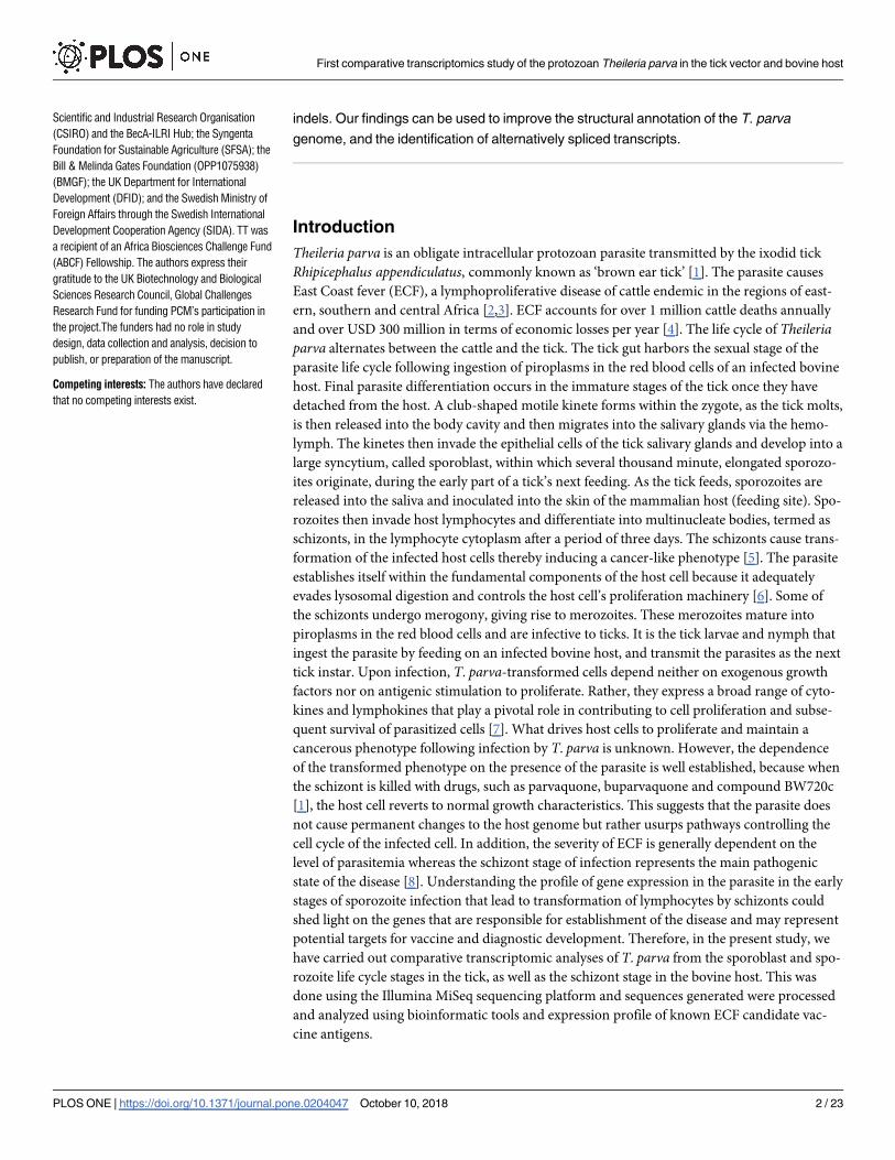
TpMuguga_04g00640 is considerably longer than TP04_0640 (in the original 2005
genome annotation), with one extra exon where an intron was previously annotated, and a
much longer last (4th) exon. It now encodes a small protein of 114 amino acid residues (com-
pared to 50 residues in TP04_0640), without a signal peptide but with two putative transmem-
brane domains. Furthermore, this protein contains two casein kinase II phosphorylation sites
(SMID and STVE) located in its intracellular region. BLASTX search revealed a significant
homology with the fission yeast endoplasmic reticulum membrane meiotically up-regulated
gene 84 protein, MUG84_SCHPO (BLAST expectation value 6e-21), which plays a role in the
meiotic cell cycle. TpMuguga_04g00640 showed strong homology to proteins in other piro-
plasms, including Theileria, Babesia and Cytauxzoon species.
TpMuguga_03g00299 has similar structure to the original protein annotated in this geno-
mic region, TP03_0299. It encodes a hypothetical protein of 770 amino acids containing a sig-
nal peptide, which suggests that it is released in the host cytosol. In contrast, the absence of a
classical nuclear localization signal suggests that this protein is not directed to the host nucleus
but may either be confined in the host cytoplasm or could be processed through ER/Golgi-
dependent secretion pathways where it would be processed and presented on the surface of the
infected host cell as an antigen epitope in the context of the major histocompatibility complex
(MHC) class I. We also assessed whether this protein had other known motifs and identified
several potential post-translational modification sites including one N-glycosylation (NSTD),
one cyclic cAMP- and cGMP-dependent protein kinase phosphorylation (KRKT), seven pro-
tein kinase C phosphorylation (TSK, TPR, TRK, TDR, SER, SEK and SSR), five casein kinase
II phosphorylation sites (SPLD, SESD, TSKD, SNED and STGE), and eleven N-myristoylation
(GSGIGI, GNSDNF, GVTVTQ, GVTVTQ, GTTPSV, GAIASS, GTTAAS, GLGMGG,
Fig 4. A pairwise comparison of sporoblast against sporozoite and schizont stages. 2845 genes differentially expressed in
sporozoite and schizont stages in comparison to sporoblast, plus 647 and 310 only differentially expressed relative to the sporozoite or
the schizont stages, respectively.
https://doi.org/10.1371/journal.pone.0204047.g004
First comparative transcriptomics study of the protozoan Theileria parva in the tick vector and bovine host
PLOS ONE | https://doi.org/10.1371/journal.pone.0204047 October 10, 2018 10 / 23

Fig 5. Distribution of differentially expressed genes (p-adj< 0.01). A). Total number of genes upregulated (coral) and downregulated
(green) in the three life cycle stages of the parasite. B) Venn diagram comparing the upregulated genes in sporozoite and schizont to the
downregulated genes in sporozoite. C) Venn diagram comparing the upregulated genes in sporoblast and sporozoite to the downregulated
genes in schizont. D) List of four downregulated genes in the schizont stage of the parasite. TMD, transmembrane domain; SP, signal peptide;
(-), none.
https://doi.org/10.1371/journal.pone.0204047.g005
First comparative transcriptomics study of the protozoan Theileria parva in the tick vector and bovine host
PLOS ONE | https://doi.org/10.1371/journal.pone.0204047 October 10, 2018 11 / 23

GTGAAS, GVSVGT and GIMLGE) sites as well as one cell attachment sequence (RGD). An
ortholog has been detected in T. annulata only.
TpMuguga_01g00541 is identical to TP01_0541. It encodes a protein of 209 amino acid
residues in length with neither a classical signal peptide of secreted proteins nor a transmem-
brane domain or a GPI anchor motif of surface proteins. SecretomeP 2.0 software predicted
NN-score of 0.57 for TP01_0541 protein. Using the PredictProtein program, we found that the
TP01_0541 protein contains two potential disulfide bonds from the cysteine residues 17/33
and 115/164, respectively. It also contains two N-glycosylation (NYSY and NETE), two protein
kinase C phosphorylation (SLK and TRK), seven casein kinase II phosphorylation (SLVE,
SALE, TNFD, SEGE, SLSD, SQED and TIED), one tyrosine kinase phosphorylation (KEFD-
NENY) and one N-myristoylation (GSLKSL) sites, suggesting its involvement in numerous
protein-protein activities. In addition, this protein also contains a leucine zipper pattern
(LYDYGTSLVEYYRCLFQLYFNL). Most interestingly, no orthologs have been identified in
other piroplasm genomes, and our updated tblastn searches against all Piroplasma genomes in
EuPathDB confirm this result, having returned no matches other than in T. parva.
TpMuguga_03g00193 is again identical to TP03_0193. It encodes a 346 amino acid-long
protein. Much like the TP01_0541-encoded protein, this protein also contains no signal pep-
tide, transmembrane domains or a GPI anchor motif. It contains several potential post-transla-
tional modification motifs including three N-glycosylation (NNSS, NVTD and NVTM), one
cAMP- and cGMP-dependent protein kinase phosphorylation (RRLT), eleven protein kinase
C phosphorylation (TKK, TRK, STK, SQK, THK, TEK, SLK, SQR, TLR, TTK and SKK), five
casein kinase II phosphorylation (TKKD, SEQE, TYGD, TYED and TTKD), one tyrosine
kinase phosphorylation (RTLERRY) and three N-myristoylation (GLHISQ, GCERCW
and GTSITK) sites. Also of interest is the presence on a non-ordinary secondary structure
(NORS) spanning amino acids 1 to 90. An ortholog is present in T. annulata but not in other
piroplasms.
Functional annotation of upregulated genes reveals activation of plasma
membrane components and translation signals
We submitted the upregulated genes for each infection stage to DAVID for gene-annotation
enrichment analysis. Genes with the same associated Gene Ontology (GO) term were clustered
Table 2. The top 10 most highly expressed genes.
GenBank
Acc. No.
Locus Tag / 2005 Locus Tag IDa Average TPM Product name Domain
Splast Spzoit Schiz
XM_758212.1 TP03_0287 TpMuguga_03g00287 17464 840 21 p67 antigen SP, 1 TMD
XM_758077.1 TP03_0152 TpMuguga_03g00152 15623 8710 3212 Histone H3 (-)
XM_758613.1 TP04_0071 TpMuguga_04g00071 12948 2795 1695 Histone H2B (-)
XM_758224.1 TP03_0299 TpMuguga_03g00299 11757 413 3 Hypothetical protein SP
XM_760084.1 TP02_0611 TpMuguga_02g00611 9103 1848 1798 Histone H2A (-)
XM_761498.1 TP01_1070 TpMuguga_01g01070 8512 2152 2226 Ubiquitin/ ribosomal fusion protein (-)
XM_758979.1 TP04_0437 TpMuguga_04g00437 15 11151 539 p104 antigen SP, GPI
XM_760968.1 TP01_0541 TpMuguga_01g00541 0 9630 5 Hypothetical protein (-)
XM_759182.1 TP04_0640 TpMuguga_04g00640 136 8957 671 Hypothetical protein 2 TMD
XM_758118.1 TP03_0193 TpMuguga_03g00193 45 8806 10 Hypothetical protein (-)
aLocus Tag Identifiers according to the updated whole genome gene structural re-annotation (Tretina et al., in preparation).
Splast, sporoblast; Spzoit, sporozoite; Schiz, schizont. GPI, GPI anchor; SP, signal peptide; TMD, transmembrane domain; (-), none.
https://doi.org/10.1371/journal.pone.0204047.t002
First comparative transcriptomics study of the protozoan Theileria parva in the tick vector and bovine host
PLOS ONE | https://doi.org/10.1371/journal.pone.0204047 October 10, 2018 12 / 23

[27]. A high stringency cut-off was used to filter the clusters and the counts (number of genes
with the specific associated GO term) retrieved. The GO categories that were most broadly
enriched included ribosomal proteins and those involved in translation and integral compo-
nents of the plasma membrane; ATP-binding was also enriched but only in sporozoite and
schizont (Fig 6). In the sporozoite, we saw an enrichment in genes involved in zinc ion and
nucleic acid binding, ribosomal activity, transcription, transmembrane transport, GTPase,
ligase and protein kinase activities, glycolytic process, RNA processing, protease activity and
translation elongation. Functions only enriched during sporoblast included vesicle-mediated
transport (ER to Golgi), intracellular ribonucleoprotein complex and proteasome activity.
Genes upregulated during sporozoite were mainly involved in zinc-ion and nucleic acid bind-
ing, ribosome and transcription. In the schizont stage, we see GO term enrichment for some
genes with activities mainly in translation, DNA replication and cell division.
Fig 6. Heatmap showing the log10 (TPM values) for enriched GO terms enriched in the upregulated genes in the different infection stages obtained from DAVID.
https://doi.org/10.1371/journal.pone.0204047.g006
First comparative transcriptomics study of the protozoan Theileria parva in the tick vector and bovine host
PLOS ONE | https://doi.org/10.1371/journal.pone.0204047 October 10, 2018 13 / 23

This study found no variations in the sequence of transcripts throughout the three life cycle
stages analysed and, in general, our data confirmed the recent reannotation of the T. parvagenome (Tretina et al., in preparation). Nevertheless, we discovered some transcripts that were
different in size from those in the original 2005 genome annotation and/or the re-annotation
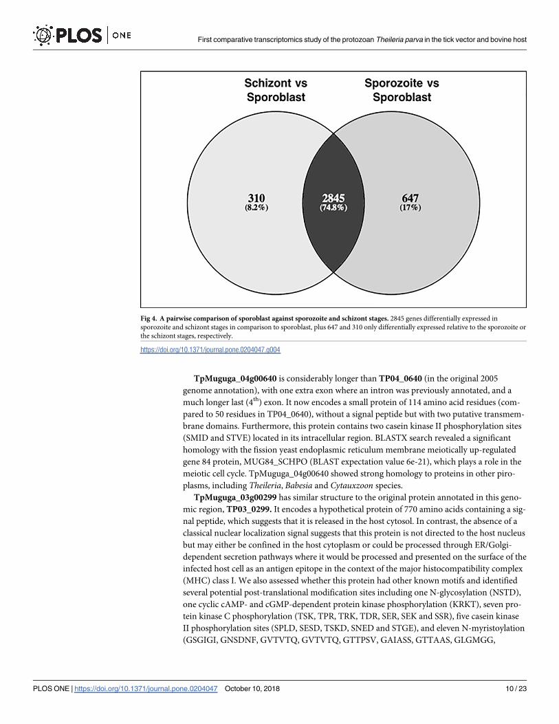
(Fig 7). The difference was due to indels, which we confirmed by gel analysis of PCR products
(Fig 8) and Sanger sequencing of amplicons (Supplementary S2 File) for two genes. Thus,
using gene-specific primers, the DNA region amplified from TpMuguga_01g00193 mRNA
was 129 bp longer, whereas the one from TpMuguga_04g00272 mRNA was 72 bp shorter,
than their respective sequences in the original and new annotations. We also observed that
TP04_0251 was shorter in the re-annotation than in the original genome annotation and had
been renamed TpMuguga_04g02465. All these genes have now been corrected in the new ver-
sion of the re-annotation.
Antigen expression is not conserved across the three infection stages
Several antigens have been characterized in T. parva [28, 29]. The antigens with low expression
during the sporoblast stage (Tp2, Tp9, p104, p150 and PIM) are activated during the sporozo-
ite stage (Fig 9). On the other hand, the antigens with a higher expression in sporoblast, such
as p67 and the proliferating cell nuclear antigens TP02_0600 and TP03_0445, are downregu-
lated during sporozoite although their expression increases again slightly in the schizont. Tp7
and Tp8 are highly and constantly expressed during all three life cycle stages. In general, most
of the antigens have reduced expression during the schizont stage. Tp8, Tp9 and PIM are the
only exceptions, with expression maintained or increased in that stage.
The R package Cluster was used to find genes with similar expression to the known anti-
gens. To compare the three infection stages, TPM values were considered for each gene and
the genes falling in the same cluster as the antigens were plot together. Up to seven most
similar genes to the antigen in each cluster were considered in each case (Fig 10). The cluster
information can be found in the supplementary S1 Table. Only a few genes with a similar
expression to p67 could be identified (TP03_0299, TP02_0425 and to a lesser extent TP01_
0178), probably due to the very high expression of p67 in the sporoblast (Fig 10); like p67,
these three contain a signal peptide. It is worth mentioning that genes like TP01_0380,
TP01_1225 and TP01_1227 have similar expression profile to the antigen p104 (TP04_0437)
and also contain a signal peptide.
We searched for orthologs of genes known in other apicomplexans to encode proteins asso-
ciated with sporozoite invasion organelles. Structural properties of proteins encoded by nine
rhoptry-, five microneme- and one dense granules-associated genes of T. parva are summa-
rized in the supplementary S1 Table. They are differentially expressed and at relatively very
low or moderate levels, except for the dense granules gene (TP02_0607) and four rhoptry
genes (TP01_0701, TP02_0645, TP03_0067 and TP03_0655), with expression higher than
1,000 TPM units in at least one of the three parasite life cycle stages.
Discussions
The intracellular parasite Theileria parva infects and reversibly transforms the bovine lympho-
cytes [7], using strategies that help the parasite survive, establish itself and proliferate within
the host [30, 31, 32]. Some of these strategies involve modulation of the host immune response
as well as molecular mimicry through host-parasite interaction [6]. In the present study of
gene expression profiling, we demonstrated reproducible performance in our assays and, as
shown in Fig 2B, there was no significant difference between replicates for each life cycle stage.
Because protein coding housekeeping genes routinely used as endogenous controls in
First comparative transcriptomics study of the protozoan Theileria parva in the tick vector and bovine host
PLOS ONE | https://doi.org/10.1371/journal.pone.0204047 October 10, 2018 14 / 23

First comparative transcriptomics study of the protozoan Theileria parva in the tick vector and bovine host
PLOS ONE | https://doi.org/10.1371/journal.pone.0204047 October 10, 2018 15 / 23
quantification studies of mRNA transcripts were shown to vary considerably between different
T. parva stocks [33], they were not used in this study. They include genes encoding glyceralde-
hyde-3-phosphate dehydrogenase (GAPDH), cytochrome b and fructose-2.6-biphosphate
aldolase (F6P) proteins. We did not use β-actin gene as control because it may be highly
demanded in actively dividing parasites like sporoblast and schizont for motility, structure and
integrity whereas its demand may be low in the non-replicating cells such as the sporozoite. In
this regard, our study showed that β-actin (TP02_0903) was more expressed in the replicative
sporoblast and schizont forms (5974 and 2499 counts, respectively) than in the non-replicating
sporozoite form (1114 counts). Lastly, by using Transcripts Per Kilobase Million (TPM) in our
study, we normalized both for gene length and sequencing depth, which improved compari-
sons across genes and across samples. We observed that the parasite development from the
sporoblast, its transformation into sporozoite and establishment in the host cells as schizont is
accompanied by generalized increase of differential gene expression. Most of these changes in
gene expression may be associated with the ability of the parasite to gain entry into the host,
and its persistence as well as interference with the signal transduction pathways that govern
important functions of the infected cell [34]. The drastic increase of the number of upregulated
genes in the schizont stage of the parasite shown here could be related to their key role in
Fig 7. Indels identified in the transcripts. Multiple nucleotide and amino acid sequences alignments of T. parva gene
regions with indels using the GT-AG dinucleotides DNA sequence requirement at the first two (GT) and last two (AG)
positions of introns in pre-mRNAs (highlighted in bold). (a) indicates difference in both original genome annotation
and re-annotation; # indicates introns not in-frame.
https://doi.org/10.1371/journal.pone.0204047.g007
Fig 8. PCR verification of deletion (TP04_0272) and insertion (TP01_0193) identified by MiSeq. Samples are as follows: Lanes 1–4,
genomic DNA from T. parva strain Muguga 3087 (lane 1); T. parva strain Marikebuni 3292 (lane 2); T. parva strain Kiambu5 (lane 3); T.
parva F100 TpM (lane 4); Lanes 5–8, mRNA from sporozoite of T. parva Muguga 3087 (lanes 5, 5a and 5b); mRNA schizont T. parvaMuguga 3087 (lane 6); schizont of T. parva Muguga 3087 (lane 7); piroplasm of T. parva Muguga 3087 (lane 8). Also shown are DNA size
markers (lanes M), with length in base pair (pb).
https://doi.org/10.1371/journal.pone.0204047.g008
First comparative transcriptomics study of the protozoan Theileria parva in the tick vector and bovine host
PLOS ONE | https://doi.org/10.1371/journal.pone.0204047 October 10, 2018 16 / 23

promoting the establishment of the parasite in the host cytosol. Interestingly, the four genes
not expressed in the schizont stage (Fig 5D) were upregulated in the sporozoite stage. These
tick stage-specific genes may play a critical role in the survival of the parasite in the tick vector,
and not in the parasite establishment in, and/or transformation of, the host cell.
In the present study, the sequence of vast majority of the parasite transcripts matched per-
fectly well with the genes in the re-annotated genome available in GenBank. The very few
sequence differences observed were confirmed by gene fragment analysis and corroborated
our results, hence validating the power of the approach we used and the high level of accuracy
of our sequencing data. Moreover, multiple sequence alignments of the mRNA with the geno-
mic DNA sequences showed that, in general, the observed indels conform to the eukaryotic
canonical splice site composition, i.e., the GT-AG dinucleotides DNA sequence requirement
at the first two and last two positions of introns in pre-mRNAs. These findings reveal a usefull
approach to identify incorrect annotations, and resulted in the improvement of the T. parvagenome annotation.
It is reported that p67 expression is stage-specific, and restricted to the sporozoite [29]. In
our study, we found that p67 is also expressed in the schizont, although at a very low level (21
TPM units) as compared to sporozoite (840 TPM). Interestingly, we found that p67 is also
expressed in the sporoblast and 20 times more than in the sporozoite. In addition, we found
Fig 9. Heatmap showing the log10 of normalized TPM values for known Theileriaparva antigens in samples from
three parasite life cycle stages. Yellowish is high expression and red is low expression. Selected known candidate
vaccine antigens are listed.
https://doi.org/10.1371/journal.pone.0204047.g009
First comparative transcriptomics study of the protozoan Theileria parva in the tick vector and bovine host
PLOS ONE | https://doi.org/10.1371/journal.pone.0204047 October 10, 2018 17 / 23
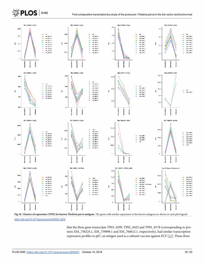
that the three gene transcripts TP03_0299, TP02_0425 and TP01_0178 (corresponding to pro-
teins XM_758224.1, XM_759898.1 and XM_760612.1, respectively), had similar transcription
expression profiles to p67, an antigen used as a subunit vaccine against ECF [35]. These three
Fig 10. Clusters of expression (TPM) for known Theileria parva antigens. The genes with similar expression to the known antigens are shown in each plot legend.
https://doi.org/10.1371/journal.pone.0204047.g010
First comparative transcriptomics study of the protozoan Theileria parva in the tick vector and bovine host
PLOS ONE | https://doi.org/10.1371/journal.pone.0204047 October 10, 2018 18 / 23

antigens also have a signal peptide (but lack a transmembrane domain) suggesting that they
are directed to the secretory pathway from the parasite secreted where they could reside in par-
asite organelles such the endoplasmic reticulum or golgi, be inserted into parasite cellular
membrane, or be secreted from the parasite. Furthermore, the XM_760612.1 protein has a
nuclear localization signal, suggesting a possible interaction with the host cell nucleus [36].
The high level of expression of the three genes in the tick stages of the parasite make them
potential candidate antigens for the development of a transmission blocking subunit vaccine.
Several genes with similar expression to other known T. parva vaccine antigens were also iden-
tified, some containing transmembrane domains and/or a signal peptide. The expression pro-
file of these selected genes could be further evaluated using quantitative reverse transcription
PCR; they could also be evaluated further for their potential as vaccine antigens. Additionally,
the genes with a nuclear localization signal can be explored further for their potential role in
host cell transformation. The two proliferating cell nuclear antigens (PCNA) (TP02_0600 and
TP03_0445), homologs of which are known to be abundant in proliferating cells [37], were
also more highly transcribed in the schizont than in the sporozoite. They might also have a
role in host cell transformation by interacting with host cell regulatory proteins.
Using protein prediction software, we examined critically the four most highly expressed
genes encoding hypothetical proteins. TP04_0640 protein showed homology and structural
similarity with meiotically up-regulated gene 84 protein (MUG84) [38], i.e. with two trans-
membrane domains, a small loop-like structure on the membrane and casein kinase II phos-
phorylation sites, suggesting that it is associated with organellar membrane like ER. MUG84 is
also a putative phosphatidylinositol N-acetylglucosaminyltransferase subunit P (PIG-P)
domain-containing protein. Therefore, TP04_0640 protein is likely to have similar function as
MUG84, i.e.: being involved in meiotic events and GPI anchor biosynthesis. The strong
homology to proteins in other piroplasms, including Theileria, Babesia and Cytauxzoon spe-
cies, suggests that this is likely a conserved protein involved in cellular metabolic processes. Its
high level of expression in the sporozoite suggest a potential important role for this protein in
that stage and during infection. Therefore, TP04_0640 protein appears as a potential target for
the development of anti Theileria drugs.
The presence of several post-translational modification sites and a cell attachment motif
suggests that the TP03_0299 protein may be involved in parasite-bovine and parasite-tick pro-
tein-protein interactions. Considering that it can also be secreted by the parasite, as suggested
by the presence of a signal peptide, the TP03_0299 protein could serve as a potential disease
biomarker or protein therapeutic target. Moreover, the fact that it is expressed almost exclu-
sively in the arthropod stages makes TP03_0299 a good candidate for the development of a
transmission blocking subunit vaccine to prevent parasite transmission by the tick vector.
The numerous post-translational modification motifs, especially the leucine zipper pattern,
present in the TP01_0541 protein, suggests that it might interact with other proteins, with
DNA motifs that have a binding affinity for leucine zippers, or both. In that regard, TP01_
0541 protein may be strongly involved in transcriptional gene regulation in the sporozoite
stage in which its expression is very high and to which it is confined. In addition, our analyses
suggest the TP01_0541 protein is a non-classical secreted protein. This structural characteristic
and the high expression level in the sporozoite stage make TP01_0541 a potential target for
antitherapeutic drugs, and/or another potential candidate antigen for the development of a
transmission blocking subunit vaccine.
Regarding the TP03_0193, the protein contained several post-translational modification
sites, an indication that it may be involved in protein-proteins interactions. In addition, the
presence of a non-ordinary secondary structure and the high level of expression in the sporo-
zoite strongly suggest that the TP03_0193 protein may play a crucial function, especially in
First comparative transcriptomics study of the protozoan Theileria parva in the tick vector and bovine host
PLOS ONE | https://doi.org/10.1371/journal.pone.0204047 October 10, 2018 19 / 23

that tick stage, such as protein stability, folding, function and recognition. In this regard,
TP03_0193 would be a potential target for the development of antiparasitic drugs.
The restricted taxonomic distribution of three of these four hypothetical proteins makes
them potentially very interesting. While the core gene set is fairly conserved among Theileriaspecies, antigens tend to be considerably more species-specific [14, 39]. Alternatively, their taxo-
nomic restriction could be explained by a role of these proteins in parasite-vector interactions,
rather than in host-parasite processes. This is particularly the case of TpMuguga_03g00299,
which is highly expressed in the sporoblast but not in the sporozoite or the schizont stages.
From the present study, the panel of contigs that were unassigned to any species during
mapping will be explored further to discover eventual novel parasite genes and their functions.
The fraction of reads corresponding to mitochondrial genes (cytochrome oxidase) will also be
studied further to gain more insight into mtDNA variations in the evolution and adaptation of
T. parva in infection.
In summary, RNASeq analysis of the three T. parva developmental stages has revealed a
drastic increase in the number of upregulated genes as T. parva sporozoites infect bovine cells
and differentiate into the schizont forms. In contrast, the highest expressed genes occur in the
sporoblast stage. The information generated here will help in enhancing knowledge into the
parasite genes involved in infection process and transformation of host cells and contribute to
the identification of additional vaccine antigens for the control of the ECF disease.
Conclusion
The work presented here is a representation of the first, albeit partial, analysis of the gene
expression profiles of T. parva developmental stages from the sporoblast through the sporozo-
ite to the schizont stages. From our study, we were able to identify genes that are differentially
expressed across those parasite life cycle stages. In particular, we identified genes that have an
expression profile and protein domains similar to those of known T. parva antigens, such as
p67. They can be explored further for their potential as candidate vaccine antigens or their role
in parasite development. Our results are also likely to provide insight into the fascinating phe-
nomenon of reversible host cell transformation, that could be of great importance in the con-
trol of ECF and similar diseases.
Supporting information
S1 File. Differentially expressed genes of unmapped reads.
(CSV)
S2 File. Confirmation of indels in the transcripts by PCR and sequencing.
(DOCX)
S1 Table. Average Transcripts Per Kilobase Million (TPM) of genes, genes hit by
unmapped reads and, T. parva orthologs of apicomplexan genes encoding proteins of spo-
rozoite invasion organelles.
(XLSX)
S2 Table. Differentially expressed genes between the three stages studied.
(XLSX)
Acknowledgments
This work was supported by BecA-ILRI Hub through the ABCF Program. The ABCF Program
is funded by the Australian Agency for International Development (AusAID) through a
First comparative transcriptomics study of the protozoan Theileria parva in the tick vector and bovine host
PLOS ONE | https://doi.org/10.1371/journal.pone.0204047 October 10, 2018 20 / 23

partnership between Australia’s Commonwealth Scientific and Industrial Research Organisa-
tion (CSIRO) and the BecA-ILRI Hub; the Syngenta Foundation for Sustainable Agriculture
(SFSA); the Bill & Melinda Gates Foundation (BMGF); the UK Department for International
Development (DFID); and the Swedish Ministry of Foreign Affairs through the Swedish Inter-
national Development Cooperation Agency (SIDA). TT was a recipient of an Africa Biosci-
ences Challenge Fund (ABCF) Fellowship. The authors express their gratitude to the UK
Biotechnology and Biological Sciences Research Council, Global Challenges Research Fund
for funding PCM’s participation in the project.
Author Contributions
Conceptualization: Appolinaire Djikeng, Roger Pelle.
Data curation: Triza Tonui.
Formal analysis: Triza Tonui, Pilar Corredor-Moreno, Esther Kanduma, Joyce Njuguna,
Joana C. Silva, Appolinaire Djikeng, Roger Pelle.
Investigation: Triza Tonui, Roger Pelle.
Methodology: Triza Tonui, Esther Kanduma, Moses N. Njahira, Appolinaire Djikeng, Roger
Pelle.
Software: Pilar Corredor-Moreno, Joyce Njuguna.
Supervision: Esther Kanduma, Steven G. Nyanjom, Roger Pelle.
Validation: Roger Pelle.
Writing – original draft: Triza Tonui, Esther Kanduma.
Writing – review & editing: Pilar Corredor-Moreno, Joyce Njuguna, Steven G. Nyanjom,
Joana C. Silva, Appolinaire Djikeng, Roger Pelle.
References1. Norval R. A. I., Perry B. D., & Young A. S. (1992). The epidemiology of theileriosis in Africa. ILRI (aka
ILCA and ILRAD).
2. Irvin A.D. and Morrison W.I., 1987. Immunopathology, immunology, and immunoprophylaxis of Thei-
leria infections. Immune responses in parasitic infections: immunology, immunopathology, and immu-
noprophylaxis. Volume III: Protozoa, pp.223–274.
3. Morrison W.I., Goddeeris B.M., Brown W.C., Baldwin C.L. and Teale A.J., 1989. Theileria parva in cat-
tle: characterization of infected lymphocytes and the immune responses they provoke. Veterinary
immunology and immunopathology, 20(3), pp.213–237. PMID: 2497579
4. Vollmer D., 2010. Enhancing the effectiveness of sustainability partnerships: Summary of a workshop.
National Academies Press.
5. Tretina K., Gotia H.T., Mann D.J. and Silva J.C., 2015. Theileria-transformed bovine leukocytes have
cancer hallmarks. Trends in parasitology, 31(7), pp.306–314. https://doi.org/10.1016/j.pt.2015.04.001
PMID: 25951781
6. Dobbelaere D.A., Fernandez P.C. and Heussler V.T., 2000. Theileria parva: taking control of host cell
proliferation and survival mechanisms. Cellular microbiology, 2(2), pp.91–99. PMID: 11207566
7. Dobbelaere D. and Heussler V., 1999. Transformation of leukocytes by Theileria parva and T. annulata.
Annual Reviews in Microbiology, 53(1), pp.1–42.
8. Dolan T.T., Young A.S., Losos G.J., McMilian I., Minder C.E., et al., 1984. Dose dependent responses of
cattle to Theileria parva stabilate. International journal for parasitology, 14(1), pp.89–95. PMID: 6423557
9. Pelle R., Graham S.P., Njahira M.N., Osaso J., Saya R.M., et al., 2011. Two Theileria parva CD8 T cell
antigen genes are more variable in buffalo than cattle parasites, but differ in pattern of sequence diver-
sity. PLoS One, 6(4), p.e19015. https://doi.org/10.1371/journal.pone.0019015 PMID: 21559495
First comparative transcriptomics study of the protozoan Theileria parva in the tick vector and bovine host
PLOS ONE | https://doi.org/10.1371/journal.pone.0204047 October 10, 2018 21 / 23

10. Morzaria S.P., Dolan T.T., Norval R.A.I., Bishop R.P. and Spooner P.R., 1995. Generation and charac-
terization of cloned Theileria parva parasites. Parasitology, 111(1), pp.39–49.
11. Sugimoto C., Conrad P.A., Ito S., Brown W.C. and Grab D.J., 1988. Isolation of Theileria parva schiz-
onts from infected lymphoblastoid cells. Acta tropica, 45(3), pp.203–216. PMID: 2903622
12. Pelle R., & Murphy N. B. (1993). Northern hybridization: rapid and simple electrophoretic conditions.
Nucleic acids research, 21(11), 2783–2784. PMID: 8332486
13. Bray N.L., Pimentel H., Melsted P. and Pachter L., 2016. Near-optimal probabilistic RNA-seq quantifica-
tion. Nature biotechnology, 34(5), p.525. https://doi.org/10.1038/nbt.3519 PMID: 27043002
14. Gardner M.J., Bishop R., Shah T., De Villiers E.P., Carlton J.M., et al., 2005. Genome sequence of
Theileria parva, a bovine pathogen that transforms lymphocytes. Science, 309(5731), pp.134–137.
https://doi.org/10.1126/science.1110439 PMID: 15994558
15. Pimentel H., Bray N., Puente S., Melsted P. and Pachter L., 2016. Differential analysis of RNA-Seq
incorporating quantification uncertainty. bioRxiv.
16. Benjamini Y. and Hochberg Y., 1995. Controlling the false discovery rate: a practical and powerful
approach to multiple testing. Journal of the royal statistical society. Series B (Methodological), pp.289–
300.
17. Kim D., Pertea G., Trapnell C., Pimentel H., Kelley R., et al., 2013. TopHat2: accurate alignment of tran-
scriptomes in the presence of insertions, deletions and gene fusions. Genome biology, 14(4), p.R36.
https://doi.org/10.1186/gb-2013-14-4-r36 PMID: 23618408
18. Trapnell C., Roberts A., Goff L., Pertea G., Kim D., et al., 2012. Differential gene and transcript expres-
sion analysis of RNA-seq experiments with TopHat and Cufflinks. Nature protocols, 7(3), p.562. https://
doi.org/10.1038/nprot.2012.016 PMID: 22383036
19. Maechler M., Rousseeuw P., Struyf A., Hubert M. and Hornik K., 2016. Cluster Analysis Basics and
Extensions. R package version 2.0. 4.
20. Love M.I., Huber W. and Anders S., 2014. Moderated estimation of fold change and dispersion for
RNA-seq data with DESeq2. Genome biology, 15(12), p.550. https://doi.org/10.1186/s13059-014-
0550-8 PMID: 25516281
21. Petersen T.N., Brunak S., von Heijne G. and Nielsen H., 2011. SignalP 4.0: discriminating signal pep-
tides from transmembrane regions. Nature methods, 8(10), p.785. https://doi.org/10.1038/nmeth.1701
PMID: 21959131
22. Omasits U., Ahrens C.H., Muller S. and Wollscheid B., 2013. Protter: interactive protein feature visuali-
zation and integration with experimental proteomic data. Bioinformatics, 30(6), pp.884–886. https://doi.
org/10.1093/bioinformatics/btt607 PMID: 24162465
23. Yachdav G., Kloppmann E., Kajan L., Hecht M., Goldberg T., et al., 2014. PredictProtein—an open
resource for online prediction of protein structural and functional features. Nucleic acids research, 42
(W1), pp.W337–W343.
24. Pierleoni A., Martelli P.L. and Casadio R., 2008. PredGPI: a GPI-anchor predictor. BMC bioinformatics,
9(1), p.392.
25. Rost B., Yachdav G. and Liu J., 2004. The PredictProtein server Nucleic Acids Res,. 32. W321 W326.
https://doi.org/10.1093/nar/gkh377 PMID: 15215403
26. Bendtsen J.D., Jensen L.J., Blom N., Von Heijne G. and Brunak S., 2004. Feature-based prediction of
non-classical and leaderless protein secretion. Protein Engineering Design and Selection, 17
(4), pp.349–356.
27. Ashburner M., Ball C.A., Blake J.A., Botstein D., Butler H., et al., 2000. Gene Ontology: tool for the unifi-
cation of biology. Nature genetics, 25(1), p.25. https://doi.org/10.1038/75556 PMID: 10802651
28. Sitt T., Pelle R., Chepkwony M., Morrison W.I. and Toye P., 2018. Theileria parva antigens recognized
by CD8+ T cells show varying degrees of diversity in buffalo-derived infected cell lines.
Parasitology, pp.1–10.
29. Nene V., Musoke A., Gobright E. and Morzaria S., 1996. Conservation of the sporozoite p67 vaccine
antigen in cattle-derived Theileria parva stocks with different cross-immunity profiles. Infection and
immunity, 64(6), pp.2056–2061. PMID: 8675307
30. Palmer G.H., Machado J., Fernandez P., Heussler V., Perinat T., et al., 1997. Parasite-mediated
nuclear factor κB regulation in lymphoproliferation caused by Theileria parva infection. Proceedings of
the National Academy of Sciences, 94(23), pp.12527–12532.
31. Ivanov V.L.A.D.I.M.I.R., Stein B., Baumann I., Dobbelaere D.A., Herrlich P. et al., 1989. Infection with
the intracellular protozoan parasite Theileria parva induces constitutively high levels of NF-kappa B in
bovine T lymphocytes. Molecular and cellular biology, 9(11), pp.4677–4686. PMID: 2513476
First comparative transcriptomics study of the protozoan Theileria parva in the tick vector and bovine host
PLOS ONE | https://doi.org/10.1371/journal.pone.0204047 October 10, 2018 22 / 23

32. Heussler V.T., Rottenberg S., Schwab R., Kuenzi P., Fernandez P.C., et al., 2002. Hijacking of host cell
IKK signalosomes by the transforming parasite Theileria. Science, 298(5595), pp.1033–1036. https://
doi.org/10.1126/science.1075462 PMID: 12411708
33. Tsotetsi T.N., Collins N.E., Oosthuizen M.C. and Sibeko-Matjila K.P., 2018. Selection and evaluation of
housekeeping genes as endogenous controls for quantification of mRNA transcripts in Theileria parva
using quantitative real-time polymerase chain reaction (qPCR). PloS one, 13(5), p.e0196715. https://
doi.org/10.1371/journal.pone.0196715 PMID: 29727459
34. Beverley S.M., 1996. Hijacking the cell: parasites in the driver’s seat. Cell, 87(5), pp.787–789. PMID:
8945504
35. Musoke A., Morzaria S., Nkonge C., Jones E. and Nene V., 1992. A recombinant sporozoite surface
antigen of Theileria parva induces protection in cattle. Proceedings of the National Academy of Sci-
ences, 89(2), pp.514–518.
36. Bilgic H.B., Karagenc T., Bakırcı S., Shiels B., Tait A., et al., 2016. Identification and Analysis of Immu-
nodominant Antigens for ELISA-Based Detection of Theileria annulata. PloS one, 11(6), p.e0156645.
https://doi.org/10.1371/journal.pone.0156645 PMID: 27270235
37. Johnson A. and O’Donnell M., 2005. Cellular DNA replicases: components and dynamics at the replica-
tion fork. Annu. Rev. Biochem., 74, pp.283–315. https://doi.org/10.1146/annurev.biochem.73.011303.
073859 PMID: 15952889
38. Guelette B.S., Benning U.F. and Hoffmann-Benning S., 2012. Identification of lipids and lipid-binding
proteins in phloem exudates from Arabidopsis thaliana. Journal of experimental botany, 63
(10), pp.3603–3616. https://doi.org/10.1093/jxb/ers028 PMID: 22442409
39. Knowles D.P., Kappmeyer L.S., Haney D., Herndon D.R., Fry L.M., et al., 2018. Discovery of a novel
species, Theileria haneyi n. sp., infective to equids.
First comparative transcriptomics study of the protozoan Theileria parva in the tick vector and bovine host
PLOS ONE | https://doi.org/10.1371/journal.pone.0204047 October 10, 2018 23 / 23
Recommended