ORIGINAL PAPER
Enhancing somatic embryogenesis of Malaysian rice cultivarMR219 using adjuvant materials in a high-efficiency protocol
R. Abiri1,2 • M. Maziah1,3,4 • N. A. Shaharuddin1,4 • Z. N. B. Yusof1 •
N. Atabaki5 • M. M. Hanafi4 • M. Sahebi4 • P. Azizi4 • N. Kalhori6 •
A. Valdiani1
Received: 12 July 2016 / Revised: 11 November 2016 / Accepted: 21 December 2016 / Published online: 18 January 2017
� The Author(s) 2017. This article is published with open access at Springerlink.com
Abstract Enhancing of the efficient tissue culture protocol
for somatic embryos would facilitate the engineered
breeding plants program. In this report, we describe the
reproducible protocol of Malaysian rice (Oryza sativa L.)
cultivar MR219 through somatic embryogenesis. Effect of
a wide spectrum of exogenesis materials was assessed in
three phases, namely callogenesis, proliferation and
regeneration. Initially, rice seeds were subjected under
various auxin treatments. Secondly, the effect of different
concentrations of 2,4-D on callus induction was evaluated.
In the next step, the efficiency of different explants was
identified. Subsequently, the effects of different auxins,
cytokinins, L-proline, casein hydrolysate and potassium
metasilicate concentrations on the callus proliferation and
regeneration were considered. For the callogenesis phase,
2 mg L-1of 2,4-D and roots were chosen as the best auxin
and explant. In the callus proliferation stage, the highest
efficiency was observed at week eight in the MS media
supplemented with 2 mg L-1 of 2,4-D, 2 mg L-1 of
kinetin, 50 mg L-1 of L-proline, 100 mg L-1 of casein
hydrolysate and 30 mg L-1 of potassium metasilicate. In
the last phase of the research, the MS media added with
3 mg L-1 of kinetin, 30 mg L-1of potassium metasilicate
and 2 mg L-1 of NAA were selected. Meanwhile, to pro-
mote the roots of regenerated explants, 0.4 mg L-1 of IBA
has shown potential as an appropriate activator.
Keywords Callogenesis � Proliferation � Root explants �Potassium metasilicate and regeneration
Introduction
The production of high productive engineered plants needs
appropriate strategies. The genetic transformation mecha-
nism involves pivotal steps such as preparation of the ini-
tial conditions, usage of an efficient DNA delivery method,
establishment of effective growth and selection of medium
as well as maintenance of transformants (Abiri et al. 2015).
Notwithstanding improvement of gene transformation
efficiency in some plant species, the pre- and post-trans-
formation conditions of Indica rice have been a matter of
concern (Visarada and Sarma 2002). The efficiency ratio of
engineered plants is severely genotype dependent (Yinxia
and Te-chato 2012). On the other hand, presenting proto-
cols of pre-transformation phases for the same species have
revealed the vast effects of other factors on the plant’s
adaptation to in vitro experiments. In this regard, size,
source and age of explants, seasonal variation, oxygen
gradient, intensity as well as quality of light, temperature
and ploidy level are effective endogenous or exogenous
factors which may change the genotype feedback in dif-
ferent experiments (Aggarwal et al. 2012). Therefore, the
Editorial responsibility: Xu Han.
& N. A. Shaharuddin
1 Department of Biochemistry, Faculty of Biotechnology and
Biomolecular Sciences, Universiti Putra Malaysia (UPM),
43400 Serdang, Selangor DE, Malaysia
2 Young Researchers and Elite Club of IAU, Kermanshah, Iran
3 Institute of Bioscience, Universiti Putra Malaysia (UPM),
43400 Serdang, Selangor DE, Malaysia
4 Institute of Tropical Agriculture, Universiti Putra Malaysia
(UPM), 43400 Serdang, Selangor DE, Malaysia
5 IAU of Tehran Science and Research Branch, Tehran, Iran
6 Department of Biology, Faculty of Science, University Putra
Malaysia (UPM), 43400 Serdang, Selangor, Malaysia
123
Int. J. Environ. Sci. Technol. (2017) 14:1091–1108
DOI 10.1007/s13762-016-1221-y
evaluation of useful endogenous or exogenous factors for
differentiation and regeneration of Indica rice in vitro are
pre-requirements in genetic transformation programs
(Haque et al. 2003).
Plant tissue culture is a symphony of art and science,
which develops genetic diversity, produces virus-free plants
and improves micropropagation under aseptic conditions in
the short term (Birch 1997). Plant cells possess high plas-
ticity potential for cell differentiation. Stresses such as
pathogen infection or wounding may lead to the production
of tumours or callus. The history of the first callus growth
traced back to 1979 when Neely described a massive and
disorganized cell mass in debarked trees (Neely 1979).
Embryogenic calli, rather than direct tissues such as imma-
ture inflorescences, shoot spices, leaves and roots, is an
effective and safe tool for regeneration of wild and modified
plants in vitro conditions (Benlioglu et al. 2015). Interest-
ingly, calli are divided to various subgroups according to
their microscopic traits. For instance, calli with some organ
regeneration are named embryonic, shooty or rooty calli,
whereas calli without organ regeneration are called compact
or friable callus (Ikeuchi et al. 2013). Callogenesis and
growth highly depend on genotype, basal saltmediums, plant
growth regulators (PGRs), organic components, carbohy-
drate, explants and adjuvant materials (Pawar et al. 2015).
Additionally, media strength is another essential factor to
regulate callus’ growth and regeneration (Din et al. 2016).
A single or combination of PGRs plays a pivotal role in
tissue culture growth, cell division and morphogenesis
(Dahot 2007). Auxins are involved in several develop-
mental pathways of crops, including rooting, abscission,
internodes and stem elongation, apical dominance and
tropisms (Visarada and Sarma 2002; Roy et al. 2015; Din
et al. 2016). Auxins in tissue culture induce embryogenic
and organogenic differentiation, cyto-differentiation and
cell division. Natural auxins such as indole-3-acetic acid
(IAA), indole-3-butyric acid (IBA), a-naphthaleneacetic
acid (NAA), para-chlorophenoxyacetic acid (p-CPA),
4-amino-3, 5,6-tricholoropyridinecarboxylic acid (piclo-
ram), 2,4-dicholorophenoxyacetic acid (2,4-D), 2,4,5-tri-
cholorophenoxyacetic acid (2,4,5-T), naphthoxyacetic acid
(NOA) and 3,6-dichloro-o-anisic acid (dicamba) are
examples of synthetic auxins in plants. IAA, IBA and NAA
mainly interact with cytokinin to proliferate shoots and
induce roots. The three auxins have been implicated in
tracheid differentiation of callus and cell culture (Bhojwani
and Dantu 2013). 2,4,-D and 2,4,5-T (systematic herbi-
cides) promote callogenesis, differentiation and growth.
Noticeably, 2,4-D, dicamba and picloram are the pivotal
auxins for somatic embryogenesis. Although 2,4-D is used
for the majority of plants, dicamba and picloram are par-
ticularly used for monocots and legumes, respectively
(Konate et al. 2013; Egan et al. 2014).
Cytokinins are N6-substituted adenine derivatives
which naturally affect shoot differentiation, apical domi-
nance modification and cell division. BAP [(benzylamino)
purine, kinetin (6-furfurylamino) purine (KIN), 6-(c,c-
dimethylallyl amino) purine)], Thidiazuron (TDZ), 2iP [(2-
isopentenyl)-adenine, a dipheny1-substituted urea and
zeatin [6-(4-hydroxy-3- methyl but-2-enyl amino)-purine)]
are common sorts of cytokinins (Bhojwani and Dantu
2013). Cytokinins decrease the apical meristem dominance
and induce adventitious shoots formation and auxiliary
from meristematic explants (Ngomuo and Ndakidemi
2013). The ratio of auxin/cytokinin in the medium may
lead to somatic embryo’s development as well as root and
shoot induction (Azizi et al. 2015).
The explant’s type and age, physiological stage and
differentiation degree of tissues have been recognized as
the most important factors influencing regeneration via
somatic embryogenesis. Generally speaking, meristematic
tissues and immature organs which possess undifferenti-
ated cells are appropriate explants for plant regeneration.
Reportedly, immature embryos are the suitable responsive
explants for rice tissue culture (Haque et al. 2003; Konate
et al. 2013; Vennapusa et al. 2015). Due to some restriction
factors such as a shorter period of time for the immature
embryos’ cycle, seasonal limitations and dormancy, other
sorts of explants which are available during the year may
be the appropriate choice (Hoque and Mansfield 2004; Wu
et al. 2013). In comparison with immature seeds, mature
seeds are not appropriate explants for embryogenic callus
for recalcitrant plants in tissue culture. Seeds of various
recalcitrant plant species like Indica rice may possess
restrictions in promoting callus in vitro culture (Thokozani
et al. 2013). Efforts have been made to find appropriate rice
explants to induce embryogenic calli under suitable in vitro
conditions (Zuraida et al. 2011).
Artificial media are not a fully autotrophic method. For
the plant to survive, it needs energy and the osmotic
potential needs a carbohydrate source in the culture media
itself (Yaseen et al. 2013). Water movement and potential
and mineral movements in the media are the most impor-
tant factors that are affected by carbon sources (Buah et al.
2011). Based on plants spices’ genotype and explants,
many carbon sources have been used to develop somatic
embryogenesis (Buah et al. 2011; Yaseen et al. 2013).
Growth, development and morphogenesis of plant organs,
tissues and cells in vitro culture are importantly affected by
the composition of mineral nutrients (Sivanesan and Park
2014). Nutritional supplements including proline and
casein hydrolysate have been reported to increase callus
induction (Lin and Zhang 2005). Proline is an a-amino acid
that is essential for embryogenic callus growth, formation
and primary metabolism (Szabados and Savoure 2010; Che
Radziah et al. 2012; Pawar et al. 2015). Interaction with
1092 Int. J. Environ. Sci. Technol. (2017) 14:1091–1108
123
nitrogen sources such as ammonium and nitrate is the main
function of proline in plant tissue culture (Holme et al.
1997). Silicon (Si), the most abundant mineral and organic
nutrient in soil, is beneficial for growth, improvement and
production of plant spices (Sahebi et al. 2015). Addition-
ally, Si in tissue culture may improve embryogenesis,
organogenesis, and the physiological and anatomical
characteristics of explants. Accordingly, Si may participate
in various crop tissue cultures including secondary
metabolites production, micropropagation, organogenesis,
somatic embryogenesis and cryopreservation (Sivanesan
and Park 2014). Although silicon is not an essential
nutrient for all plants, many plants accumulate higher sil-
icon levels than essential macronutrients (Abro et al. 2009).
Rice (Oryza sativa L.) is the main staple food source for
more than half of the world’s population (Zuraida et al.
2010). Asian cultivated rice comprise of two main classes,
the Japonica (Oryza sativa ssp. Japonica) and Indica (Oryza
sativa ssp.Indica). In South-East and South Asia countries,
the Indica species is the most cultivated rice (Zhang et al.
2005). Some Malaysian rice cultivars have been shown to
have poor responses to callus induction, growth, prolifera-
tion and regeneration. Diverse tissue culture protocols in
different rice cultivars were mostly because of different
developmental stages of explants and genotypes potential
(Saharan et al. 2004; Islam et al. 2005; Khaleda and Al-
Forkan 2006; Zuraida et al. 2010; Islam et al. 2014).
Azizi et al. (2015) showed that two Malaysian cultivars,
namely MARDI’s Quality rice 74 (MRQ74) and MRQ50,
also had the lowest number of callus induction from seed,
proliferation and regeneration frequency. Among the four
Malaysian upland rice cultivars, Kusan and Siam cultivars
showed lowest response in terms of callus induction fre-
quency and regeneration capability of the embryogenic
callus (Shahsavari 2010). MR219 cultivar is an Indica rice
developed via a cross between MR151 and MR137 by the
Malaysian Agricultural Research and Development Institute
(MARDI), in 2001 (Talei et al. 2013). The cultivar has been
considered as a high production rice with an appropriate
quality in taste and shape, but sensitive to environmental
changes (Panjaitan et al. 2009). In spite of some agricultural
advantages of MR219, the rice cultivar is regarded as a
recalcitrant variety. Although various protocols have been
reported for micropropagation of MR219, its callogenesis
and regeneration ratio are still limited. However, it seems
that by evaluating the effect of other factors such as 2,4,5-T,
other explants and the suitable time to add adjuvant mate-
rials as well as using specific PGRs in the relevant stages,
researchers can find new insights into the tissue culture of
recalcitrant plant species such as MR219.
Keeping in view the above facts, in the present inves-
tigation we attempted to develop a robust protocol for
callogenesis, proliferation and plant regeneration of
MR219 by using different concentrations of PGRs, inter-
actions of auxin/cytokinin, explants and adjuvant materials.
Materials and methods
Plant materials and culture conditions
MR219 seeds were obtained from the Malaysian Agricul-
tural Research and Development Institute (MARDI),
Seberang Perai, Malaysia. The mature seeds were dehusked
and rinsed with running water for 1 h. The dehulled seeds
were surface sterilized with 70% (v/v) ethanol for 3 min and
rinsed with double distilled water to remove ethanol traces.
Then, the dehusked seeds were shaken in 40% (v/v) sodium
hypochlorite for 20 min. The seeds were rinsed thoroughly
with double distilled water before being dried on sterilized
filter papers in a petri dish. TheMurashige and Skoog (1962)
medium supplemented with 2.75 g L-1gelrite, 30 g L-1
sucrose and modified B5 vitamin (Nwe et al. 2011) was used
as the basal medium. The medium’s pH was adjusted to 5.8,
then autoclaved and cooled under a laminar flow cabinet.
Callus frequency players
Optimization of auxins application on callus frequency
MS basal media supplemented with 1 mg L-1 of different
types of auxins, 2,4-D, NAA, IBA, picloram, dicamba and
2,4,5-T were used for rice callogenesis. In the experiment,
callus induction frequency (Azizi et al. 2015), days to
callus induction and rooty or shooty callus induction per-
centages (R/S CI %) were evaluated after four weeks.
Then, to optimize the concentration of auxin used, MS
media were supplemented with different concentrations of
2,4-D (control, 0.5, 1, 1.5, 2, 2.5, 3, 5, 10, 15 and
20 mg L-1). Parameters recorded were days to callus
induction and dead explants/no callus induction.
Effect of different explants on callus frequency
To find out the effects of explants on callus frequency,
different explants involving seeds, roots, shoots and leave
were used. To obtain the roots, shoots and leaves explants,
the rice seeds were cultured in MSO (MS media-free
hormone) media. Two weeks after germination of seeds in
the media, the explants were excised and transferred to MS
media supplemented with the best 2,4-D concentration
(2 mg L-1) achieved form the previous step. In the current
experiment, various traits such as callus induction fre-
quency, days to callus induction and dead explants were
evaluated after 4 weeks. For callus frequency experiments,
three replicates with 40 samples were implemented.
Int. J. Environ. Sci. Technol. (2017) 14:1091–1108 1093
123

Callus growth orchestration
To find out the most effective adjuvant materials on callus
growth, MS media supplemented with 2 mg L-1 of 2,4-D
were used as selection media. In the current step, various
cytokinins, casein hydrolysate, proline and silicon con-
centrations were evaluated.
Effect of various cytokinin concentrations on callus growth
To optimize the callus growth pattern, 200 mg callus
achieved from the previous steps was cultured in the
selected MS media supplemented with 0, 1, 2 and 3 mg
L-1 of BAP, KIN, TDZ and Zeatin. MS media added with
2 mg L-1 of 2,4-D were used as control. After culturing
calli in the selection media added with cytokinins, the petri
dishes were kept in the dark place in the chamber room at
25 �C. For callus growth curve identification, the calluses’
fresh and dry weight was measured at weekly intervals for
7 weeks.
Effect of various adjuvant materials and concentrations
on callus growth
To find out the effect of different adjuvant materials and
concentrations, experiments were implemented using
casein hydrolysate, L-proline and potassium metasilicate.
To run the investigation, MS proliferation media (MS
selection media added with 2 mg L-1 of 2,4-D and
2 mg L-1 of KIN) were applied simultaneously in three
separate steps. To this end, the four-week-old clump callus
(200 mg) was transferred to MS proliferation media sup-
plemented with 0, 50, 100, 150 and 200 mg L-1 of casein
hydrolysate, L-proline and 0, 10, 20, 30, 40 mg L-1 of
potassium metasilicate (K2SiO3). To identify the effect of
each treatment on callus growth, the fresh and dry weights
of calli were evaluated in week 4 (the highest fresh and dry
weight callus achieved from the last step).
Plant regeneration symphony
To find out the optimum regeneration media, regeneration
experiments were implemented by using different con-
centrations of cytokinins, auxins, L- proline, casein
hydrolysate and potassium metasilicate.
Effect of various cytokinin concentrations on plant
regeneration
Two grams of seven-week-old clump calluses was trans-
ferred to the initial MS regeneration media supplemented
with 2 mg L-1 of 2,4-D, 0, 1, 2, 3, 4 and 5 mg L-1 of
BAP, KIN, TDZ and Zeatin. The culture media were
placed under light conditions at 27 �C with a 16-h photo
period (110 mmol/m2/s) for 20 days.
Effect of various auxin concentrations on plant
regeneration
Two grams of seven-week-old clump calluses was trans-
ferred to the MS regeneration media supplemented with
2 mg L-1 of 2,4-D, 3 mg L-1 of KIN, 0, 1, 2 and
3 mg L-1 of IAA, ABA, NAA. As a control treatment, a
MS medium added with 2 mg L-1 of 2,4-D, 3 mg L-1of
KIN was used. The culture media were placed under light
conditions at 27 �C with a 16-h photo period (110 mmol/
m2/s) for 20 days.
Effect of various adjuvant material concentrations on plant
regeneration
Two grams of seven-week-old clump calli was transferred
to MS regeneration media supplemented with 2 mg L-1 of
2,4-D, 3 mg L-1 of KIN, 2 mg L-1 NAA, 0, 50, 100, 150
and 200 mg L-1 of casein hydrolysate and L-proline and 0,
10, 20, 30, 40 mg L-1 of potassium metasilicate (K2SiO3)
simultaneously. The culture media were placed under light
conditions at 27 �C with a 16-h photo period (110 mmol/
m2/s) for 20 days.
For all the regeneration experiments, the green plantlet
differentiation rate was measured as follows.
Green plantlet differentiation rate %ð Þ
¼ number of green planlet differentiation
number of callus inoculated� 100
Regenerated plantlets’ protection art
One-month-old regenerated plants were transferred to
various root induction media. To this end, MS media added
with diverse concentrations of IAA, IBA and ABA, 0, 0.1,
0.2, 0.3 and 0.4 were used. Before transferring the regen-
erated plants to the new media, the mean of root length was
measured. After one month, the root length mean was
calculated again and the percentage of increasing root
length was evaluated. Root segments above 2 mm were
assumed as induced roots. After root and shoot induction,
the explants were transferred to Yoshida media and then
transferred to the soil.
Localization of potassium metasilicate in treated
samples
Quantitative analysis and images of samples under potas-
sium metasilicate (K2SiO3) were obtained using energy-
dispersive X-ray spectroscopy (EDX) (LEO 1455 VPSEM,
New England) and scanning electron microscopy (SEM).
1094 Int. J. Environ. Sci. Technol. (2017) 14:1091–1108
123
To evaluate the amount of silicon by EDX, energy to
wavelengths were measured as follows
Wavelength Að Þ ¼ 12:3983
Energy keVð Þ
Three independent replicates of control and treated
samples were used to display the effect of potassium
metasilicate accumulation in calli and roots of the individual
samples. To evaluate the silicon content of samples, three
different spectrums of roots were randomly measured for
each image. The magnification and accelerating voltage of
images are 350 9 and 20.00 kV, respectively.
Statistical analysis method
To analyse the data, the SAS software version 9.4 was
used. The level of significance was evaluated from the
analysis of variance (ANOVA). Duncan’s multiple range
was used to compare the mean values, and interpretations
were made accordingly.
Results and discussion
Callus induction frequency
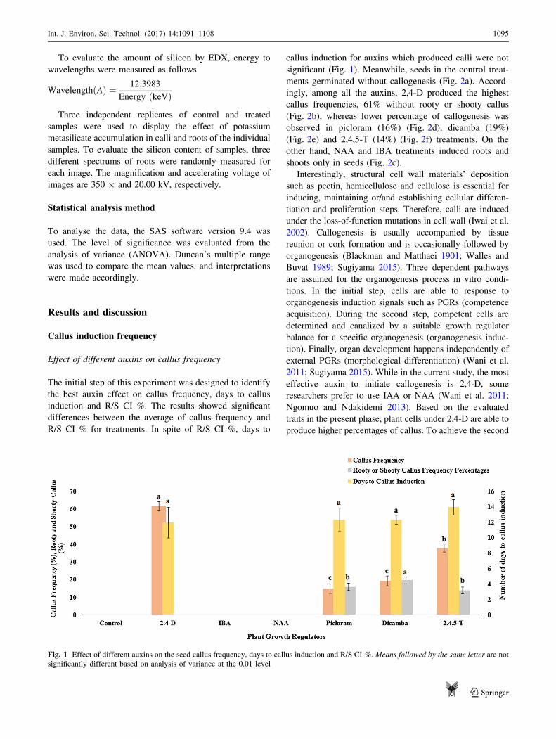
Effect of different auxins on callus frequency
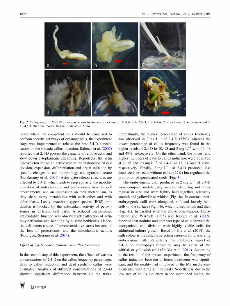
The initial step of this experiment was designed to identify
the best auxin effect on callus frequency, days to callus
induction and R/S CI %. The results showed significant
differences between the average of callus frequency and
R/S CI % for treatments. In spite of R/S CI %, days to
callus induction for auxins which produced calli were not
significant (Fig. 1). Meanwhile, seeds in the control treat-
ments germinated without callogenesis (Fig. 2a). Accord-
ingly, among all the auxins, 2,4-D produced the highest
callus frequencies, 61% without rooty or shooty callus
(Fig. 2b), whereas lower percentage of callogenesis was
observed in picloram (16%) (Fig. 2d), dicamba (19%)
(Fig. 2e) and 2,4,5-T (14%) (Fig. 2f) treatments. On the
other hand, NAA and IBA treatments induced roots and
shoots only in seeds (Fig. 2c).
Interestingly, structural cell wall materials’ deposition
such as pectin, hemicellulose and cellulose is essential for
inducing, maintaining or/and establishing cellular differen-
tiation and proliferation steps. Therefore, calli are induced
under the loss-of-function mutations in cell wall (Iwai et al.
2002). Callogenesis is usually accompanied by tissue
reunion or cork formation and is occasionally followed by
organogenesis (Blackman and Matthaei 1901; Walles and
Buvat 1989; Sugiyama 2015). Three dependent pathways
are assumed for the organogenesis process in vitro condi-
tions. In the initial step, cells are able to response to
organogenesis induction signals such as PGRs (competence
acquisition). During the second step, competent cells are
determined and canalized by a suitable growth regulator
balance for a specific organogenesis (organogenesis induc-
tion). Finally, organ development happens independently of
external PGRs (morphological differentiation) (Wani et al.
2011; Sugiyama 2015). While in the current study, the most
effective auxin to initiate callogenesis is 2,4-D, some
researchers prefer to use IAA or NAA (Wani et al. 2011;
Ngomuo and Ndakidemi 2013). Based on the evaluated
traits in the present phase, plant cells under 2,4-D are able to
produce higher percentages of callus. To achieve the second
Fig. 1 Effect of different auxins on the seed callus frequency, days to callus induction and R/S CI %. Means followed by the same letter are not
significantly different based on analysis of variance at the 0.01 level
Int. J. Environ. Sci. Technol. (2017) 14:1091–1108 1095
123
phase where the competent cells should be canalized to
perform specific pathways of organogenesis, the experiment
stage was implemented to release the best 2,4-D concen-
tration on the somatic callus induction. Rahman et al. (2007)
reported that 2,4-D possess the capacity to remove actin and
slow down cytoplasmatic streaming. Reportedly, the actin
cytoskeleton shows an active role in the elaboration of cell
division, expansion, differentiation and organ initiation by
specific changes in cell morphology and cytoarchitecture
(Kandasamy et al. 2001). Actin cytoskeleton structures are
affected by 2,4-D, which leads to crop epinasty, the mobility
alteration of mitochondria and peroxisomes into the cell
environments, and an impression on their metabolism, as
they share many metabolites with each other and with
chloroplasts. Lastly, reactive oxygen species (ROS) pro-
duction is blocked by the antioxidant activity of peroxi-
somes in different cell parts. A reduced peroxisomes
antioxidative function was observed after affection of actin
polymerization and bundling by auxinic herbicides. Hence,
the cell enters a state of severe oxidative stress because of
the loss of peroxisomes and the mitochondria actions
(Rodrıguez-Serrano et al. 2014).
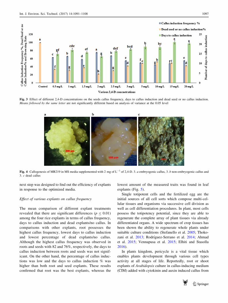
Effect of 2,4-D concentrations on callus frequency
In the second step of this experiment, the effects of various
concentrations of 2,4-D on the callus frequency percentage,
days to callus induction and dead seeds/no callus were
evaluated. Analysis of different concentrations of 2,4-D
showed significant differences between all the traits.
Interestingly, the highest percentage of callus frequency
was observed in 2 mg L-1 of 2,4-D (75%), whereas the
lowest percentage of callus frequency was found in the
higher levels of 2,4-D in 10, 15 and 5 mg L-1 with 44, 48
and 49%, respectively. On the other hand, the lowest and
highest numbers of days to callus induction were observed
at 2, 15 and 20 mg L-1 of 2,4-D at 13, 21 and 20 days,
respectively. Finally, 2 mg L-1 of 2,4-D produced less
dead seeds or seeds without callus (22%) but regulated the
promotion of germinated seeds (Fig. 3).
The embryogenic calli produced in 2 mg L-1 of 2,4-D
were compact, nodular, dry, iso-diametric, big and rather
regular in size and were tightly held together, relatively
smooth and yellowish to whitish (Fig. 4a). In contrast, non-
embryogenic calli were elongated, soft and loosely held
cells on the surface (Fig. 4b), which turned brown and died
(Fig. 4c). In parallel with the above observations, Chris-
tianson and Warnick (1985) and Rashid et al. (2009)
reported that nodular and compact type of calli showed the
unorganized cell division with highly viable cells for
additional culture growth. Based on Jila et al. (2014), the
calli colour is the suitable selection criterion for classifying
embryogenic calli. Reportedly, the inhibitory impact of
2,4-D on chlorophyll formation may be cause of the
whitish or yellowish calli (Shukla et al. 2014). According
to the results of the present experiment, the frequency of
callus induction between different treatments was signifi-
cant, and the quality had improved in the MS media sup-
plemented with 2 mg L-1 of 2,4-D. Nonetheless, due to the
low rate of callus induction in the mentioned media, the
Fig. 2 Callogenesis of MR219 in various auxins treatments. 2. a Control (MSO), 2. b 2,4-D, 2. c NAA, 2. d picloram, 2. e dicamba and 2.
f 2,4,5-T after one month. Red bar indicates 0.5 cm
1096 Int. J. Environ. Sci. Technol. (2017) 14:1091–1108
123
next step was designed to find out the efficiency of explants
in response to the optimized media.
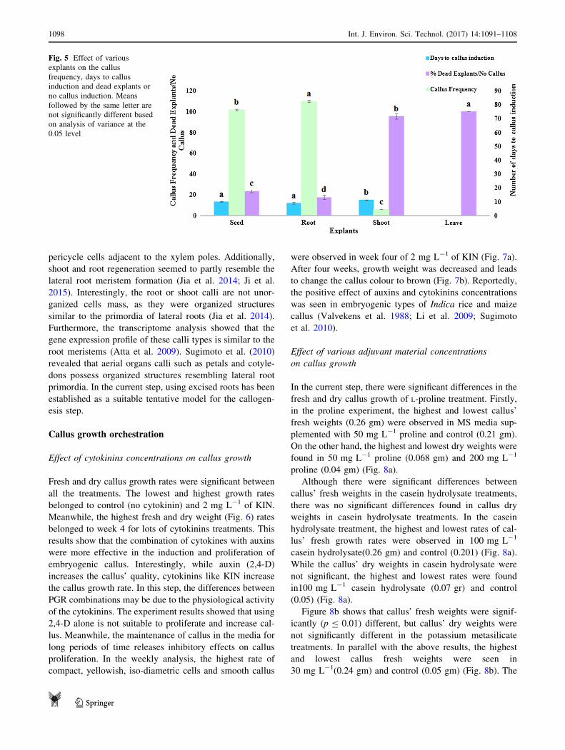
Effect of various explants on callus frequency
The mean comparison of different explant treatments
revealed that there are significant differences (p B 0.01)
among the four rice explants in terms of callus frequency,
days to callus induction and dead explants/no callus. In
comparisons with other explants, root possesses the
highest callus frequency, lowest days to callus induction
and lowest percentage of dead explants/no callus.
Although the highest callus frequency was observed in
roots and seeds with 82 and 76%, respectively, the days to
callus induction between roots and seeds was not signif-
icant. On the other hand, the percentage of callus induc-
tions was low and the days to callus induction % was
higher than both root and seed explants. These results
confirmed that root was the best explants, whereas the
lowest amount of the measured traits was found in leaf
explants (Fig. 5).
Single totipotent cells and the fertilized egg are the
initial sources of all cell sorts which compose multi-cel-
lular tissues and organisms via successive cell division as
well as cell differentiation procedures. In plant, most cells
possess the totipotency potential, since they are able to
regenerate the complete array of plant tissues via already
differentiated organs. A wide spectrum of crop tissues has
been shown the ability to regenerate whole plants under
suitable culture conditions (Stefanello et al. 2005; Thoko-
zani et al. 2013; Rodrıguez-Serrano et al. 2014; Ahmad
et al. 2015; Vennapusa et al. 2015; Elhiti and Stasolla
2016).
In plants kingdom, pericycle is a vital tissue which
enables plants development through various cell types
activity at all stages of life. Reportedly, root or shoot
explants of Arabidopsis culture in callus-inducing medium
(CIM) added with cytokinin and auxin induced callus from
Fig. 3 Effect of different 2,4-D concentrations on the seeds callus frequency, days to callus induction and dead seed or no callus induction.
Means followed by the same letter are not significantly different based on analysis of variance at the 0.05 level
Fig. 4 Callogenesis of MR219 in MS media supplemented with 2 mg of L-1 of 2,4-D. 3. a embryogenic callus, 3. b non-embryogenic callus and
3. c dead callus
Int. J. Environ. Sci. Technol. (2017) 14:1091–1108 1097
123
pericycle cells adjacent to the xylem poles. Additionally,
shoot and root regeneration seemed to partly resemble the
lateral root meristem formation (Jia et al. 2014; Ji et al.
2015). Interestingly, the root or shoot calli are not unor-
ganized cells mass, as they were organized structures
similar to the primordia of lateral roots (Jia et al. 2014).
Furthermore, the transcriptome analysis showed that the
gene expression profile of these calli types is similar to the
root meristems (Atta et al. 2009). Sugimoto et al. (2010)
revealed that aerial organs calli such as petals and cotyle-
dons possess organized structures resembling lateral root
primordia. In the current step, using excised roots has been
established as a suitable tentative model for the callogen-
esis step.
Callus growth orchestration
Effect of cytokinins concentrations on callus growth
Fresh and dry callus growth rates were significant between
all the treatments. The lowest and highest growth rates
belonged to control (no cytokinin) and 2 mg L-1 of KIN.
Meanwhile, the highest fresh and dry weight (Fig. 6) rates
belonged to week 4 for lots of cytokinins treatments. This
results show that the combination of cytokines with auxins
were more effective in the induction and proliferation of
embryogenic callus. Interestingly, while auxin (2,4-D)
increases the callus’ quality, cytokinins like KIN increase
the callus growth rate. In this step, the differences between
PGR combinations may be due to the physiological activity
of the cytokinins. The experiment results showed that using
2,4-D alone is not suitable to proliferate and increase cal-
lus. Meanwhile, the maintenance of callus in the media for
long periods of time releases inhibitory effects on callus
proliferation. In the weekly analysis, the highest rate of
compact, yellowish, iso-diametric cells and smooth callus
were observed in week four of 2 mg L-1 of KIN (Fig. 7a).
After four weeks, growth weight was decreased and leads
to change the callus colour to brown (Fig. 7b). Reportedly,
the positive effect of auxins and cytokinins concentrations
was seen in embryogenic types of Indica rice and maize
callus (Valvekens et al. 1988; Li et al. 2009; Sugimoto
et al. 2010).
Effect of various adjuvant material concentrations
on callus growth
In the current step, there were significant differences in the
fresh and dry callus growth of L-proline treatment. Firstly,
in the proline experiment, the highest and lowest callus’
fresh weights (0.26 gm) were observed in MS media sup-
plemented with 50 mg L-1 proline and control (0.21 gm).
On the other hand, the highest and lowest dry weights were
found in 50 mg L-1 proline (0.068 gm) and 200 mg L-1
proline (0.04 gm) (Fig. 8a).
Although there were significant differences between
callus’ fresh weights in the casein hydrolysate treatments,
there was no significant differences found in callus dry
weights in casein hydrolysate treatments. In the casein
hydrolysate treatment, the highest and lowest rates of cal-
lus’ fresh growth rates were observed in 100 mg L-1
casein hydrolysate(0.26 gm) and control (0.201) (Fig. 8a).
While the callus’ dry weights in casein hydrolysate were
not significant, the highest and lowest rates were found
in100 mg L-1 casein hydrolysate (0.07 gr) and control
(0.05) (Fig. 8a).
Figure 8b shows that callus’ fresh weights were signif-
icantly (p B 0.01) different, but callus’ dry weights were
not significantly different in the potassium metasilicate
treatments. In parallel with the above results, the highest
and lowest callus fresh weights were seen in
30 mg L-1(0.24 gm) and control (0.05 gm) (Fig. 8b). The
Fig. 5 Effect of various
explants on the callus
frequency, days to callus
induction and dead explants or
no callus induction. Means
followed by the same letter are
not significantly different based
on analysis of variance at the
0.05 level
1098 Int. J. Environ. Sci. Technol. (2017) 14:1091–1108
123
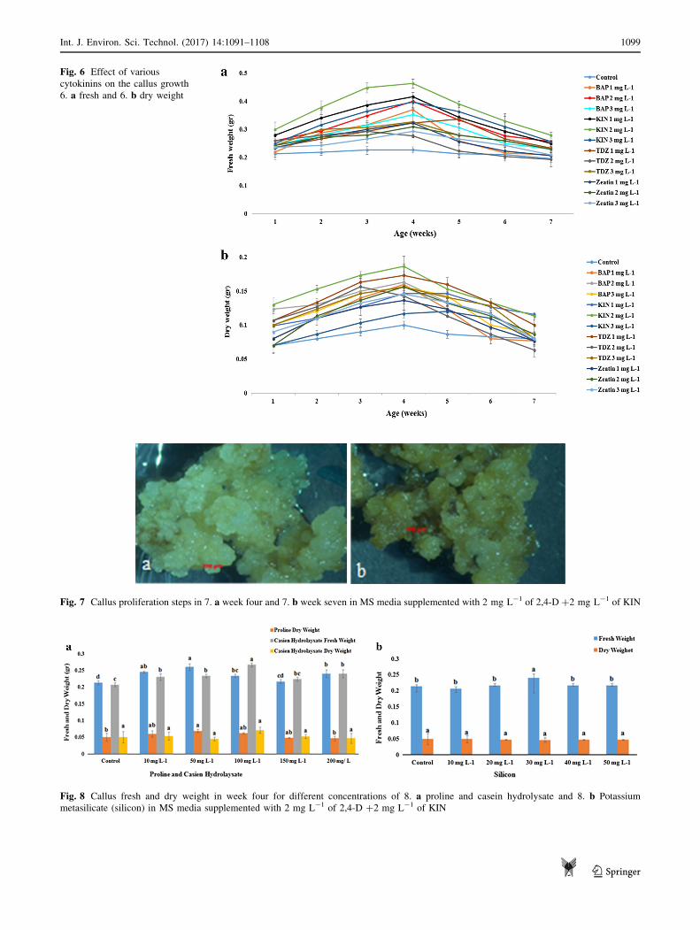
Fig. 6 Effect of various
cytokinins on the callus growth
6. a fresh and 6. b dry weight
Fig. 7 Callus proliferation steps in 7. a week four and 7. b week seven in MS media supplemented with 2 mg L-1 of 2,4-D ?2 mg L-1 of KIN
Fig. 8 Callus fresh and dry weight in week four for different concentrations of 8. a proline and casein hydrolysate and 8. b Potassium
metasilicate (silicon) in MS media supplemented with 2 mg L-1 of 2,4-D ?2 mg L-1 of KIN
Int. J. Environ. Sci. Technol. (2017) 14:1091–1108 1099
123
highest and lowest callus dry weights were also found
in30 mg L-1 (0.05 gm) and control (0.04 gm).
Nutritional additions such as proline and casein
hydrolysate have been reported to improve callusing
reaction (Lin and Zhang 2005). Accumulation of proline
occurs in plants, marine invertebrates, eubacteria and
protozoa after adverse conditions (Pawar et al. 2015).
Accumulation of this anti-stress has been described after
drought, low temperature, high temperature, salt heavy
metal, UV irradiation, anaerobiosis, atmospheric pollution
and pathogen infection (Hare and Cress 1997; Rach-
mawati and Anzai 2006; Narciso and Hattori 2010; Pawar
et al. 2015). Generally speaking, proline accumulation in
plants leads to enzymatic regulator functions, osmo-pro-
tection, carbon reverse and nutritional nitrogen storage
under stress (Holme et al. 1997). Proline is added to the
media as an organic nitrogen source. The promotive
effects of L-proline on inducing and proliferating
embryogenesis callus were observed in different plants
such as Oryza sativa, Dactylis glomerata and Zea maize
(Siripornadulsil et al. 2002; Pawar et al. 2015). Report-
edly, the proline’s presence in the medium appears to
produce a required stress condition where there is a
decrease in water potential, increasing the nutritional
elements accumulation in cells, callus and enhance
embryogenesis. Although the biochemical and physio-
logical roles of proline in tissue culture are still unclear,
the interaction of proline with nitrogen sources of media
such as nitrate and ammonium content has been reported.
Hence, proline accumulation is effective for the prolifer-
ation, initiation and maintenance of embryogenic callus
(Chowdhry et al. 1993; Verbruggen and Hermans 2008).
Nonetheless, high accumulation of proline causes toxicity
affects, which leads to negative impact on callus growth
rate (Hare and Cress 1997).
Casein hydrolysate is a rich source of phosphate, cal-
cium, vitamins, several microelements and a mixture of up
to 18 amino acids. This nutrient may overcome the short-
age or lack of glutamine while there is inadequate phos-
phorus for adequate biosynthesis. It has been reported that
the addition of casein hydrolysate is more effective than the
addition of main amino acids alone (Verbruggen and
Hermans 2008; Pawar et al. 2015) due to the fact that
proline and casein hydrolysate provide a more accessible to
nitrogen source. A low concentration of proline and casein
hydrolysate is non-toxic which helps maintain the cell for a
longer period (Siripornadulsil et al. 2002; Verbruggen and
Hermans 2008).
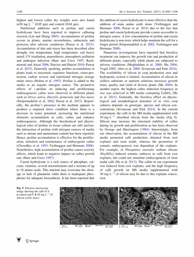
Numerous investigations have reported that biosilica
treatment can improve the growth rate and production of
different plants, especially while plants are subjected to
adverse conditions (Moghaddam et al. 2000; Ma 2004;
Vogel 2005; Abro et al. 2009; Sivanesan and Park 2014).
The availability of silicon in crop production area and
hydroponic system is limited. Accumulation of silicon in
soilless substrate or nutrient solutions increases growth
traits, quality and yield of diverse plants (Ma 2004). In
another report, the highest callus induction frequency in
rice was achieved in MS media containing CaSiO3 (He
et al. 2013). Generally, the biosilica effect on physio-
logical and morphological potential of in vitro crop
cultures depends on genotype, species and silicon con-
centrations (Sivanesan and Park 2014). In the current
experiment, the calli in the MS media supplemented with
30 mg L-1 absorbed silicon from the media (Fig. 9).
Silicon may increase the structural stability of callus
during its growth and proliferation as has been observed
by George and Sherrington (1984). Interestingly, from
our observation, the accumulation of silicon in the MS
media promoted calli production obtained from root
explants and stem nodal, whereas the promotion of
somatic embryogenesis was dependent of the explants.
For example, in Phragmites australis sodium silicate
(Na2SiO3) induced somatic embryos in calli from root
explants, but could not stimulate embryogenesis of stem
nodal calli (He et al. 2013). The callus in our experiment
was induced from root explants, and the high frequency
of calli growth on MS media supplemented with
30 mg L-1 of silicon may be due to this explants source,
root.
Fig. 9 Electron microscopy
image showing the calli in 9.
a control and 9. b 30 mg L-1 of
silicon (white mass)
1100 Int. J. Environ. Sci. Technol. (2017) 14:1091–1108
123
Plant regeneration symphony
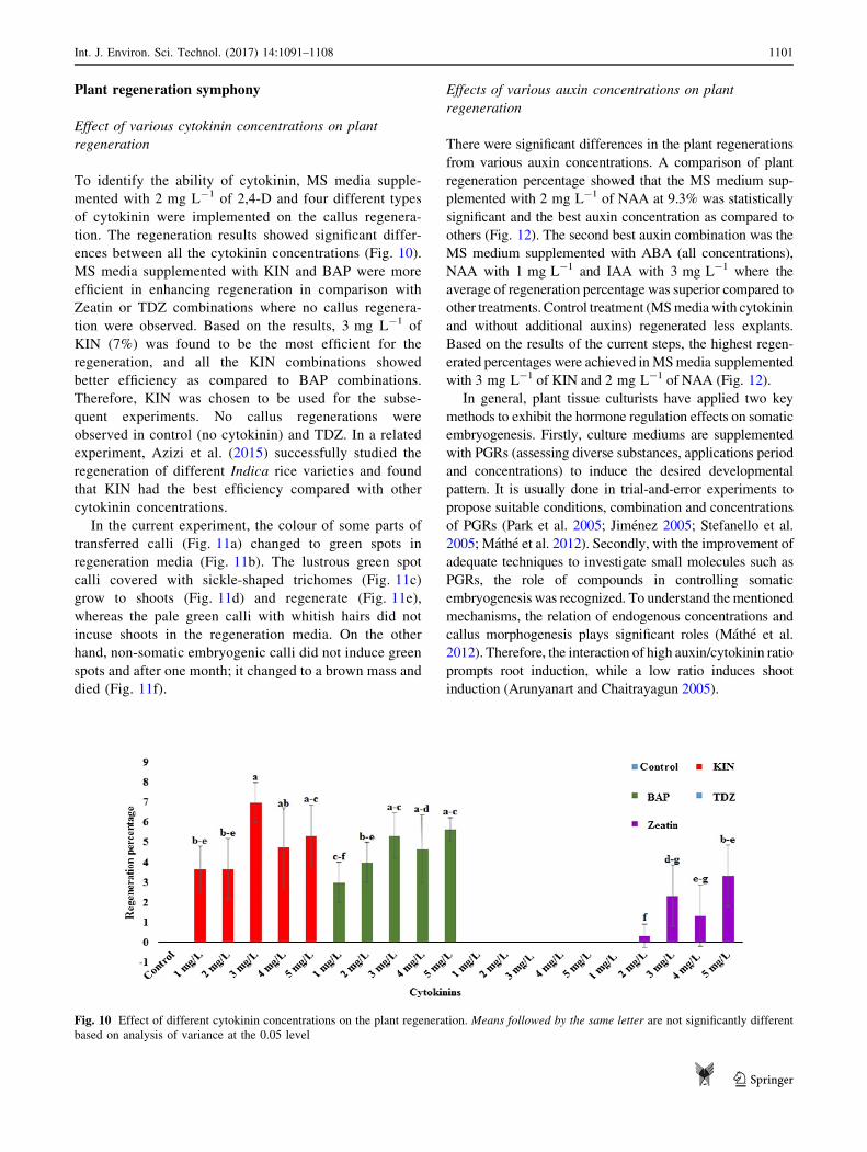
Effect of various cytokinin concentrations on plant
regeneration
To identify the ability of cytokinin, MS media supple-
mented with 2 mg L-1 of 2,4-D and four different types
of cytokinin were implemented on the callus regenera-
tion. The regeneration results showed significant differ-
ences between all the cytokinin concentrations (Fig. 10).
MS media supplemented with KIN and BAP were more
efficient in enhancing regeneration in comparison with
Zeatin or TDZ combinations where no callus regenera-
tion were observed. Based on the results, 3 mg L-1 of
KIN (7%) was found to be the most efficient for the
regeneration, and all the KIN combinations showed
better efficiency as compared to BAP combinations.
Therefore, KIN was chosen to be used for the subse-
quent experiments. No callus regenerations were
observed in control (no cytokinin) and TDZ. In a related
experiment, Azizi et al. (2015) successfully studied the
regeneration of different Indica rice varieties and found
that KIN had the best efficiency compared with other
cytokinin concentrations.
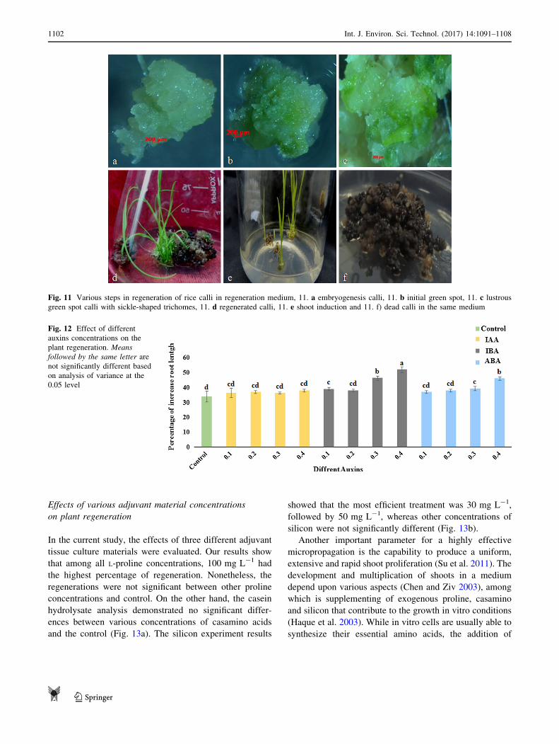
In the current experiment, the colour of some parts of
transferred calli (Fig. 11a) changed to green spots in
regeneration media (Fig. 11b). The lustrous green spot
calli covered with sickle-shaped trichomes (Fig. 11c)
grow to shoots (Fig. 11d) and regenerate (Fig. 11e),
whereas the pale green calli with whitish hairs did not
incuse shoots in the regeneration media. On the other
hand, non-somatic embryogenic calli did not induce green
spots and after one month; it changed to a brown mass and
died (Fig. 11f).
Effects of various auxin concentrations on plant
regeneration
There were significant differences in the plant regenerations
from various auxin concentrations. A comparison of plant
regeneration percentage showed that the MS medium sup-
plemented with 2 mg L-1 of NAA at 9.3% was statistically
significant and the best auxin concentration as compared to
others (Fig. 12). The second best auxin combination was the
MS medium supplemented with ABA (all concentrations),
NAA with 1 mg L-1 and IAA with 3 mg L-1 where the
average of regeneration percentage was superior compared to
other treatments. Control treatment (MSmediawith cytokinin
and without additional auxins) regenerated less explants.
Based on the results of the current steps, the highest regen-
erated percentages were achieved inMSmedia supplemented
with 3 mg L-1 of KIN and 2 mg L-1 of NAA (Fig. 12).
In general, plant tissue culturists have applied two key
methods to exhibit the hormone regulation effects on somatic
embryogenesis. Firstly, culture mediums are supplemented
with PGRs (assessing diverse substances, applications period
and concentrations) to induce the desired developmental
pattern. It is usually done in trial-and-error experiments to
propose suitable conditions, combination and concentrations
of PGRs (Park et al. 2005; Jimenez 2005; Stefanello et al.
2005; Mathe et al. 2012). Secondly, with the improvement of
adequate techniques to investigate small molecules such as
PGRs, the role of compounds in controlling somatic
embryogenesis was recognized. To understand thementioned
mechanisms, the relation of endogenous concentrations and
callus morphogenesis plays significant roles (Mathe et al.
2012). Therefore, the interaction of high auxin/cytokinin ratio
prompts root induction, while a low ratio induces shoot
induction (Arunyanart and Chaitrayagun 2005).
Fig. 10 Effect of different cytokinin concentrations on the plant regeneration. Means followed by the same letter are not significantly different
based on analysis of variance at the 0.05 level
Int. J. Environ. Sci. Technol. (2017) 14:1091–1108 1101
123
Effects of various adjuvant material concentrations
on plant regeneration
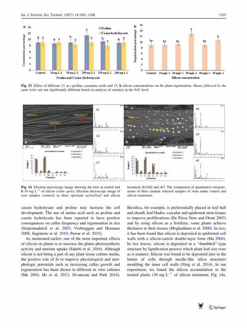
In the current study, the effects of three different adjuvant
tissue culture materials were evaluated. Our results show
that among all L-proline concentrations, 100 mg L-1 had
the highest percentage of regeneration. Nonetheless, the
regenerations were not significant between other proline
concentrations and control. On the other hand, the casein
hydrolysate analysis demonstrated no significant differ-
ences between various concentrations of casamino acids
and the control (Fig. 13a). The silicon experiment results
showed that the most efficient treatment was 30 mg L-1,
followed by 50 mg L-1, whereas other concentrations of
silicon were not significantly different (Fig. 13b).
Another important parameter for a highly effective
micropropagation is the capability to produce a uniform,
extensive and rapid shoot proliferation (Su et al. 2011). The
development and multiplication of shoots in a medium
depend upon various aspects (Chen and Ziv 2003), among
which is supplementing of exogenous proline, casamino
and silicon that contribute to the growth in vitro conditions
(Haque et al. 2003). While in vitro cells are usually able to
synthesize their essential amino acids, the addition of
Fig. 11 Various steps in regeneration of rice calli in regeneration medium, 11. a embryogenesis calli, 11. b initial green spot, 11. c lustrous
green spot calli with sickle-shaped trichomes, 11. d regenerated calli, 11. e shoot induction and 11. f) dead calli in the same medium
Fig. 12 Effect of different
auxins concentrations on the
plant regeneration. Means
followed by the same letter are
not significantly different based
on analysis of variance at the
0.05 level
1102 Int. J. Environ. Sci. Technol. (2017) 14:1091–1108
123
casein hydrolysate and proline may increase the cell
development. The use of amino acid such as proline and
casein hydrolysate has been reported to have positive
consequences on callus frequency and regeneration in rice
(Siripornadulsil et al. 2002; Verbruggen and Hermans
2008; Sugimoto et al. 2010; Pawar et al. 2015).
As mentioned earlier, one of the most important effects
of silicon on plants is to increase the plants photosynthetic
activity and nutrient uptake (Sahebi et al. 2016). Although
silicon is not being a part of any plant tissue culture media,
the positive role of Si to improve physiological and mor-
phologic potentials such as increasing callus growth and
regeneration has been shown in different in vitro cultures
(Ma 2004; He et al. 2013; Sivanesan and Park 2014).
Biosilica, for example, is preferentially placed in leaf hull
and sheath, leaf blades, vascular and epidermal stem tissues
to improve proliferations (De Paiva Neto and Otoni 2003)
and by using silicon as a fertilizer, some plants achieve
thickness in their tissues (Moghaddam et al. 2000). In rice,
it has been found that silicon is deposited in epidermal cell
walls with a silicon-cuticle double-layer form (Ma 2004).
In rice leaves, silicon is deposited in a ‘‘dumbbell’’-type
structure by lignification process which plant leaf size rises
as it matures. Silicon was found to be deposited also in the
lumen of cells through needle-like silica structures
moulding the inner cell walls (Ning et al. 2014). In our
experiment, we found the silicon accumulation in the
treated plants (30 mg L-1 of silicon treatment; Fig. 14a,
Fig. 13 Effect of different 13. a L-proline, casamino acids and 13. b silicon concentrations on the plant regeneration. Means followed by the
same letter are not significantly different based on analysis of variance at the 0.01 level
Fig. 14 Electron microscopy image showing the root. a control and
b 30 mg L-1 of silicon (white spots). Electron microscopy image of
root samples (control) in three spectrum sa1/sa2/sa3 and silicon
treatment sb1/sb2 and sb3. The comparison of quantitative measure-
ments of three random selected samples of roots under control and
silicon treatments
Int. J. Environ. Sci. Technol. (2017) 14:1091–1108 1103
123
b). The differences between the regenerations may occur
due to the decrease in hyperhydric shoot induction and
increase in mechanical strength. Silicon has been reported
to decrease the hydrogen peroxide content and oxidative
reductive enzyme activity such as ascorbate oxidase, APX
and GPX. The energy-dispersive X-ray spectroscopy
(EDX) and scanning electron microscopy (SEM) results
confirmed the existence of silicon in the regenerated root
explants (Fig. 14). The biosilica accumulation in the root’s
cell as well as cell walls’ inner space showed the positive
effects of silicon on the regeneration and development of
the plants. Silicon is usually transferred from the rice roots
to shoots and is unloaded into stem and leaf. Still, addi-
tional research is necessary to understand the molecular
and biochemical network of biosilica on somatic embryo-
genesis and organogenesis.
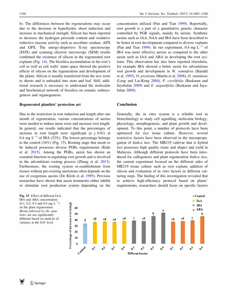
Regenerated plantlets’ protection art
Due to the restriction in root induction and length after one
month of regeneration, various concentrations of auxins
were needed to induce more roots and increase root length.
In general, our results indicated that the percentages of
increase in root length were significant (p B 0.01) at
0.4 mg L-1 of IBA (52%). The lowest percentage belongs
to the control (34%) (Fig. 15). Rooting stage that needs to
be induced possesses diverse PGRs requirements (Kido
et al. 2015). Among the PGRs, auxin has shown an
essential function in regulating root growth and is involved
in the adventitious rooting process (Zhang et al. 2013).
Furthermore, the rooting system re-establishment from
tissues without pre-existing meristems often depends on the
use of exogenous auxins (De Klerk et al. 1995). Previous
researches have shown that auxin treatments either inhibit
or stimulate root production system depending on the
concentration utilized (Pan and Tian 1999). Reportedly,
root growth is a part of a quantitative genetic character
controlled by PGR signals, mainly by auxins. Synthetic
auxins such as IAA, NAA and IBA have been described to
be better in root development compared to diverse explants
(Pan and Tian 1999). In our experiment, 0.4 mg L-1 of
IBA was more effective auxins as compared to the other
auxin such as IAA and ABA in developing the root sys-
tems. This observation has also been reported elsewhere,
for example IBA showed a better auxin for adventitious
root growth and development in W. somnifera (Baraldi
et al. 1995), O. prostrata (Martin et al. 2008), O. stamineus
(Leng and Lai-Keng 2004), P. corylifolia (Baskaran and
Jayabalan 2009) and E. angustifolia (Baskaran and Jaya-
balan 2009).
Conclusion
Generally, the in vitro system is a reliable tool in
biotechnology to study cell signalling, molecular biology,
physiology, morphogenesis, and plant growth and devel-
opment. To this point, a number of protocols have been
optimized for rice tissue culture. However, several
restrictive factors have been observed in the micropropa-
gation of Indica rice. The MR219 cultivar that is hybrid
rice possesses high quality (taste and shape) and yield in
Malaysia. Although different protocols have been intro-
duced for callogenesis and plant regeneration Indica rice,
the current experiment focused on the different sides of
MR219 tissue culture such as root explant, addition of
silicon and evaluation of in vitro factors in different cul-
turing steps. The finding of this investigation revealed that
to achieve high-efficiency protocol based on plants’
requirements, researchers should focus on specific factors
Fig. 15 Effect of different IAA,
IBA and ABA concentration
(0.1, 0.2, 0.3 and 0.4 mg L-1)
on the plant regeneration.
Means followed by the same
letter are not significantly
different based on analysis of
variance at the 0.01 level
1104 Int. J. Environ. Sci. Technol. (2017) 14:1091–1108
123
separately. Although 2,4,5-T is an Agent Orange like 2,4-
D, the reaction of these auxins was significantly different in
response to callogenesis. In this investigation, we intro-
duced root as an appropriate explant for callogenesis of
MR219. We have found also L-proline and casein hydro-
lysate are showing reliable effects on tissue culture pro-
cedures, and as far as usage of silicon, this is the first report
to show the suitability of silicon as adjuvant for MR219
micropropagation. In conclusion, for the callogenesis of
MR219 the best recipe is MS media added with 2 mg L-1
of 2,4-D with root as the explant. For the proliferation
phase, the highest efficiency was observed at week eight in
the MS media supplemented with 2 mg L-1 of 2,4-D,
2 mg L-1 of kinetin, 50 mg L-1 of L-proline, 100 mg L-1
of casein hydrolysate and 30 mg L-1 of potassium
metasilicate. Finally, MS media supplemented with
3 mg L-1 of KIN, 30 mg L-1 of potassium metasilicate
and 2 mg L-1 of NAA were selected as the best media
condition for regeneration section. To promote the roots of
regenerated explants, 0.4 mg L-1 of IBA has shown
potential as an appropriate activator.
Acknowledgements The authors would like to appreciate the Long-
term Research Grants Scheme (LRGS), Food Security Rice Research
Program of the Ministry of Higher Education, Malaysia, for creating
an opportunity to conduct the present research article.
Open Access This article is distributed under the terms of the
Creative Commons Attribution 4.0 International License (http://
creativecommons.org/licenses/by/4.0/), which permits unrestricted
use, distribution, and reproduction in any medium, provided you give
appropriate credit to the original author(s) and the source, provide a
link to the Creative Commons license, and indicate if changes were
made.
References
Abiri R, Valdiani A, Maziah M, Shaharuddin NA, Sahebi M, Yusof
ZN, Atabaki N, Talei D (2015) A critical review of the concept
of transgenic plants: insights into pharmaceutical biotechnology
and molecular farming. Curr Issues Mol Biol 18:21–42
Abro SA, Qureshi R, Soomro FM, Mirbahar AA, Jakhar G (2009)
Effects of silicon levels on growth and yield of wheat in silty
loam soil. Pak J Bot 41:1385–1390
Aggarwal D, Kumar A, Sharma J, Reddy MS (2012) Factors affecting
micropropagation and acclimatization of an elite clone of
Eucalyptus tereticornis Sm. In Vitro Cell Dev Biol Plant
48:521–529. doi:10.1007/s11627-012-9446-z
Ahmad N, Fatima N, Ahmad I, Anis M (2015) Effect of PGRs in
adventitious root culture in vitro: present scenario and future
prospects. Rend Lincei 26:307–321. doi:10.1007/s12210-015-
0445-y
Arunyanart S, Chaitrayagun M (2005) Induction of somatic embryo-
genesis in lotus (Nelumbo nucifera Geartn.). Sci Hortic
105:411–420. doi:10.1016/j.scienta.2005.01.034
Atta R, Laurens L, Boucheron Dubuisson E, Guivarc’h A, Carnero E,
Giraudat Pautot V, Rech P, Chriqui D (2009) Pluripotency of
Arabidopsis xylem pericycle underlies shoot regeneration from
root and hypocotyl explants grown in vitro. Plant J 57:626–644.
doi:10.1111/j.1365-313X.2008.03715.x
Azizi P, Rafii MY, Mahmood M, Hanafi MM, Abdullah SNA, Abiri
R, Sahebi M (2015) Highly efficient protocol for callogenesis,
somagenesis and regeneration of Indica rice plants. C R Biol
338:463–470. doi:10.1016/j.crvi.2015.04.004
Baraldi P, Bertazza G, Bregoli A, Fasolo F, Rotondi A, Predieri S,
Serafini-Fracassini D, Slovin J, Cohen J (1995) Auxins and
polyamines in relation to differential in vitro root induction on
microcuttings of two pear cultivars. J Plant Growth Regul
14:49–59. doi:10.1007/BF00212646
Baskaran P, Jayabalan N (2009) Psoralen production in hairy roots
and adventitious roots cultures of Psoralea coryfolia. Biotechnol
Lett 31:1073–1077. doi:10.1007/s10529-009-9957-9
Benlioglu B, Tuna D, Birsin M, Ozgen A (2015) Effect of growth
regulators on tissue culture parameters in rice (Oryza sativa L.).
Ekin J Crop Breed and Gen 2:43–46
Bhojwani SS, Dantu PK (2013) Plant tissue culture: an introductory
text. Springer, Berlin. doi:10.1007/978-81-322-1026-9
Birch RG (1997) Plant transformation: problems and strategies for
practical application. Annu Rev Plant Biol 48:297–326. doi:10.
1146/annurev.arplant.48.1.297
Blackman FF, Matthaei GL (1901) On the reaction of leaves to
traumatic stimulation. Ann Bot 3:533–546
Buah J, Tachie-Menson J, Addae G, Asare P (2011) Sugarcane juice
as an alternative carbon source for in vitro culture of plantains
and bananas. Am J Food Technol 6:685–694. doi:10.3923/ajft.
2011.685.694
Che Radziah C, Siti Nurkhalida A, Zamri Z, Ismanizan I (2012)
Effect of illumination, casein hydrolysate and proline on callus
induction of Oryza sativa L. var. MR219. Malays Appl Biol
41:37–41
Chen J, Ziv M (2003) Carbohydrate, metabolic, and osmotic changes
in scaled-up liquid cultures of Narcissus leaves. In Vitro Cell
Dev Biol Plant 39:645–650. doi:10.1079/IVP2003451
Chowdhry CN, Tyagi A, Maheshwari N, Maheshwari S (1993) Effect
of L-proline and L-tryptophan on somatic embryogenesis and
plantlet regeneration of rice (Oryza sativa L. cv. Pusa 169). Plant
Cell Tissue Organ Cult 32:357–361. doi:10.1007/BF00042300
Christianson M, Warnick D (1985) Temporal requirement for
phytohormone balance in the control of organogenesis in vitro.
Dev Biol 112:494–497. doi:10.1016/0012-1606(85)90423-3
Dahot MU (2007) Morpho-physiological aspects of micro-propagat-
ing banana under different hormonal conditions. Asian J Plant
Sci 6:496–501
De Klerk GJ, Keppel M, Ter Brugge J, Meekes H (1995) Timing of
the phases in adventitous root formation in apple microcuttings.
J Exp Bot 46:965–972. doi:10.1093/jxb/46.8.965
De Paiva Neto VB, Otoni WC (2003) Carbon sources and their
osmotic potential in plant tissue culture: does it matter? Sci
Hortic 97:193–202. doi:10.1016/S0304-4238(02)00231-5
Int. J. Environ. Sci. Technol. (2017) 14:1091–1108 1105
123
Din ARJM, Ahmad FI, Wagiran A, Samad AA, Rahmat Z, Sarmidi
MR (2016) Improvement of efficient in vitro regeneration
potential of mature callus induced from Malaysian upland rice
seed (Oryza sativa cv. Panderas). Saudi J Biol Sci 23:S69–S77.
doi:10.1016/j.sjbs.2015.10.022
Egan JF, Barlow KM, Mortensen DA (2014) A meta-analysis on the
effects of 2,4-D and dicamba drift on soybean and cotton. Weed
Sci 62:193–206. doi:10.1614/WS-D-13-00025.1
Elhiti M, Stasolla C (2016) Somatic embryogenesis: the molecular
network regulating embryo formation, somatic embryogenesis in
ornamentals and its applications, Springer, pp. 217–229. doi: 10.
1007/978-81-322-2683-3-14
George EF, Sherrington PD (1984) Plant propagation by tissue
culture. Exegetics Ltd, Eversley
Haque MS, Wada T, Hattori K (2003) Effects of sucrose, mannitol
and KHsub2/subPOsub4/sub on proliferation of root tip derived
shoots and subsequent bulblet formation in garlic. Asian J Plant
Sci 2:903–908
Hare P, Cress W (1997) Metabolic implications of stress-induced
proline accumulation in plants. Plant Growth Regul 21:79–102.
doi:10.1023/A:1005703923347
He C, Wang L, Liu J, Liu X, Li X, Ma J, Lin Y, Xu F (2013) Evidence
for ‘silicon’ within the cell walls of suspension cultured rice
cells. New Phytol 200:700–709. doi:10.1111/nph.12401
Holme IB, Krogstrup P, Hansen J (1997) Embryogenic callus
formation, growth and regeneration in callus and suspension
cultures ofMiscanthus x ogiformis Honda Giganteus’ as affected
by proline. Plant Cell Tiss Org Cult 50:203–210. doi:10.1023/A:
1005981300847
Hoque ME, Mansfield JW (2004) Effect of genotype and explant
age on callus induction and subsequent plant regeneration
from root-derived callus of Indica rice genotypes. Plant Cell
Tiss Org Cult 78:217–223. doi:10.1023/B:TICU.0000025640.
75168.2d
Ikeuchi M, Sugimoto K, Iwase A (2013) Plant callus: mechanisms of
induction and repression. Plant Cell 25:3159–3173. doi:10.1105/
tpc.113.116053
Islam MM, Ahmed M, Mahaldar D (2005) In vitro callus induction
and plant regeneration in seed explants of rice (Oryza Sativa L.).
Res J Agric Biol Sci 1:72–75
Islam MM, Roly ZY, Lee Y, Khalekuzzaman M (2014) In vitro
propagation and genetic transformation system using immature
embryo in elite rice (Oryza sativa L.) cultivars. Plant Breed
Biotechnol 2:88–96. doi:10.9787/PBB.2014.2.1.088
Iwai H, Masaoka N, Ishii T, Satoh S (2002) A pectin glucuronyl-
transferase gene is essential for intercellular attachment in the
plant meristem. Proc Natl Acad Sci 99:16319–16324. doi:10.
1073/pnas.252530499
Ji XH, Wang YT, Zhang R, Wu SJ, An MM, Li M, Wang CZ, Chen
XL, Zhang YM, Chen XS (2015) Effect of auxin, cytokinin and
nitrogen on anthocyanin biosynthesis in callus cultures of red-
fleshed apple (Malus sieversii f. niedzwetzkyana). Plant Cell.
Tissue and Organ Cult (PCTOC) 120:325–337. doi:10.1007/
s11240-014-0609-y
Jia Y, Zhang QX, Pan HT, Wang SQ, Liu QL, Sun LX (2014) Callus
induction and haploid plant regeneration from baby primrose
(Primula forbesii Franch.) anther culture. Sci Hortic
176:273–281. doi:10.1016/j.scienta.2014.07.018
Jimenez VM (2005) Involvement of plant hormones and plant growth
regulators on in vitro somatic embryogenesis. Plant Growth
Regul 47:91–110. doi:10.1007/s10725-005-3478-x
Kandasamy MK, Gilliland LU, McKinney EC, Meagher RB (2001)
One plant actin isovariant, ACT7, is induced by auxin and
required for normal callus formation. Plant Cell 13:1541–1554.
doi:10.1105/TPC.010026
Khaleda L, Al-Forkan M (2006) Genotypic variability in callus
induction and plant regeneration through somatic embryogenesis
of five deepwater rice (Oryza sativa L.) cultivars of Bangladesh.
Afr J Biotechnol 17:5–16
Kido N, Yokoyama R, Yamamoto T, Furukawa J, Iwai H, Satoh S,
Nishitani K (2015) The matrix polysaccharide (1; 3, 1; 4)-b-D-glucan is involved in silicon-dependent strengthening of rice cell
wall. Plant Cell Physiol 56:268–276. doi:10.1093/pcp/pcu162Konate S, Kone M, Kouakou H, Kouadio J, Zouzou M (2013) Callus
induction and proliferation from cotyledon explants in Bambara
groundnut. Afr Crop Sci J 21:255–263
Leng LW, Lai-Keng C (2004) Plant regeneration from stem nodal
segments of Orthosiphon stamineus Benth., a medicinal plant
with diuretic activity. In Vitro Cell Dev Biol Plant 40:115–118.
doi:10.1079/IVP2003500
Li Y, Gao J, Sz Fei (2009) High frequency in vitro embryogenic callus
induction and plant regeneration from indiangrass mature cary-
posis. Sci Hortic 119:306–309. doi:10.1016/j.scienta.2008.07.035
Lin YJ, Zhang Q (2005) Optimising the tissue culture conditions for
high efficiency transformation of indica rice. Plant Cell Rep
23:540–547. doi:10.1007/s00299-004-0843-6
Ma JF (2004) Role of silicon in enhancing the resistance of plants to
biotic and abiotic stresses. Soil Sci Plant Nutr 50:11–18. doi:10.
1080/00380768.2004.10408447
Martin KP, Zhang CL, Hembrom ME, Slater A, Madassery J (2008)
Adventitious root induction in Ophiorrhiza prostrata: a tool for
the production of camptothecin (an anticancer drug) and rapid
propagation. Plant Biotechnol Rep 2:163–169. doi:10.1007/
s11816-008-0057-4
Mathe C, Mosolygo A, Suranyi G, Beke A, Demeter Z, Toth VR,
Beyer D, Meszaros I, Marta M (2012) Genotype and explant-
type dependent morphogenesis and silicon response of common
reed (Phragmites australis) tissue cultures. Aquat Bot 97:57–63.
doi:10.1016/j.aquabot.2011.11.005
Moghaddam BE, Mesbah M, Yavari N (2000) The effect of in planta
TIBA and proline treatment on somatic embryogenesis of sugar
beet (Beta vulgaris L.). Euphytica 112:151–156. doi:10.1023/A:
1003879914856
Murashige T, Skoog F (1962) A revised medium for rapid growth and
bio assays with tobacco tissue cultures. Physiol Plant
15:473–497. doi:10.1111/j.1399-3054.1962.tb08052.x
Narciso JO, Hattori K (2010) Genotypic differences in morphology
and ultrastructures of callus derived from selected rice varieties.
Phillipine Sci Lett 3:59–65
Neely D (1979) Tree wounds and wound closure. J Arboricult (USA)
5:135–140
Ngomuo M, Ndakidemi P (2013) The effects of auxins and cytokinin
on growth and development of (Musa sp.) var. ‘‘Yangambi’’
explants in tissue culture. American. J Plant Sci 4:2174. doi:10.
4236/ajps.2013.411269
Ning D, Song A, Fan F, Li Z, Liang Y (2014) Effects of slag-based
silicon fertilizer on rice growth and brown-spot resistance. PLoS
ONE 9:e102681. doi:10.1371/journal.pone.0102681
Nwe NH, Mahmood M, Ho CL, Qamaruz Zaman F, Md Zain A
(2011) Regeneration capacity of cell suspension culture in
Malaysian rice genotypes under salinity stress. Asian J Biotech-
nol 3:357–367. doi:10.3923/ajbkr.2011.357.367
1106 Int. J. Environ. Sci. Technol. (2017) 14:1091–1108
123
Pan R, Tian X (1999) Comparative effect of IBA, BSAA and 5,
6-Cl2-IAA-Me on the rooting of hypocotyl in mung bean. Plant
Growth Regul 27:91–98. doi:10.1023/A:1006154426941
Panjaitan SB, Abdullah SNA, Abdul Aziz M, Meon S, Omar O (2009)
Somatic embryogenesis from scutellar embryo of Oryza sativa
L. var. MR219. Pertanika J Trop Agric Sci 32:185–194
Park SY, Ahn JK, Lee WY, Murthy HN, Paek KY (2005) Mass
production of Eleutherococcus koreanum plantlets via somatic
embryogenesis from root cultures and accumulation of
eleutherosides in regenerants. Plant Sci 168:1221–1225. doi:10.
1016/j.plantsci.2004.12.023
Pawar B, Prashant K, Bahurupe J, Jadhav A, Anil K, Pawar S (2015)
Proline and Glutamine Improve in vitro Callus Induction and
Subsequent Shooting in Rice. Rice Sci 22:283–289. doi:10.1016/
j.rsci.2015.11.001
Rachmawati D, Anzai H (2006) Studies on callus induction, plant
regeneration and transformation of Javanica rice cultivars. Plant
Biotechnol 23:521–524. doi:10.5511/plantbiotechnology.23.521
Rahman A, Bannigan A, Sulaman W, Pechter P, Blancaflor EB,
Baskin TI (2007) Auxin, actin and growth of the Arabidopsis
thaliana primary root. Plant J 50:514–528. doi:10.1111/j.1365-
313X.2007.03068.x
Rashid U, Ali S, Ali GM, Ayub N, Masood MS (2009) Establishment
of an efficient callus induction and plant regeneration system in
Pakistani wheat (Triticum aestivum) cultivars. Electron J
Biotechnol 12:4–5. doi:10.4067/S0717-34582009000300004
Rodrıguez-Serrano M, Pazmino D, Sparkes I, Rochetti A, Hawes C,
Romero-Puertas M, Sandalio L (2014) 2, 4-Dichlorophenoxy-
acetic acid promotes S-nitrosylation and oxidation of actin
affecting cytoskeleton and peroxisomal dynamics. J Exp
Bot:eru237. doi: 10.1093/jxb/eru237
Roy P, Hasan M, Rasul M, Hossain M (2015) Seed culture of
aromatic rice varieties under salt stress. Am J Biol Life Sci 3:260
Saharan V, Yadav RC, Yadav NR, Chapagain BP (2004) High
frequency plant regeneration from desiccated calli of indica rice
(Oryza sativa L.). Afr J Biotechnol 3:256–259
Sahebi M, Hanafi MM, Siti Nor Akmar A, Rafii MY, Azizi P,
Tengoua FF, Nurul Mayzaitul Azwa J, Shabanimofrad M (2015)
Importance of silicon and mechanisms of biosilica formation in
plants. BioMed Res Int 2015:1–16. doi:10.1155/2015/396010
Sahebi M, Hanafi MM, Azizi P (2016) Application of silicon in plant
tissue culture. In Vitro Cell Dev Biol Plant 2016:1–7. doi:10.
1007/s11627-016-9757-6
Shahsavari E (2010) Evaluation and optimizations of media on the
tissue culture system of upland rice. Int J Agric Biol Eng
12:537–540
Shukla R, Dube A, Koshy E (2014) Production of high quality
embryogenic callus of rice. Int Quarterly J Life Sci 9:1077–1080
Siripornadulsil S, Traina S, Verma DPS, Sayre RT (2002) Molecular
mechanisms of proline-mediated tolerance to toxic heavy metals
in transgenic microalgae. Plant Cell 14:2837–2847. doi:10.1105/
tpc.004853
Sivanesan I, Park SW (2014) The role of silicon in plant tissue
culture. Front Plant Sci 5:571. doi:10.3389/fpls.2014.00571
Stefanello S, Dal Vesco LL, Ducroquet JPH, Nodari RO, Guerra MP
(2005) Somatic embryogenesis from floral tissues of feijoa
(Feijoa sellowiana Berg). Sci Hortic 105:117–126. doi:10.1016/
j.scienta.2004.11.006
Su YH, Liu YB, Zhang XS (2011) Auxin–cytokinin interaction
regulates meristem development. Mol Plant 4:616–625. doi:10.
1093/mp/ssr007
Sugimoto K, Jiao Y, Meyerowitz EM (2010) Arabidopsis regenera-
tion from multiple tissues occurs via a root development
pathway. Dev Cell 18:463–471. doi:10.1016/j.devcel.2010.02.
004
Sugiyama M (2015) Historical review of research on plant cell
dedifferentiation. J Plant Res 128:349–359. doi:10.1007/s10265-
015-0706-y
Szabados L, Savoure A (2010) Proline: a multifunctional amino acid.
Trends Plant Sci 15:89–97. doi:10.1016/j.tplants.2009.11.009
Talei D, Valdiani A, Maziah M, Mohsenkhah M (2013) Germination
response of MR 219 rice variety to different exposure times and
periods of 2450 MHz microwave frequency. Sci World J. doi:10.
1155/2013/408026
Thokozani BL, Zulu D, Sileshi C, Teklehaimanot Z, Gondwe DS,
Sarasan V, Stevenson P (2013) Seed germination and in vitro
regeneration of the African medicinal and pesticidal plant,
Bobgunnia madagascariensis. Afr J Biotechnol 10:5959–5966
Valvekens D, Van Montagu M, Van Lijsebettens M (1988)
Agrobacterium tumefaciens-mediated transformation of Ara-
bidopsis thaliana root explants by using kanamycin selection.
Proc Natl Acad Sci 85:5536–5540. doi:10.1073/pnas.85.15.5536
Vennapusa AR, Vemanna RS, Rajashekar RBH, Babitha K, Kiranmai
K, Nareshkumar A, Sudhakar C (2015) An efficient callus
induction and regeneration protocol for a drought tolerant rice
indica genotype AC39020. J plant Sci 3:248–254
Verbruggen N, Hermans C (2008) Proline accumulation in plants: a
review. Amino Acids 35:753–759. doi:10.1007/s00726-008-
0061-6
Visarada K, Sarma N (2002) Qualitative assessment of tissue culture
parameters useful in transformation of indica rice. Curr Sci
82:343–346
Vogel G (2005) How does a single somatic cell become a whole
plant? Science 309:86. doi:10.1126/science.309.5731.86
Walles B, Buvat R (1989) Ontogeny, cell differentiation, and
structure of vascular plants. Nord J Bot 9:498–499. doi:10.
1111/j.1756-1051.1990.tb00540.x
Wani SH, Sanghera GS, Gosal SS (2011) An efficient and
reproducible method for regeneration of whole plants from
mature seeds of a high yielding Indica rice (Oryza sativa L.)
variety PAU 201. New Biotech 28:418–422. doi:10.1016/j.nbt.
2011.02.006
Wu H-X, Ma Y-Z, Xiao J-P, Zhang Z-H, Shi Z-H (2013) Photosyn-
thesis and root characteristics of rice (Oryza sativa L.) in floating
culture. Photosynthetica 51:231–237. doi:10.1007/s11099-013-
0015-4
Yaseen M, Ahmad T, Sablok G, Standardi A, Hafiz IA (2013)
Review: role of carbon sources for in vitro plant growth and
development. Mol Biol Rep 40:2837–2849. doi:10.1007/s11033-
012-2299-z
Yinxia Z, Te-chato S (2012) Callus induction and plantlet regener-
ation from mature embryos of indica rice (Oryza Sativa L.)
cultivar Kra Dang Ngah. J AgricTech 8:2423–2433
Zhang J, Feng Q, Jin C, Qiu D, Zhang L, Xie K, Yuan D, Han B,
Zhang Q, Wang S (2005) Features of the expressed sequences
revealed by a large scale analysis of ESTs from a normalized
cDNA library of the elite indica rice cultivar Minghui 63. Plant J
42:772–780. doi:10.1111/j.1365-313X.2005.02408.x
Zhang G, Cui Y, Ding X, Dai Q (2013) Stimulation of phenolic
metabolism by silicon contributes to rice resistance to sheath
blight. J Plant Nutr Soil Sci 176:118–124. doi:10.1002/jpln.
201200008
Int. J. Environ. Sci. Technol. (2017) 14:1091–1108 1107
123

Zuraida A, Rahiniza K, Nurul Hafiza M, Roowi S, Zamri Z,
Subramaniam S (2010) Factors affecting delivery and transient
expression of gusA gene in Malaysian indica rice MR 219 callus
via biolistic gun system. Afr J Biotechnol 9:8810. doi:10.5897/
AJB10.1467
Zuraida A, Naziah B, Zamri Z, Sreeramanan S (2011) Efficient plant
regeneration of Malaysian indica rice MR 219 and 232 via
somatic embryogenesis system. Acta Physiol Plant
33:1913–1921. doi:10.1007/s11738-011-0739-3
1108 Int. J. Environ. Sci. Technol. (2017) 14:1091–1108
123
Recommended