ENVIRONMENTAL SANITATION AND WATER BORNE DISEASES
Dr.Harpreet Singh
MD, DM (Gastroenterology)
COMPONENTS OF ENVIRNOMENTAL SANITATION
• WATER SANITATION
• FOOD AND MILK SANITATION
• EXCRETA DISPOSAL
• SEWAGE DISPOSAL
• REFUSE DISPOSAL
• VECTOR AND VERMIN CONTROL
• HOUSING
• AIR SANITATION
WATER SANITATION
WATER ANALYSIS CONSISTS OF:
• PHYSICAL
• CHEMICAL
• RADIOLOGICAL
• BIOLOGICAL
• BACTERIOLOGICAL

WATER SANITATION
• PUBLIC WATER SUPPLY MUST BE-
– SAFE
–REASONABLY SOFT
–PLENTIFUL
–CHEAP
WATER SANITATION
• HOUSEHOLD TREATMENT OF WATER
– BOILING, i.e., beyond 2 minutes
– CHLORINATION- 1-5ppm
– IODINE TREATMENT- 10 drops per gallon
– FILTRATION
– AERATION
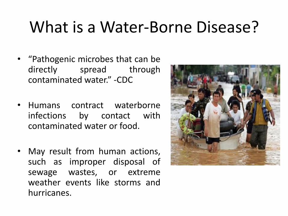
What is a Water-Borne Disease?
• “Pathogenic microbes that can be directly spread through contaminated water.” -CDC
• Humans contract waterborne infections by contact with contaminated water or food.
• May result from human actions, such as improper disposal of sewage wastes, or extreme weather events like storms and hurricanes.
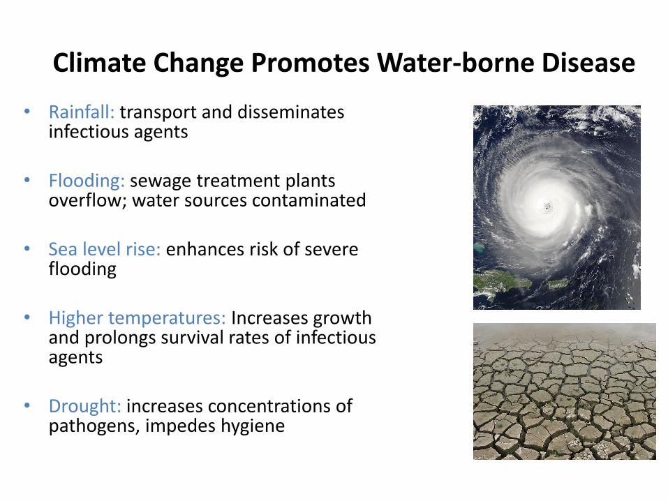
Climate Change Promotes Water-borne Disease
• Rainfall: transport and disseminates infectious agents
• Flooding: sewage treatment plants
overflow; water sources contaminated • Sea level rise: enhances risk of severe
flooding • Higher temperatures: Increases growth
and prolongs survival rates of infectious agents
• Drought: increases concentrations of
pathogens, impedes hygiene
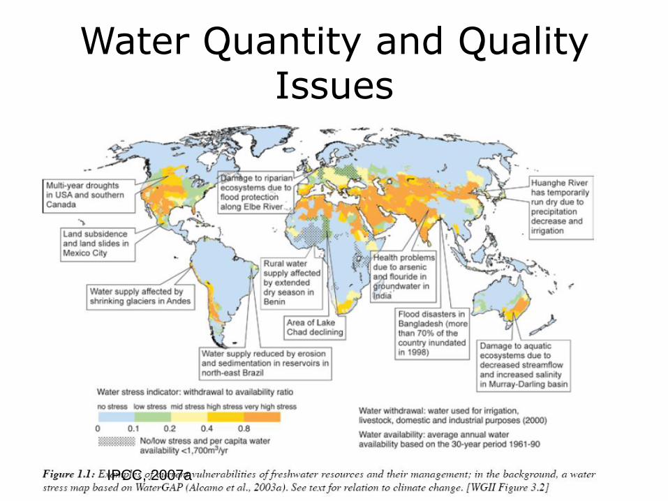
Water Quantity and Quality Issues
IPCC, 2007a
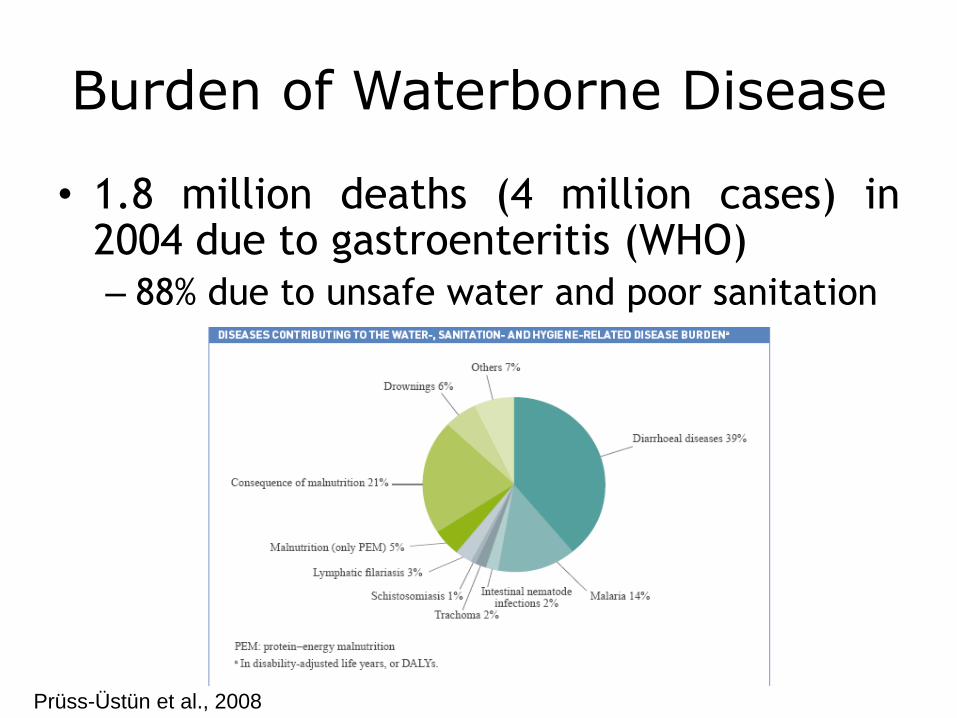
Burden of Waterborne Disease
• 1.8 million deaths (4 million cases) in 2004 due to gastroenteritis (WHO)
– 88% due to unsafe water and poor sanitation
Prüss-Üstün et al., 2008
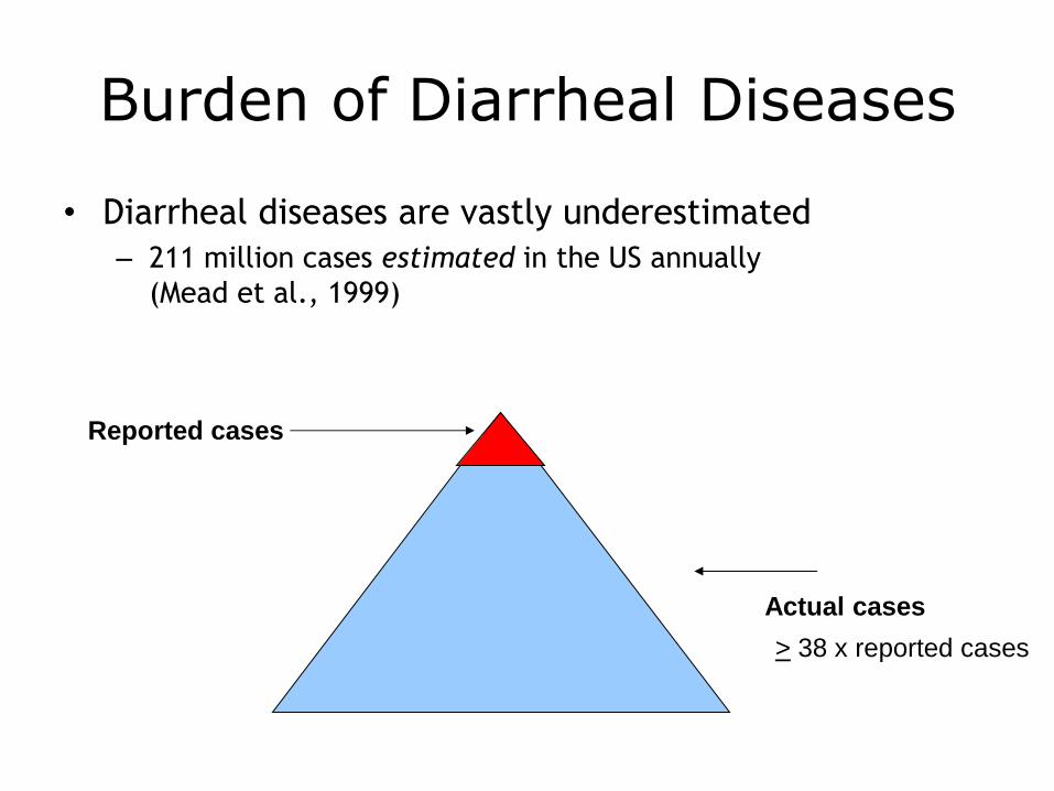
Burden of Diarrheal Diseases
• Diarrheal diseases are vastly underestimated
– 211 million cases estimated in the US annually
(Mead et al., 1999)
Reported cases
Actual cases
> 38 x reported cases
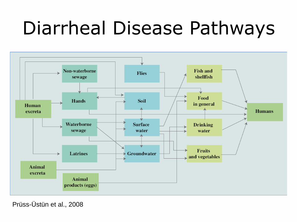
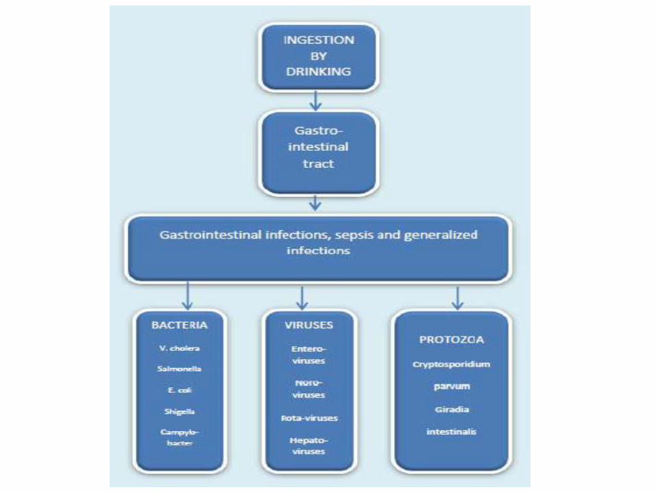
Diarrheal Disease Pathways
Prüss-Üstün et al., 2008
CHOLERA
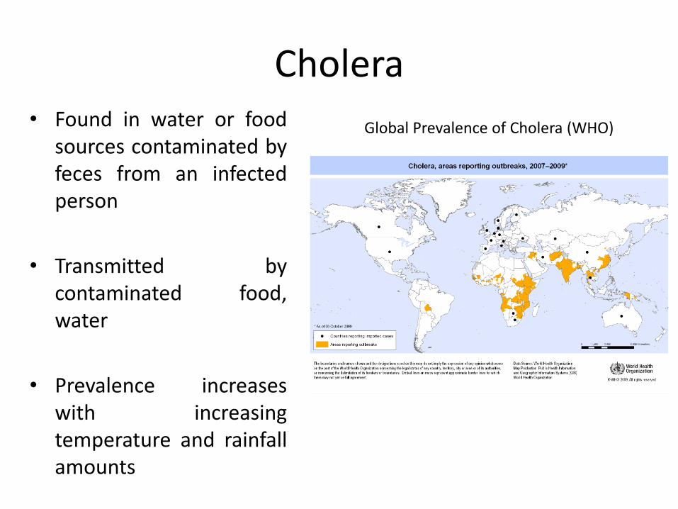
Cholera • Found in water or food
sources contaminated by feces from an infected person
• Transmitted by contaminated food, water
• Prevalence increases with increasing temperature and rainfall amounts
Global Prevalence of Cholera (WHO)
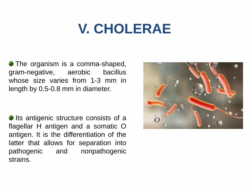
The organism is a comma-shaped,
gram-negative, aerobic bacillus
whose size varies from 1-3 mm in
length by 0.5-0.8 mm in diameter.
V. CHOLERAE
Its antigenic structure consists of a
flagellar H antigen and a somatic O
antigen. It is the differentiation of the
latter that allows for separation into
pathogenic and nonpathogenic
strains.
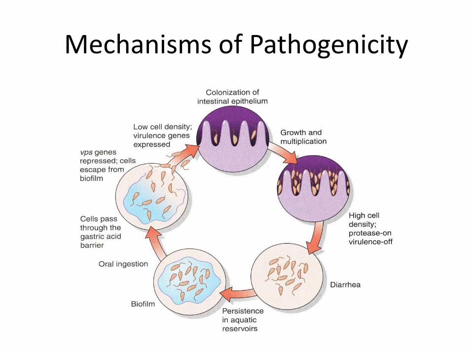
Mechanisms of Pathogenicity
V cholerae cause clinical disease by producing an enterotoxin that
promotes the secretion of fluid and electrolytes into the lumen of the
gut.
The result is watery diarrhea with electrolyte concentrations
isotonic to those of plasma.
The enterotoxin acts locally & does not invade the intestinal wall. As
a result few WBC & no RBC are found in the stool.
PATHOGENESIS

Fluid loss originates in the duodenum and upper jejunum; the
ileum is less affected.
The large volume of fluid produced in the upper intestine,
however, overwhelms the absorptive capacity of the lower
bowel, which results in severe diarrhea.
The colon is usually in a state of absorption because it is
relatively insensitive to the toxin.
PATHOGENESIS/2
TRANSMISSION
Cholera is transmitted by the fecal-oral route through
contaminated water & food.
The infectious dose of bacteria required to cause clinical disease
varies with the source. If ingested with water the dose is in the order
of 103-106 organisms. When ingested with food, fewer organisms are
required to produce disease, namely 102-104.
Person to person infection is rare.
V. cholerae is a saltwater organism & it is primary habitat is the
marine ecosystem.
Cholera has 2 main reservoirs, man & water. Animals do not play a
role in transmission of disease.
V. cholerae is unable to survive in an acid medium. Therefore, any
condition that reduces gastric acid production increases the risk of
acquisition.
TRANSMISSION/2
AT RISK GROUPS
All ages but children & elderly are more severely affected.
Subjects with blood group “O” are more susceptible; the
cause is unknown.
Subjects with reduced gastric acid.
CLINICAL PICTURE
Incubation period is 24-48 hours.
Symptoms begin with sudden onset of watery diarrhea, which may
be followed by vomiting. Fever is typically absent.z
The diarrhea has fishy odor in the beginning, but became less
smelly & more watery over time.
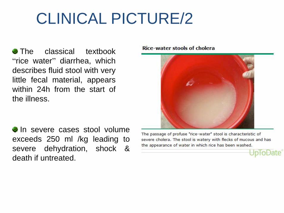
In severe cases stool volume
exceeds 250 ml /kg leading to
severe dehydration, shock &
death if untreated.
The classical textbook
“rice water” diarrhea, which
describes fluid stool with very
little fecal material, appears
within 24h from the start of
the illness.
CLINICAL PICTURE/2
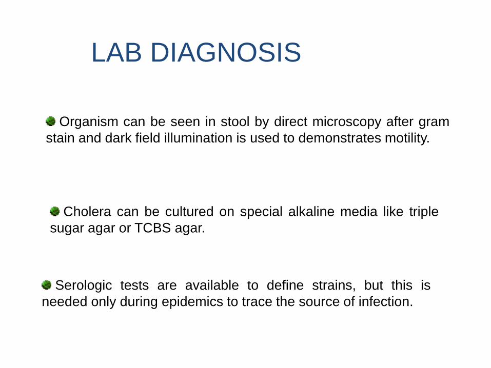
LAB DIAGNOSIS
Organism can be seen in stool by direct microscopy after gram
stain and dark field illumination is used to demonstrates motility.
Cholera can be cultured on special alkaline media like triple
sugar agar or TCBS agar.
Serologic tests are available to define strains, but this is
needed only during epidemics to trace the source of infection.
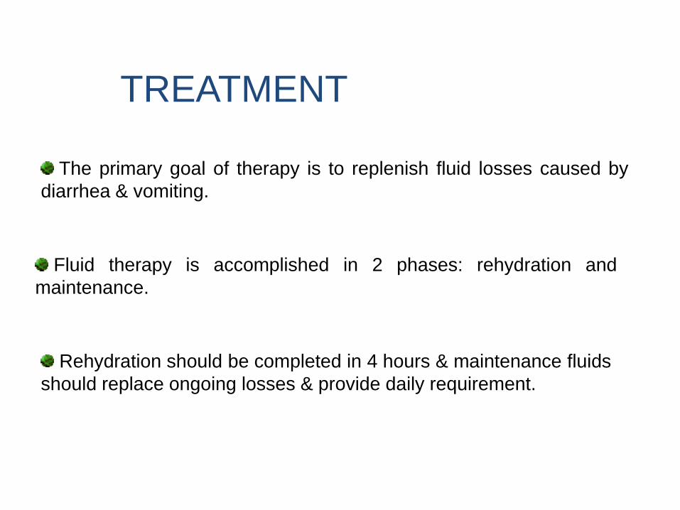
TREATMENT
The primary goal of therapy is to replenish fluid losses caused by
diarrhea & vomiting.
Fluid therapy is accomplished in 2 phases: rehydration and
maintenance.
Rehydration should be completed in 4 hours & maintenance fluids
should replace ongoing losses & provide daily requirement.
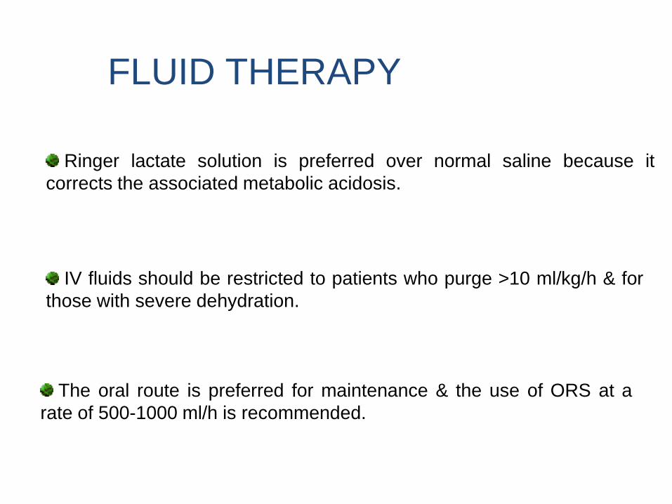
FLUID THERAPY
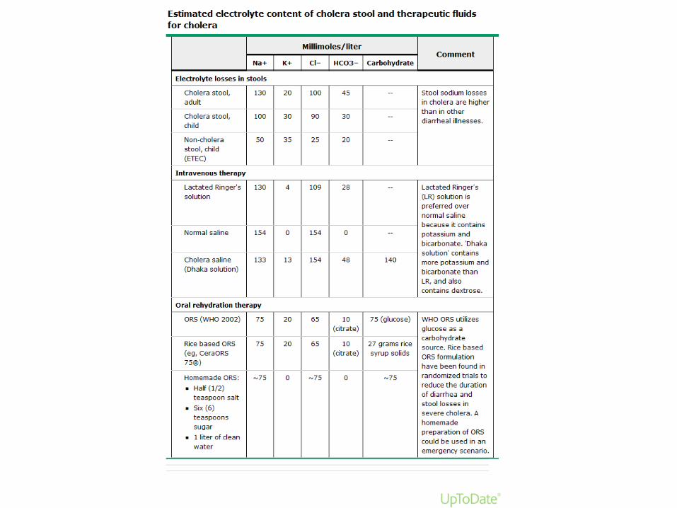
Ringer lactate solution is preferred over normal saline because it
corrects the associated metabolic acidosis.
IV fluids should be restricted to patients who purge >10 ml/kg/h & for
those with severe dehydration.
The oral route is preferred for maintenance & the use of ORS at a
rate of 500-1000 ml/h is recommended.
DRUG THERAPY
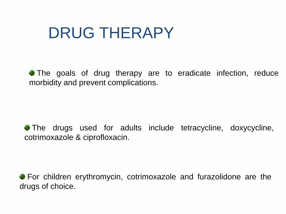
The goals of drug therapy are to eradicate infection, reduce
morbidity and prevent complications.
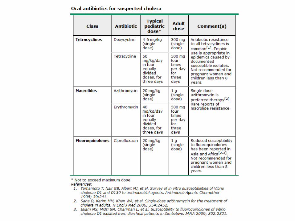
The drugs used for adults include tetracycline, doxycycline,
cotrimoxazole & ciprofloxacin.
For children erythromycin, cotrimoxazole and furazolidone are the
drugs of choice.
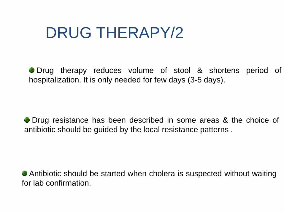
DRUG THERAPY/2
Drug therapy reduces volume of stool & shortens period of
hospitalization. It is only needed for few days (3-5 days).
Drug resistance has been described in some areas & the choice of
antibiotic should be guided by the local resistance patterns .
Antibiotic should be started when cholera is suspected without waiting
for lab confirmation.
COMPLICATIONS
If dehydration is not corrected adequately & promptly it can lead to
hypovolemic shock, acute renal failure & death.
Electrolyte imbalance is common.
Hypoglycemia occurs in children.
Complications of therapy like over hydration & side effects of drug
therapy are rare.
PUBLIC HEALTH ASPECTS
Isolation & barrier nursing is indicated
Trace source of infection.
Resume feeding with normal diet when vomiting has stopped &
continue breastfeeding infants & young children.
Notification of the case to local authorities & WHO.
PREVENTION
Education on hygiene practices.
Provision of safe, uncontaminated, drinking water to the people.
Antibiotic prophylaxis to house-hold contacts of index cases.
Vaccination against cholera to travellers to endemic countries & during
public gatherings.
CHOLERA VACCINES
The old killed injectable vaccine is obsolete now because it is not
effective.
Two new oral vaccines became available in 1997. A Killed & a live
attenuated types.
Both provoke a local immune response in the gut & a blood
immune response.
Cholera vaccination is no more required for international travellers
because risk is small.
Giardiasis
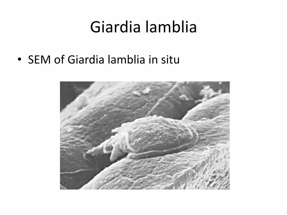
Giardia lamblia
• SEM of Giardia lamblia in situ
Clinical Disease
• 1. Diarrhea (steatorrhea)
• 2. Weight loss
• 3. Constipation
• 4. Fatigue
COMPLICATIONS
• Rarely giardia can spread from the duodenum to the biliary and
pancreatic ducts, leading to cholecystitis, cholangitis or
granulomatous hepatitis.
• Impaired exocrine pancreatic function with diminished secretion of
trypsin and lipase has also been described.
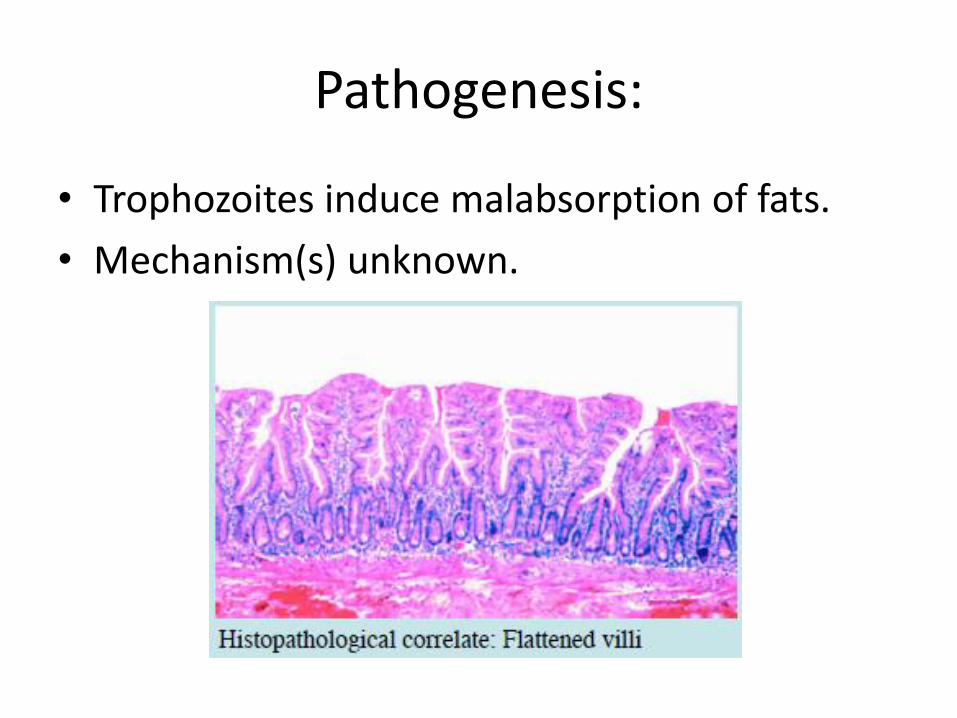
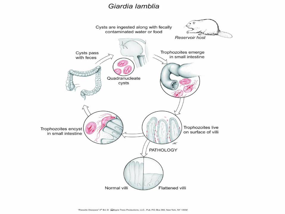
Pathogenesis:
• Trophozoites induce malabsorption of fats.
• Mechanism(s) unknown.
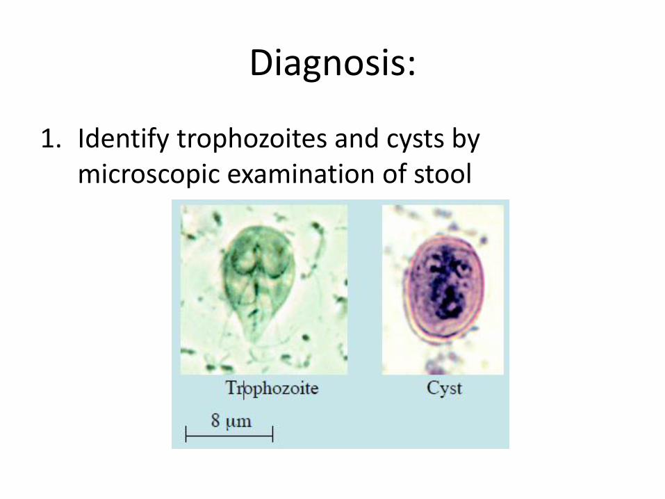
Diagnosis:
1. Identify trophozoites and cysts by microscopic examination of stool
Diagnosis
2. Antigen Capture ELISA using stool sample
3. PCR
4. IHA serology:
– Intestinal - 95% predictive of active infection
– Extra-intestinal - 100% predictive of active infection
Treatment
• Supportive measures for the treatment of children with symptomatic giardiasis include correction of fluid and electrolyte abnormalities.
• We recommend antimicrobial therapy for symptomatic patients with giardiasis (Grade 1A).
Treatment
• Treatment is not indicated for most patients with Giardia who are asymptomatic.
• To prevent the spread of infection, however, we suggest treatment of asymptomatic carriers who are food handlers, household contacts of pregnant women or immunocompromised individuals, or children in a day care or other setting who might transmit infection to others
Treatment
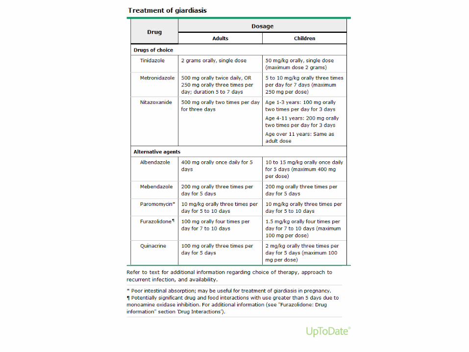
• We suggest metronidazole, tinidazole, or nitazoxanide as the drugs of choice for initial therapy (Grade 2A).
• Alternative- albendazole, mebendazole, paromomycin, furazolidone, and quinacrine.
• Patients should be counseled to avoid lactose-containing foods for one month after therapy
Follow up
• There is no need to repeat the stool examination for parasite clearance in patients whose symptoms resolve.
• Patients with recurrent diarrhea should undergo reevaluation of the stool for parasites before empiric retreatment since the diarrhea may be related to lactose intolerance rather than recurrent Giardia
Relapse
• The optimal approach to relapse after treatment is uncertain.
• We suggest treatment with a drug from a different class (Grade 2C).
• Alternative approaches include treatment with a second course of the original agent, treatment with a longer course or higher dose of the original agent, or treatment with a combination of drugs
PREVENTION
• Person-to-person spread of giardiasis can be prevented through strict handwashing, care with diaper disposal, and treatment of symptomatic patients.
• The local health department should be contacted when an outbreak of giardiasis is suspected.
• Waterborne Giardia infection can be prevented through effective treatment of drinking water.
• Individuals with giardiasis should refrain from using recreational water venues until they have been asymptomatic for two weeks

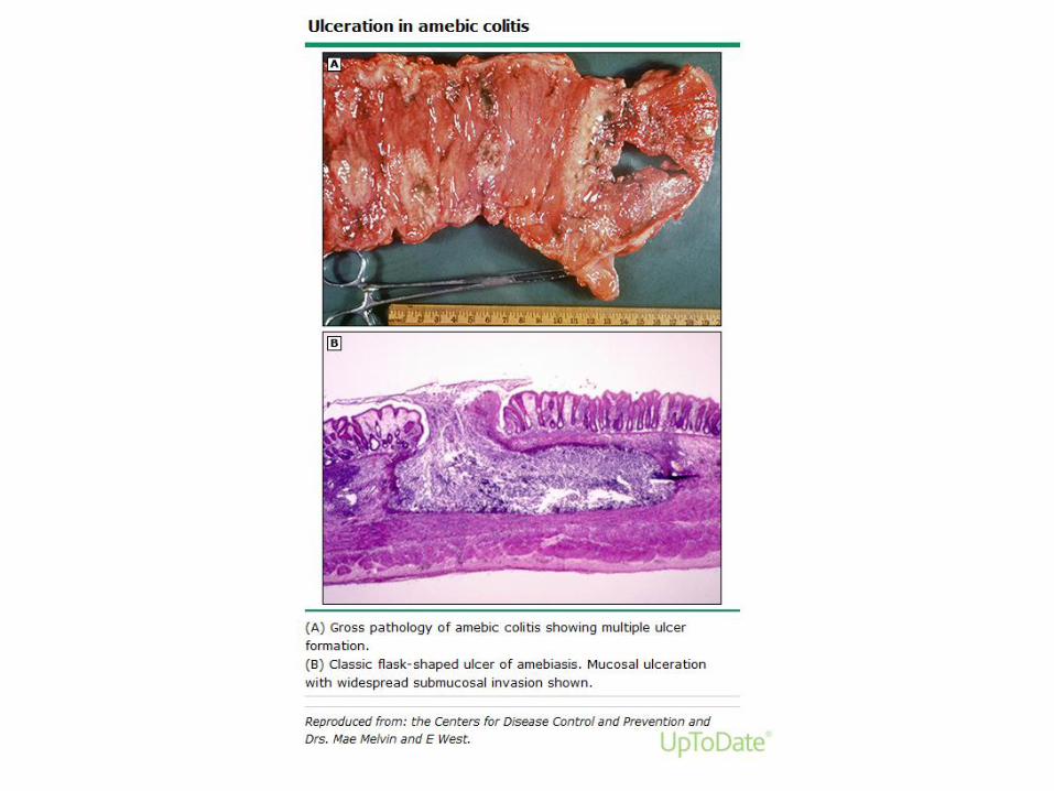
E.Histolytica
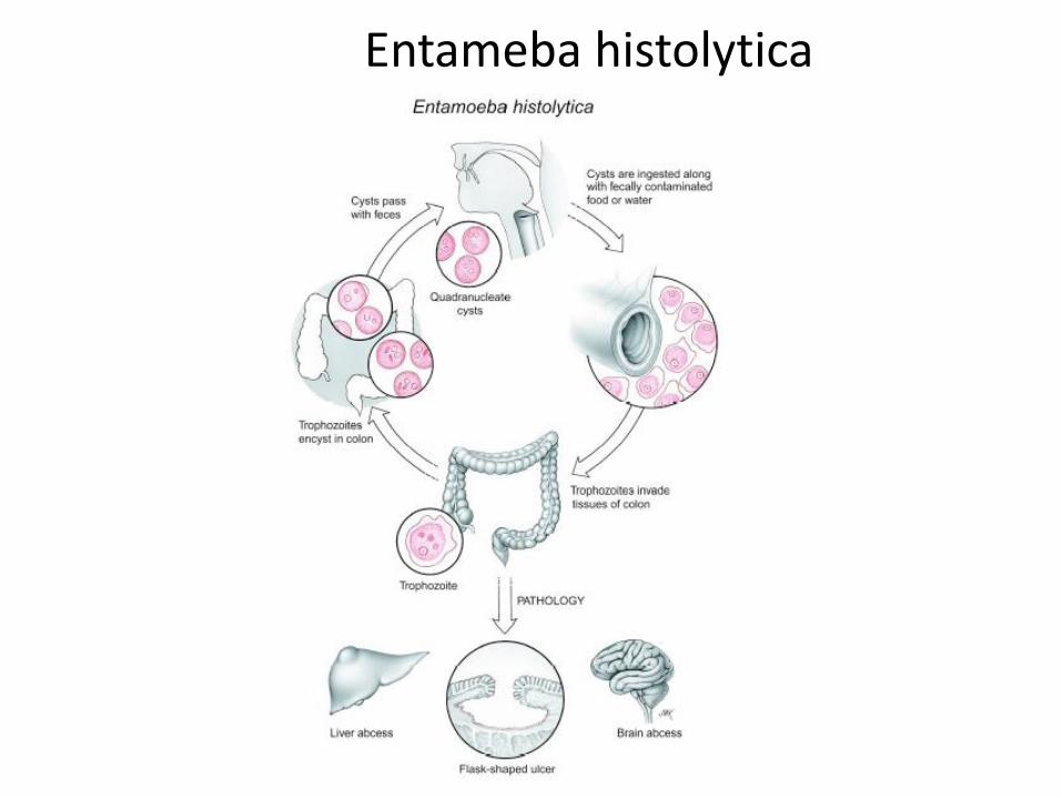
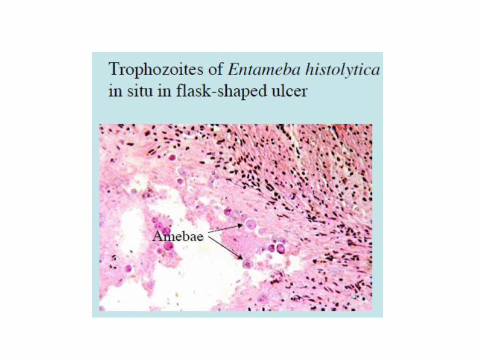
Entameba histolytica
Pathogenesis
• Attachment of amebae to target cells mediated by galactose, then pore-forming protein disrupts target cell membrane:
• Cell-cell contact induces synthesis of lysosomal enzymes in amebae at interface with target cells.
• Cell death ensues.
Clinical symptoms
• Intestinal:
– Diarrhea
– Dysentery (bloody diarrhea)
• B. Extra-intestinal:
– Liver abscess (most common site)
– Lung abscess
– Brain abscess (usually fatal)
Diagnosis
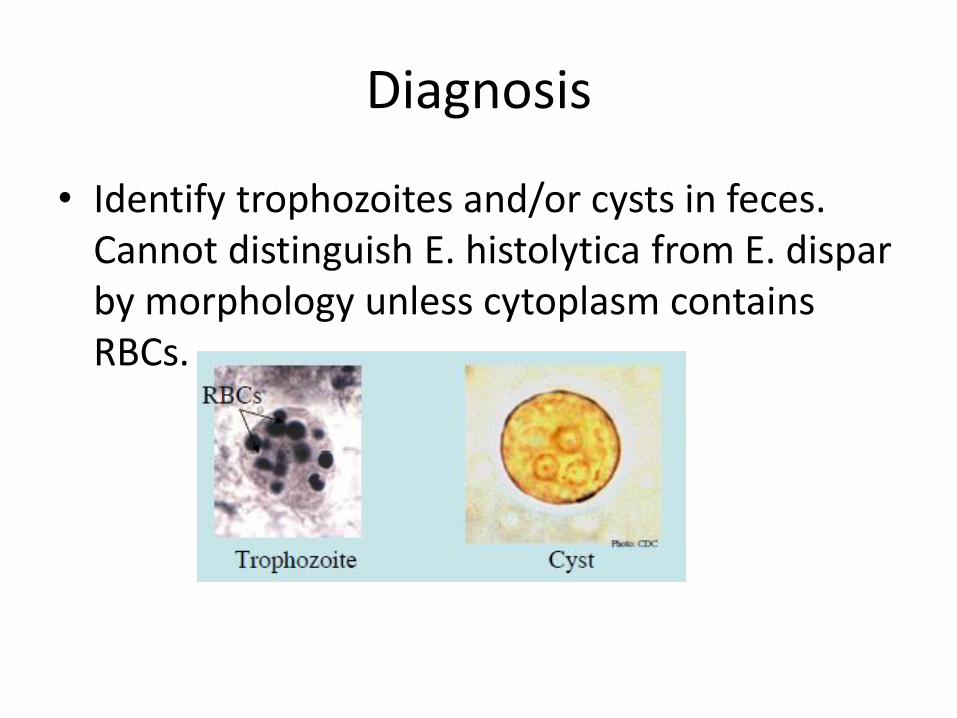
• Identify trophozoites and/or cysts in feces. Cannot distinguish E. histolytica from E. dispar by morphology unless cytoplasm contains RBCs.
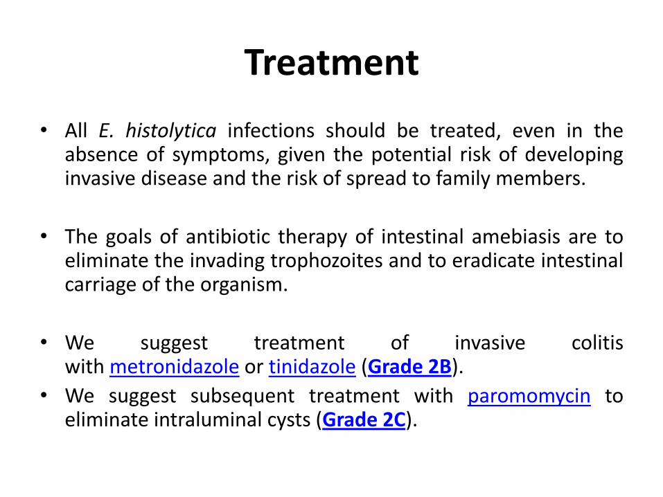
Treatment
• All E. histolytica infections should be treated, even in the absence of symptoms, given the potential risk of developing invasive disease and the risk of spread to family members.
• The goals of antibiotic therapy of intestinal amebiasis are to eliminate the invading trophozoites and to eradicate intestinal carriage of the organism.
• We suggest treatment of invasive colitis with metronidazole or tinidazole (Grade 2B).
• We suggest subsequent treatment with paromomycin to eliminate intraluminal cysts (Grade 2C).
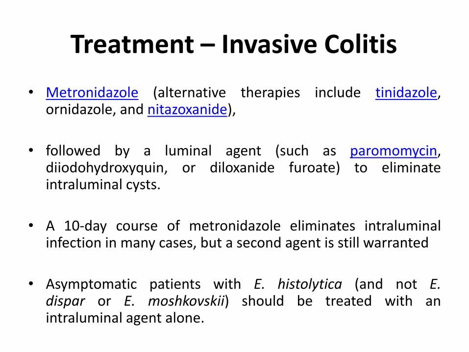
Treatment – Invasive Colitis
• Metronidazole (alternative therapies include tinidazole, ornidazole, and nitazoxanide),
• followed by a luminal agent (such as paromomycin, diiodohydroxyquin, or diloxanide furoate) to eliminate intraluminal cysts.
• A 10-day course of metronidazole eliminates intraluminal infection in many cases, but a second agent is still warranted
• Asymptomatic patients with E. histolytica (and not E. dispar or E. moshkovskii) should be treated with an intraluminal agent alone.
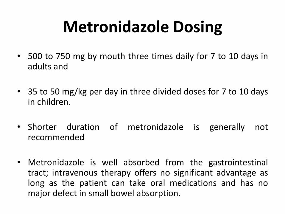
Metronidazole Dosing
• 500 to 750 mg by mouth three times daily for 7 to 10 days in adults and
• 35 to 50 mg/kg per day in three divided doses for 7 to 10 days in children.
• Shorter duration of metronidazole is generally not recommended
• Metronidazole is well absorbed from the gastrointestinal tract; intravenous therapy offers no significant advantage as long as the patient can take oral medications and has no major defect in small bowel absorption.
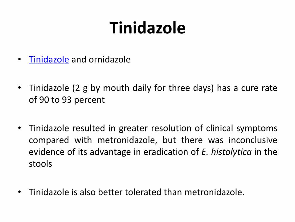
Tinidazole
• Tinidazole and ornidazole
• Tinidazole (2 g by mouth daily for three days) has a cure rate of 90 to 93 percent
• Tinidazole resulted in greater resolution of clinical symptoms compared with metronidazole, but there was inconclusive evidence of its advantage in eradication of E. histolytica in the stools
• Tinidazole is also better tolerated than metronidazole.
Nitazoxanide
• Nitazoxanide has been proposed as an alternative agent;
• it is likely to be effective at reducing clinical treatment failure
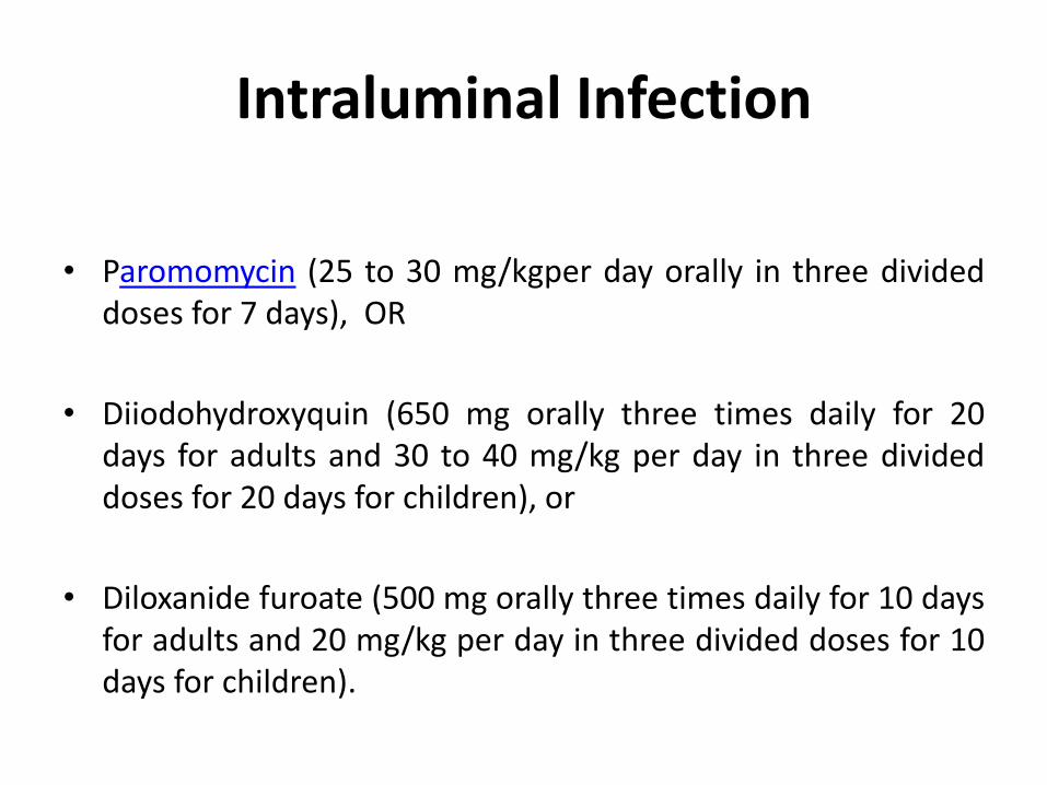
Intraluminal Infection
• Paromomycin (25 to 30 mg/kgper day orally in three divided doses for 7 days), OR
• Diiodohydroxyquin (650 mg orally three times daily for 20 days for adults and 30 to 40 mg/kg per day in three divided doses for 20 days for children), or
• Diloxanide furoate (500 mg orally three times daily for 10 days for adults and 20 mg/kg per day in three divided doses for 10 days for children).
PREVENTION
• Prevention of amebic infection in travelers to endemic areas involves avoidance of untreated water in endemic areas and uncooked food, such as fruit and vegetables, that may have been washed in local water.
• Amebic cysts are resistant to chlorine at the levels used in water supplies, but disinfection with iodine may be effective.
Hepatitis A and E
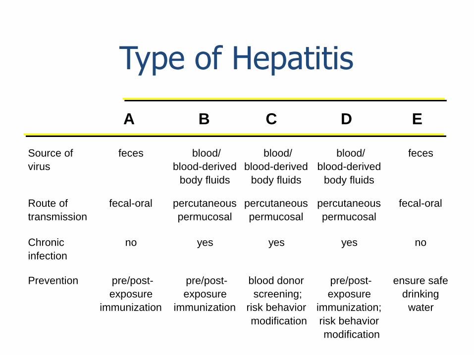
Source of
virus
feces blood/
blood-derived
body fluids
blood/
blood-derived
body fluids
blood/
blood-derived
body fluids
feces
Route of
transmission
fecal-oral percutaneous
permucosal
percutaneous
permucosal
percutaneous
permucosal
fecal-oral
Chronic
infection
no yes yes yes no
Prevention pre/post-
exposure
immunization
pre/post-
exposure
immunization
blood donor
screening;
risk behavior
modification
pre/post-
exposure
immunization;
risk behavior
modification
ensure safe
drinking
water
Type of Hepatitis
A B C D E
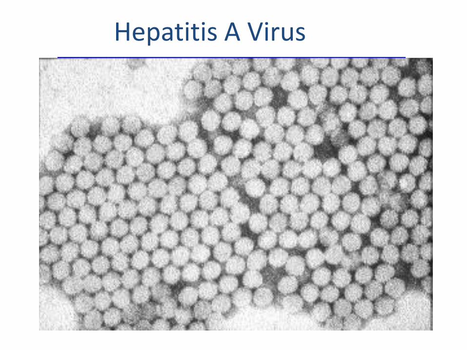
Hepatitis A Virus

Hepatitis A Virus
• Naked RNA virus
• Related to enteroviruses, formerly known as enterovirus 72, now put in its own family: heptovirus
• One stable serotype only
• Difficult to grow in cell culture: primary marmoset cell culture and also in vivo in chimpanzees and marmosets
• 4 genotypes exist, but in practice most of them are group 1
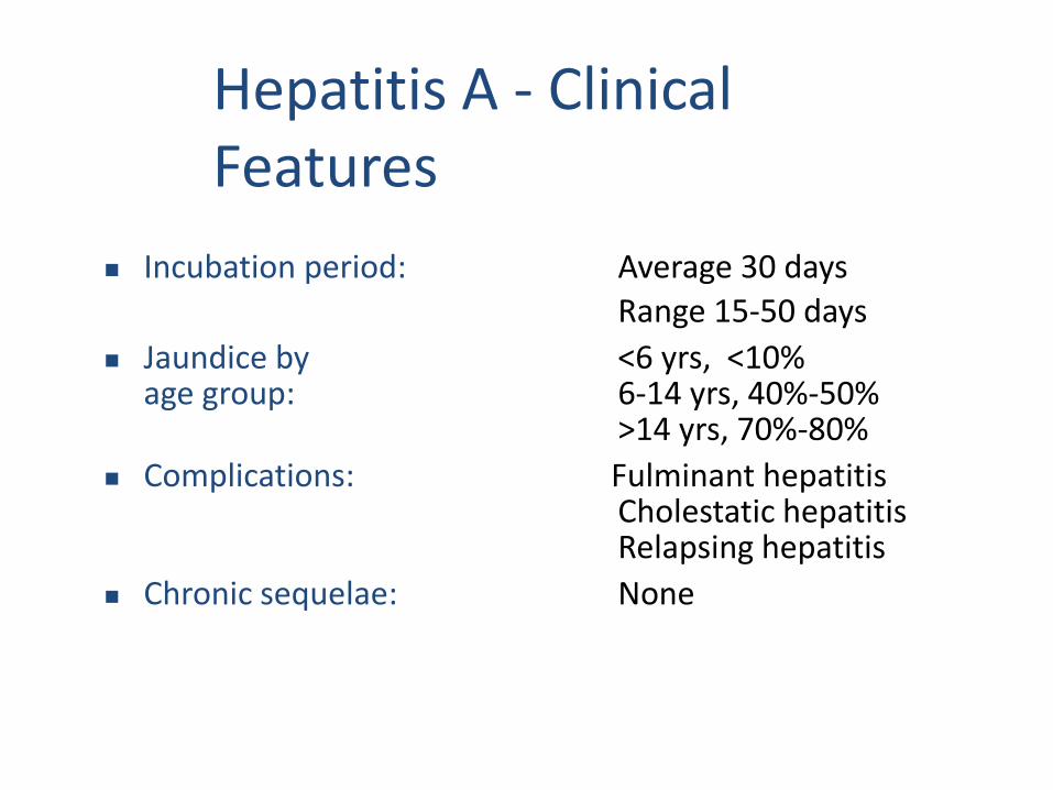
Incubation period: Average 30 days
Range 15-50 days
Jaundice by <6 yrs, <10% age group: 6-14 yrs, 40%-50% >14 yrs, 70%-80%
Complications: Fulminant hepatitis Cholestatic hepatitis Relapsing hepatitis
Chronic sequelae: None
Hepatitis A - Clinical Features
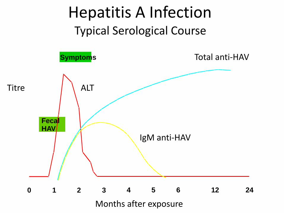
Fecal HAV
Symptoms
0 1 2 3 4 5 6 12 24
Hepatitis A Infection
Total anti-HAV
Titre ALT
IgM anti-HAV
Months after exposure
Typical Serological Course
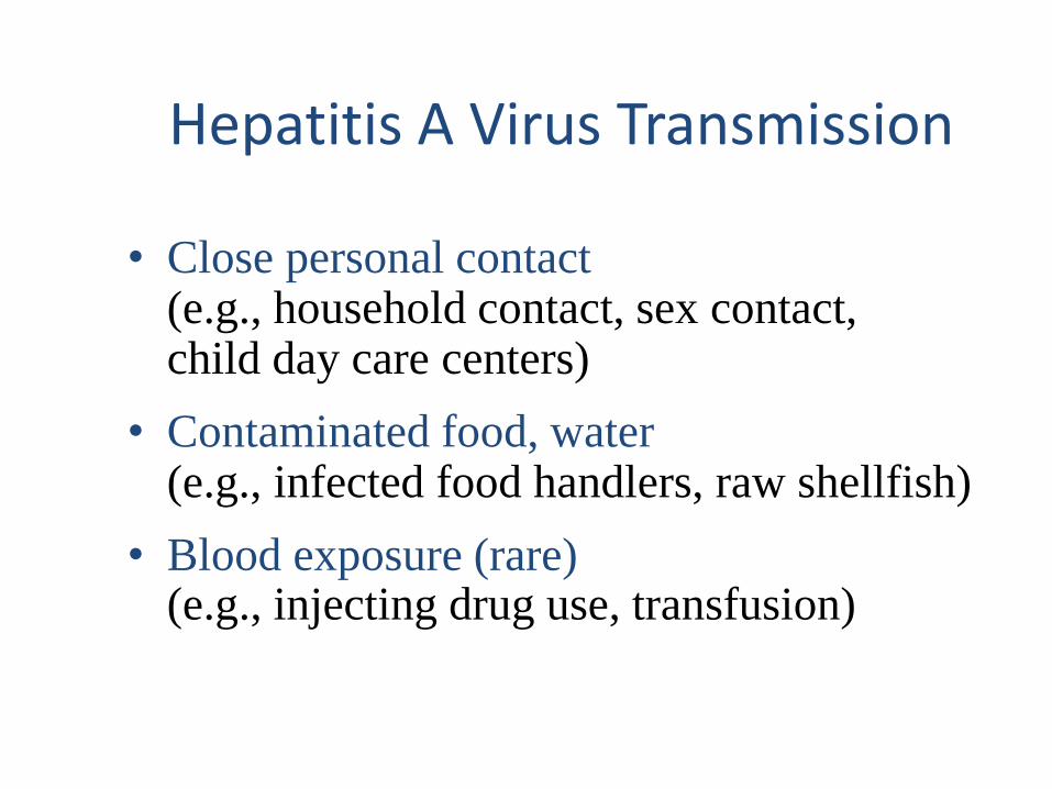
• Close personal contact (e.g., household contact, sex contact, child day care centers)
• Contaminated food, water (e.g., infected food handlers, raw shellfish)
• Blood exposure (rare) (e.g., injecting drug use, transfusion)
Hepatitis A Virus Transmission
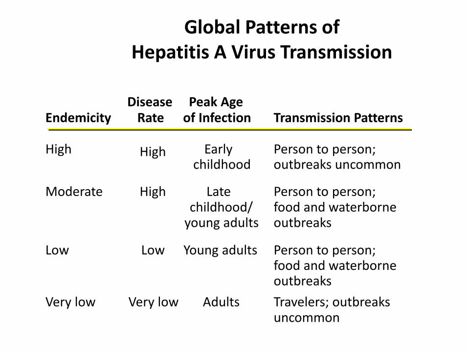
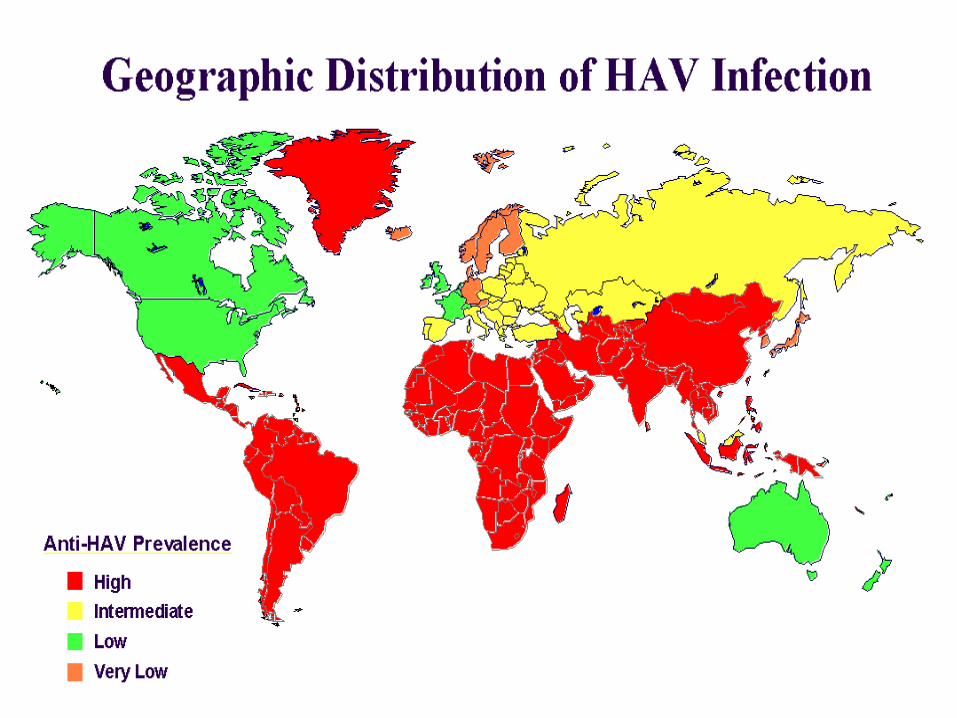
Endemicity Disease
Rate Peak Age
of Infection Transmission Patterns
High High Early childhood
Person to person; outbreaks uncommon
Moderate High Late childhood/
young adults
Person to person; food and waterborne outbreaks
Low Low Young adults Person to person; food and waterborne outbreaks
Very low Very low Adults Travelers; outbreaks uncommon
Global Patterns of Hepatitis A Virus Transmission
Laboratory Diagnosis
• Acute infection is diagnosed by the detection of HAV-IgM in serum by EIA.
• Past Infection i.e. immunity is determined by the detection of HAV-IgG by EIA.
• Cell culture – difficult and take up to 4 weeks, not routinely performed
• Direct Detection – EM, RT-PCR of faeces. Can detect illness earlier than serology but rarely performed.
• Many cases occur in community-wide outbreaks
– no risk factor identified for most cases
– highest attack rates in 5-14 year olds
– children serve as reservoir of infection
• Persons at increased risk of infection
– travelers
– homosexual men
– injecting drug users
Hepatitis A Vaccination Strategies
Epidemiologic Considerations
• Pre-exposure
– travelers to intermediate and high
HAV-endemic regions
• Post-exposure (within 14 days) Routine
– household and other intimate contacts
Selected situations
– institutions (e.g., day care centers)
– common source exposure (e.g., food prepared by infected food handler)
Hepatitis A Prevention - Immune Globulin
Hepatitis A Vaccine
• Hepatitis A vaccine is an inactivated (killed) vaccine.
• 2 Doses are needed that are given at least 6 months apart.
• It can be considered for
– those traveling to countries where hepatitis A is common,
– Homosexuals
– Drug Abusers
– have a chronic liver disease such as hepatitis B or hepatitis C
– are being treated with clotting-factor concentrates
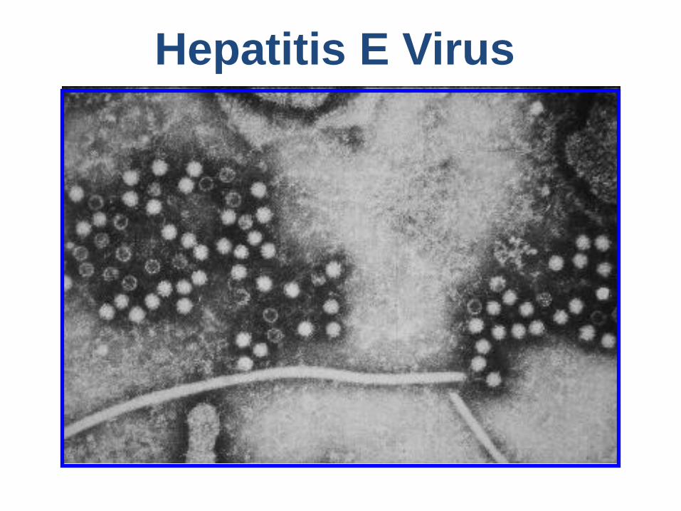
Hepatitis E Virus
Hepatitis E Virus
• now classified as a member of the genus Orthohepevirus in the
Hepeviridae family
• unenveloped RNA virus, 32-34nm in diameter
• +ve stranded RNA genome, 7.6 kb in size.
• 4 genotypes: genotype 1 (Asia), genotype 2 (Africa and
Mexico), genotype 3 (Europe and North America) and
genotype 4 (Asia)
• very labile and sensitive
• Can only be cultured recently
Incubation period: Average 40 days Range 15-60 days
Case-fatality rate: Overall, 1%-3% Pregnant women, 15%-25%
Illness severity: Increased with age
Chronic sequelae: None identified
Hepatitis E - Clinical Features
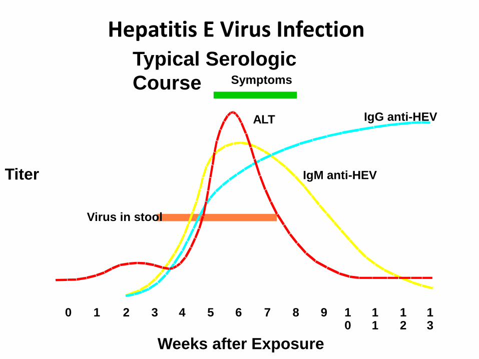
Symptoms
ALT IgG anti-HEV
IgM anti-HEV
Virus in stool
0 1 2 3 4 5 6 7 8 9 10
11
12
13
Hepatitis E Virus Infection Typical Serologic
Course
Titer
Weeks after Exposure
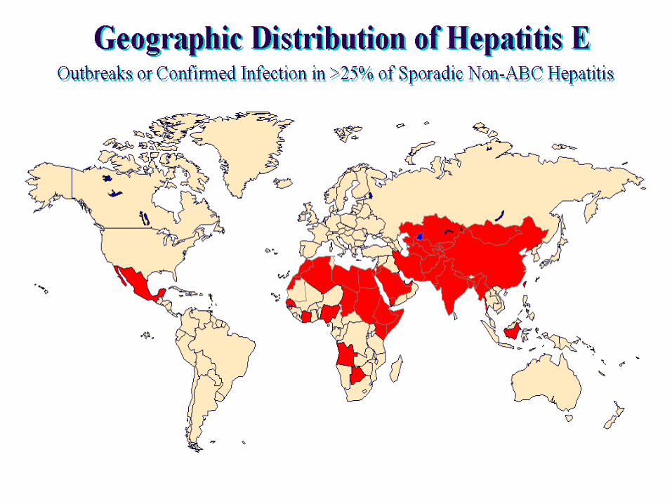
Most outbreaks associated with faecally contaminated drinking water.
Several large epidemics have occurred since in the Indian subcontinent and the USSR, China, Africa and Mexico.
In the United States and other nonendemic areas, a low prevalence of anti-HEV (<2%) has been found in healthy populations. The source of infection for these persons is unknown.
Minimal person-to-person transmission.
Hepatitis E -
Epidemiologic Features
Avoid drinking water (and beverages with ice) of unknown purity, uncooked shellfish, and uncooked fruit/vegetables not peeled or prepared by traveler.
IG prepared from donors in Western countries does not prevent infection.
Unknown efficacy of IG prepared from donors in endemic areas.
. A recombinant vaccine called HEV 239 has been developed and is licensed for use in China.
Prevention and Control Measures for Travelers to HEV-Endemic Regions
Enteric Fever
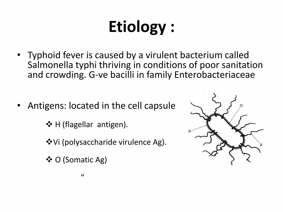
Etiology :
• Typhoid fever is caused by a virulent bacterium called Salmonella typhi thriving in conditions of poor sanitation and crowding. G-ve bacilli in family Enterobacteriaceae
• Antigens: located in the cell capsule
H (flagellar antigen).
Vi (polysaccharide virulence Ag).
O (Somatic Ag)
“
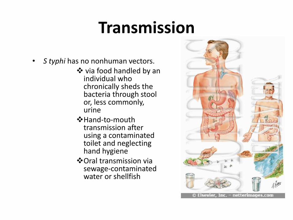
Transmission • S typhi has no nonhuman vectors.
via food handled by an individual who chronically sheds the bacteria through stool or, less commonly, urine
Hand-to-mouth transmission after using a contaminated toilet and neglecting hand hygiene
Oral transmission via sewage-contaminated water or shellfish
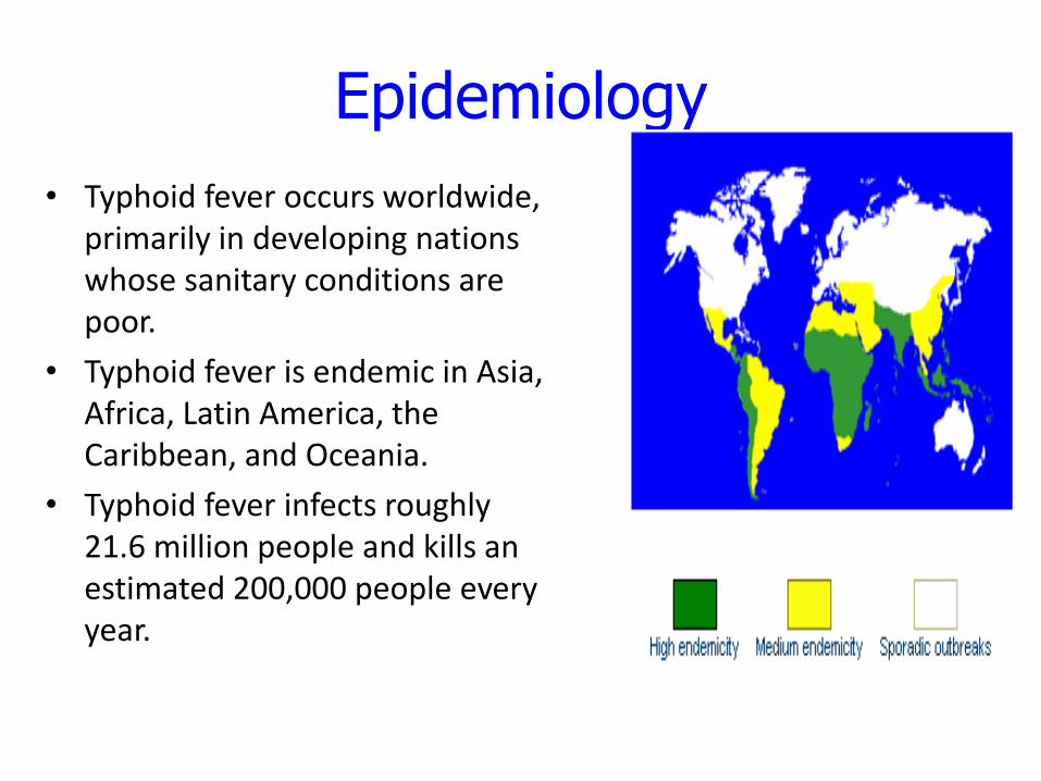
Epidemiology
• Typhoid fever occurs worldwide, primarily in developing nations whose sanitary conditions are poor.
• Typhoid fever is endemic in Asia, Africa, Latin America, the Caribbean, and Oceania.
• Typhoid fever infects roughly 21.6 million people and kills an estimated 200,000 people every year.
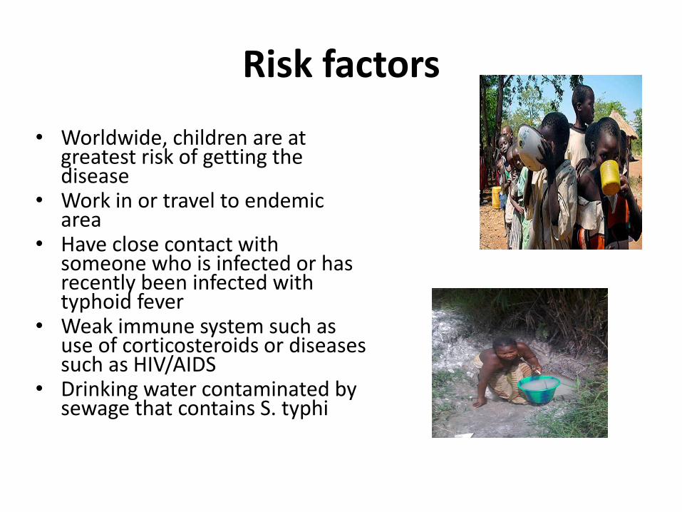
Risk factors • Worldwide, children are at
greatest risk of getting the disease
• Work in or travel to endemic area
• Have close contact with someone who is infected or has recently been infected with typhoid fever
• Weak immune system such as use of corticosteroids or diseases such as HIV/AIDS
• Drinking water contaminated by sewage that contains S. typhi
Pathogenesis
• The organisms penetrate ileal mucosa reach mesentric lymph nodes via Lymphatics , Multiply,
• Invade Blood stream via thoracic duct
• In 7 – 10 days through blood stream infect
• Liver, Gall Bladder,, spleen, Kidney, Bone marrow.
• After multiplication, bacilli pass into blood causing secondary and heavier bacteremia
Pathology
Essential lesion:
• proliferation of RES
• specific changes in lymphoid tissues
• and mesenteric lymph nodes. "typhoid nodules“
Most characteristic lesion:
• ulceration of mucous membrane in the region of the Peyer’s patches of the small intestine
Clinical presentation
The incubation period for typhoid fever is 7-14 days (range 3-60 days)
If not treated, the symptoms develop over four weeks, with new symptoms appearing each week but with treatment, symptoms should quickly improve.
Clinical manifestations
The initial period (early stage due to bacteremia)
• First week: non-specific, insidious onset of fever
Fever up to 39-400C in 5-7 days, step-ladder (seen in < 12%),
Headache, chills, toxic, tired, sore throat, cough,
abdominal pain
and diarrhea or constipation.
The fastigium stage
• second and third weeks.
• fever reaches a plateau at 39-40. Last 10-14 days.
• more toxic and anorexic with significant weight loss. The
conjunctivae are injected, and the patient is tachypnoeic with a
thredy pulse and crackles over the lung bases. Abdominal
distension is severe. Some patients experience foul, green-
yellow, liquid diarrohea (pea soup diarrohea).
• The( typhoid state) is characterized by apathy, confusion, and
even psychosis. Necrotic Payer patches may cause bowel
perforation and peritonitis. This complication may be masked
by corticosteroids. At this point, overwhelming toxaemia,
myocarditis, or intestinal hemorrhage may cause death.
• Signs and symptoms:
relative bradycardia.
• Splenomegaly, hepatomegaly
• rash ( rose-spots):30%, maculopapular
a faint pale color, slightly raised
round or lenticular, fade on pressure
2-4 mm in diameter, less than 10 in No.
on the trunk, disappear in 2-3 days.
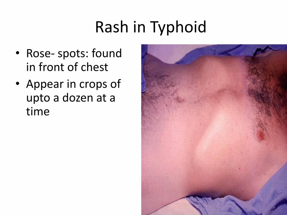
Rash in Typhoid
• Rose- spots: found in front of chest
• Appear in crops of upto a dozen at a time
defervescence stage
• By the fourth week of infection:
If the individual survives , the fever, mental state, and abdominal distension slowly improve over a few days. Intestinal and neurologic complications may still occur. Weight loss and debilitating weakness last months. Some survivors become asymptomatic carriers and have the potential to transmit the bacteria indefinitely
convalescence stage
• the fifth week: disappearance of all symptoms, but
can relapse
Atypical manifestations :
• Mild infection:
very common seen recently
symptom and signs are mild
good general condition
temperature is 380C
short period of disease
recovery expected in 1~3 weeks
seen in early antibiotic users
in young children more common
easy to misdiagnose
• Persistent infection:
disease continue > 5weeks
Ambulatory infection:
mild symptoms, early intestinal bleeding or
perforation.
• Fulminant infection:
rapid onset, severe toxemia and
septicemia.
High fever, chill, circulatory failure,
shock, delirium, coma, myocarditis,
bleeding and other complications, DIC.
• In the aged
temperature not high, weakness common.
More complications.
High mortality.
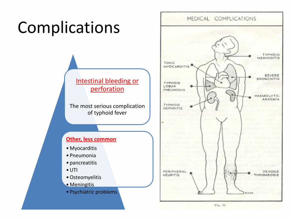
Complications
Intestinal bleeding or perforation
The most serious complication
of typhoid fever
Other, less common
• Myocarditis
• Pneumonia
• pancreatitis
• UTI
• Osteomyelitis
• Meningitis
• Psychiatric problems
Complications
Intestinal hemorrhage
Commonly appear during the second-third week
may be mild or severe bleeding
often caused by unsuitable food, and diarrhea
serious bleeding in about 2~8%
clues: sudden drop in temperature, rise in pulse, and
signs of shock followed by dark or fresh blood in the
stool.
Intestinal perforation:
• more serious. Incidence:1-4%
• Commonly appear during 2nd-3rd week.
• Take place at the lower end of ileum.
• Before perforation, abdominal pain or diarrhea, intestinal bleeding .
• When perforation: ↑ abdominal pain, sweating, drop in temperature,
and increase in pulse rate, then rebound tenderness +ve
reduce or disappear in the dullness of liver, leukocytosis .
• Temperature rise when peritonitis appear.
• free air in abdominal x-ray.
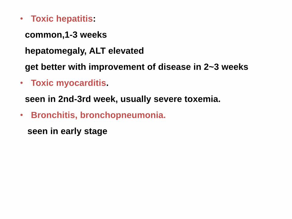
• Toxic hepatitis:
common,1-3 weeks
hepatomegaly, ALT elevated
get better with improvement of disease in 2~3 weeks
• Toxic myocarditis.
seen in 2nd-3rd week, usually severe toxemia.
• Bronchitis, bronchopneumonia.
seen in early stage
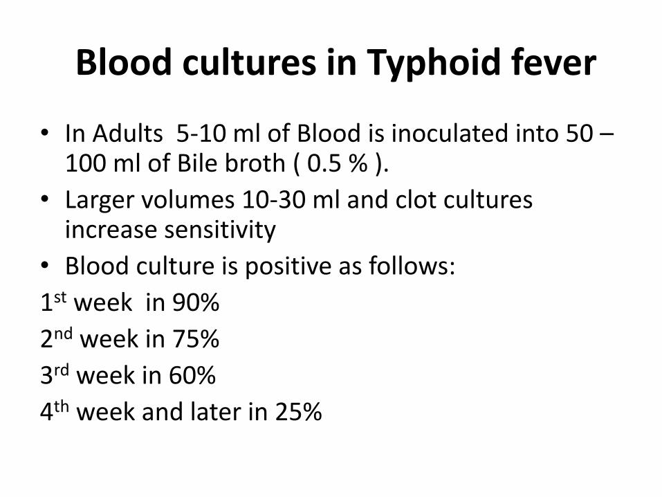
Blood cultures in Typhoid fever
• In Adults 5-10 ml of Blood is inoculated into 50 – 100 ml of Bile broth ( 0.5 % ).
• Larger volumes 10-30 ml and clot cultures increase sensitivity
• Blood culture is positive as follows:
1st week in 90%
2nd week in 75%
3rd week in 60%
4th week and later in 25%
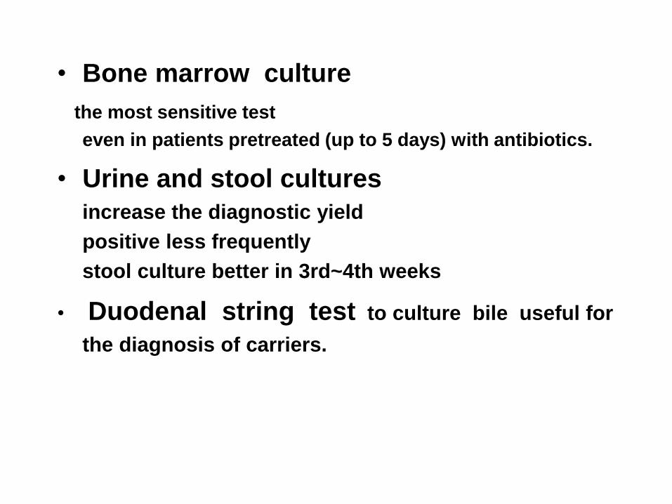
• Bone marrow culture
the most sensitive test
even in patients pretreated (up to 5 days) with antibiotics.
• Urine and stool cultures
increase the diagnostic yield
positive less frequently
stool culture better in 3rd~4th weeks
• Duodenal string test to culture bile useful for
the diagnosis of carriers.
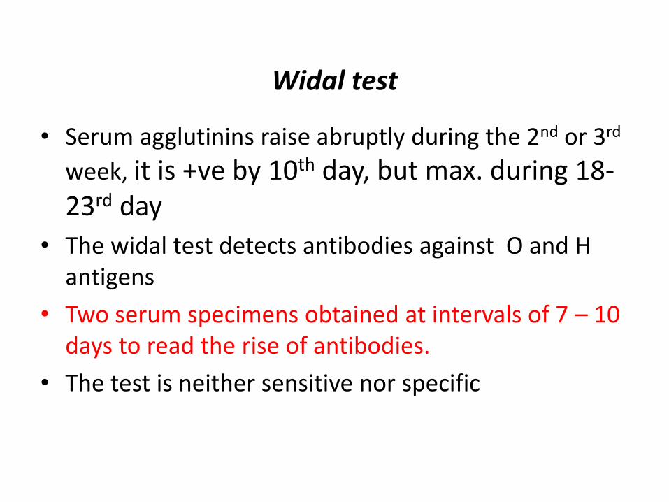
Widal test
• Serum agglutinins raise abruptly during the 2nd or 3rd
week, it is +ve by 10th day, but max. during 18-23rd day
• The widal test detects antibodies against O and H antigens
• Two serum specimens obtained at intervals of 7 – 10 days to read the rise of antibodies.
• The test is neither sensitive nor specific
TREATMENT
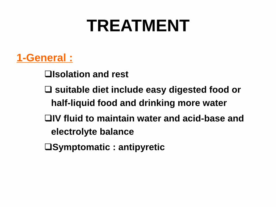
1-General :
Isolation and rest
suitable diet include easy digested food or
half-liquid food and drinking more water
IV fluid to maintain water and acid-base and
electrolyte balance
Symptomatic : antipyretic
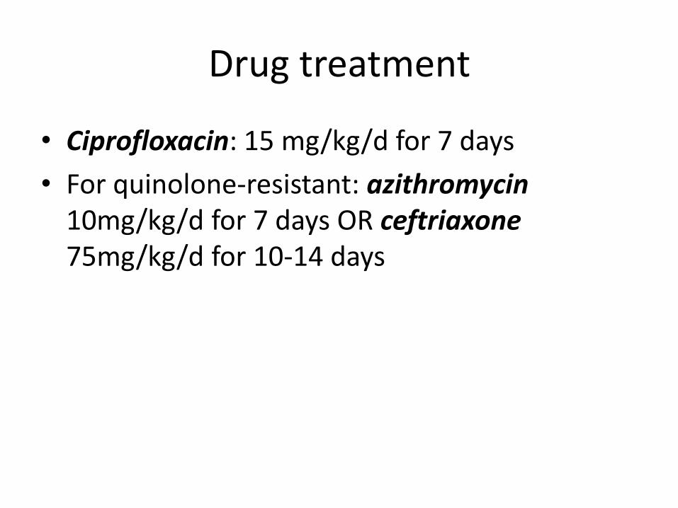
Drug treatment
• Ciprofloxacin: 15 mg/kg/d for 7 days
• For quinolone-resistant: azithromycin 10mg/kg/d for 7 days OR ceftriaxone 75mg/kg/d for 10-14 days
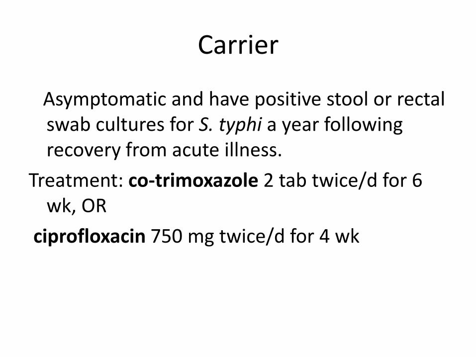
Carrier
Asymptomatic and have positive stool or rectal swab cultures for S. typhi a year following recovery from acute illness.
Treatment: co-trimoxazole 2 tab twice/d for 6 wk, OR
ciprofloxacin 750 mg twice/d for 4 wk
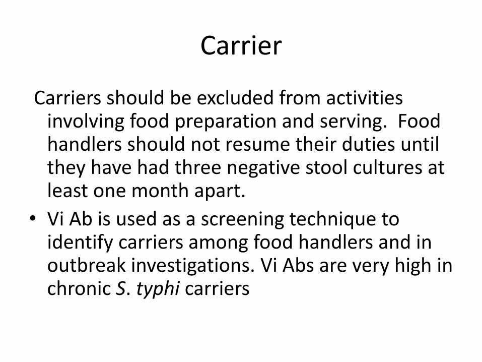
Carrier
Carriers should be excluded from activities involving food preparation and serving. Food handlers should not resume their duties until they have had three negative stool cultures at least one month apart.
• Vi Ab is used as a screening technique to identify carriers among food handlers and in outbreak investigations. Vi Abs are very high in chronic S. typhi carriers
Relapse
• Apparent recovery can be followed by relapse in 5 – 10 % of untreated patient
• culture +ve of S.typhi after 1-3 wks of defervescence
• Symptom and signs reappear
• the bacilli have not been completely eradicated
• Some cases relapse more than once
• On few occasions relapses can be severe and may be fatal.
Prognosis:
• Case fatality 0.5-1%.
• but high in old ages, infant, and serious complications
• About 3% of patients become fecal carriers .
Vaccines for Typhoid Prevention
• Two types :
1. Oral – A live vaccine ( typhoral )
One capsule given orally taken before food, with a glass of water or milk, on day 1, 3, 5 ( three doses )
No antibiotics should be taken during the period of administration of vaccine
2. The injectable vaccine, ( typhim –vi)
Given as single sc or im injection
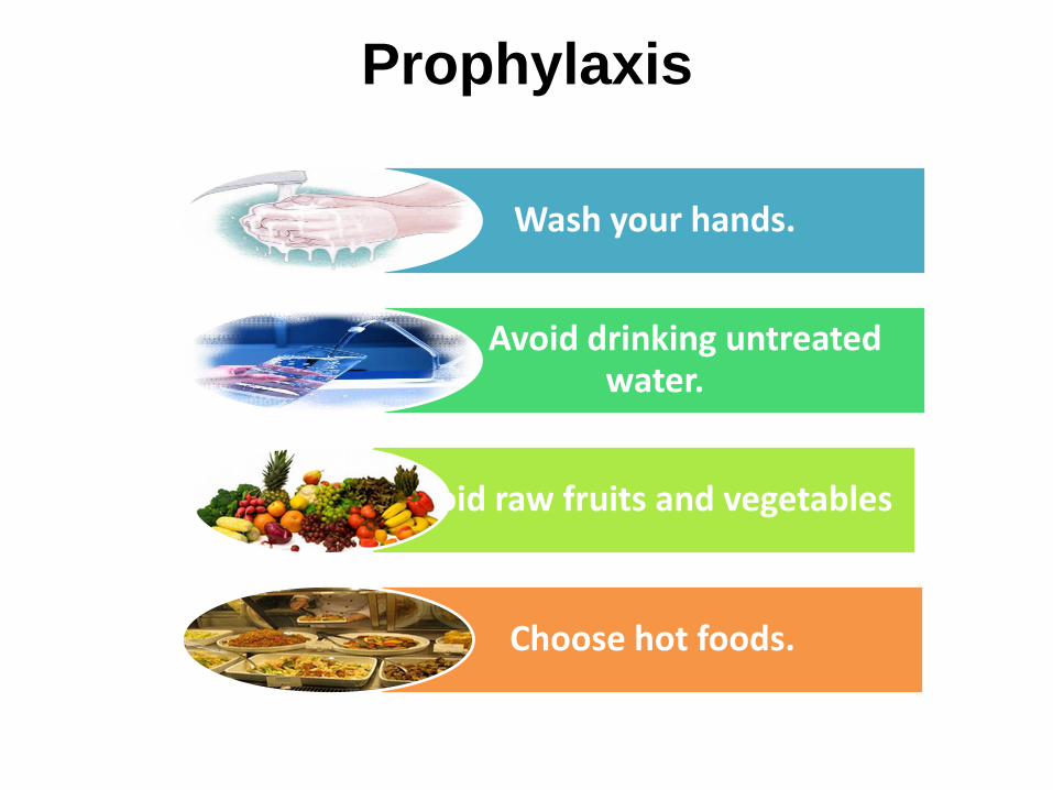
Prophylaxis
Wash your hands.
Avoid drinking untreated water.
Avoid raw fruits and vegetables
Choose hot foods.
Prevention: Water-borne Disease
• Improve quality and quantity of drinking water at source, at the tap, or in the storage vessel
• Interrupt routes of transmission by emptying accumulated water sources
• Chlorinate water
• Change hygiene behavior (ex. Hand washing)
• Take care in disposing of waste and human and animal feces
• Proper use of latrines by adults and children
• Proper use and maintenance of water supply, sanitation systems, pumps and wells
• Good food hygiene (ex. protect food from flies)
Recommended