Functional Brain Signal Processing: Current Trends and
Future Directions
Kaushik Majumdar
Indian Statistical Institute Bangalore Center
National Conference on Brain and Consciousness, 20 – 21 September 2013, ISI Kolkata

Functional Brain Signals
EEG ECoG LFP Single Cell Electrophysiology MEG fMRI PET SPECT
• Two Photon Microscopy

Functional Brain Regions
http://spot.colorado.edu/~dubin/talks/brodmann/brodmann.html
By fundamental premise of deductive science it is to be determined how each area works and how different areas work together, that is, how the areas couple and decouple among themselves.
The gold-standard signals are electrophysiological signals from single cells to scalp EEG.

Electrophysiological Signals at Different Scales
Single cell recording Local filed potential (LFP) Electrocorticogram (ECoG) Electroencephalogram (EEG)
Buzsaki et al., Nat. Rev. Neurosci., 13: 407 – 420, 2012

EEG, LFP, Spikes
Buzsaki et al., Nat. Rev. Neurosci., 13: 407 – 420, 2012

Information Richness
EEG – least informative, source ambiguous, full of artifacts.
ECoG – mainly excitatory postsynaptic potential in layer VI of the cortex, has less artifacts and more informative than EEG.
LFP – is the most information rich brain signal, superposition of almost all sorts of membrane potentials.

Oscillation and Synchrony: Two Major Paradigms for Studying Brain Functions
Oscillating band components in EEG are delta (0 – 4 Hz), theta (4 – 8 Hz), alpha (8 – 12 Hz), beta (12 – 30 Hz) and gamma (30 – 80 Hz).
LFP in mammalian forebrain can oscillate between 0.05 to 500 Hz (Buzsaki & Draguhn, 2004).
Power of oscillation of frequency ƒ varies as ƒ-2.
Brain Oscillations (cont.)
The higher the frequency the more confined the oscillation is locally.
The lower the frequency the more widespread the oscillation is.

Neuronal Oscillation: Functions
Modulates synaptic plasticity. Influence reaction time. Correlates with attention. Modulates perceptual binding. Coordinate among brain regions far apart. Consolidate memory.
Canolty et al., Science., 313: 1626 – 1628, 2006

Cortical Oscillation: Frequency Bands
Delta (0 – 4 Hz) Theta (4 – 8 Hz) Alpha (8 – 12 Hz), Mu (8 – 12 Hz) Beta (12 – 30 Hz) Gamma (30 – 80 Hz) High gamma (80 – 150 Hz)
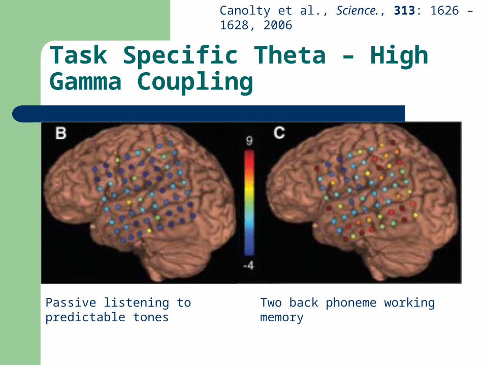
Task Specific Theta – High Gamma Coupling
Passive listening to predictable tones Two back phoneme working memory
Canolty et al., Science., 313: 1626 – 1628, 2006

Theta – High Gamma Coupling
Canolty et al., Science., 313: 1626 – 1628, 2006
Phase of 4 – 8 Hz (theta) modulates amplitude of 80 – 150 Hz (high gamma).

Neuronal Synchronization
Gray et al., Nature, 338: 334 – 337, 23 March 1989
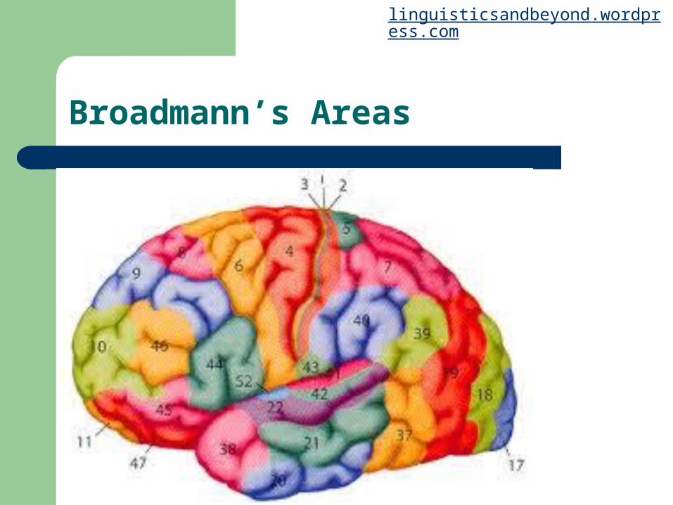
Broadmann’s Areas
linguisticsandbeyond.wordpress.com

Binding Problem
Engel et al. Nat. Rev. Neurosci., 2: 704-716, 2001

Phase Synchronization
-0.8 -0.6 -0.4 -0.2 0 0.2 0.4 0.6 0.8-20
-15
-10
-5
0
5
10
15
20
time
ampl
itude

Phase Synchronization in Face Perception
Rodriguez et al., Nature, 397: 430 – 433, 1999
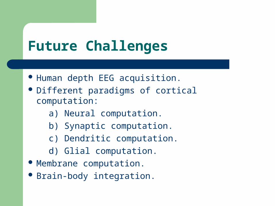
Future Challenges
Human depth EEG acquisition. Different paradigms of cortical computation:
a) Neural computation.
b) Synaptic computation.
c) Dendritic computation.
d) Glial computation. Membrane computation. Brain-body integration.

References
G. Buzsaki, C. A. Anastassiou and C. Koch, The origin of extracellular fields and currents – EEG, ECoG, LFP and spikes, Nat. Rev. Neurosci., 13: 407 – 420, 2012.
X.-J. Wang, Neurophysiological and computaitonal principles of cortical rhythms in cognition, Physiological Rev., 90(3): 1195 – 1268, 2010.

THANK YOU
This lecture is available at http://www.isibang.ac.in/~kaushik
Recommended