
General anatomy of skeletal muscle
its innervation and blood supply
General anatomy of spinal nerve
Miloš Grim
Institute of Anatomy
First Faculty of Medicine,
Charles University
Winter semester 2017/2018
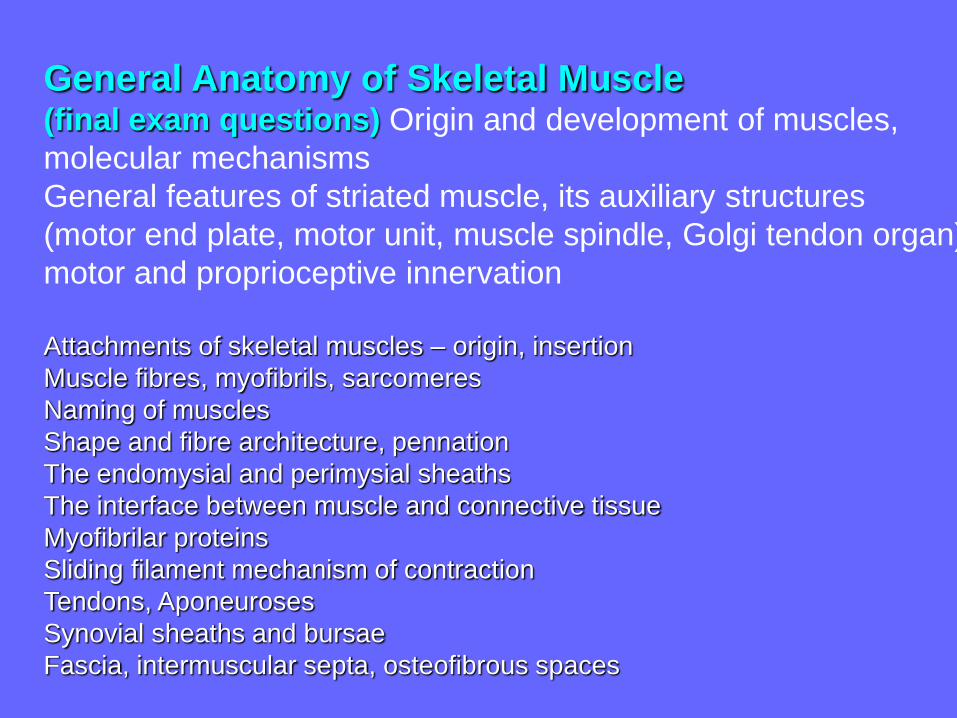
General Anatomy of Skeletal Muscle(final exam questions) Origin and development of muscles,
molecular mechanisms
General features of striated muscle, its auxiliary structures
(motor end plate, motor unit, muscle spindle, Golgi tendon organ),
motor and proprioceptive innervation
Attachments of skeletal muscles – origin, insertion
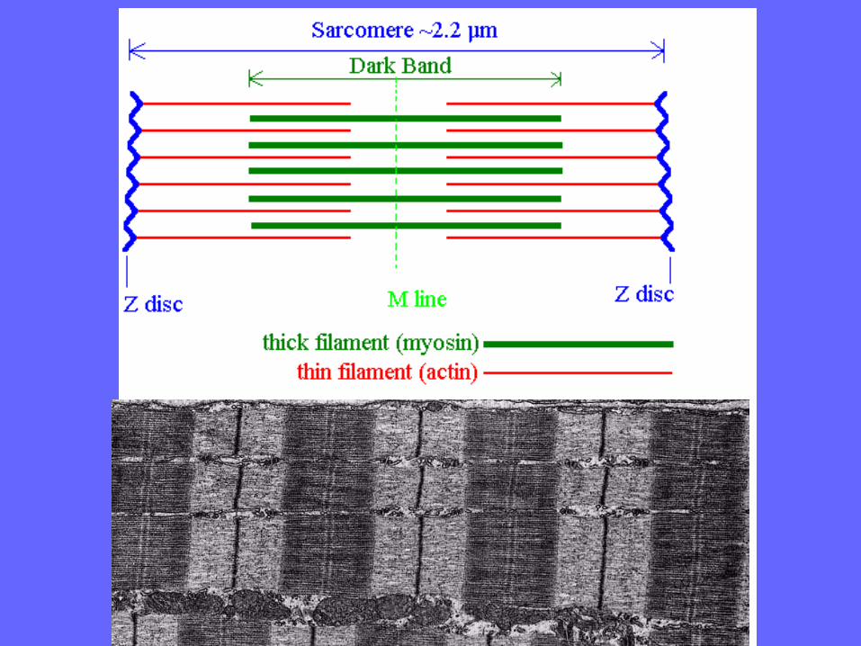
Muscle fibres, myofibrils, sarcomeres
Naming of muscles
Shape and fibre architecture, pennation
The endomysial and perimysial sheaths
The interface between muscle and connective tissue
Myofibrilar proteins
Sliding filament mechanism of contraction
Tendons, Aponeuroses
Synovial sheaths and bursae
Fascia, intermuscular septa, osteofibrous spaces

How to study skeletal muscles: identification, muscle groups,
innervation, function, origo, insertion,
position (scheme, tables), osteofascial
spaces (compartments),
transverse sections of limb segments,
dissection

R.S.Snell: Clinical Anatomy. Lippincott Williams Wilkins
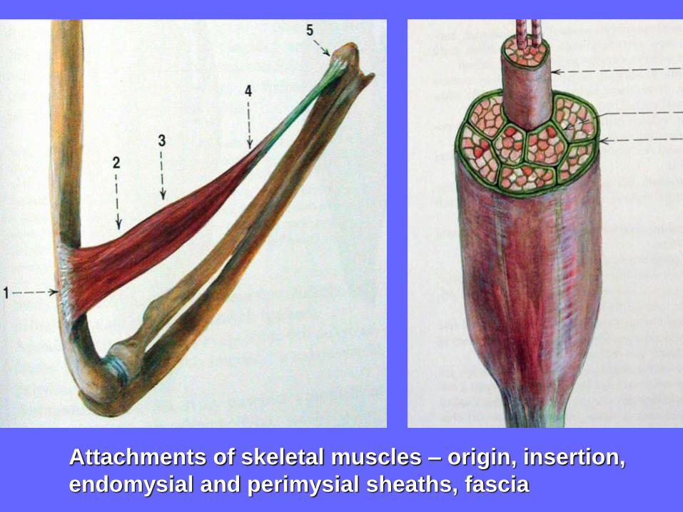
Attachments of skeletal muscles – origin, insertion,
endomysial and perimysial sheaths, fascia
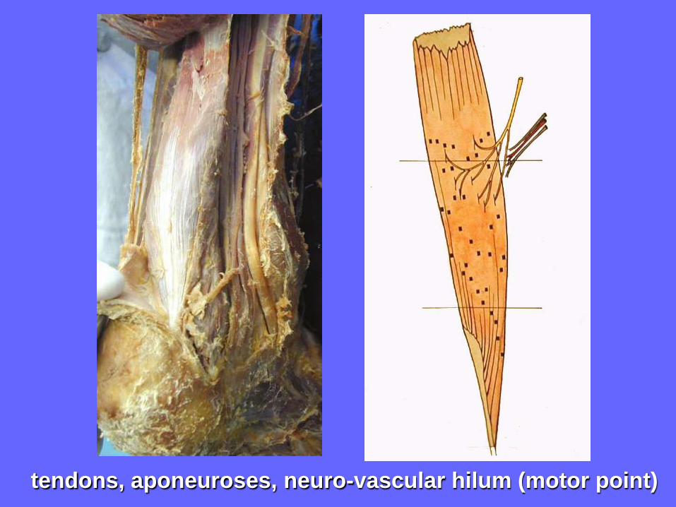
tendons, aponeuroses, neuro-vascular hilum (motor point)


Naming of Muscles
Shape:
deltoid (= triangular), quadratus (= square), rhomboid
(= diamond-shaped)
teres (= round), gracilis (= slender), rectus (= straight),
lumbrical (= worm-like)
Size : major, minor, longus (= long), brevis (= short),
latissimus (= broadest), longissimus (= longest)
Number of Heads or Bellies:
biceps (= 2 heads), triceps (= 3 heads), quadriceps (= 4 heads)
digastric (= 2 bellies), biventer (= 2 bellies),
Position:
anterior, posterior, interosseous (= between bones)
supraspinatus (= above spine of scapula),
infraspinatus (= below spine),
dorsi (= of the back), abdominis (= of the abdomen)
pectoralis (= of the chest), brachii (= of the arm)
femoris (= of the thigh), oris (= of the mouth)
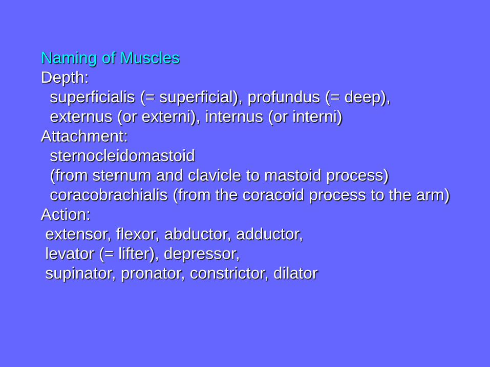
Naming of Muscles
Depth:
superficialis (= superficial), profundus (= deep),
externus (or externi), internus (or interni)
Attachment:
sternocleidomastoid
(from sternum and clavicle to mastoid process)
coracobrachialis (from the coracoid process to the arm)
Action:
extensor, flexor, abductor, adductor,
levator (= lifter), depressor,
supinator, pronator, constrictor, dilator
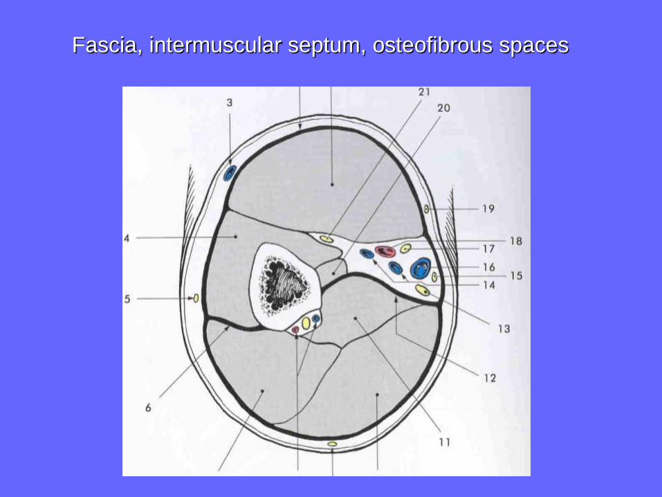
Fascia, intermuscular septum, osteofibrous spaces

Osteofasciál
spaces of
the upper limb

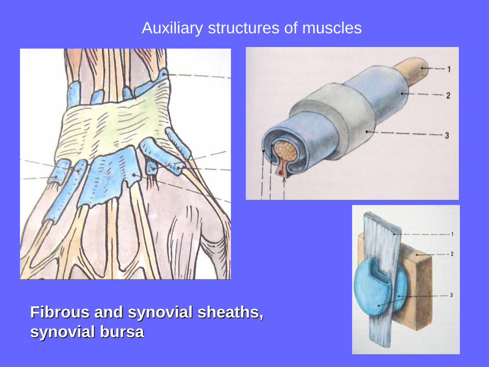
Fibrous and synovial sheaths,
synovial bursa
Auxiliary structures of muscles

Retinaculum musculorum extensorum
m. extensor hallucis longus (Vesalius, 1555)

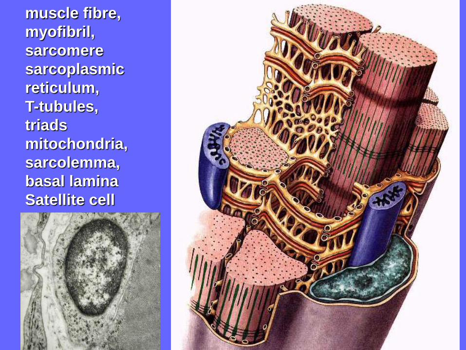
muscle fibre,
myofibril,
sarcomere
sarcoplasmic
reticulum,
T-tubules,
triads
mitochondria,
sarcolemma,
basal lamina
Satellite cell

http://fig.cox.miami.edu/~cmallery/150/neuro/muscle.htm
striated muscle fibres, sarkomere

Innervation of skeletal muscle
Neurovascular hilum
Blood supply
Motor innervation
Alfa motoneurons: slow and fast,
gamma motoneurons,
motor end-plate, ACh
motor unit, zone of motor end-plates,
polyneural innervation, segmental
innervation
Sensory (proprioceptive) innervation
muscle spindle, Golgi tendon organ,
proprioceptive reflexes
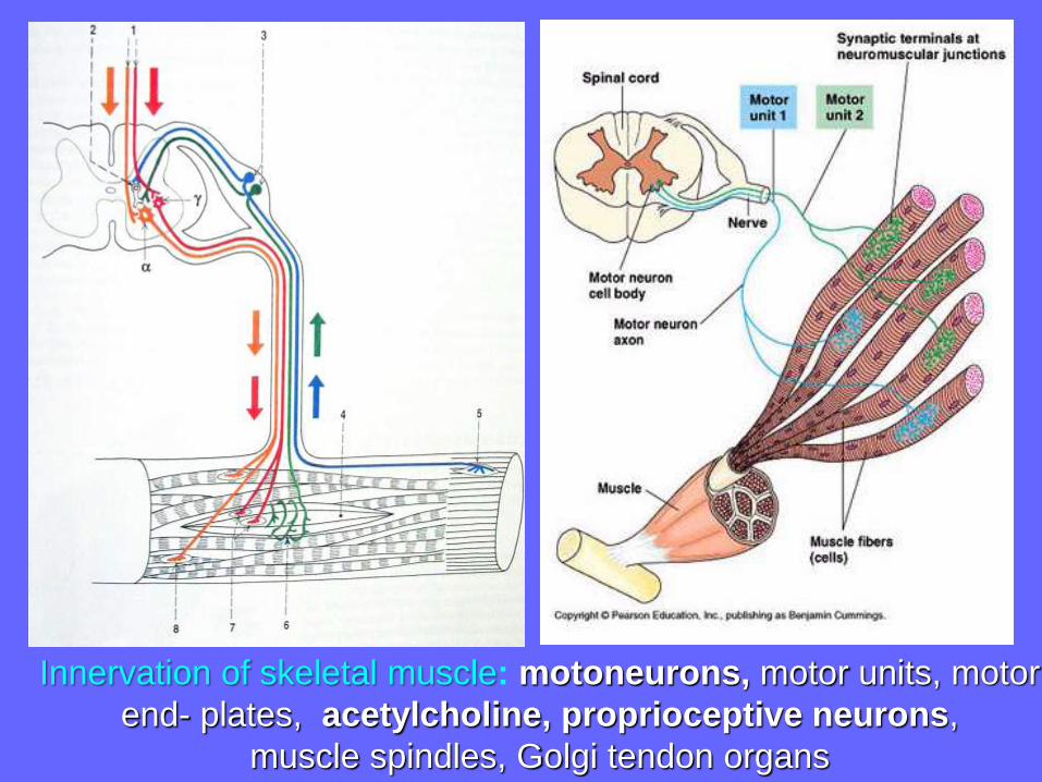
Innervation of skeletal muscle: motoneurons, motor units, motor
end- plates, acetylcholine, proprioceptive neurons,
muscle spindles, Golgi tendon organs
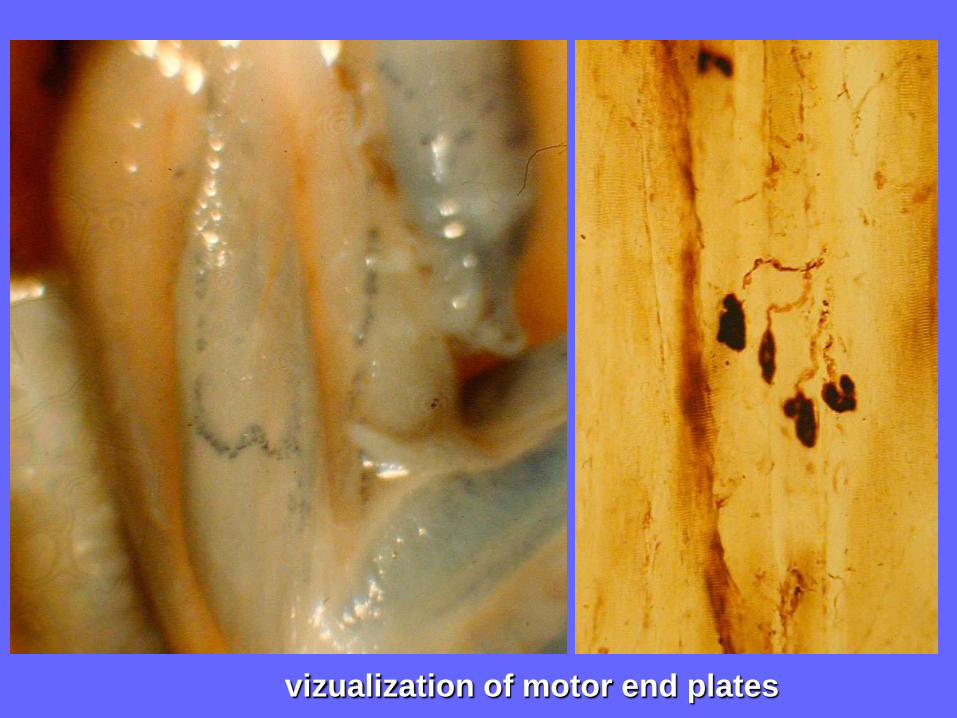
vizualization of motor end plates
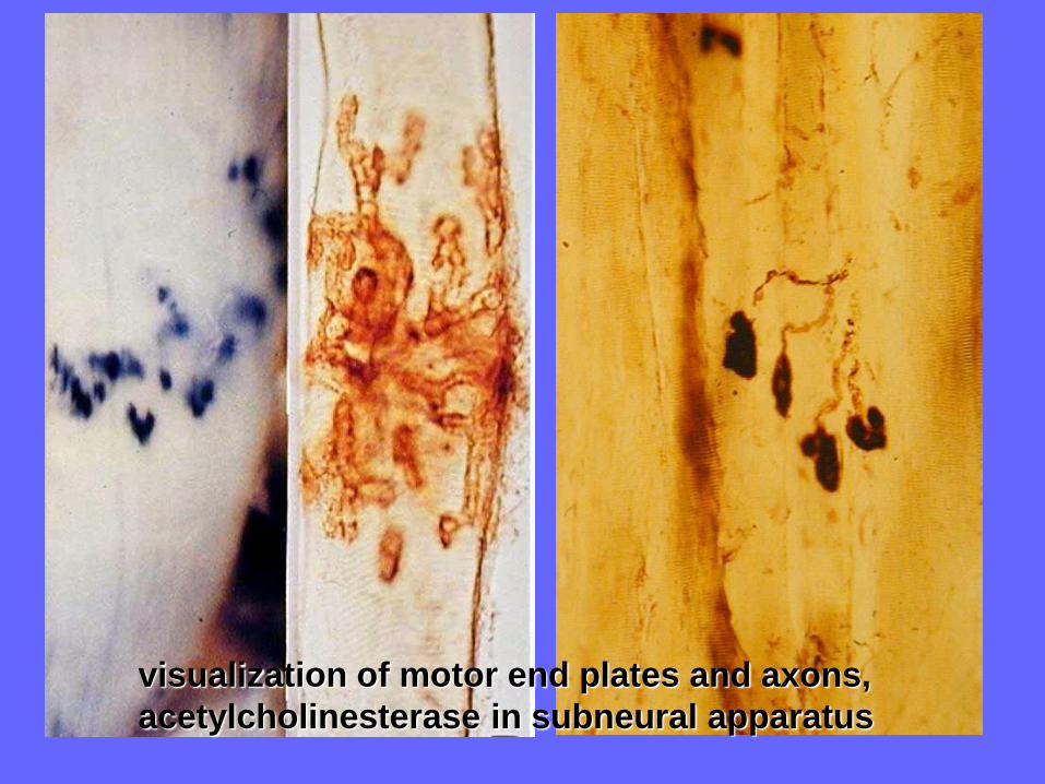
visualization of motor end plates and axons,
acetylcholinesterase in subneural apparatus
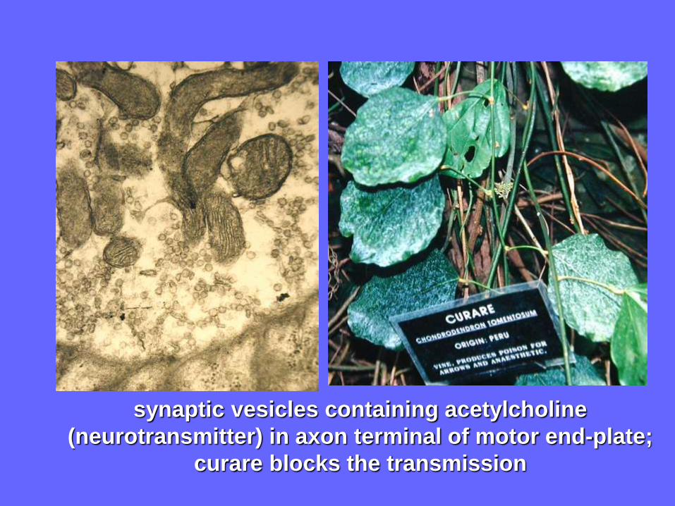
synaptic vesicles containing acetylcholine
(neurotransmitter) in axon terminal of motor end-plate;
curare blocks the transmission

Muscle spindle
Golgi tendon organ
Elektromyography (EMG)

A young women with sensory neuropathy of unknown
origin who completely lost proprioceptive sensation:
She could not stand without watching her feet,
she could not held anything in her hands, and they
wandered around without her awareness…
„Something awful´s happened, I can´t feel my body.
I feel weird-disembodied“, she said, and „I may lose
my arms. I think they´re one place and I find they´re
another“.
After having proprioception explained, she said:
„This proprioception is like the eyes of the body,
the way the body sees itself. And if it goes, as it´s gone
with me, it´s like the body is blind…so I have
to watch it - be its eyes. Right?“ (Dr. O.Sacks, neurologist)

pennation of muscles
arrangement of parallel
running muscle fibres

myo-tendinous junction
Terms related to muscle functionIsotonic and isometric contraction, reciprocal
innervation, synergists, antagonists, resting tension,
postural muscles, electromyography,
denervation atrophy
Muscle activity controls motor systems of CNS
according to information from mechanoreceptors and
proprioceptors and according to motivation
processed by the limbic system

Posture, isometric contraction
Motion, isotonic contraction
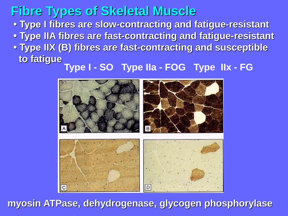
Fibre Types of Skeletal Muscle• Type I fibres are slow-contracting and fatigue-resistant
• Type IIA fibres are fast-contracting and fatigue-resistant
• Type IIX (B) fibres are fast-contracting and susceptible
to fatigue
myosin ATPase, dehydrogenase, glycogen phosphorylase
Type I - SO Type IIa - FOG Type IIx - FG

Capillaries of skeletal muscle
FG IIx FOG IIa SO I
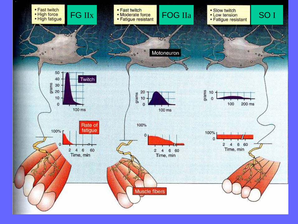
FG IIx FOG IIa SO I
A gene for speed? The evolution and function of a-actinin-3
Summary
The a-actinins are an ancient family of actin-binding proteins that play
structural and regulatory roles in cytoskeletal organisation and muscle
contraction. Alfa-actinin-3 is the most-highly specialised of the four
mammalian a-actinins, with its expression restricted largely to fast
glycolytic fibres in skeletal muscle. Intriguingly, a significant proportion (
18%) of the human population is totally deficient in a-actinin-3 due to
homozygosity for a premature stop codon polymorphism (R577X) in the
ACTN3 gene. Recent work in our laboratory has revealed a strong
association between R577X genotype and performance in a variety of
athletic endeavours. We are currently exploring the function and
evolutionary history of the ACTN3 gene and other a-actinin family
members. The a-actinin family provides a fascinating case study in
molecular evolution, illustrating phenomena such as functional redundancy
in duplicate genes, the evolution of protein function, and the action of
natural selection during recent h uman evolution. BioEssays 26:786–795,
2004. ß 2004 Wiley Periodicals, Inc.

DG MacArthur and KN North:
BioEssays 26:786–795, 2004
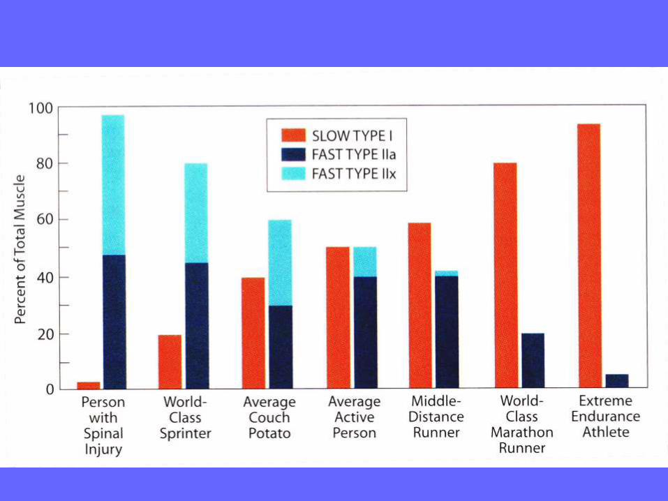
Only the R allele allows the synthesis of alpha actinin 3, a protein that is
predominantly present in FG (IIx) muscle fibers responsible for rapid and intense
muscle contraction
Development and Differentiation
of Skeletal MuscleMyogenesis,
Myogenic Determination Factors
Myf-5, myogenin, MyoD and Myf-6 (herculin)
Myostatin
Growth of Skeletal Muscle
Hypertrophy, not hyperplasia
Anabolic Steroids
Regeneration of Skeletal MuscleSatellite cells

Langman´s Medical Embryology
Paraxial mesoderm: somites, somatopleura, splanchnopleura
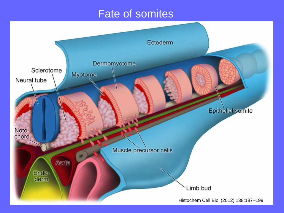
Histochem Cell Biol (2012) 138:187–199
Fate of somites

HH 25
MyoD

Epaxial and hypaxial musculature and its innervation
from dorsal and ventral branches of spinal nerves

Myogenese - diferenciace svalového vlákna
myoD
myf-5
mrf-4
Myogenin
myostatin

Mutation of myostatine gene resulting in overproduction of myogenic cells

Segmental
pattern
of muscle
innervation
The origin of the muscles of the limb
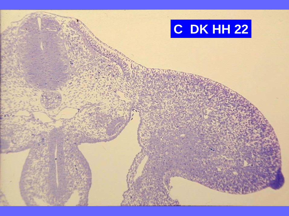
C DK HH 22
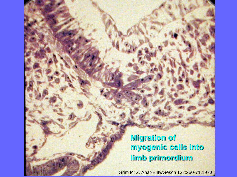
Migration of
myogenic cells into
limb primordium
Grim M: Z. Anat-EntwGesch 132:260-71,1970
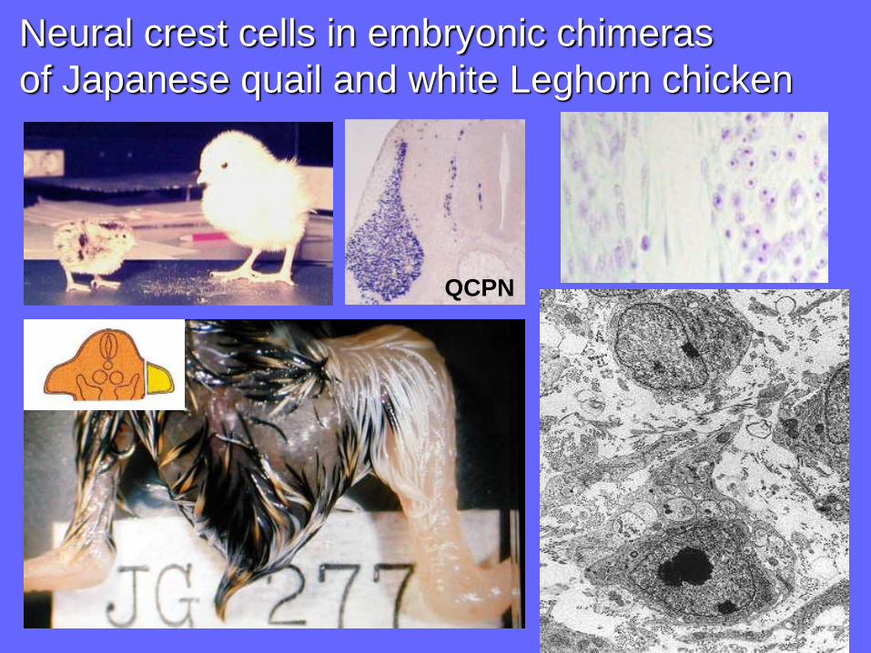
Neural crest cells in embryonic chimeras
of Japanese quail and white Leghorn chicken
QCPN
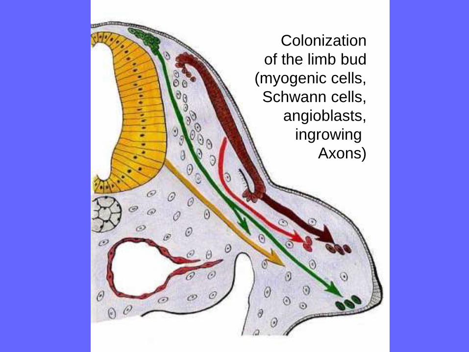
Colonization
of the limb bud
(myogenic cells,
Schwann cells,
angioblasts,
ingrowing
Axons)

Ventral and dorsal muscle blastema in limb primordium
Desmin
Morfogenesis of limb muscles

Morfogenesis of limb muscles
Regeneration of skeletal muscle
Satellite cells are resident myogenic progenitors
in postnatal skeletal muscle involved in muscle
postnatal growth and adult regenerative capacity
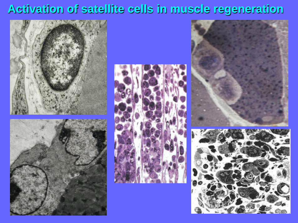
Activation of satellite cells in muscle regeneration

Fate of the free muscle graft in the rat
1.day
3. day
5. day
7. day
30. day
60. day Intact muscle

General anatomy of spinal nerves
Regeneration of peripheral nerve
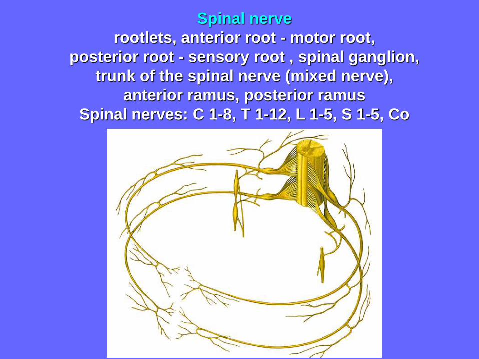
Spinal nerve
rootlets, anterior root - motor root,
posterior root - sensory root , spinal ganglion,
trunk of the spinal nerve (mixed nerve),
anterior ramus, posterior ramus
Spinal nerves: C 1-8, T 1-12, L 1-5, S 1-5, Co
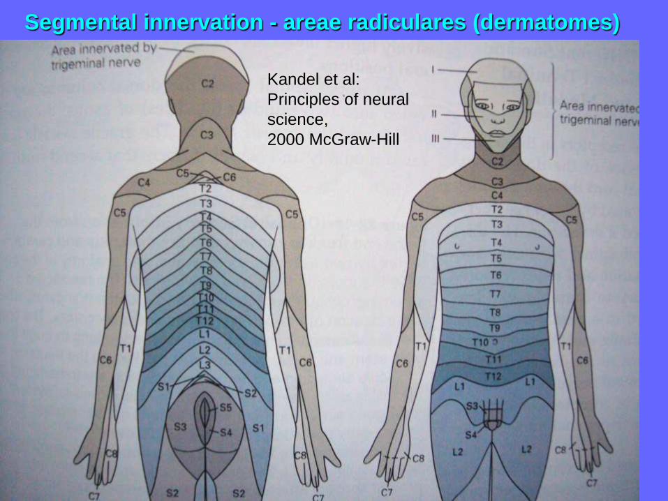
Kandel et al:
Principles of neural
science,
2000 McGraw-Hill
Segmental innervation - areae radiculares (dermatomes)
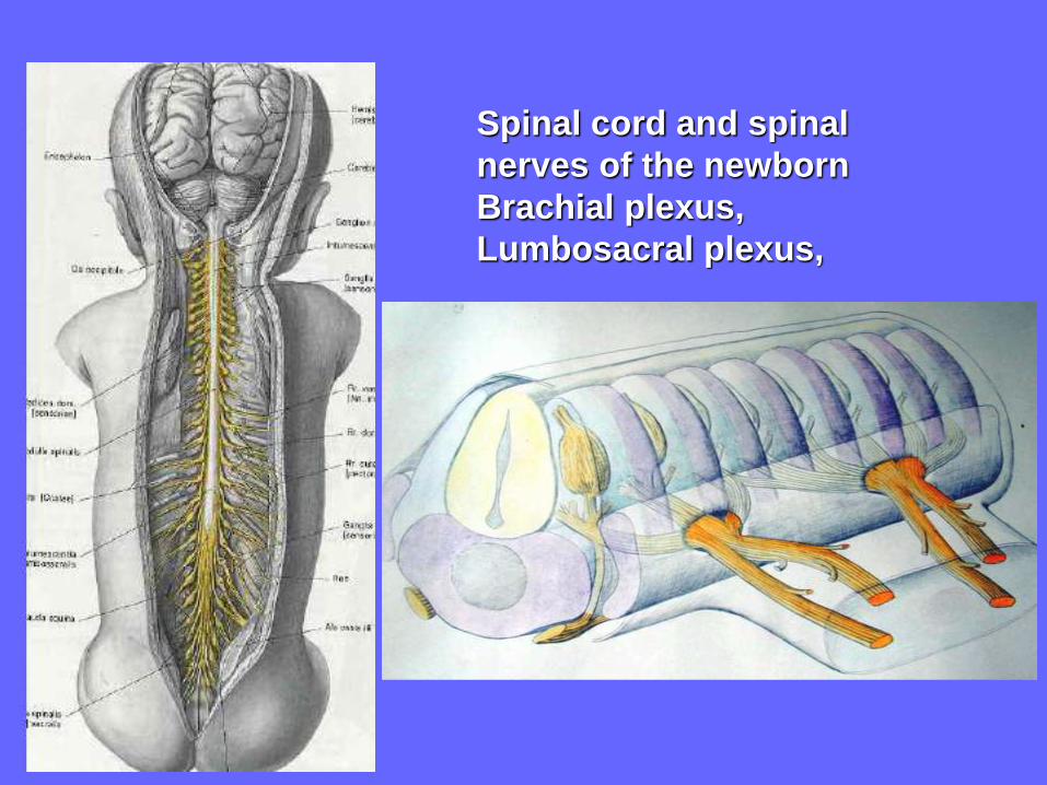
Spinal cord and spinal
nerves of the newborn
Brachial plexus,
Lumbosacral plexus,

Development of segmental innervation of limbs
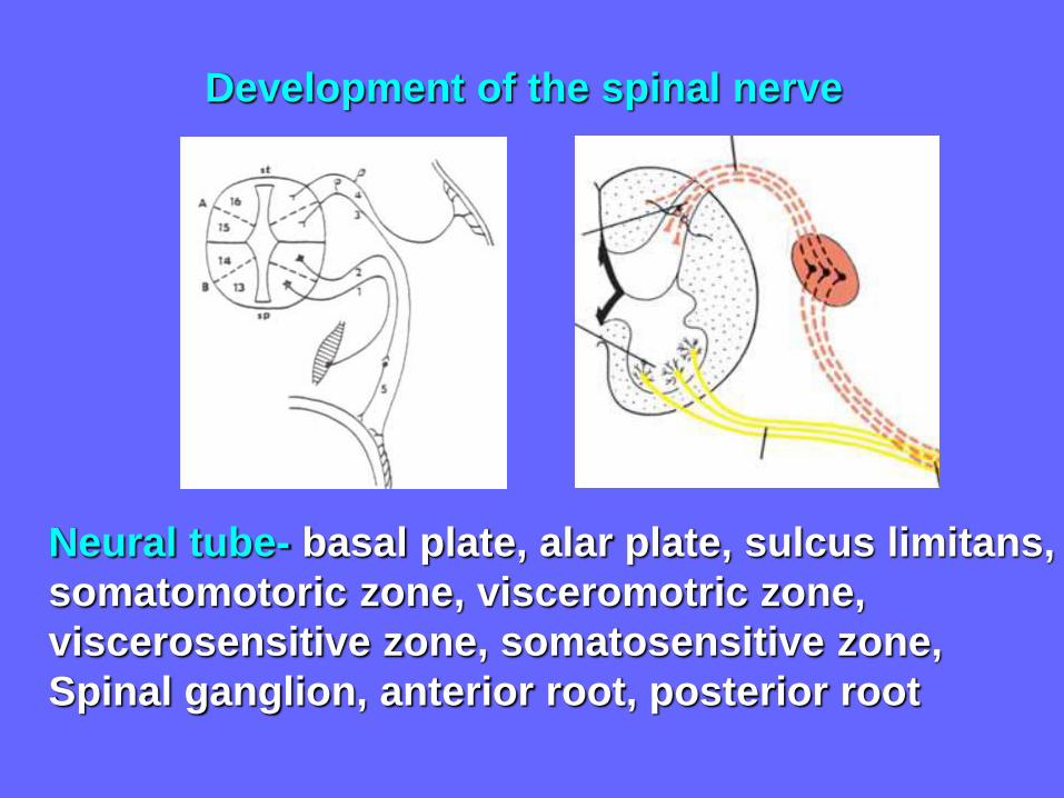
Neural tube- basal plate, alar plate, sulcus limitans,
somatomotoric zone, visceromotric zone,
viscerosensitive zone, somatosensitive zone,
Spinal ganglion, anterior root, posterior root
Development of the spinal nerve

Nerve graft bridging the partial defect, suture
of perineurium without tension, wavy course of axons
in the peripheral nerve can not be eliminated
peripheral nerve, endoneurium, perineurium,
perineural epithelium, epineurium, denervation atrophy

Regeneration of interrupted axon

General terms of angiology
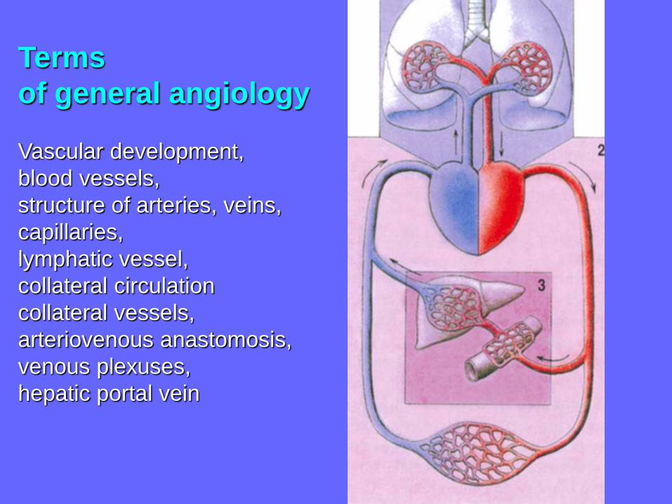
Terms
of general angiology
Vascular development,
blood vessels,
structure of arteries, veins,
capillaries,
lymphatic vessel,
collateral circulation
collateral vessels,
arteriovenous anastomosis,
venous plexuses,
hepatic portal vein
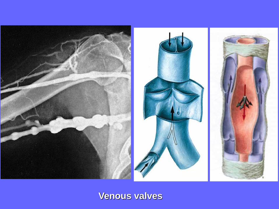
Venous valves
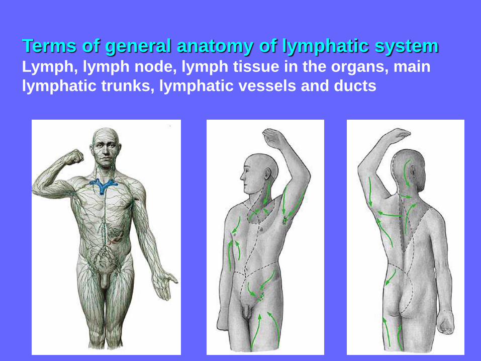
Terms of general anatomy of lymphatic systemLymph, lymph node, lymph tissue in the organs, main
lymphatic trunks, lymphatic vessels and ducts

Literature and sources of illustrations used:
Mescher: Junqueira´s Basic Histology 12th Edit., 2010
Sadler: Langman´s Medical Embryology, 11th Edit. 2009
Gilroy, MacPherson, Schuenke, Schulte, Schumacher: Atlas of
Anatomy, Thieme 3 ed. 2016
Platzer: Color Atlas and Textbook of Human Anatomy –
Vol.1 Locomotor System, Thieme 2008
Kahle, Frotscher: Color Atlas and Textbook of Human Anatomy – Vol.
3 Nervous System and Sensory Organs, Thieme 2010
Stingl, Grim, Druga: Regional Anatomy, 2012
Netter: Atlas of Human Anatomy, Icon 2003
Sobotta: Atlas of Human Anatomy Vol.1+2, Williams and Wilkins 2000
Snell: Clinical Anatomy by systems, Lippincott Williams and Wilkins
Grim M, Naňka O, Helekal I: Atlas of Human Anatomy I, II , 2014,
2017

Beware of muscle atrophy from inactivity

Recommended