Acc
epte
d A
rticl
e
W Ganzevoort1, N Mensing van Charante1, B Thilaganathan2, F Prefumo3, B Arabin4, CM Bilardo5, C Brezinka6, JB Derks7, A Diemert8, JJ Duvekot9, E Ferrazzi10, T Frusca11, K Hecher8, N Marlow12, P Martinelli13, E Ostermayer14, AT Papageorghiou2, D Schlembach15, KTM Schneider14, T Todros16, A Valcamonico11, GHA Visser17, A van Wassenaer-Leemhuis18, CC Lees19* and H Wolf1* on behalf of the TRUFFLE Group
1Department of Obstetrics and Gynecology, Academic Medical Centre, Amsterdam, The Netherlands; 2Fetal Medicine Unit, St George’s Hospital, London, UK; 3Maternal Fetal Medicine Unit, University of Brescia, Brescia, Italy; 4Department of Perinatology, Isala Clinics Zwolle, Utrecht, The Netherlands; 5Fetal Medicine Unit, Department of Obstetrics and Gynaecology, University Medical Centre Groningen, Groningen, The Netherlands; 6Obstetrics and Gynecology, Medical University of Innsbruck, Innsbruck, Austria; 7Perinatal Center, Wilhelmina Children’s Hospital, Utrecht, The Netherlands; 8Department of Obstetrics and Fetal Medicine, University Medical Center Hamburg-Eppendorf, Hamburg, Germany; 9Division of Obstetrics and Prenatal Medicine, Department of Obstetrics and Gynaecology, Erasmus MC, Rotterdam, The Netherlands; 10Children’s Hospital, Buzzi, University of Milan, Milan, Italy; 11Maternal-Fetal Medicine Unit, University of Brescia, Brescia, Italy; 12University College London Institute for Women's Health Ringgold standard institution – Neonatology, London, UK; 13Department of Gynecology and Obstetrics, University Federico II of Naples, Naples, Italy; 14Section of Perinatal Medicine, Department of Obstetrics and Gynecology,Technical University, Munich, Germany; 15Department of Obstetrics, Vivantes Clinic Neukölln, Berlin, Germany; 16Department of Obstetrics and Gynecology, University of Turin, Turin, Italy; 17Department of Perinatology, University Medical Center, Utrecht, Netherlands; 18Department of Neonatology, Emma Children’s Hospital Academic Medical Centre, Amsterdam, The Netherlands; 19Centre for Fetal Care, Queen Charlotte's and Chelsea Hospital, Imperial College London, London, UK and Department of Development and Regeneration, KU Leuven, Leuven, Belgium
Corresponding Authors:
Christoph C. Lees MD
Centre for Fetal Care, Queen Charlotte's and Chelsea Hospital, Du Cane Road, Imperial College Health NHS Trust, London, W12 0HS, UK
This article is protected by copyright. All rights reserved.
This article has been accepted for publication and undergone full peer review but has not been through the copyediting, typesetting, pagination and proofreading process, which may lead to differences between this version and the Version of Record. Please cite this article as doi: 10.1002/uog.17433
How to monitor pregnancies complicated by fetal growth restriction and delivery below 32 weeks: a post-hoc sensitivity analysis of the TRUFFLE-
study
Acc
epte
d A
rticl
eHans Wolf MD
Department of Obstetrics and Gynecology, Academic Medical Centre, H4-278, Meibergdreef 15, 1007 MB Amsterdam, Netherlands
Abstract
Objectives
In the recent TRUFFLE study it appeared that, in pregnancies complicated by fetal growth
restriction (FGR) between 26 and 32 weeks, monitoring of the ductus venosus (DV) combined
with computerised cardiotocography (cCTG) as a trigger for delivery, increased the chance of
infant survival without neurological impairment. However, concerns in interpretation were raised
as DV monitoring appeared associated with a non-significant increase in fetal death, and part of
the infants were delivered after 32 weeks, after which the study protocol was no longer applied.
This secondary sensitivity analysis focuses on women who delivered before 32 completed weeks,
and analyses fetal death cases in detail.
Methods
We analysed the monitoring data of 317 women who delivered before 32 weeks, excluding
women with absent infant outcome data or inevitable perinatal death. The association of the last
monitoring data before delivery and infant outcome was assessed by multivariable analysis.
Results
The primary outcome (two year survival without neurological impairment) occurred more often in
the two DV groups (both 83%) than in the CTG-STV group (77%), however the difference was not
statistically significant (p=0.21). Nevertheless, in surviving infants 93% was free of neurological
impairment in the DV groups versus 85% in the CTG-STV group (p=0.049). All fetal deaths (n=7)
occurred in women allocated to DV monitoring, which explains this difference. Assessment of the
monitoring parameters that were obtained shortly before fetal death in these 7 cases showed an
abnormal CTG in only one.
This article is protected by copyright. All rights reserved.

Acc
epte
d A
rticl
eMultivariable regression analysis of factors at study entry demonstrated that higher gestational
age, larger estimated fetal weight 50th percentile ratio and lower U/C ratio were significantly
associated with the (normal) primary outcome. Allocation to the DV groups had a smaller effect,
but remained in the model (p<0.1). Assessment of the last monitoring data before delivery showed
that in the CTG-STV group abnormal fetal arterial Doppler was significantly associated with
adverse outcome. In contrast, in the DV groups an abnormal DV was the only fetal monitoring
parameter that was associated with adverse infant outcome, while fetal arterial Doppler, STV
below CTG-group cut-off or recurrent fetal heart rate decelerations were not.
Conclusions
In accordance with the results of the overall TRUFFLE study of the monitoring-intervention
management of very early severe FGR we found that the difference in the proportion of infants
surviving without neuroimpairment (the primary endpoint) was non-significant when comparing
timing of delivery with or without changes in the DV waveform. However, the uneven distribution of
fetal deaths towards the DV groups was likely by chance, and among surviving children
neurological outcomes were better. Before 32 weeks, delaying delivery until abnormalities in DVPI
or STV and/or recurrent decelerations occur, as defined by the study protocol, is therefore
probably safe and possibly benefits long-term outcome.
Keywords
Fetal growth restriction; intra-uterine growth restriction, ductus venosus, cardiotocography
This article is protected by copyright. All rights reserved.

Acc
epte
d A
rticl
eIntroduction and objective
No cure exists for fetal growth restriction (FGR). Only timely diagnosis, fetal surveillance and the
decision to deliver the baby when fetal condition deteriorates can reduce the risk of mortality and
neurological impairments. No consensus exists as to the best way of monitoring and triggering
delivery in early preterm FGR, although optimal timing of delivery could be crucial for the chance
of healthy survival.
The TRUFFLE study, carried out in 20 European perinatal centres, explored if a monitoring
method that uses abnormal ductus venosus (DV) measurements (‘early’ at 95th percentile or ‘late’
at absent A-wave) or abnormal findings of computerised cardiotocography (cCTG) as a trigger for
delivery, could increase the chance of healthy infant survival compared to the standard monitoring
method by cCTG in pregnancies complicated by FGR between 26 and 32 weeks.(1, 2) Survival
without neurological impairment occurred more often in the group using late ductus and cCTG
changes as trigger for delivery compared to the cCTG group, while differences between the early
and late DV group were minimal.(2) However, reservations in data interpretation were raised by
the fact that only a proportion of the fetuses allocated to delivery based on ductus venosus
changes actually delivered according to this criterion, the majority having been delivered on
cardiotocography safety net criteria. Also, all fetal deaths were in the DV groups. Differences in
outcome between the DV groups were minimal and part of the infants were delivered after 32
weeks, the term after which the study protocol was no longer followed. This sensitivity analysis
aims to focus on women delivered according to the formal protocol, thus restricted to women who
delivered before 32 completed weeks, to pinpoint the effect of ductus venosus added to CTG in
the monitoring of growth-restricted fetuses. The second aim is to analyse the monitoring data in
the fetal deaths.
This article is protected by copyright. All rights reserved.

Acc
epte
d A
rticl
eMethods
The study design has been described earlier.(1, 2) In short, women with singleton fetuses at 26-32
weeks of gestation, with abdominal circumference <10th percentile and umbilical artery Doppler
pulsatility index (PI) >95th percentile, were included in a twenty centre European study (ISRCTN
56204499). Baseline maternal and fetal data were collected via a secure internet data entry page.
Study group allocation was performed in an even ratio from randomly sized blocks, stratified for
gestational age (lower or higher than 29 weeks of gestation) and for participating centres. Eligible
women were allocated for delivery according to one of three monitoring arms: reduced
cardiotocography (cCTG) fetal heart rate short-term variation (STV<3.5 ms at a gestational age
below 29 weeks and <4.0 ms thereafter), early ductus venosus (PI>95th percentile; ‘DV p95’) or
late ductus venosus changes (‘a’ wave at or below baseline; ‘DV no A’). Abnormal DV
measurements should be repeated within 24 hours, if cCTG results allowed this, to demonstrate
consistency. In all groups delivery could also be decided on ‘safety net criteria’ when the cCTG
showed recurrent decelerations, or in the DV groups when STV was very low (STV<2.6 ms at a
gestational age below 29 weeks and <3.0 ms thereafter).
The primary outcome was survival without cerebral palsy, severe neurosensory impairment or low
Bayley Scales of Infant Developmental score <85 at 2 years of age.
This post-hoc analysis targets at the association of fetal monitoring data (CTG STV, DV PI, DV a-
wave, umbilical artery PI, middle cerebral artery PI and the umbilical artery / middle cerebral artery
ratio (UC ratio), that were available shortly before delivery, and outcome (two years
neurodevelopmental outcome, fetal, neonatal, infant death).
Because the study protocol was restricted to management before 32 weeks and monitoring data
thereafter were not stored (and DV was not used), we analysed the data of women who delivered
before 32 weeks only. Seven women with inevitable fetal or neonatal death, and one with absent
neonatal data, who had remained in the primary published intention to treat analysis, were
excluded in this analysis as these circumstances precluded any exploration of the association
between monitoring data and outcome. In five of these women fetal death occurred due to refusal
of intervention, and in two the babies died shortly after delivery because of lethal congenital This article is protected by copyright. All rights reserved.

Acc
epte
d A
rticl
eabnormality (one trisomy 18, one complex cardiac defect). In one no neonatal data could be
provided after neonatal transfer immediately after delivery. Furthermore short-term data of 33
surviving infants (9%), who could not be examined after two years, were excluded from endpoint
data analysis.
Cut-off values for fetal monitoring data were as defined by the study protocol. For reference of
estimated fetal weight and birth weight a ratio was calculated with a 50th percentile (p50) weight
adjusted for gestational age, maternal ethnic descent, weight and length and infant sex.(3)
The effect of the most recent monitoring registration before birth on long-term outcome was
evaluated by univariable and multivariable analysis. Univariable analysis used Anova, Chi-square
or Kruskal-Wallis as appropriate. Multivariable analysis allowed the adjustment for relevant clinical
details that were significantly different between outcome categories in univariable analysis. Criteria
for inclusion and exclusion of potential variables were set at 0.05 and 0.10 respectively. IBM SPSS
version 22 (New York, USA) was used for statistics.
Results
Three-hundred and seventeen (of the original 503) FGR infants with known outcome (either with
perinatal death or follow-up examination at 2 years) were delivered before 32 weeks and included
in this post-hoc analysis (Table 1). For the purposes of further analysis the two DV groups were
combined to assess more precisely the association of abnormal DV PI with infant outcome. The
primary outcome (two-year survival without neurological impairment) occurred more often in the
DV groups (both 83%) than in the CTG-STV group (77%), although this difference is not
statistically significant (p=0.21). Nevertheless, when analysing the group of surviving infants, the
prevalence of neurological impairment in the DV groups was half of the prevalence in the CTG
groups (14/190 (7%) versus 14/95 (15%); RR 0.50 (0.25 to 1.00); p=0.049, numbers-needed-to-
treat [NNT] 13).
Table 2A and 2B demonstrate demographic, obstetric and neonatal information specified for
normal or abnormal neurological infant development at the corrected age of 2 years, and perinatal This article is protected by copyright. All rights reserved.
Acc
epte
d A
rticl
emortality. Thirty-two infants (10%) had died (7 antenatal, 25 neonatal). Causes for neonatal death
that were not included in the study definition of severe neonatal morbidity were acute respiratory
distress, multi-organ failure or clinical sepsis. Twenty-eight infants (9%) had impaired neurological
development at two years corrected age.
Women with a normal infant outcome had been randomised at a higher gestational age and the
estimated fetal weight and birthweight p50 ratio were larger than in women with an infant who died
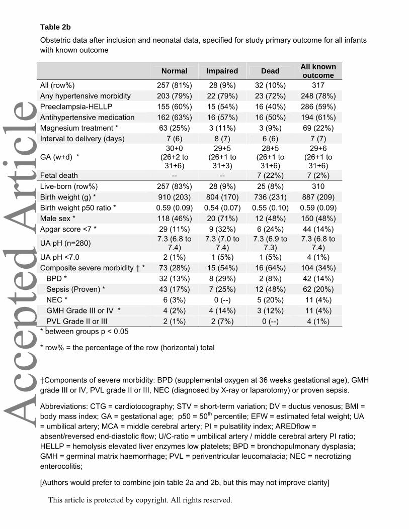
or had abnormal development. All fetal deaths (n=7) occurred in women allocated to DV
monitoring. Assessment of the monitoring parameters obtained shortly before fetal death showed
that in only one case STV was below the CTG group cut-off, whereas DV was still normal (Table
3). All other fetal death cases had either no STV assessment within 24 hours before fetal death or
a normal CTG according to CTG-group protocol. In two cases the most recent DV PI before death
had been higher than 95th percentile (but these cases had been allocated to the DV no A group).
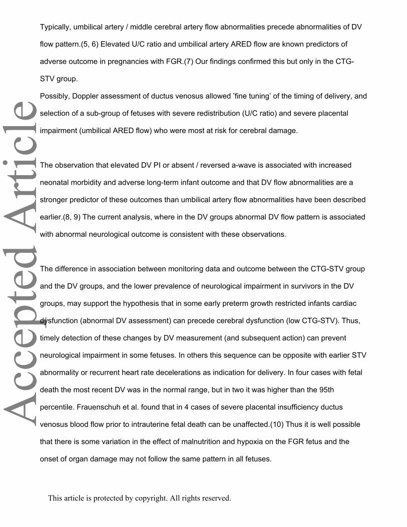
At delivery, infants with a normal outcome were born at later gestational age, with a higher birth
weight and birth weight ratio, and less frequently had a low APGAR score. At discharge, severe
composite morbidity was less likely in infants with a normal outcome (definition of composite
morbidity and results given in Table 2b). Especially cerebral haemorrhage and periventricular
leucomalacia (PVL) were more frequent in infants with an abnormal outcome (17%) than in the
normal outcome group (2%). Eighty-three percent of the live-born infants survived without
neurological impairment, although 28% of these infants had severe morbidity in the neonatal
period. In contrast, thirteen of the 28 infants (46%) with neurological impairment did not have
severe morbidity during the neonatal period.
There were no differences in inclusion characteristics, or in obstetric and neonatal details between
the randomisation groups and between infants with or infants without follow-up at two years age
corrected for prematurity (data published previously(2)). Infants delivered after 32 weeks were
included at a later gestational age, and a larger estimated fetal weight and had better outcomes
than those included earlier, as can be expected.
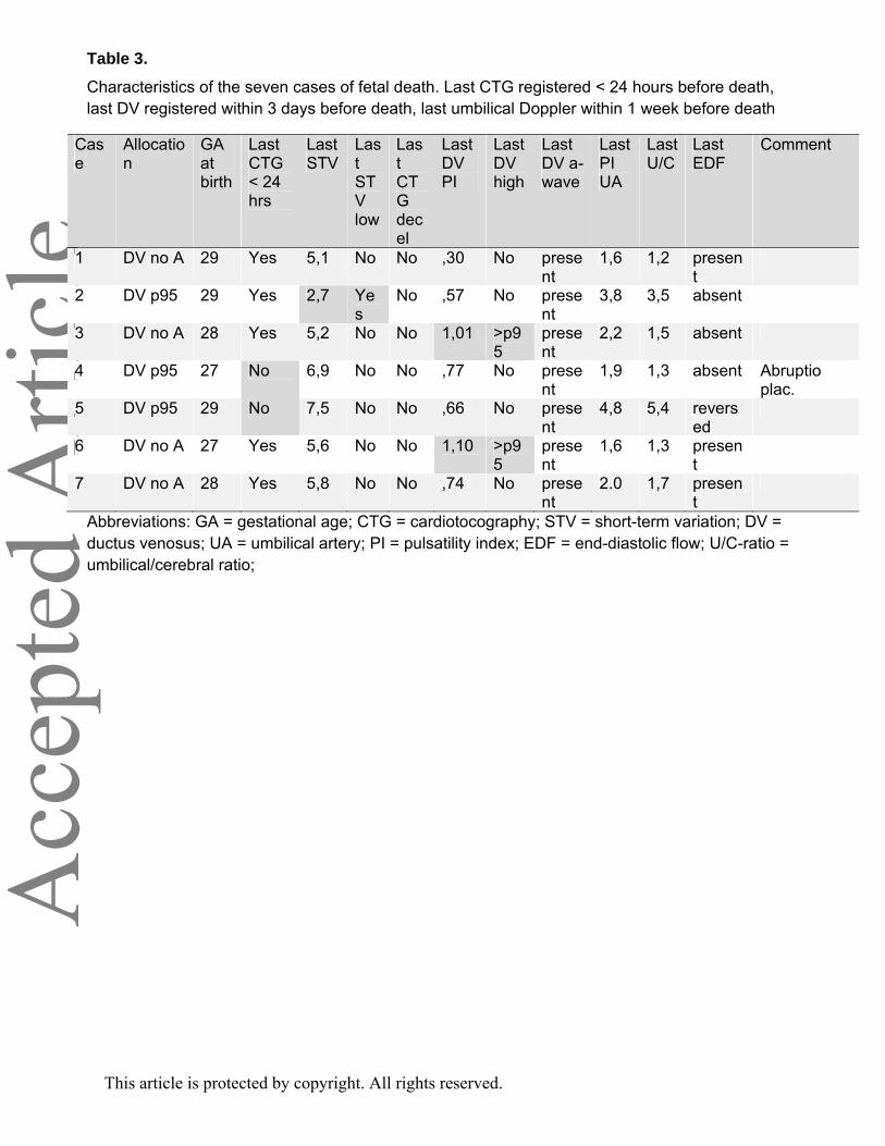
Multivariable regression analysis of factors at study entry demonstrated that gestational
hypertension, larger estimated fetal weight p50 ratio and lower U/C ratio were significantly This article is protected by copyright. All rights reserved.

Acc
epte
d A
rticl
eassociated with the (normal) primary outcome (Figure 1A). Allocation to the DV groups had a
smaller effect, but remained in the model (p<0.1). Multivariable analysis with parameters known at
delivery demonstrated that women with a normal outcome were more likely to have been allocated
to the DV-groups, had a lower U/C ratio, and their babies had a higher birth weight, a higher
APGAR score and were more often female (Figure 1B). If this analysis was repeated including DV
PI only in those women who had a last DV measurement within 3 days of delivery or fetal death
(n=180) then only a last DV measurement higher than 95th percentile was negatively associated
with the primary endpoint and a larger birth weight p50 ratio had a positive effect, while the other
parameters were ejected from the model (Figure 1C).
The association between the last fetal monitoring before birth (or fetal death) and primary outcome
is specified in greater detail in Table 4 separately for participants of the CTG-STV group and the
DV groups. There appeared a difference of the impact of abnormal monitoring results on the
primary outcome between these groups. In the CTG group, umbilical artery Doppler ARED flow
and high U/C-ratio were negatively associated with the primary outcome (Figure 2A). In the DV
groups an abnormal DV PI > 95th percentile was negatively associated with the primary outcome
and this effect was more pronounced after recurrent elevated DV PI > 95th percentile with a longer
interval than 1 day (which was allowed by the study protocol) (Figure 2B). The negative effect of
an abnormal DV PI >95th percentile was not further affected by a combination with a STV below
CTG-STV group cut-off, below DV safety-net cut-off or decelerations. Although the U/C ratio in the
DV groups was of the same value as in the CTG group and the incidence of umbilical artery ARED
flow was similar, the negative association of these parameters with the primary endpoint that was
observed in the CTG-STV group was absent in the DV groups.
Discussion This secondary sensitivity analysis of TRUFFLE strengthens the conclusion of the primary
intention-to-treat analysis, that perinatal outcomes are improved if ductus venosus measurements
are added to CTG-STV in the monitoring of fetuses with severe early-onset FGR. The analysis
targeted those infants that were delivered before 32 weeks to focus on the effect of the different This article is protected by copyright. All rights reserved.
Acc
epte
d A
rticl
emonitoring techniques and looked in-depth at the perinatal deaths and the association between
the most recent fetal monitoring parameters with the primary outcome.
In this post-hoc analysis both DV groups were combined because we wanted to explore the
association of 2-year infant outcome with DV measurement results. It was deemed justified
because survival with normal neurodevelopment at 2 years corrected for prematurity was equal in
both DV groups (both 83% in infants with known outcome). Normal 2 year outcome was less
frequent in the CTG-STV group (77%), but this difference was not statistically significant.
Perinatal mortality was similar between the CTG-STV and the DV groups (10%), however all
antenatal deaths (n=7) occurred in the DV groups. Analysis of this antenatal mortality suggested
this was a spurious result: 6 of 7 cases would probably also not have been delivered timely if they
had been allocated to the CTG-STV group as the last STV results were above the CTG-group cut-
off limits. Two cases of fetal death might have benefited from a DVPI cut-off at the 95th percentile
instead of absent a-wave.
Multivariable analysis did not demonstrate a significant benefit for normal primary outcome in
those randomised to the DV groups after adjustment for gestational age and fetal weight p50 ratio.
If the analysis is restricted to those that are born alive, assuming that fetal death had occurred
unbalanced by chance, there is a statistically significant benefit for DV monitoring. This is in line
with aggregated cohort evidence as systematically reviewed by Morris et al.(4) who showed
moderate predictive accuracy of longitudinal DV measurements for fetal/neonatal wellbeing in
high-risk pregnancies (likelihood ratio 3.15; 95% CI 2.19-4.54).
Analysis of the results of the different monitoring techniques shows that with CTG monitoring
decelerations and umbilical artery ARED flow are negatively associated with outcome while this is
not so with combined DV and CTG monitoring. It might be that those at risk for neurological
impairment with ARED flow are delivered more timely in the DV groups because of abnormal DV
measurement, although we cannot prove this because DV was not measured in the CTG-STV
group after inclusion. This article is protected by copyright. All rights reserved.
Acc
epte
d A
rticl
eTypically, umbilical artery / middle cerebral artery flow abnormalities precede abnormalities of DV
flow pattern.(5, 6) Elevated U/C ratio and umbilical artery ARED flow are known predictors of
adverse outcome in pregnancies with FGR.(7) Our findings confirmed this but only in the CTG-
STV group.
Possibly, Doppler assessment of ductus venosus allowed ’fine tuning’ of the timing of delivery, and
selection of a sub-group of fetuses with severe redistribution (U/C ratio) and severe placental
impairment (umbilical ARED flow) who were most at risk for cerebral damage.
The observation that elevated DV PI or absent / reversed a-wave is associated with increased
neonatal morbidity and adverse long-term infant outcome and that DV flow abnormalities are a
stronger predictor of these outcomes than umbilical artery flow abnormalities have been described
earlier.(8, 9) The current analysis, where in the DV groups abnormal DV flow pattern is associated
with abnormal neurological outcome is consistent with these observations.
The difference in association between monitoring data and outcome between the CTG-STV group
and the DV groups, and the lower prevalence of neurological impairment in survivors in the DV
groups, may support the hypothesis that in some early preterm growth restricted infants cardiac
dysfunction (abnormal DV assessment) can precede cerebral dysfunction (low CTG-STV). Thus,
timely detection of these changes by DV measurement (and subsequent action) can prevent
neurological impairment in some fetuses. In others this sequence can be opposite with earlier STV
abnormality or recurrent heart rate decelerations as indication for delivery. In four cases with fetal
death the most recent DV was in the normal range, but in two it was higher than the 95th
percentile. Frauenschuh et al. found that in 4 cases of severe placental insufficiency ductus
venosus blood flow prior to intrauterine fetal death can be unaffected.(10) Thus it is well possible
that there is some variation in the effect of malnutrition and hypoxia on the FGR fetus and the
onset of organ damage may not follow the same pattern in all fetuses.
This article is protected by copyright. All rights reserved.

Acc
epte
d A
rticl
eWe only included babies delivered before 32 weeks because by protocol and in practice, ductus
venosus Doppler only contributed to the delivery decision before 32 weeks gestation. The potential
bias introduced by excluding differential delivery after 32 weeks by trial allocation group is likely to
be small, since the post 32 week numbers were equally distributed between the groups. Results
from this analysis can therefore only be applied to women with FGR before 32 weeks.
This post-hoc analysis highlights some of the effects of DV-monitoring that were obscured by the
original intention-to-treat analysis. However, just like all post-hoc analyses there should be caution
regarding the possibility of bias. Nonetheless, the current findings are consistent with the original
data.
Conclusions In accordance with the results of the overall TRUFFLE study of the monitoring-intervention
management of very early severe FGR we found that the difference in the proportion of infants
surviving without neuroimpairment (the primary endpoint) was non-significant when comparing
timing of delivery with or without changes in the DV waveform. However, we speculate that the
uneven distribution of fetal deaths towards the DV groups was likely by chance, and we found that
among surviving children the neurological outcomes at two years of age were better. Adverse
neurodevelopmental outcome was significantly associated with abnormal DVPI before delivery
and lower birth weight in surviving babies. Before 32 weeks, delaying delivery until abnormalities
in DVPI or STV and/or recurrent decelerations occur, as defined by the study protocol, is therefore
probably safe and possibly benefits long-term outcome.
References
1. Lees C, Marlow N, Arabin B, Bilardo CM, Brezinka C, Derks JB, Duvekot J, Frusca T, Diemert A, Ferrazzi E, Ganzevoort W, Hecher K, Martinelli P, Ostermayer E, Papageorghiou AT, Schlembach D, Schneider KT, Thilaganathan B, Todros T, van Wassenaer-Leemhuis A, Valcamonico A, Visser GH and Wolf H. Perinatal morbidity and mortality in early-onset fetal growth restriction: cohort outcomes of the trial of randomized umbilical and fetal flow in Europe (TRUFFLE). Ultrasound Obstet Gynecol 2013; 42: 400-408. This article is protected by copyright. All rights reserved.

Acc
epte
d A
rticl
e2. Lees CC, Marlow N, van Wassenaer-Leemhuis A, Arabin B, Bilardo CM, Brezinka C, Calvert S, Derks JB, Diemert A, Duvekot JJ, Ferrazzi E, Frusca T, Ganzevoort W, Hecher K, Martinelli P, Ostermayer E, Papageorghiou AT, Schlembach D, Schneider KT, Thilaganathan B, Todros T, Valcamonico A, Visser GH and Wolf H. 2 year neurodevelopmental and intermediate perinatal outcomes in infants with very preterm fetal growth restriction (TRUFFLE): a randomised trial. Lancet 2015; 385: 2162-2172. 3. Gardosi J, Chang A, Kalyan B, Sahota D and Symonds EM. Customised antenatal growth charts. Lancet 1992; 339: 283-287. 4. Morris RK, Selman TJ, Verma M, Robson SC, Kleijnen J and Khan KS. Systematic review and meta-analysis of the test accuracy of ductus venosus Doppler to predict compromise of fetal/neonatal wellbeing in high risk pregnancies with placental insufficiency. Eur J Obstet Gynecol Reprod Biol 2010; 152: 3-12. 5. Hecher K, Bilardo CM, Stigter RH, Ville Y, Hackeloer BJ, Kok HJ, Senat MV and Visser GH. Monitoring of fetuses with intrauterine growth restriction: a longitudinal study. Ultrasound Obstet Gynecol 2001; 18: 564-570. 6. Baschat AA, Gembruch U, Harman CR. The sequence of changes in Doppler and biophysical parameters as severe fetal growth restriction worsens. Ultrasound Obstet Gynecol 2001; 18: 571-577. 7. Flood K, Unterscheider J, Daly S, Geary MP, Kennelly MM, McAuliffe FM, O'Donoghue K, Hunter A, Morrison JJ, Burke G, Dicker P, Tully EC and Malone FD. The role of brain sparing in the prediction of adverse outcomes in intrauterine growth restriction: results of the multicenter PORTO Study. Am J Obstet Gynecol 2014; 211: 288 e281-285. 8. Baschat AA, Viscardi RM, Hussey-Gardner B, Hashmi N and Harman C. Infant neurodevelopment following fetal growth restriction: relationship with antepartum surveillance parameters. Ultrasound Obstet Gynecol 2009; 33: 44-50. 9. Bilardo CM, Wolf H, Stigter RH, Ville Y, Baez E, Visser GH and Hecher K. Relationship between monitoring parameters and perinatal outcome in severe, early intrauterine growth restriction. Ultrasound Obstet Gynecol 2004; 23: 119-125. 10. Frauenschuh I, Frambach T, Karl S, Dietl J and Muller T. [Ductus venosus blood flow prior to intrauterine foetal death in severe placental insufficiency can be unaffected as shown by doppler sonography]. Z Geburtshilfe Neonatol 2014; 218: 218-222.
TRUFFLE GROUP COLLABORATING AUTHORS
A Aktas (Marburg), S Borgione (Turin), R Chaoui (Berlin), JMJ Cornette (Rotterdam), T Diehl (Hamburg), J van Eyck (Zwolle), N Fratelli (Brescia), IC van Haastert (Utrecht), S Lobmaier (Munich), E Lopriore (Leiden), H Missfelder-Lobos (Cambridge), G Mansi (Naples), P Martelli (Brescia), G Maso (Trieste), U Maurer-Fellbaum (Graz), S Mulder-de Tollenaer (Zwolle), R Napolitano (Naples), M Oberto (Turin), D Oepkes (Leiden), G Ogge (Turin), JAM van der Post (Amsterdam); L Preston (Cambridge), F Raimondi (Naples), H Rattue (London), IKM Reiss (Rotterdam), LS Scheepers (Nijmegen/Maastricht), A Skabar (Trieste), M Spaanderman (Nijmegen), N Weisglas–Kuperus (Rotterdam), A Zimmermann (Munich)
This article is protected by copyright. All rights reserved.

Acc
epte
d A
rticl
eTable 1 Randomisation allocation, with selection for post-hoc analysis and primary study outcome.
CTG-STV DV p95 DV noA Total
Total study group (row%) 166 (33%)
167 (33%)
170 (34%) 503
Excluded Inevitable perinatal death 2 1 4 7
Neonatal data missing 1 -- -- 1 Alive, but no follow-up at 2 years 21 (13%) 25 (15%) 13 (8%) 59 (12%)
Delivered at 32 weeks or later 37 (22%) 39 (23%) 43 (25%) 119 (24%)
Delivered before 32 weeks 105 (63%)
102 (61%)
110 (65%) 317 (63%)
Gestational age (median (IQR)) 29.7
(28.5 to 30.9)
29.9 (28.7 to
30.9)
29.9 (28.7 to
30.7)
29.9 (28.6 to 30.9)
Birthweight (mean (SD)) 888 (202)
887 (220)
876 (208) 884 (209)
Fetal death 0 (--) 3 (3%) 4 (4%) 7 (2%)
Neonatal death 10 (10%) 6 (6%) 9 (8%) 25 (8%)
Alive and evaluated at 2 years 95 (90%) 93 (91%) 97 (88%) 285 (90%)
Alive without neurological impairment % of evaluated live infants (p=0.049)* % of all infants with known outcome (p=0.21)*
81 (85%) (77%)
85 (91%) (83%)
91 (94%) (83%)
257 (90%) (81%)
*Pearson’s chi-square for comparison of the CTG-STV group with both DV groups combined
This article is protected by copyright. All rights reserved.

Acc
epte
d A
rticl
eTable 2a. Demographic and obstetric characteristics at study entry, specified for study primary outcome for all infants with known outcome (omitting 33 lost to follow-up) – percentages for each row.
Normal Impaired Dead All known outcome
Delivered <32 weeks (row%) 257 (81%) 28 (9%) 32 (10%) 317
Maternal age 31 (5) 31 (5) 30 (5) 31 (5) Caucasian ethnicity 220 (86%) 25 (89%) 29 (91%) 274 (86%) Nulliparity * 159 (62%) 14 (50%) 27 (84%) 200 (63%) BMI (kg/m2) 25 (6) 25 (6) 25 (5) 25 (6) Smoking 30 (12%) 6 (21%) 4 (13%) 40 (13%)
GA (w+d) * 28+6
(26+0 to 31+5)
28+1 (26+0 to
31+0)
27+6 (26+0 to
31+4)
28+4 (26+0 to
31+5) EFW (g) * 852 (193) 778 (180) 703 (178) 833 (202) EFW p50 ratio * 0.65 (0.09) 0.62 (0.08) 0.60 (0.08) 0.64 (0.09) Uterine artery notch 131 (51%) 19 (68%) 20 (63%) 170 (54%) UA PI 2.0 (0.5) 2.2 (0.7) 2.2 (0.7) 2.1 (0.6) UA ARED flow 111 (43%) 15 (54%) 18 (56%) 144 (45%) UA RED flow 15 (6%) 1 (4%) 3 (9%) 19 (6%) U/C ratio* 1.5 (0.5) 1.8 (0.6) 1.7 (0.8) 1.5 (0.6) * between groups p < 0.05;
Abbreviations: BMI = body mass index; GA = gestational age; EFW = estimated fetal weight; p50 = 50th percentile; UA = umbilical artery; PI = pulsatility index; AREDflow = absent/reversed end-diastolic flow; U/C-ratio = umbilical artery / middle cerebral artery PI ratio;
This article is protected by copyright. All rights reserved.
Acc
epte
d A
rticl
eTable 2b Obstetric data after inclusion and neonatal data, specified for study primary outcome for all infants with known outcome
Normal Impaired Dead All known outcome
All (row%) 257 (81%) 28 (9%) 32 (10%) 317 Any hypertensive morbidity 203 (79%) 22 (79%) 23 (72%) 248 (78%) Preeclampsia-HELLP 155 (60%) 15 (54%) 16 (40%) 286 (59%) Antihypertensive medication 162 (63%) 16 (57%) 16 (50%) 194 (61%) Magnesium treatment * 63 (25%) 3 (11%) 3 (9%) 69 (22%) Interval to delivery (days) 7 (6) 8 (7) 6 (6) 7 (7)
GA (w+d) * 30+0
(26+2 to 31+6)
29+5 (26+1 to 31+3)
28+5 (26+1 to 31+6)
29+6 (26+1 to 31+6)
Fetal death -- -- 7 (22%) 7 (2%) Live-born (row%) 257 (83%) 28 (9%) 25 (8%) 310 Birth weight (g) * 910 (203) 804 (170) 736 (231) 887 (209) Birth weight p50 ratio * 0.59 (0.09) 0.54 (0.07) 0.55 (0.10) 0.59 (0.09) Male sex * 118 (46%) 20 (71%) 12 (48%) 150 (48%) Apgar score <7 * 29 (11%) 9 (32%) 6 (24%) 44 (14%)
UA pH (n=280) 7.3 (6.8 to 7.4)
7.3 (7.0 to 7.4)
7.3 (6.9 to 7.3)
7.3 (6.8 to 7.4)
UA pH <7.0 2 (1%) 1 (5%) 1 (5%) 4 (1%) Composite severe morbidity † * 73 (28%) 15 (54%) 16 (64%) 104 (34%) BPD * 32 (13%) 8 (29%) 2 (8%) 42 (14%) Sepsis (Proven) * 43 (17%) 7 (25%) 12 (48%) 62 (20%) NEC * 6 (3%) 0 (--) 5 (20%) 11 (4%) GMH Grade III or IV * 4 (2%) 4 (14%) 3 (12%) 11 (4%) PVL Grade II or III 2 (1%) 2 (7%) 0 (--) 4 (1%) * between groups p < 0.05
* row% = the percentage of the row (horizontal) total
†Components of severe morbidity: BPD (supplemental oxygen at 36 weeks gestational age), GMH grade III or IV, PVL grade II or III, NEC (diagnosed by X-ray or laparotomy) or proven sepsis.
Abbreviations: CTG = cardiotocography; STV = short-term variation; DV = ductus venosus; BMI = body mass index; GA = gestational age; p50 = 50th percentile; EFW = estimated fetal weight; UA = umbilical artery; MCA = middle cerebral artery; PI = pulsatility index; AREDflow = absent/reversed end-diastolic flow; U/C-ratio = umbilical artery / middle cerebral artery PI ratio; HELLP = hemolysis elevated liver enzymes low platelets; BPD = bronchopulmonary dysplasia; GMH = germinal matrix haemorrhage; PVL = periventricular leucomalacia; NEC = necrotizing enterocolitis;
[Authors would prefer to combine join table 2a and 2b, but this may not improve clarity]
This article is protected by copyright. All rights reserved.
Acc
epte
d A
rticl
eTable 3. Characteristics of the seven cases of fetal death. Last CTG registered < 24 hours before death, last DV registered within 3 days before death, last umbilical Doppler within 1 week before death
Case
Allocation
GA at birth
Last CTG < 24 hrs
Last STV
Last STV low
Last CTG decel
Last DV PI
Last DV high
Last DV a-wave
Last PI UA
Last U/C
Last EDF
Comment
1 DV no A 29 Yes 5,1 No No ,30 No present
1,6 1,2 present
2 DV p95 29 Yes 2,7 Yes
No ,57 No present
3,8 3,5 absent
3 DV no A 28 Yes 5,2 No No 1,01 >p95
present
2,2 1,5 absent
4 DV p95 27 No 6,9 No No ,77 No present
1,9 1,3 absent Abruptio plac.
5 DV p95 29 No 7,5 No No ,66 No present
4,8 5,4 reversed
6 DV no A 27 Yes 5,6 No No 1,10 >p95
present
1,6 1,3 present
7 DV no A 28 Yes 5,8 No No ,74 No present
2.0 1,7 present
Abbreviations: GA = gestational age; CTG = cardiotocography; STV = short-term variation; DV = ductus venosus; UA = umbilical artery; PI = pulsatility index; EDF = end-diastolic flow; U/C-ratio = umbilical/cerebral ratio;
This article is protected by copyright. All rights reserved.

Acc
epte
d A
rticl
eTable 4. Last results of fetal monitoring before delivery or fetal death, for participants allocated to the CTG-STV group or to either of the DV groups. The latter groups have been combined. Participants are included in the table if the last CTG was within 24 hours, the last DV within 3 days or the last fetal arterial Doppler within 1 week of delivery or fetal death. Percentages are calculated from column totals. Numbers add up to more than 100 because many foetuses had multiple test results recorded within the relevant time period. 8 cases from STV group and 8 cases from DV groups excluded because they had no last CTG < 24 hours before birth. 20 cases from DV groups excluded because they had no last DV PI within 3 days before delivery.
Group allocation Normal neuro dev.
Abnormal neuro
dev
Neonatal death
Fetal death
Total
CTG N with last STV<24 hrs 78 13 8 -- 99 STV (range) 4.5 (1.8) 5.0 (2.8) 3.7 (1.9) -- 4.5 (2.0) Low STV† 43 (55%) 6 (46%) 4 (50%) 53 (55%) Decelerations (%) 26 (33%) 7 (54%) 3 (38%) -- 36 (37%) Low STV† or/and
decelerations 53 (68%) 11 (85%) 7 (88%) -- 71 (73%)
UA PI 2.0 (0.6) 2.8 (1.3) 2.6 (1.0) -- 2.2 (0.8) U/C ratio * 1.6 (0.5) 2.0 (1.2) 2.0 (1.1) -- 1.7 (0.7) UA ARED flow * 32 (41%) 8 (62%) 7 (88%) -- 47 (49%) DV PI N with last STV<24 hrs 172 13 15 5 205 STV (range) 4.6 (2.0) 4.5 (2.0) 5.1 (2.2) 4.9 (1.2) 46 (2.0) Low STV† 70 (41%) 6 (46%) 4 (27%) 1 (20%) 81 (40%) Decelerations (%) 69 (40%) 4 (31%) 5 (33%) 0 (--) 78 (38%) Low STV† or/and
decelerations 97 (56%) 9 (70%) 9 (60%) 1 (20%) 116 (57%)
UA PI 2.3 (0.9) 2.1 (0.8) 2.7 (1.4) 2.2 (0.9) 2.3 (0.9) U/C ratio 1.8 (1.0) 1.9 (0.6) 2.0 (1.0) 1.8 (0.9) 1.8 (1.0) UA ARED flow 104
(60%) 10 (77%) 10 (67%) 2 (40%) 126 (62%)
N with last DVPI ≤3 days 152 14 14 7 187 DV PI 0.80
(0.45) 1.00
(0.32) 1.03
(0.36) 0.84
(0.27) 0.82
(0.44) DV PI > p95 * 50 (33%) 8 (57%) 9 (64%) 2 (29%) 69 (37%) DV A wave abs/rev 12 (8%) 1 (7%) 1 (7%) 0 (--) 14 (8%) DV PI >p95 + (Low STV†
or/and decelerations) * 21 (14%) 4 (29%) 6 (43%) 0 (--) 31 (17%)
Recurrent DV PI >p95 > 1 day before birth * 12 (8%) 4 (29%) 3 (14%) 1 (14%) 20 (11%)
DV PI > p95 once, later normal 4 (2%) 2 (14%) 0 (---) 0 (---) 6 (3%)
DV PI <p95 + (Low STV† or/and decelerations) 62 (41%) 5 (36%) 4 (29%) 1 (14%) 72 (39%)
DV PI <p95 + (Low STV‡ or/and decelerations) 54 (36%) 4 (29%) 3 (14%) 0 (--) 61 (33%)
* P<0.05
† STV cut-off by CTG-STV group criteria This article is protected by copyright. All rights reserved.

Acc
epte
d A
rticl
e‡ STV cut-off by DV groups safety-net criteria
Abbreviations: CTG = cardiotocography; STV = short-term variation; DV = ductus venosus; UA = umbilical artery; PI = pulsatility index; AREDflow = absent/reversed end-diastolic flow; U/C-ratio = umbilical/cerebral ratio.
This article is protected by copyright. All rights reserved.
Acc
epte
d A
rticl
e
Figu
calc
0.05
mat
1A.
1B.
1C.
deli
ure 1. Gra
culated by
5, p out 0.1
terial.
analysis w
analysis w
analysis 1
ivery (n=18
aphic repre
multivaria
10; Group
with factors
with factors
1B. restrict
80; AUC 0
esentations
ble analys
allocation
s at inclusi
s at deliver
ted to wom
.75).
s of odds ra
is, in infan
forced to s
on; AUC 0
ry; AUC 0.7
men in the D
atios for no
ts with feta
stay in the
0.69
75
DV groups
ormal outc
al growth re
model; Un
s with a DV
come at co
estriction b
nderlying d
V measurem
rrected ag
before 32 w
data as sup
ment <3 da
e of 2 year
weeks. P in
pplemental
ays before
rs,
n
l
This article is protected by copyright. All rights reserved.

Acc
epte
d A
rticl
e
Figu
2A:
yea
day
or t
ure 2
Univariab
ars specifie
ys or the la
o DVPI + c
ble Odds ra
ed for the la
ast DVPI w
cCTG mon
atios with 9
ast CTG w
ithin 3 day
nitoring (B)
95% confid
within 24 ho
ys before d
.
ence limits
ours before
elivery for
s for norma
e delivery,
women ra
al outcome
the last fet
ndomised
e at correct
tal arterial
to cCTG m
ted age of
PI within 7
monitoring
2
7
(A)
This article is protected by copyright. All rights reserved.
Recommended