“Identification of a novel drug target protein against Haemophilus
influenzae Rd KW20: an insilico approach”
A Project
Thesis Submitted in Partial Fulfillment of
The Requirement for the Degree in
Bachelor of Technology
In Biotechnology Engineering
Submitted by:-
Om Bikash Kumar Das
111BT0571
Under the Supervision of:-
Dr. Nandini Sarkar
Assistant Professor
Department of Biotechnology and Medical Engineering
National Institute of Technology, Rourkela
Odisha-769008
i
National Institute of Technology, Rourkela
Odisha-769008
CERTIFICATE
This is to certify that the project report entitle “Identification of a novel drug target protein
against Haemophilus influenzae Rd KW20: an In silico approach” submitted by OM
BIKASH KUMAR DAS (111BT0571) in the partial fulfillment of the required for the
degree of the B.Tech in Biotechnology Engineering in Department of Biotechnology and
Medical Engineering, National Institute of Technology, Rourkela is an authentic work
carried out by him under my supervision. To the best of my knowledge the content in the
report has not been submitted to any other Institute/University for any degree.
Date: - 8th May, 2015 Place: - Rourkela
Dr. Nandini Sarkar (Supervisor)
Assistant Professor
Department of Biotechnology and Medical Engineering
National Institute of Technology, Rourkela, Odisha-769008

ii
ACKNOWLEDGEMENTS
I would like to convey the opportunity to extent my hearty gratitude to my guide and
advisor Dr. Nandini Sarkar, Assistant Professor; Department of Biotechnology and
Medical Engineering; National Institute of Technology-Rourkela,Odisha-769008, whose
regular guidance and encouragement helped me a lot for the completion of my B.Tech
thesis possible.
I would also like to thank my friend Ajeet Singh who helped me in my project work.
I would also like to thank National Institute of Technology Rourkela, for permitting to use
all the required facilities in laboratories to carry out my project work.
Submitted by
Om Bikash Kumar Das
Roll No.-111BT0571
Department of Biotechnology & Medical Engineering
National Institute of Technology-Rourkela, Odisha-769008
iii
Contents
Sl No Title Page No
1. Certificate i
2. Acknowledgment ii
3. List of figures Iv
4. List of tables V
5. Abstract 1
1 Chapter 1
Introduction
2
1.1 Literature and reviews 5
1.2 Mode of infection and symptoms 5
1.1.1 Earlier Therapeutic Approach 6
1.1.3 Tools used for study 8
2 Chapter 2
Objective and work plan
15
2.1 Objective 16
2.2 work plan 17
3 Chapter 3 18
iv
Materials and method
3.1 Retrieval of proteome from NCBI 19
3.2 Identification of unique metabolic
pathways
19
3.3 Selection of essential genes 19
3.4 Identification of non-homologous
genes
20
3.5 Homology modelling of identified
protein
20
3.6 Modelled protein structure validation
using Ramachandran plot
21
4 Chapter 4
Result and Description
22
4.1 List of genes from unique metabolic
pathways
23
4.2 Identification of unique metabolic
pathways
27
4.3 List of Essential Genes 35
v
4.4 List of essential of non-homologous
genes
37
4.5 Result of Homology modeling 42
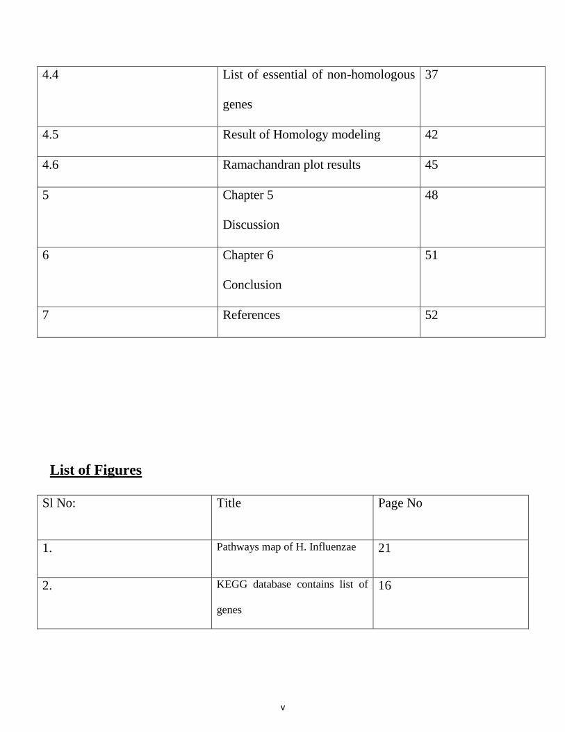
4.6 Ramachandran plot results 45
5 Chapter 5
Discussion
48
6 Chapter 6
Conclusion
51
7 References 52
List of Figures
Sl No: Title Page No
1. Pathways map of H. Influenzae 21
2. KEGG database contains list of
genes
16
vi
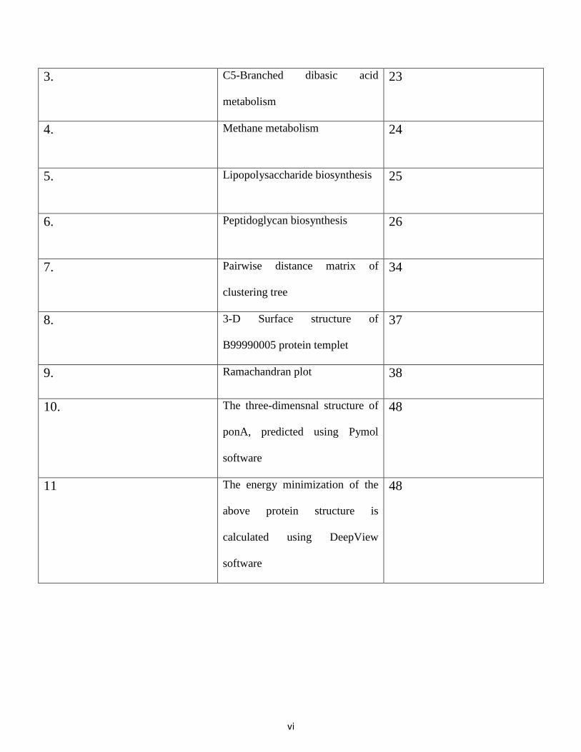
3. C5-Branched dibasic acid
metabolism
23
4. Methane metabolism 24
5. Lipopolysaccharide biosynthesis 25
6. Peptidoglycan biosynthesis 26
7. Pairwise distance matrix of
clustering tree
34
8. 3-D Surface structure of
B99990005 protein templet
37
9. Ramachandran plot 38
10. The three-dimensnal structure of
ponA, predicted using Pymol
software
48
11 The energy minimization of the
above protein structure is
calculated using DeepView
software
48
vii
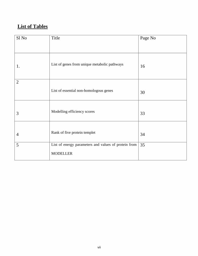
List of Tables
Sl No Title Page No
1.
List of genes from unique metabolic pathways
16
2
List of essential non-homologous genes
30
3
Modelling efficiency scores
33
4
Rank of five protein templet
34
5 List of energy parameters and values of protein from
MODELLER
35
1
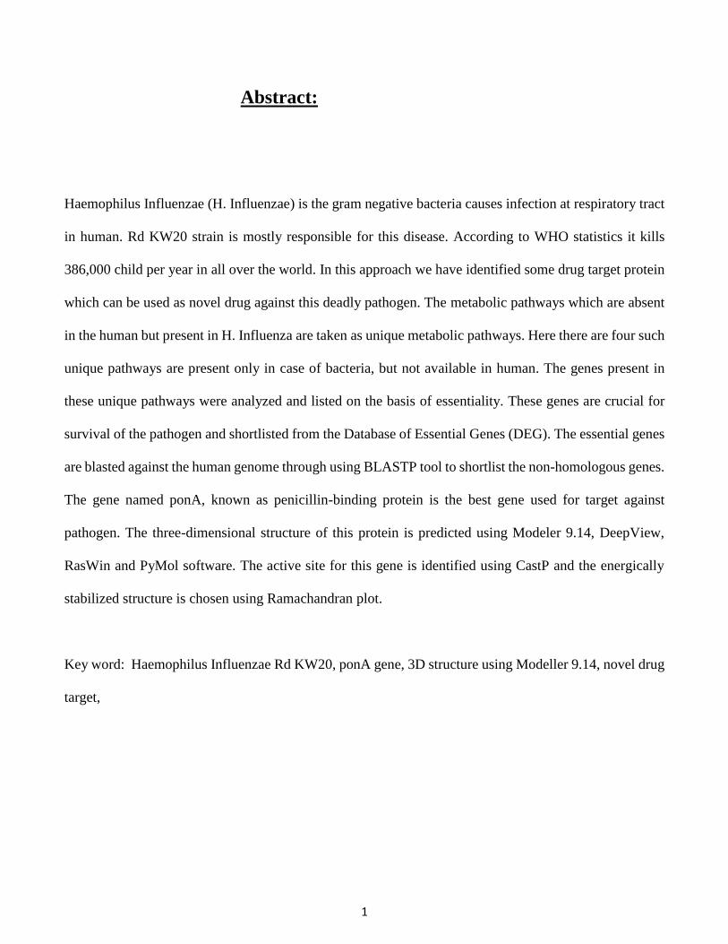
Abstract:
Haemophilus Influenzae (H. Influenzae) is the gram negative bacteria causes infection at respiratory tract
in human. Rd KW20 strain is mostly responsible for this disease. According to WHO statistics it kills
386,000 child per year in all over the world. In this approach we have identified some drug target protein
which can be used as novel drug against this deadly pathogen. The metabolic pathways which are absent
in the human but present in H. Influenza are taken as unique metabolic pathways. Here there are four such
unique pathways are present only in case of bacteria, but not available in human. The genes present in
these unique pathways were analyzed and listed on the basis of essentiality. These genes are crucial for
survival of the pathogen and shortlisted from the Database of Essential Genes (DEG). The essential genes
are blasted against the human genome through using BLASTP tool to shortlist the non-homologous genes.
The gene named ponA, known as penicillin-binding protein is the best gene used for target against
pathogen. The three-dimensional structure of this protein is predicted using Modeler 9.14, DeepView,
RasWin and PyMol software. The active site for this gene is identified using CastP and the energically
stabilized structure is chosen using Ramachandran plot.
Key word: Haemophilus Influenzae Rd KW20, ponA gene, 3D structure using Modeller 9.14, novel drug
target,
2
1. Introduction:
Haemophilus influenza is a hazardous bacterial pathogen, bringing about respiratory tract infections in
both kids and grown-ups [1]. This pathogen is present in nasopharynx, the upper respiratory tract of
human body. It causes serious incursive infections to human body by extending the pathogen from
nasopharnyx to the lower respiratory system. According to the survey done by World Health Organization
(WHO), around 386,000 child deaths occur annually caused by H. influenza all over the world [2].
Haemophilus influenzae (H. influenzae) is a Gram-negative bacteria categorized to Pasteurellaceae
family. It was first discovered in 1892 by Richard Pfeiffer. It is the first free living pathogen, whose entire
genome project is sequenced and finished during 1995. It has both capsulated and unencapsulated strains.
There are about eight different phenotypic characteristics and six different capsular antigen types, a-f
categorized. Our current research is on Haemophilus influenzae Rd KW20 and to develop effective drug
target using computational tools and technique. H. influenzae strain Rd KW20 has conventional been
considered avirulent, when it cannot survive in the bloodstream of animals. The pathogen can be killed by
normal adult human sera and very difficult to colonize the nasopharynx of infant rats. H. influenza strain
KW20 is grown as monolayers of differentiated epithelium at the air liquid interface [3] & [4].
Several research work are going on progressively to develop the effective drugs by genetic or genomic
approaches. Novel drug target are design to defend against antibiotic sensitive bacteria. New effective
method has been developed in bioinformatics for finding organized targets antecedently from unexplored
cellular functions and to empathize the inner biological process of pathogen. The complete genome
information is also crucial for selection of accurate approach to check essentiality and selectivity pattern
of the microbe. The target of the approach should be substantive encoded gene for the replication, growth
and survival of pathogen. This target should not create any cytotoxicity damage to host. The genes called
3
as “essential genes” that are present in different conserved domain of genome and essential for the survival
of the organism. These essential genes cannot endure inactivation through the mutation process [5]. The
contingent pernicious mutants assist to adapt the status of these genes. Terminating the function of
essential genes results death-dealing constitution inside bacteria. So it will be not worthless by addressing
these drugs as “super bullet” against pathogen. This will not only help to avert cost but also very easy to
detect virulent inhibitors by recognizing extend drug targets [2] & [5].
Now a days it’s very easy to recognize the targets by insilico-genomic approaches. “Differential genome
method” is one of the beneficial approach for the anticipation of likely drug targets. This method offers
detail genomic information of pathogens i.e. how the complete set of genes and protein are encoded inside
the small genome [6] & [7]. The genes which present in pathogen, but absent in human are called non-
homologous genes. These are most fundamental components for insilico-genome analysis. Using
bioinformatics tools and techniques, the drug targets can be recognized so easily from these genes. The
genes which are responsible for the foundation of life are known as the essential genes [8]. The function
of essential genes are common to all cells. For the sustainment of infections is based to work out for anti-
microbial agents against bacteria. The characterization of particular essential genes for specific pathogen
can be used as drug target in several conserve domain of that bacteria. Database of Essential Genes (DEG)
incorporates the list of essential genes of some limited pathogen. It is very easy to encounter the
essentiality of genes after the successful development and implementation of human genome project
databases. So it is tending to one step ahead development for novel drug target approaches. Anti-bacterial
drug targets can be done by recognizing the specific essential genes by “subtractive genome approaches”
[9], [10] & [11].
4
Subtractive genome approaches is successful implemented in this research paper to identify the potential
drug targets for Haemophilus influenza. The essential genes for Haemophilus influenzae Rd KW20 are
listed successfully by assisting Database of Essential Genes (DEG) against human genome. The genes
present in Haemophilus influenzae Rd KW20, closely related to human genome are called as homologous
genes and these genes are discarded [12], [13], [14] & [16].
The potential drug targets are effectively used in vaccination purposes. Vaccine provides procure
immunity for the prevention of specific infection. Vaccine contains agents, which are part of an organism
used to kill that organism. Vaccines may be toxins, surface proteins or inactive part of the organism which
triggers the immune system to demolish the pathogen by identifying and recording the threat [17].
Operative vaccines can be developed by targeting the genes present in cell wall or plasma membrane.
Kyoto Encyclopedia of Genes and Genomes (KEGG) database provides unique metabolic pathway map
of Haemophilus influenzae Rd KW20. As we are targeting the genes located in cell wall or plasma
membrane, four important metabolic pathways are selected like c5-Branched dibasic acid metabolism
pathways in Carbohydrate metabolism, Methane metabolism pathways in energy metabolism,
Lipopolysaccharide biosynthesis and Peptidoglycan biosynthesis [18] & [19].
For novel antibiotic development ponA protein, which is also known as penicillin-binding protein of
Haemophilus influenzae Rd KW20 is select for drug target. The structure of ponA can be predicted using
various computational and bioinformatics approaches. Using Homology modeling, we can develop
energycally stable three-dimensional structure for ponA protein [20], [21], [22], [23] & [24].
Protein achieves functional conformation by interacting with different molecules like ligand, substrate,
DNA and other proteins. It’s very crucial to obtain the specific three-dimensional protein structure for the
5
identification of proper interaction by visualizing the shape, physical, chemical and biological properties.
By the enactment of protein surface characterization assist to analyze specification of binding, enzyme
mechanism and examine for mutation.
Another important approach is by visualizing activity of protein using structure-based drug design
(SBDD). The substrate binding site of protein helps in conformational changes and chemical
modifications. This specific binding site of protein assist to trigger implementing the therapeutics
approach for disruption in biological processes of pathogen.
1.1. Literature and review:
1.1.1 Life Cycle of H. Influanzae:
Interesting features about the cell structure of H. Influnzae; how it picks up energy; what essential
molecules it it delivers. haemophilus influenzae is a microorganisms and consequently shows
characteristics of a prokaryotic cell. It was distinguished as a gram negative microorganisms on account
of its reaction to Gram staining techniques, as it stains red [1]. The gram negative coccobacillus has
imperative cell wall components that assume a part in its survival and its pathogenicity. H. influenzae
microbes comprise of different strains taking into account the presence or absence of an external covering
called capsules. Haemophilus influenzae, the significant pathogen, can be differentiated into epitomized
or typable strains, of which there are seven sorts (a-f) in light of the antigenic structure of the capsular
polysaccharide, and unencapsulated or nontypable strains [2]. By segregating H. influenzae it was
observed that some were indicated to have pili structures, which help in connection to the oropharyngeal
6
epithelial cell of human. Another essential properties of the H. influenzae cell structure is the rough
lipopolysaccharide (LPS) which stretches out from the cell surface. There are varieties in the LPS from
specie to specie and it has been recommended to be vital in the life cycle of the Haemophilus influenzae.
Haemophilus influenzae metabolizes sugar as its wellspring of vitality, however there is minimal thought
about this metabolic ability of the H. influenzae. It is a facultative anaerobe and along these lines makes
ATP by high-impact breath when oxygen is present and is likewise capable for metabolizing its sugar
source without oxygen by fermentation. it was discovered that more than 90% of H. influenzae separated,
digests sugars, for example, maltose glucose, galactose and ribose by fermentation and the remaining
percent ferment fructose, mannose, or glycerol [3], [11] & [26].
Haemophilus influenzae reproduces by asexual procedure called binary fission which is characteristic to
microscopic organisms. At binary fission, the H. influenzae starts replication at the source of replication
site. As the chromosome is reproduced, proteins help in the development of the chromosome to inverse
shafts of the cell and the extension of the cell. Septum formation and invagination of the cell layer divides
the chromosomes into two different cells that are fit for developing to the shape of the first parent cell [3].
1.1.2 Mode of infection and symptoms:
H. Influnzae mostly affects the children below five years age. Haemophilus influenzae bacterias, are
spread individual to-individual by direct contact or through respiratory droplets like by sneezing and
coughing. Normally the microorganisms stay in the nose and throat-creating no problem. In some cases
the microorganisms can enter the blood and spread, creating genuine disease in the person. More often
7
than not, Haemophilus influenzae microorganisms are spread by individuals who have the microbes in
their noses and throats yet who are not sick (asymptomatic). The incubation period (time between first
symptoms and exposure) of Haemophilus influenzae infection is not sure, but rather could be as short as
a couple of days [3].
Infrequently Haemophilus influenzae microorganisms spread to other individuals who have had close or
extensive contact with a patient with Haemophilus influenzae infection. In specific cases, individuals in
close contact with that patient should get anti-microbial to keep them from getting the infection [1].
As of late there has been expanding recognition that this bacterium has a part in chronic lower
inflammation of respiratory tract. However the interaction between H. influenzae and the lung is still not
very much characterized. A combination of bacterial pathogenic character and deficiency of host defense
may allow this bacterium to build contamination in the lower respiratory tract bringing about inflammation
and clinical infection [9]. The other diseases caused by pathogen:
1. Bacteremia.
2. Pneumonia.
3. Epiglottitis.
4. Sinusitis.
5. Infectious arthritis.
6. Infect the host by attaching to the host using Trimeric Autotransporter Adhesins.
1.1.3 Earlier Therapeutic Approach:
Successful vaccines for Haemophilus influenzae have been discovered since the mid-1990s, and is
suggested for kids under five age and asplenic patients. The World Health Organization suggests a
8
precautionary vaccine, consolidating vaccines against diphtheria, tetanus, pertussis, hepatitis B and Hib.
There is not yet adequate confirmation on how viable this preventive vaccine is in connection to the
individual vaccine [25], [26], [27], [28] & [29].
The available vaccines are very expansive compare to tuberculosis, diphtheria, measles, polio tetanus, and
pertussis. Subsequently, though 92% of 92% of the populations of developed nations was vaccinated at
the starting of 2003, vaccination scope was 42% for developing nations, and 8% for least-developed
nations. The disadvantages of these vaccines are:
i. very expansive.
ii. Unfavorable reactions.
iii. Vaccine recipients ~30%.
iv. Causes swelling, or pain at the injection site.
9
1.1.4. Tools used for study:
NCBI:
Sequence alignment tools are used for comparability of amino acid sequences and characterized query
genes. Basic Local Alignment Search Tool (BLAST) used to compare and quick search of protein and
nucleotide sequences from databases. BLAST provides both local and global search alignment algorithm
facilities to find the similarities from conserved domains of sequences. BLAST provides much faster
alignment process implementing Smith–Waterman algorithm. There are five different version of BLAST
like BLASTn, BLASTp, BLASTx, tBLASTn, tBLASTx. BLASTn assists to compare nucleotide
sequences nucleotide databases. BLASTp assists to compare amino acid sequences from protein
databases. BLASTx is used to compare six entrapped transcription product of a nucleotide sequences vs
protein sequences. tBLASTx is used to compare six entrapped translation nucleotide sequence vs 6
entrapped sequence of nucleotide from database. tBLASTn is used to compare six entrapped translation
nucleotide sequence vs six protein sequences from database [31].
KEGG:
Kyoto Encyclopedia of Genes and Genomes is a set of database of biological pathways, diseases, drugs,
chemical substances, utilized for identification of genomics, metagenomics and metabolomics. It is an
aggregation of pathway maps fusing various substances including qualities, proteins, RNAs, substance
mixes, glycans, and compound responses, and furthermore infection qualities and targets, which are
secured as individual doorways in exchange databases of KEGG [32].
10
DEG:
Database of Essential Genes, is a database and give tools to investigate the essentiality of the genes.
Essential genes are those genes of an organism entity that are thought to be discriminating for its survival
of the organism. Essential genes in a bacterium constitute a minimal genome, forming an arrangement of
functional modules, which assume key parts in the emerging field, synthetic biology [18].
UNIPORT:
It gives data of the gene about the function, sequence and location in the cell. UniPort Knowledgebase is
a protein database partially curated by specialists, comprising of two segments: UniProtKB/Swiss-Prot
(containing assessed, manually annotated entries) and UniProtKB/ TrEMBL (containing reviewed,
automatically annotated entries) [21].
CPHmodels (Computerized neural-system based protein demonstrating server):
CPHmodels is a gathering of databases and what's more, routines created to anticipate protein structure.
It performs expectation of protein structure utilizing Comparative Modeling. It doesn't acknowledge more
than 900 amino acids in the data succession. The arrangements are kept classified and are erased in the
wake of preparing. This system did not issue me fitting results. The error it showed was like the one
showed by Swiss Model [17], [30] & [31].
11
Swiss model:
It is used for automated homology modelling. It has a first approach mode that aides performs Homology
Modeling. The user needs to enter his/ her email id and information the protein arrangement in Fasta
position. It permits the user to pick as far as possible for format choice. It can seek the pdb document from
the pdb database with the user giving the name of the pdb record or the client can transfer his/ her own
pdb document. The yield record is a pdb document that is come back to the user's email address. The
outcome can be sent by Swiss Model to PHD Secondary structure forecast at Columbia University
furthermore, Fold Recognition Server (3D-pssm) of the ICRF [15] & [18].
Geno3D:
It performs Comparative protein structure modeling by spatial limitations (separations and dihedral)
fulfillment. Geno3D is most habitually utilized for Homology or Comparative protein structure Modeling.
Geno3d acknowledges information like Fasta organize yet just the one letter code must be utilized. The
outcome is gotten in the PDB file format that can be seen in any Molecular Modeling software.Geno3d
offers numerous other highlights, it permits the user to choose PDB entrances as formats for Molecular
Modeling after a 3 stage iterative PSI BLAST. It exhibits the yield for every layout, alongside the optional
structure forecast, shows percent of assertion in auxiliary structure and repartition of data from format on
inquiry succession. The final result is sent to the user's email address. It likewise informs the client when
12
its server starts the Homology Displaying. It has an alternative where the user can choose what number of
models to create. The fundamental thought behind having more than one model created is that the client
may have a superior adaptability and comprehension. It likewise gives back a superimposed PDB
document which has the models superimposed on one another. This is one of the great focuses in Geno3d
as it permits us to think about the different models created in one window [22]. All the outcomes acquired
can be downloaded as an archive.tar.Z that can be opened in WinZip in windows and in UNIX or Linux
stages. So the user does not need to spare results in site page impact or in an archive record. It likewise
shows the Ramachandran plot in the outcome.
Ramachandran plot:
The Sasisekharan-Ramakrishnan-Ramachandran plot describes permitted main chain conformations. A
Ramachandran plot is an approach to visualize dihedral angles φ against ψ of amino visualize dihedral
angles. It demonstrates the possible conformations of φ and ψ plots for a polypeptide. Rotation is allowed
around the N-Cα and Cα-C single bonds of all residues (with one special case: proline). The angles φ and
ψ around these bonds, and the angle of rotation around the peptide bond, ω, characterize the conformation
of a residue. The peptide bond itself has a tendency to be planar, with two permitted states: Trans, ω ≈
180° (generally) and cis, ω ≈ 0° (once in a while, and by and large at a proline deposit). The sequence of
φ, ψ and ω points of all residues in a protein defines the backbone conformation [9].
13
MODELLER:
Modeler is used for homology or relative modelling of protein in three-dimensional structures. It is
assembled in FORTRAN. It will runs on python script file commands. Modeler is most frequently utilized
for homology or near protein structure demonstrating. Modeler aides focus the spatial limitations from the
formats. It creates various 3D models of the arrangement you submit fulfilling the layout limitations.
Modeler naturally figure a full molecule model. Modeler models protein 3D structure keeping in the
requirements of spatial limitations. The restrictions can be gotten from various distinctive sources [29].
DeepView:
Swiss-PdbViewer is an application that gives an easy to use interface permitting to break down a few
proteins in the meantime. The proteins can be superimposed in request to derive basic arrangements and
look at their dynamic destinations or some other important parts. Amino corrosive changes, H-bonds,
angles and separations between particles are anything but difficult to get because of the natural realistic
and menu interface. DeepView - Swiss-PdbViewer was developed by Nicolas Guex (GlaxoSmithKline
R&D). Swiss-PdbViewer is hard connected to SWISS-MODEL, an automated homology modelling server
created inside the Swiss Institute of Bioinformatics (SIB) in Basel [10] & [11].

14
Chapter 2
OBJECTIVES AND WORK PLAN

15
2.1Objective
To identify a non-homologous essential gene of unique metabolism pathways, which can be used as
potential drug target against Haemophilus Influenzae.
16
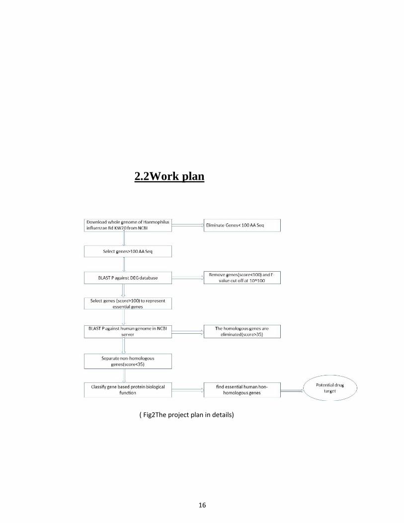
2.2Work plan
( Fig2The project plan in details)

17
Chapter 3
Material and method
18
3.1 Retrieval of proteome from NCBI:
The complete set of protein (proteome) is revived from NCBI. The sequence less than 100 amino acid
sequence are consider to be paralog or duplicate protein. The non-paralog proteins are selected and paralog
are eliminated.
3.2 Identification of unique metabolic pathways:
KEGG database is used for the selection of unique metabolic pathways from H. Influenzae and human.
Some unique metabolic pathways are selected to identify appropriate genes. For the selection of genes in
unique metabolic pathways, “pathway Entry” is selected. Then select “Metabolism” from drop down
menu. The gene from the unique metabolic pathways are listed and analyzed.
19
3.3 Selection of essential genes:
To identify essential genes the amino acid sequence are submitted for BLASTP in DEG (Database of
Essential Genes). The above genes are analyzed through DEG database. To get the essential genes, cut
off score greater than 100 are selected and non-essential genes are eliminated.
3.4 Identification of non-homologous genes:
Using BLASTP tool homologous and non-homologous genes can be differentiated. Homologous genes
are present in both human and pathogen. Elimination of homologous genes are necessary, because these
genes involves in the common biological processes and vaccination will be not effective. For the selection
of essential non-homologous genes the identity is considered below 35 % and expected threshold value is
set at 0.005. We are targeting the most conserved bacterial to get best result for multi resistant strain
pathogen.
3.5 Homology modelling of identified protein:
The homologs conserved protein coding sequence was chosen from H. Influanzae strains for drug target.
The three-dimensional structure of the targeted protein was displayed by considering the suitable all
around contemplated protein structure is recognized by closeness search with the BLASTP tool against
the protein databank. The homology modelling is done with online software like Geno3D , Swiss model,
CPHmodels by using distinctive parameters. What's more, offline homology modelling is done utilizing
profound parameters, the modeled protein was refined by the MODELER 9.14. The model is submitted
20
for the 3D-1D profile with VERIFY3D, and the stereo chemical qualities were checked with PROCHECK,
Errat, Prove and WHAT_IF (http://nihserver.mbi.ucla.edu/SAVS/). At last, the basic properties of the
target protein were visualized by using the Ramachandran plot score. The distinctive software models are
contrasted and one another last best model is chosen; it is utilized for further drug design process.
3.6 Modelled protein structure validation using Ramachandran plot:
The best PDB result after the homology modelling is selected for Ramachandran plot analysis. The PDB
file is submitted in SAVE (Structure Analysis and Verification) online server. The WHAT_CHECK tool
of SAVE server will check the validation of protein structure. The result will be sand via web showing
favoured, allowed and outlier region.

21
Chapter 4
Result and Description
22
4. Result and Description:
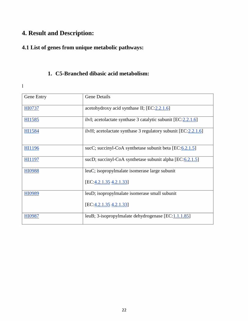
4.1 List of genes from unique metabolic pathways:
1. C5-Branched dibasic acid metabolism:
l
Gene Entry Gene Details
HI0737 acetohydroxy acid synthase II; [EC:2.2.1.6]
HI1585 ilvI; acetolactate synthase 3 catalytic subunit [EC:2.2.1.6]
HI1584 ilvH; acetolactate synthase 3 regulatory subunit [EC:2.2.1.6]
HI1196 sucC; succinyl-CoA synthetase subunit beta [EC:6.2.1.5]
HI1197 sucD; succinyl-CoA synthetase subunit alpha [EC:6.2.1.5]
HI0988 leuC; isopropylmalate isomerase large subunit
[EC:4.2.1.35 4.2.1.33]
HI0989 leuD; isopropylmalate isomerase small subunit
[EC:4.2.1.35 4.2.1.33]
HI0987 leuB; 3-isopropylmalate dehydrogenase [EC:1.1.1.85]
23
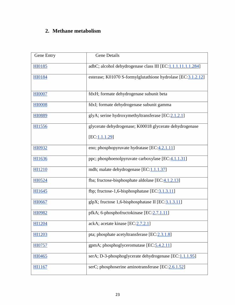
2. Methane metabolism
Gene Entry Gene Details
HI0185 adhC; alcohol dehydrogenase class III [EC:1.1.1.11.1.1.284]
HI0184 esterase; K01070 S-formylglutathione hydrolase [EC:3.1.2.12]
HI0007 fdxH; formate dehydrogenase subunit beta
HI0008 fdxI; formate dehydrogenase subunit gamma
HI0889 glyA; serine hydroxymethyltransferase [EC:2.1.2.1]
HI1556 glycerate dehydrogenase; K00018 glycerate dehydrogenase
[EC:1.1.1.29]
HI0932 eno; phosphopyruvate hydratase [EC:4.2.1.11]
HI1636 ppc; phosphoenolpyruvate carboxylase [EC:4.1.1.31]
HI1210 mdh; malate dehydrogenase [EC:1.1.1.37]
HI0524 fba; fructose-bisphosphate aldolase [EC:4.1.2.13]
HI1645 fbp; fructose-1,6-bisphosphatase [EC:3.1.3.11]
HI0667 glpX; fructose 1,6-bisphosphatase II [EC:3.1.3.11]
HI0982 pfkA; 6-phosphofructokinase [EC:2.7.1.11]
HI1204 ackA; acetate kinase [EC:2.7.2.1]
HI1203 pta; phosphate acetyltransferase [EC:2.3.1.8]
HI0757 gpmA; phosphoglyceromutase [EC:5.4.2.11]
HI0465 serA; D-3-phosphoglycerate dehydrogenase [EC:1.1.1.95]
HI1167 serC; phosphoserine aminotransferase [EC:2.6.1.52]
24
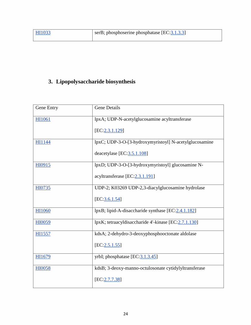
HI1033 serB; phosphoserine phosphatase [EC:3.1.3.3]
3. Lipopolysaccharide biosynthesis
Gene Entry Gene Details
HI1061 lpxA; UDP-N-acetylglucosamine acyltransferase
[EC:2.3.1.129]
HI1144 lpxC; UDP-3-O-[3-hydroxymyristoyl] N-acetylglucosamine
deacetylase [EC:3.5.1.108]
HI0915 lpxD; UDP-3-O-[3-hydroxymyristoyl] glucosamine N-
acyltransferase [EC:2.3.1.191]
HI0735 UDP-2; K03269 UDP-2,3-diacylglucosamine hydrolase
[EC:3.6.1.54]
HI1060 lpxB; lipid-A-disaccharide synthase [EC:2.4.1.182]
HI0059 lpxK; tetraacyldisaccharide 4'-kinase [EC:2.7.1.130]
HI1557 kdsA; 2-dehydro-3-deoxyphosphooctonate aldolase
[EC:2.5.1.55]
HI1679 yrbI; phosphatase [EC:3.1.3.45]
HI0058 kdsB; 3-deoxy-manno-octulosonate cytidylyltransferase
[EC:2.7.7.38]
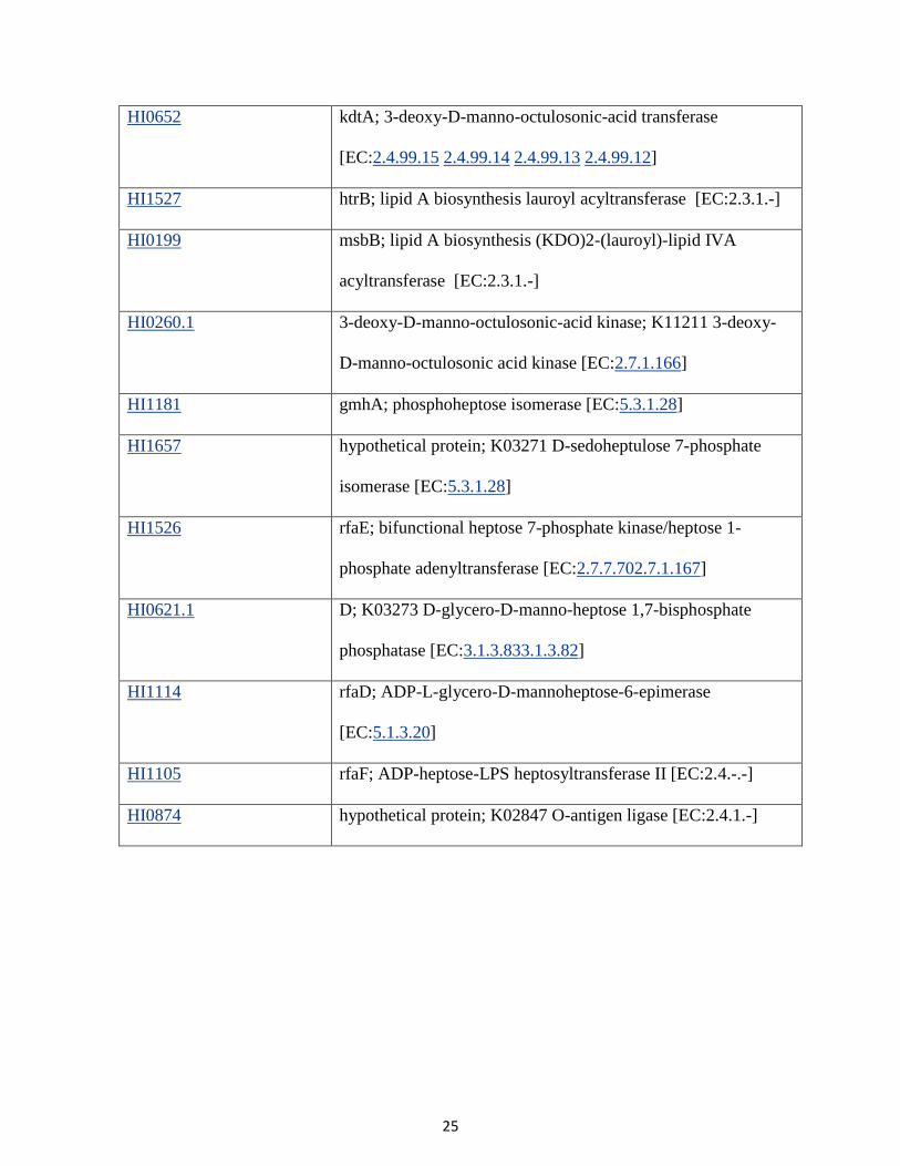
25
HI0652 kdtA; 3-deoxy-D-manno-octulosonic-acid transferase
[EC:2.4.99.15 2.4.99.14 2.4.99.13 2.4.99.12]
HI1527 htrB; lipid A biosynthesis lauroyl acyltransferase [EC:2.3.1.-]
HI0199 msbB; lipid A biosynthesis (KDO)2-(lauroyl)-lipid IVA
acyltransferase [EC:2.3.1.-]
HI0260.1 3-deoxy-D-manno-octulosonic-acid kinase; K11211 3-deoxy-
D-manno-octulosonic acid kinase [EC:2.7.1.166]
HI1181 gmhA; phosphoheptose isomerase [EC:5.3.1.28]
HI1657 hypothetical protein; K03271 D-sedoheptulose 7-phosphate
isomerase [EC:5.3.1.28]
HI1526 rfaE; bifunctional heptose 7-phosphate kinase/heptose 1-
phosphate adenyltransferase [EC:2.7.7.702.7.1.167]
HI0621.1 D; K03273 D-glycero-D-manno-heptose 1,7-bisphosphate
phosphatase [EC:3.1.3.833.1.3.82]
HI1114 rfaD; ADP-L-glycero-D-mannoheptose-6-epimerase
[EC:5.1.3.20]
HI1105 rfaF; ADP-heptose-LPS heptosyltransferase II [EC:2.4.-.-]
HI0874 hypothetical protein; K02847 O-antigen ligase [EC:2.4.1.-]
26
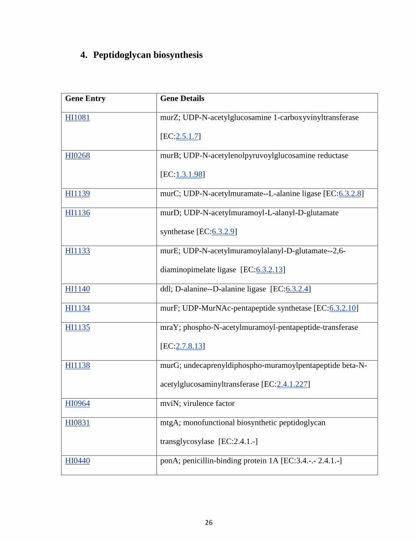
4. Peptidoglycan biosynthesis
Gene Entry Gene Details
HI1081 murZ; UDP-N-acetylglucosamine 1-carboxyvinyltransferase
[EC:2.5.1.7]
HI0268 murB; UDP-N-acetylenolpyruvoylglucosamine reductase
[EC:1.3.1.98]
HI1139 murC; UDP-N-acetylmuramate--L-alanine ligase [EC:6.3.2.8]
HI1136 murD; UDP-N-acetylmuramoyl-L-alanyl-D-glutamate
synthetase [EC:6.3.2.9]
HI1133 murE; UDP-N-acetylmuramoylalanyl-D-glutamate--2,6-
diaminopimelate ligase [EC:6.3.2.13]
HI1140 ddl; D-alanine--D-alanine ligase [EC:6.3.2.4]
HI1134 murF; UDP-MurNAc-pentapeptide synthetase [EC:6.3.2.10]
HI1135 mraY; phospho-N-acetylmuramoyl-pentapeptide-transferase
[EC:2.7.8.13]
HI1138 murG; undecaprenyldiphospho-muramoylpentapeptide beta-N-
acetylglucosaminyltransferase [EC:2.4.1.227]
HI0964 mviN; virulence factor
HI0831 mtgA; monofunctional biosynthetic peptidoglycan
transglycosylase [EC:2.4.1.-]
HI0440 ponA; penicillin-binding protein 1A [EC:3.4.-.- 2.4.1.-]
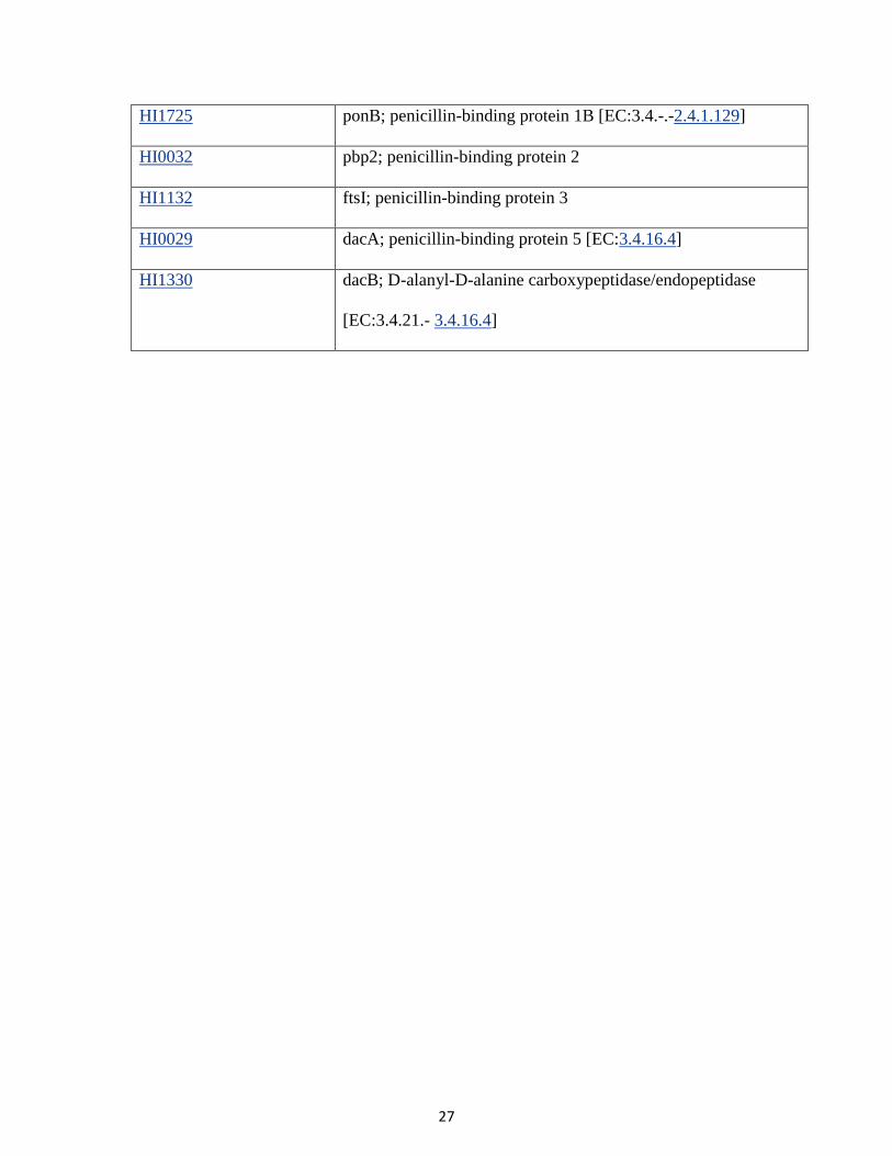
27
HI1725 ponB; penicillin-binding protein 1B [EC:3.4.-.-2.4.1.129]
HI0032 pbp2; penicillin-binding protein 2
HI1132 ftsI; penicillin-binding protein 3
HI0029 dacA; penicillin-binding protein 5 [EC:3.4.16.4]
HI1330 dacB; D-alanyl-D-alanine carboxypeptidase/endopeptidase
[EC:3.4.21.- 3.4.16.4]
28
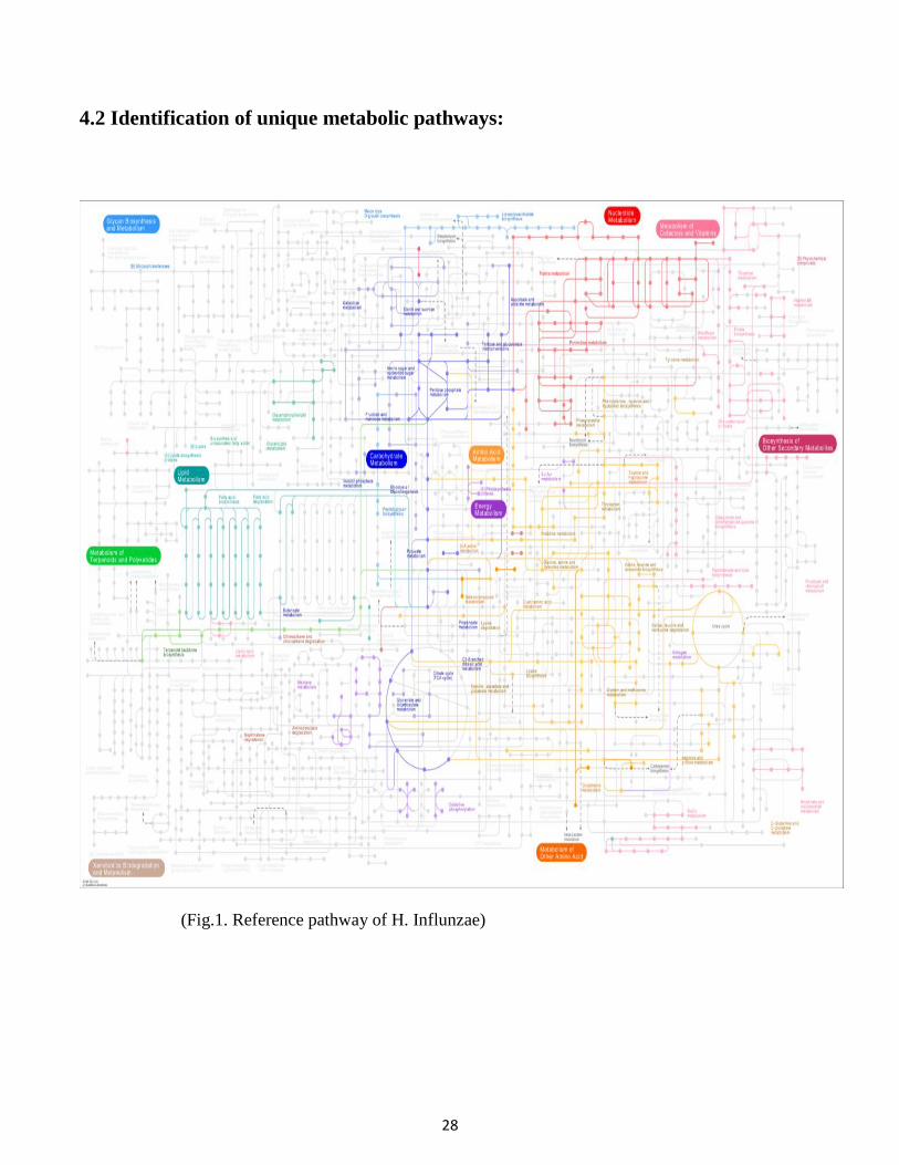
4.2 Identification of unique metabolic pathways:
(Fig.1. Reference pathway of H. Influnzae)
29
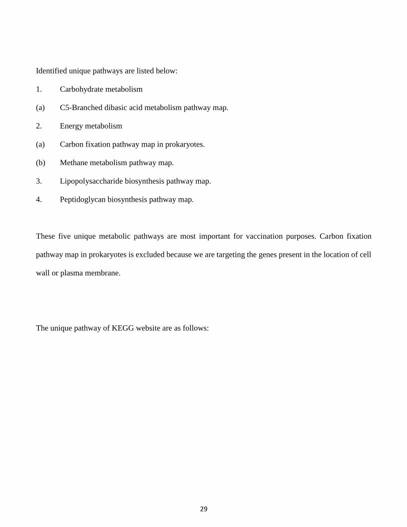
Identified unique pathways are listed below:
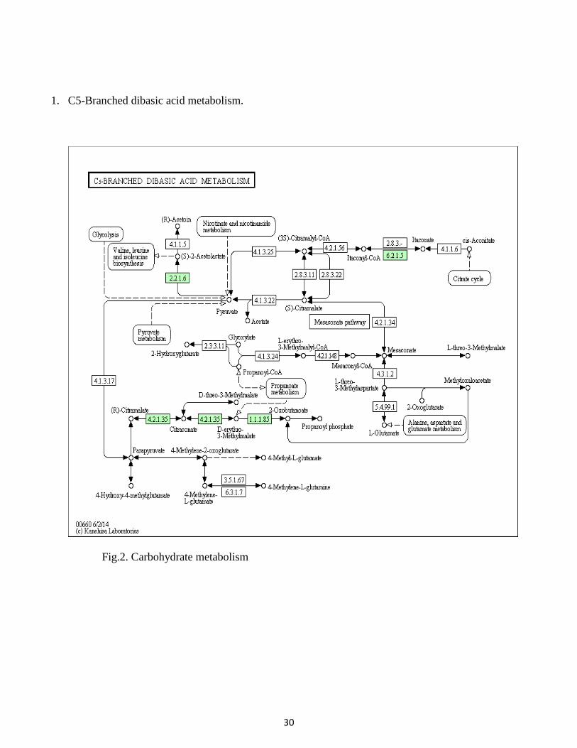
1. Carbohydrate metabolism
(a) C5-Branched dibasic acid metabolism pathway map.
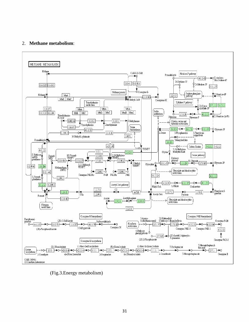
2. Energy metabolism
(a) Carbon fixation pathway map in prokaryotes.
(b) Methane metabolism pathway map.
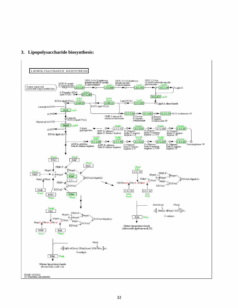
3. Lipopolysaccharide biosynthesis pathway map.
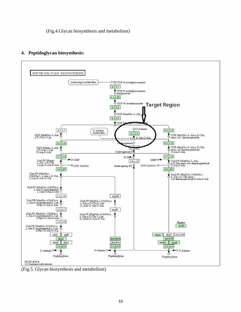
4. Peptidoglycan biosynthesis pathway map.
These five unique metabolic pathways are most important for vaccination purposes. Carbon fixation
pathway map in prokaryotes is excluded because we are targeting the genes present in the location of cell
wall or plasma membrane.
The unique pathway of KEGG website are as follows:
30
1. C5-Branched dibasic acid metabolism.
Fig.2. Carbohydrate metabolism
31
2. Methane metabolism:
(Fig.3.Energy metabolism)
32
3. Lipopolysaccharide biosynthesis:
33
(Fig.4.Glycan biosynthesis and metabolism)
4. Peptidoglycan biosynthesis:
(Fig.5. Glycan biosynthesis and metabolism)
34
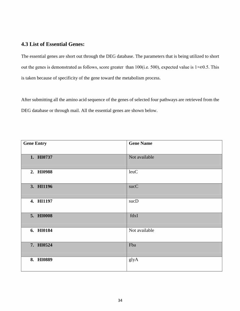
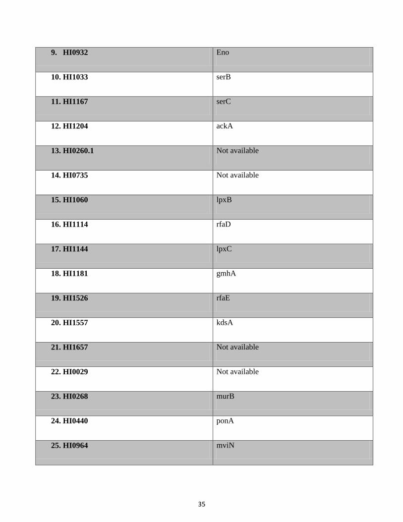
4.3 List of Essential Genes:
The essential genes are short out through the DEG database. The parameters that is being utilized to short
out the genes is demonstrated as follows, score greater than 100(i.e. 500), expected value is 1×℮0.5. This
is taken because of specificity of the gene toward the metabolism process.
After submitting all the amino acid sequence of the genes of selected four pathways are retrieved from the
DEG database or through mail. All the essential genes are shown below.
Gene Entry Gene Name
1. HI0737 Not available
2. HI0988 leuC
3. HI1196 sucC
4. HI1197 sucD
5. HI0008 fdxI
6. HI0184 Not available
7. HI0524 Fba
8. HI0889 glyA
35
9. HI0932 Eno
10. HI1033 serB
11. HI1167 serC
12. HI1204 ackA
13. HI0260.1 Not available
14. HI0735 Not available
15. HI1060 lpxB
16. HI1114 rfaD
17. HI1144 lpxC
18. HI1181 gmhA
19. HI1526 rfaE
20. HI1557 kdsA
21. HI1657 Not available
22. HI0029 Not available
23. HI0268 murB
24. HI0440 ponA
25. HI0964 mviN
36
26. HI1081 murZ
27. HI1135 mraY
28. HI1584 ilvH
29. HI1585 ilvI
30. HI1167 serC
31. HI1204 ackA
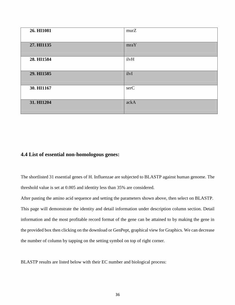
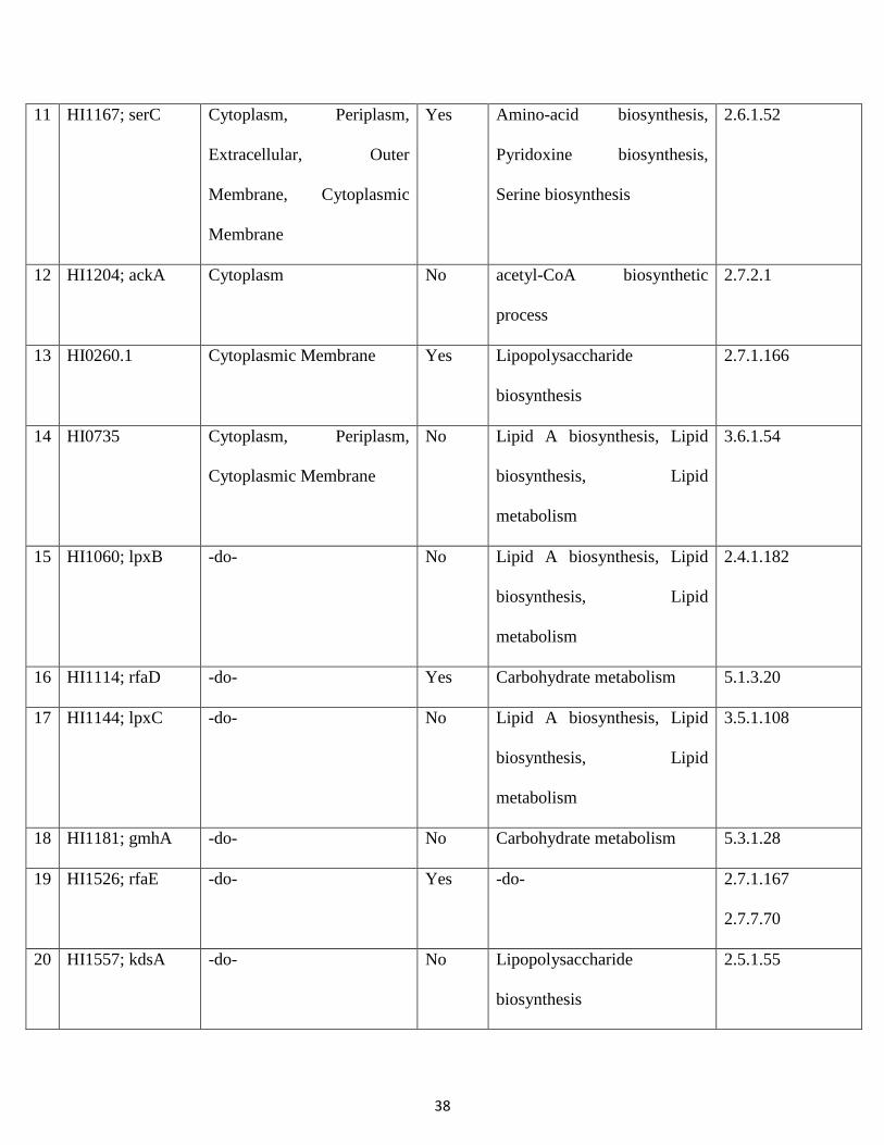
4.4 List of essential non-homologous genes:
The shortlisted 31 essential genes of H. Influenzae are subjected to BLASTP against human genome. The
threshold value is set at 0.005 and identity less than 35% are considered.
After pasting the amino acid sequence and setting the parameters shown above, then select on BLASTP.
This page will demonstrate the identity and detail information under description column section. Detail
information and the most profitable record format of the gene can be attained to by making the gene in
the provided box then clicking on the download or GenPept, graphical view for Graphics. We can decrease
the number of column by tapping on the setting symbol on top of right corner.
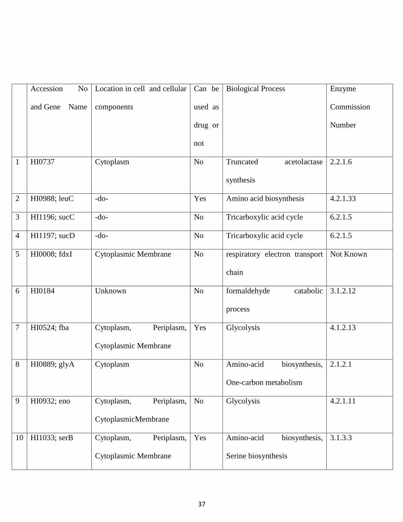
BLASTP results are listed below with their EC number and biological process:
37
Accession No
and Gene Name
Location in cell and cellular
components
Can be
used as
drug or
not
Biological Process Enzyme
Commission
Number
1
HI0737 Cytoplasm No Truncated acetolactase
synthesis
2.2.1.6
2 HI0988; leuC -do- Yes Amino acid biosynthesis 4.2.1.33
3 HI1196; sucC -do- No Tricarboxylic acid cycle 6.2.1.5
4 HI1197; sucD -do- No Tricarboxylic acid cycle 6.2.1.5
5 HI0008; fdxI Cytoplasmic Membrane No respiratory electron transport
chain
Not Known
6 HI0184 Unknown No formaldehyde catabolic
process
3.1.2.12
7 HI0524; fba Cytoplasm, Periplasm,
Cytoplasmic Membrane
Yes Glycolysis 4.1.2.13
8 HI0889; glyA Cytoplasm No Amino-acid biosynthesis,
One-carbon metabolism
2.1.2.1
9 HI0932; eno Cytoplasm, Periplasm,
CytoplasmicMembrane
No Glycolysis 4.2.1.11
10 HI1033; serB Cytoplasm, Periplasm,
Cytoplasmic Membrane
Yes Amino-acid biosynthesis,
Serine biosynthesis
3.1.3.3
38
11 HI1167; serC Cytoplasm, Periplasm,
Extracellular, Outer
Membrane, Cytoplasmic
Membrane
Yes Amino-acid biosynthesis,
Pyridoxine biosynthesis,
Serine biosynthesis
2.6.1.52
12 HI1204; ackA Cytoplasm No acetyl-CoA biosynthetic
process
2.7.2.1
13 HI0260.1 Cytoplasmic Membrane Yes Lipopolysaccharide
biosynthesis
2.7.1.166
14 HI0735 Cytoplasm, Periplasm,
Cytoplasmic Membrane
No Lipid A biosynthesis, Lipid
biosynthesis, Lipid
metabolism
3.6.1.54
15 HI1060; lpxB -do- No Lipid A biosynthesis, Lipid
biosynthesis, Lipid
metabolism
2.4.1.182
16 HI1114; rfaD -do- Yes Carbohydrate metabolism 5.1.3.20
17 HI1144; lpxC -do- No Lipid A biosynthesis, Lipid
biosynthesis, Lipid
metabolism
3.5.1.108
18 HI1181; gmhA -do- No Carbohydrate metabolism 5.3.1.28
19 HI1526; rfaE -do- Yes -do- 2.7.1.167
2.7.7.70
20 HI1557; kdsA -do- No Lipopolysaccharide
biosynthesis
2.5.1.55
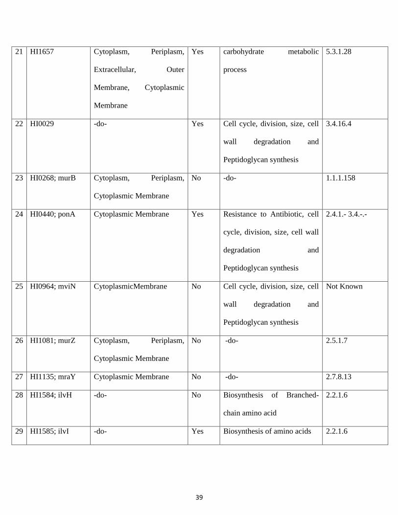
39
21 HI1657
Cytoplasm, Periplasm,
Extracellular, Outer
Membrane, Cytoplasmic
Membrane
Yes carbohydrate metabolic
process
5.3.1.28
22 HI0029 -do- Yes Cell cycle, division, size, cell
wall degradation and
Peptidoglycan synthesis
3.4.16.4
23 HI0268; murB Cytoplasm, Periplasm,
Cytoplasmic Membrane
No -do- 1.1.1.158
24 HI0440; ponA Cytoplasmic Membrane Yes Resistance to Antibiotic, cell
cycle, division, size, cell wall
degradation and
Peptidoglycan synthesis
2.4.1.- 3.4.-.-
25 HI0964; mviN CytoplasmicMembrane No Cell cycle, division, size, cell
wall degradation and
Peptidoglycan synthesis
Not Known
26 HI1081; murZ Cytoplasm, Periplasm,
Cytoplasmic Membrane
No -do- 2.5.1.7
27 HI1135; mraY Cytoplasmic Membrane No -do- 2.7.8.13
28 HI1584; ilvH -do- No Biosynthesis of Branched-
chain amino acid
2.2.1.6
29 HI1585; ilvI -do- Yes Biosynthesis of amino acids 2.2.1.6
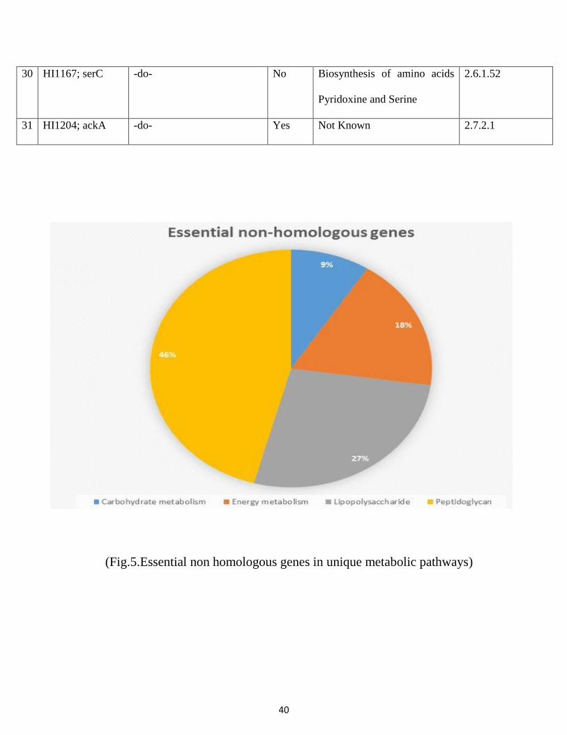
40
30 HI1167; serC -do- No Biosynthesis of amino acids
Pyridoxine and Serine
2.6.1.52
31 HI1204; ackA -do- Yes Not Known 2.7.2.1
(Fig.5.Essential non homologous genes in unique metabolic pathways)
41
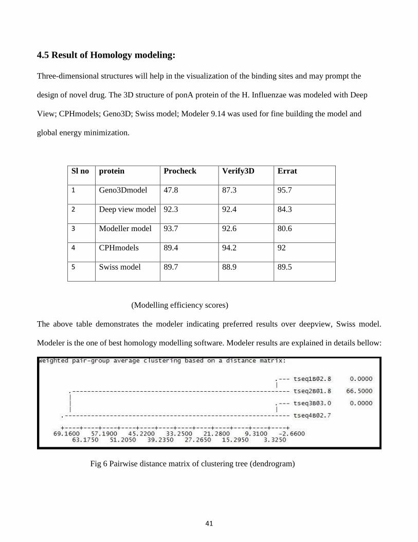
4.5 Result of Homology modeling:
Three-dimensional structures will help in the visualization of the binding sites and may prompt the
design of novel drug. The 3D structure of ponA protein of the H. Influenzae was modeled with Deep
View; CPHmodels; Geno3D; Swiss model; Modeler 9.14 was used for fine building the model and
global energy minimization.
Sl no protein Procheck Verify3D Errat
1 Geno3Dmodel 47.8 87.3 95.7
2 Deep view model 92.3 92.4 84.3
3 Modeller model 93.7 92.6 80.6
4 CPHmodels 89.4 94.2 92
5 Swiss model 89.7 88.9 89.5
(Modelling efficiency scores)
The above table demonstrates the modeler indicating preferred results over deepview, Swiss model.
Modeler is the one of best homology modelling software. Modeler results are explained in details bellow:
Fig 6 Pairwise distance matrix of clustering tree (dendrogram)
42
The first four best result from NCBI BLASTP results are named as tesq1, 2, 3&4. Tseq2 having pdb
accession no 3UDF_A is showing best crystallographic structure. So it has higher crystallographic R-
factor around 66.5 and sequence identity is around 41%.
There are five templet pdb files are generated and the best model is selected on the basic of DOPE score.
The total number of residues of the model is 864 from 6723 number of selected real atoms. There are
about 1192322 number of non-bonded pairs present in the model. The overall energy of the model is -
49257.1602 Joule. Dope score are used to predict the most stable protein templet. Less is the DOPE score
more is the stability and greater is the rank.
Rank of five protein templet are listed below on the basic of DOPE score:
File name(pdb) Identity DOPE score Rank
B99990001 41% -46309.45703 5
B99990002 40% -50459.66406 2
B99990003 53% -49490.78516 3
B99990004 53% -49256.56250 4
B99990005 36% -50748.95313 1
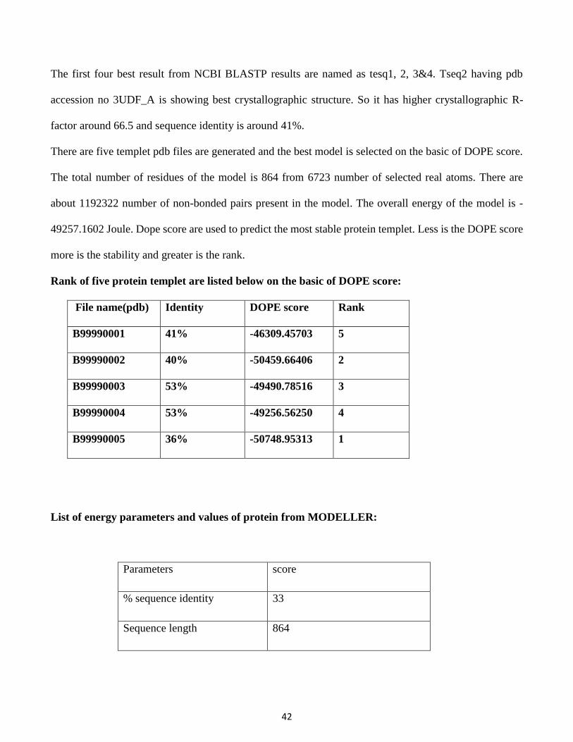
List of energy parameters and values of protein from MODELLER:
Parameters score
% sequence identity 33
Sequence length 864
43
Compactness 0.019779
Native energy (pair) -1133.825974 J
Native energy (surface) -188.171705 J
Native energy (combined) -30.176544 J
Z score (pair) -3.254623
Z score (surface) -0.983342
Z score (combined) -2.725979
Total DOPE score -50748.953125 J
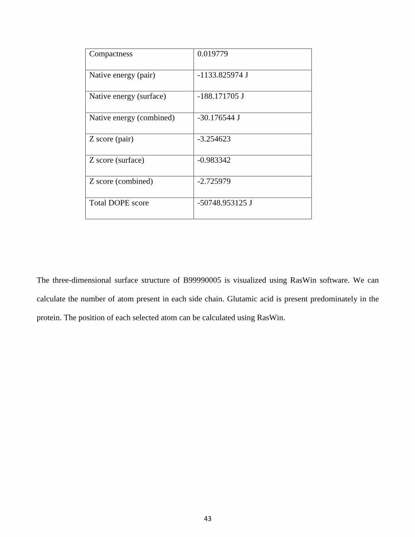
The three-dimensional surface structure of B99990005 is visualized using RasWin software. We can
calculate the number of atom present in each side chain. Glutamic acid is present predominately in the
protein. The position of each selected atom can be calculated using RasWin.
44
(Fig 7. 3-D Surface structure of B99990005 protein templet)
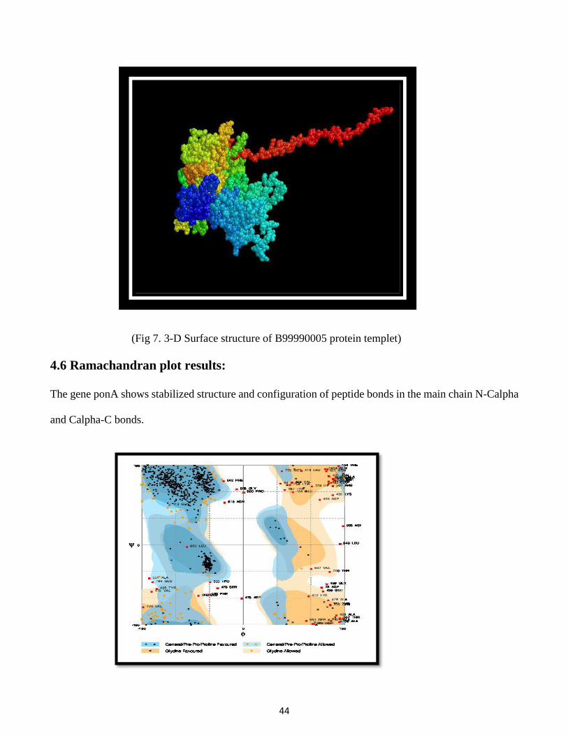
4.6 Ramachandran plot results:
The gene ponA shows stabilized structure and configuration of peptide bonds in the main chain N-Calpha
and Calpha-C bonds.
45
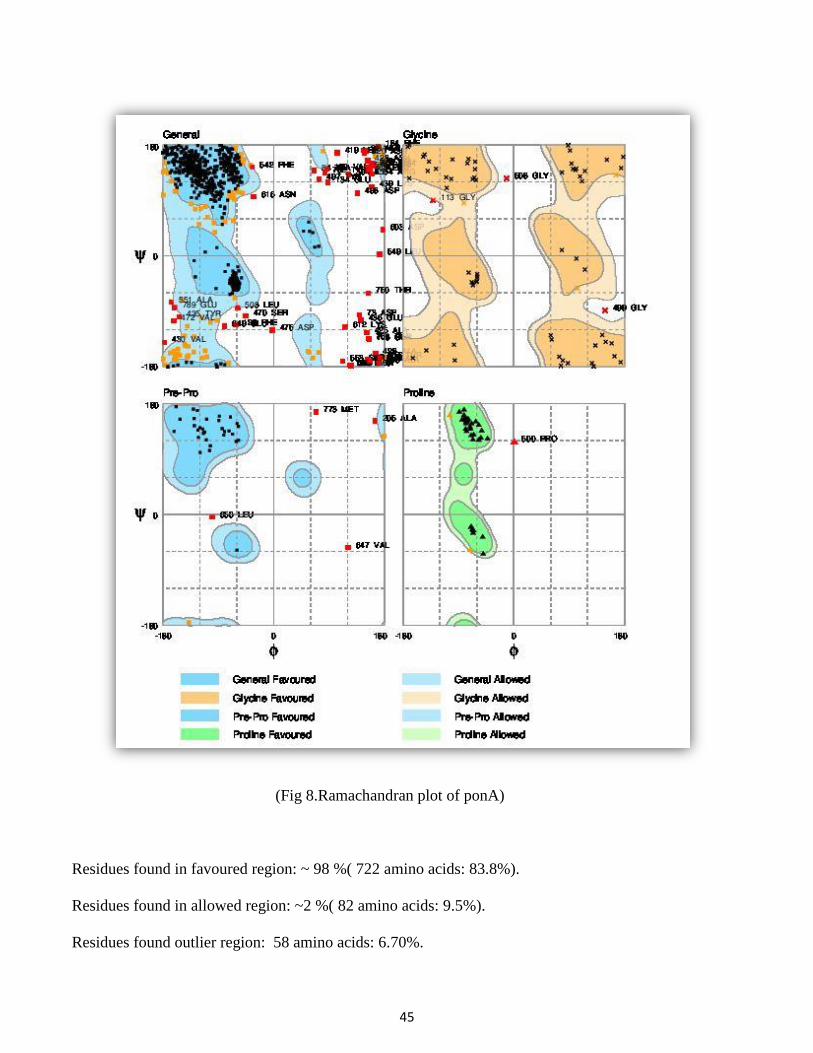
(Fig 8.Ramachandran plot of ponA)
Residues found in favoured region: ~ 98 %( 722 amino acids: 83.8%).
Residues found in allowed region: ~2 %( 82 amino acids: 9.5%).
Residues found outlier region: 58 amino acids: 6.70%.

46
Chapter 5
Discussion
47
5. Discussion:
The ponA gene having NCBI Gene ID 949537 is identified as essential hon-homologs gene, is most
preferable for vaccination purposes. This gene is also known as penicillin-binding protein of Haemophilus
influenzae Rd KW20. This gene is present in the Cytoplasmic Membrane and involved in the biological
process like resistance to Antibiotic, Cell size, Cell Lysis and Peptidoglycan synthesis. The chemical
properties of the gene is similar to the modular pieces that form the peptidoglycan. When it is used as a
drug target, blocks the enzymes that connect all the pieces together. The gene is constructed with long
chains of sugars molecules with short peptides bonds sticking out in all directions. The D-alanyl-D-alanine
carboxypeptidase region of the protein is cross-linked with these short peptides to form a three-
dimensional structure. Acyl-ester intermediate is present in 441 position of the gene. It is the active region
of the gene, because it helps in binding of metal ions. Metal ions like magnesium are crucial for drug
targets approaches.
This can be taken as target protein, because of the following points:
1. The 3D structure of the protein is known.
2. It is the essential hon-homologous gene.
3. This gene is responsible for Peptidoglycan synthesis and Cell lysis process. So this gene will function
effectively for potential drug target to disrupt the cell or plasma membrane.
4. It can block the metabolism process of pathogen, because it is not present in human.
5. The energy minimized structure is predicted.

48
Structure prediction of the gene ponA:
(Fig 9. The three-dimensnal structure of ponA, predicted using Pymol software)
(Fig 10.The energy minimized superimposed protein structure of ponA is calculated using DeepView
software)

49
Chapter 6
Conclusion
50
6. Conclusion:
In this study, the genome of H. Influenzae from four important metabolism pathways were sucessfully
analyzed, which are absent in the human. The essentiality of the genes were identified through the DEG
tool. Around 31 genes are short listed from DEG. The essential genes were subjected for BLASTP against
human genome. Using BLASTP homologous and non-homologous genes were separated. There was
around 11 essential non-homologous genes, which can be used as drug target. After implementing all the
steps successfully, we can able to identify a gene named as ponA for drug target. It is present in the
Cytoplasmic Membrane of the pathogen. The pathogen H. Influanzae can be killed by blocking the
biological function of ponA.
The future direction of this project is to perform Docking with ligands to the targeted protein, prediction
of thermodynamic activities of ligands, and study about pharmacodynamics, pharmacokinetics, and
solubility activities.
51
7. References:
1. A. E. Curr. (1998). New antibiotic discovery, novel screens, novel targets and impact of microbial
genomics. Opin Microbiol. 1, 530-534.
2. Moxon ER: The molecular basis of pathogenicity in Haemophilus influenzae: comparative virulence of
genetically-related capsular transformants and correlation with changes at the capsulation locus cap. Kroll
JS, Microb Pathog 1989, 7:225-235.
3. Morens Fauci AS: Predominant role of bacterial pneumonia as a cause of death in pandemic influenza:
implications for pandemic influenza preparedness. Taubenberger JK, J Infect Dis 2008, 198:962-970.
4. SCJ Lazaro E: Ampicillin-resistant non-beta-lactamase-producing Haemophilus influenzae in Spain:
recent emergence of clonal isolates with increased resistance to cefotaxime and cefixime. Agents
Antimicrob Chemother 2007, 51:2564-2573.
5. M. F.J Balzarini, J. Schools, (2007) “Broad Antiviral activity of Carbohydrate-binding agents against
the four serotypes of dengue virus in monocyte-derived.
6. Sakharkar &Chow, V. T. K., (2004). A novel genomics approach for the identification of drug targets
in pathogens, with special reference to Pseudomonas aeruginosa. In Silico Biol. 4, 0028.
7. Shaw, K.J & Hare, R.S., Vovis, G.F., (1999) Genomics and antimicrobial drug discovery. Antimicrob
Agents Chemother. 43:439-446.
8. Huynen & Diaz-Lazcoz, Y. and Bork, P. (1997). Differential genome display. Trends Genet. 13, 389-
390.
52
9. Ou, H.Y. and Zhang (2004). DEG: a Database of Essential Genes. Nucleic Acids Research. 32, D271-
D272.
10. P. Rost, B. (1999). Twilight zone of protein sequence alignments. Protein Eng. 12, 85-94.
11. D. Ringe, (1995). What makes a binding site a binding site? Cur. Op. Struct. Biol.5,825.
12. Welch, W. and Jain, A.N. (1997). Automatic identification and representation of protein binding sites
for molecular docking. Prot. Sci. 6,524.
13. M. L., (1983). Analytical molecular surface calculation. Journal of ApplCrystallogr. 16,548.
14. Marshall, G.R. (1990). De novo design of ligands. Journal Comput.- Aided Mol. Design, 4 337.
15. Stouten, P.F.W. (1995). Molecular Mechanics/Grid Method for the Evaluation of Ligand-Receptor
Interactions. J. Comp.Chem. 16, 454-464.
16. R. S, (1999). Chlamydophila: intracellular biology, pathogenesis, and immunity. American Society
for Microbiology. Washington, D.C.
17. Andersen (1998). Pathogenesis of lower respiratory tract infections due to Chlamydia, Mycoplasma,
Legionella and viruses. Thorax. Apr. 53(4), 302-7.
18. Lesk (2005), Introduction to Bioinformatics, Second edition. Oxford University Press Inc., New York.
19. Ramachandran, V. (1963). Stereochemistry of polypeptide chain configurations. In: J. Mol. Biol. 7,
95-99.
20. Peitsch, M.C. (2003). SWISS-MODEL: an automated protein homology-modeling server. Nucleic
Acids Res. 31, 3381-3385.
53
21. Lund and Brunak, S. (1997). Protein distance constraints predicted by neural networks and probability
density functions. Protein Engg.10, 1241-1248.
22. Peitsch, M.C. (1999). Protein modelling for all. TiBS. 24,364-367.
23. G, Vriend (1990). WHAT IF: a molecular modeling and drug design program. J. Mol. Graph. 8, 52-
56.
24. Bowie, J. U. (1997). VERIFY3D: assessment of protein models with three-dimensional profiles.
Methods Enzymol. 277, 396-404.
25. Stouten, G. P, Jr (2000). Fast Prediction and Visualization ofProtein Binding Pockets with PASS.
Journal of Computer-Aided Molecular Design, 14, 383-401.
26. A. Singh, S. K., Ghosh, and Bandyopadhyay, (2006). In silico identification of potential therapeutic
targets in the human pathogen Helicobacter pylori. Biol. 6, 0005.
27. O.N. (2006). ACD/ChemSketch Freeware, version 10.00, Adv. Chemistry Development, Inc., Canada.
28. Olson, A. J. (1998). Automated Docking Using a Lamarckian Genetic Algorithm and Empirical
Binding Free Energy Function. J. Computational Chemistry. 19, 1639- 1662.
29. S.d. frank (1991). Control of protein phosphatase 2A by simian virus 40 smalltantigen.
Mol. Cell Biol. 11(4): 1988-1995.
30. C.P H, Shinagawa and Morikawa, K. (2001). Crystal structure of the Holliday junction migration
motor protein RuvB from Thermus thermophilus HB8. Proc.Natl.Acad.Sci. 98, 1442-1447.

54
31. SD Jain C.D., Clancy, S.B, H., Gonzalez, S., Wetmur, J.G .and Tainer, J.A. (2001). Structure and
mechanism of the RuvB Holliday junction branch migration motor. J. Molecular Biol. 311,297-310.
32. JP., Cann, I.K.O., Ishino, S.P. and Morikawa, K. (2001). Atomic Structure of the Clamp Loader Small
Subunit from Pyrococcus furiosus. Mol. Cell. 8, 455-463.
Recommended