The University of Manchester Research
Intra-and inter-observer reliability of nailfoldvideocapillaroscopy - A possible outcome measure forsystemic sclerosis-related microangiopathyDOI:10.1016/j.mvr.2017.02.001
Document VersionAccepted author manuscript
Link to publication record in Manchester Research Explorer
Citation for published version (APA):Dinsdale, G., Moore, T., O'Leary, N., Tresadern, P., Berks, M., Roberts, C., Manning, J., Allen, J., Anderson, M.,Cutolo, M., Hesselstrand, R., Howell, K., Pizzorni, C., Smith, V., Sulli, A., Wildt, M., Taylor, C., Murray, A., &Herrick, A. L. (2017). Intra-and inter-observer reliability of nailfold videocapillaroscopy - A possible outcomemeasure for systemic sclerosis-related microangiopathy. Microvascular Research, 112, 1-6.https://doi.org/10.1016/j.mvr.2017.02.001Published in:Microvascular Research
Citing this paperPlease note that where the full-text provided on Manchester Research Explorer is the Author Accepted Manuscriptor Proof version this may differ from the final Published version. If citing, it is advised that you check and use thepublisher's definitive version.
General rightsCopyright and moral rights for the publications made accessible in the Research Explorer are retained by theauthors and/or other copyright owners and it is a condition of accessing publications that users recognise andabide by the legal requirements associated with these rights.
Takedown policyIf you believe that this document breaches copyright please refer to the University of Manchester’s TakedownProcedures [http://man.ac.uk/04Y6Bo] or contact [email protected] providingrelevant details, so we can investigate your claim.
Download date:28. Feb. 2022
1
INTRA-AND INTER-OBSERVER RELIABILITY OF NAILFOLD VIDEOCAPILLAROSCOPY – A
POSSIBLE OUTCOME MEASURE FOR SYSTEMIC SCLEROSIS-RELATED MICROANGIOPATHY
Graham Dinsdale, PhD1, Tonia Moore, BSc2, Neil O’Leary, PhD3, Philip Tresadern, PhD4,
Michael Berks, PhD4, Christopher Roberts, PhD3, Joanne Manning HNC2, John Allen, PhD5,
Marina Anderson, PhD6, Maurizio Cutolo, MD7, Roger Hesselstrand, MD, PhD8, Kevin Howell,
PhD9, Carmen Pizzorni, MD, PhD7, Vanessa Smith, MD, PhD10, Alberto Sulli, MD7, Marie
Wildt8, Christopher Taylor, PhD4, Andrea Murray, PhD1, Ariane L Herrick, MD1,11
1. Division of Musculoskeletal & Dermatological Sciences, University of Manchester,
Manchester Academic Health Science Centre, Manchester, UK
2. Salford Royal Hospital NHS Foundation Trust, Salford, UK
3. Centre for Biostatistics, Division of Population Health, Health Services Research &
Primary Care, University of Manchester, Manchester, UK
4. Centre for Imaging Sciences, Division of Informatics, Imaging & Data Sciences,
University of Manchester, Manchester, UK
5. Microvascular Diagnostics, Northern Medical Physics and Clinical Engineering,
Freeman Hospital, Newcastle upon Tyne, UK
6. Institute of Ageing and Chronic Disease, University of Liverpool, Liverpool, UK
7. Research Laboratory and Academic Division of Clinical Rheumatology, Dept. Internal
Medicine, University of Genova, Italy

2
8. Department of Clinical Sciences, Section of Rheumatology, Lund University, Lund,
Sweden
9. Institute of Immunity and Transplantation, University College London, Royal Free
Campus, London, UK
10. Department of Rheumatology, Ghent University Hospital, Faculty of Internal
Medicine, Ghent University, Ghent, Belgium
11. NIHR Manchester Musculoskeletal Biomedical Research Unit, Central Manchester
NHS Foundation Trust, Manchester Academic Health Science Centre, Manchester,
UK.
Corresponding author: Dr Graham Dinsdale, C214 Clinical Sciences Building, Salford Royal
Hospital, Stott Lane, Salford, M6 8HD, UK. Email: [email protected].
Telephone: (+44) 161 206 2935
Word count (excluding title page, abstract, references, figures and tables): 3,012
Competing interests: The authors declare no conflict of interests.
3
ABSTRACT
Objectives
Our aim was to assess the reliability of nailfold capillary assessment in terms of image
evaluability, image severity grade (‘normal’, 'early', 'active', 'late'), capillary density, capillary
(apex) width, and presence of giant capillaries, and also to gain further insight into
differences in these parameters between patients with systemic sclerosis (SSc), patients
with primary Raynaud's phenomenon (PRP) and healthy control subjects.
Methods
Videocapillaroscopy images (magnification 300x) were acquired from all 10 digits from 173
participants: 101 patients with SSc, 22 with PRP and 50 healthy controls. Ten capillaroscopy
experts from 7 European centres evaluated the images. Custom image mark-up software
allowed extraction of the following outcome measures: overall grade ('normal', 'early',
'active', 'late', 'non-specific', or 'ungradeable'), capillary density (vessels/mm), mean vessel
apical width, and presence of giant capillaries.
Results
Observers analysed a median of 129 images each. Evaluability (i.e. the availability of
measures) varied across outcome measures (e.g. 73.0% for density and 46.2% for overall
grade in patients with SSc). Intra-observer reliability for evaluability was consistently higher
than inter- (e.g. for density, intra-class correlation coefficient [ICC] was 0.71 within and 0.14
between observers). Conditional on evaluability, both intra- and inter-observer reliability
were high for grade (ICC 0.93 and 0.78 respectively), density (0.91 and 0.64) and width (0.91
and 0.85).

4
Conclusions
Evaluability is one of the major challenges in assessing nailfold capillaries. However, when
images are evaluable, the high intra- and inter-reliabilities suggest that overall image grade,
capillary density and apex width have potential as outcome measures in longitudinal
studies.
KEYWORDS
- Systemic sclerosis
- Nailfold videocapillaroscopy
- Reliability
- Capillaroscopic patterns
- Raynaud's phenomenon
5
INTRODUCTION
Nailfold capillaroscopy allows direct, non-invasive visualisation of the digital
microcirculation, which is abnormal in the multisystem connective tissue disease systemic
sclerosis (SSc) [1,2]: abnormalities include widened, architecturally abnormal capillaries and
areas of avascularity[3,4]. A major implication of the inclusion of abnormal nailfold
capillaroscopy into the 2013 American College of Rheumatology (ACR)/European League
Against Rheumatism (EULAR) classification criteria for SSc [5,6], is that it now behoves all
rheumatologists to have some understanding of the technique and of image interpretation
(including the challenges and caveats therein). To date, the main application of nailfold
capillaroscopy has been the identification and prediction of SSc-spectrum disease in patients
with Raynaud’s phenomenon [7-9], separating this from primary Raynaud’s phenomenon
(PRP) in which the nailfold capillaries are normal. Because Raynaud's phenomenon is often
the first symptom of SSc (a disease with high morbidity and mortality), nailfold
capillaroscopy therefore affords a window of opportunity for early diagnosis (and
intervention). However, in the research arena there is increasing interest in capillaroscopy
as an outcome measure in clinical studies of SSc-related microvascular disease. Advances in
hardware, and in image annotation software [10-12], mean that nailfold capillary changes
can be tracked over time, affording new insights into pathophysiology and potentially
reliable measurement of change.
A key requirement of any method for application in either clinical practice or research is that
of reliability. We need to know the level of agreement between observers when reporting
those parameters currently used to assess (the degree of) abnormality: these include vessel
density (number of distal row capillaries/mm of nailfold), capillary width (measured at the
6
capillary apex), presence of giant capillaries (homogenously enlarged capillaries with a
vessel diameter of >50um, generally considered pathognomonic of a SSc-spectrum
disorder), and image grading according to the ‘early’, ‘active’ and ‘late’ classification of
Cutolo et al [10]. Importantly, it must also be recognised that the distal row of capillaries
cannot always be visualised, sometimes due to the physical limitations of imaging in
optically dense tissue (meaning that a significant percentage of incident light is lost via
scattering) and sometimes (in patients with SSc) because the nailfold is so abnormal (and so
avascular) that no capillaries are present or can be adequately seen. Whether or not a
nailfold capillary image is evaluable can be subjective, and so evaluability is a further
parameter to take into account when assessing reliability [13].
Against this background, our aim was to assess the reliability of nailfold capillary assessment
in terms of image evaluability, image grade (early, active, late), capillary density, capillary
(apex) width, and presence of giant capillaries, and also to gain further insight into
differences in these parameters between patients with SSc, patients with PRP and healthy
control subjects.
7
PATIENTS AND METHODS
Patients
A total of 173 participants were recruited into the study (101 patients with SSc, 22 with PRP
and 50 healthy controls) at Salford Royal NHS Foundation Trust, a tertiary referral centre for
SSc (Table 1). Patients included in the SSc group all fulfilled the 2013 American College of
Rheumatology/European League Against Rheumatism criteria for SSc [5,6]. Patients included
in the PRP group had no clinical features of connective tissue disease, were antinuclear
antibody negative (≤ 1/100) and had no previous abnormal nailfold capillaroscopy findings.
All imaging was carried out after 20 minutes acclimatisation at 23°C in a temperature and
humidity-controlled laboratory. Participants were asked to refrain from caffeine and
smoking for 4 hours before each study visit. All participants gave informed, written consent.
The study was approved by the Greater Manchester East Research Ethics committee
(reference: 11/NW/0444).
Table 1 – Participant demographics, and numbers of images acquired and evaluated
Control (n=50) PRP (n=22) SSc (n=101)
Median age [range] (years) 44 [19-71] 46 [20-69] 62 [26-80]
Female [%] 31 [62%] 18 [82%] 85 [84%]
Current smoker [%] 4 [8%] 5 [23 %] 9 [9%]
Limited cutaneous subtype [%] - - 81 [80%]
Median time since RP* onset [range] (years) - 9 [1-45] 15 [0-64]
Median time since 1st non-RP* symptom
[range] (years) - - 12 [0-35]
History of IV iloprost use [%] - - 16 [16%]
History of debridement [%] - - 7 [7%]
History of amputation [%] - - 0 [0%]
Current vasodilator [%] - 6 [27%] 39 [39%]
Number of images 496 209 945
Number of evaluations 863 552 1986
*RP: Raynaud’s phenomenon
8
Image acquisition
Panoramic nailfold videocapillaroscopy imaging was performed on all 10 digits (including
thumbs) using a microscope with 300x magnification (approx. 1um/pixel resolution) (KK
Technology, Honiton, UK), and green LED illumination for maximum capillary contrast.
Custom capture software allowed images to be recorded as panoramic mosaics across the
entire nailfold by registering and digitally stitching individual camera frames [11,12].
Capturing panoramic mosaics in this manner insures against the possibility of false
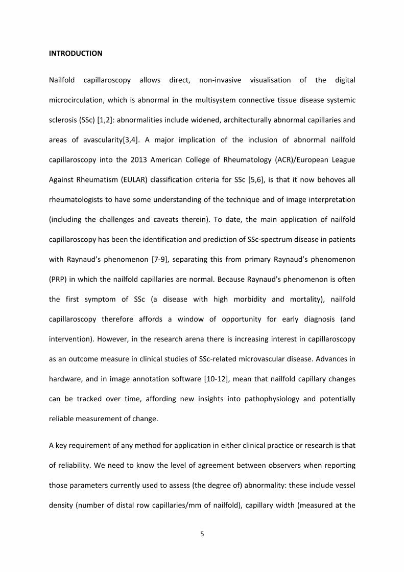
assessment due to the spatial heterogeneity of the capillary architecture, as shown in Figure
1.
Figure 1. Example mosaic nailfold images. These demonstrate the heterogeneity of capillary
architecture and why it is important to view the whole nailfold. Panel a) displays normal

9
capillaries on the left but some avascularity, capillary enlargement and distortion of the
vessel bed on the right. The lower panel, b), shows avascularity and evidence of
neoangiogenisis on the left, while on the right there are mainly greatly enlarged or giant
vessels with generally regular shape and spacing. In both examples, consideration of only
one half of the nailbed would give a different 'result' from the other half.
10
Image analysis and grading
A group of 10 capillaroscopy experts (‘observers’) from seven European centres assessed
the images. Custom image mark-up software was written, allowing detailed, quantitative
image assessment using a set of pre-determined criteria (described in detail below). The
software allowed image batches to be delivered to each observer, and results to be
returned securely, via the internet. In order to reduce the possibility of any training effects,
observers assessed a test batch of 25 video capillaroscopy images (taken from subjects in a
previous capillaroscopy study) in order to familiarise themselves with the software interface
and scoring protocol. Following successful completion of the training, blinded observers
(blinded to all subject characteristics including finger and 'patient' group) were randomly
assigned batches (stratified by patient group, and with sufficient overlap of images between
observers to allow assessment of inter-observer reliability) of around 50 images at a time
(including some repeat images to allow assessment of intra-observer reliability); on
completion further images were then made available to them. Two observers were vascular
technicians (from two different centres) and (by prior agreement) were sent larger numbers
of images than the other observers. Observers were presented with each
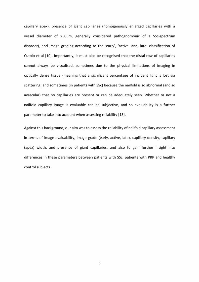
videocapillaroscopy image in turn (Figure 2) and asked to: (1) assess overall image grade, (2)
mark the location of distal capillary apices, (3) categorically grade the marked (distal) vessels
in terms of size ('Normal', 'Enlarged', 'Very enlarged' ['Giant' = homogeneously enlarged, or
'irregularly enlarged']) and shape ('Normal', 'Non-specific', or 'Angiogenic’), and (4) measure
the apical width of marked (distal) vessels using a click-and-drag tool. When assessing the
overall image grade, observers could choose from ‘Normal’, ‘Early’, ‘Active’, ‘Late’, or ‘Non-
specific’ (non-specific changes in vessel shape and/or size) categories. There were also two
11
‘Ungradeable’ categories for: (1) images considered to be so abnormal as to be
unclassifiable/unmeasurable and (2) poor image quality. It should be noted that vessels
marked as “Giant” in size, were automatically assigned a “Normal” shape.
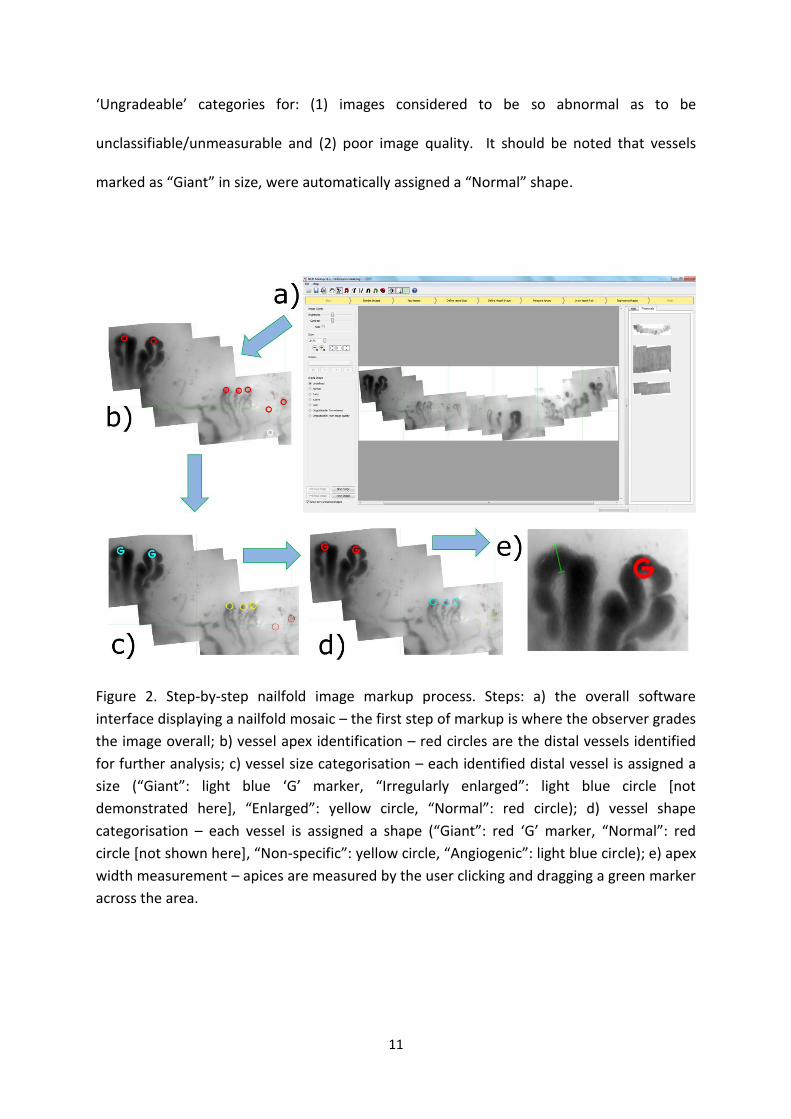
Figure 2. Step-by-step nailfold image markup process. Steps: a) the overall software
interface displaying a nailfold mosaic – the first step of markup is where the observer grades
the image overall; b) vessel apex identification – red circles are the distal vessels identified
for further analysis; c) vessel size categorisation – each identified distal vessel is assigned a
size (“Giant”: light blue ‘G’ marker, “Irregularly enlarged”: light blue circle [not
demonstrated here], “Enlarged”: yellow circle, “Normal”: red circle); d) vessel shape
categorisation – each vessel is assigned a shape (“Giant”: red ‘G’ marker, “Normal”: red
circle [not shown here], “Non-specific”: yellow circle, “Angiogenic”: light blue circle); e) apex
width measurement – apices are measured by the user clicking and dragging a green marker
across the area.

12
13
Outcome measures and measure requirements
From the returned dataset, four image-level outcome measures were extracted or derived
to allow further analysis: (1) the observer’s overall grade (hereafter grade) of the image on a
4-point severity scale ranging from “normal” (assigned a value of 1), through
“early”(assigned 2) and “active” (assigned 3), to “late” (assigned 4). Images graded as “non-
specific”, or in either of the “ungradeable categories” were not considered; (2) capillary
density (hereafter density; vessels/mm) was calculated as the total number of distal vessels
divided by the Euclidean distance between the leftmost and rightmost vessels marked in the
image. Density was only calculated when 2 or more vessels where identified within an
image; (3) mean apex width (hereafter width) was defined as the arithmetic mean of the
measured apex width of all vessels measured in an image, conditional on at least one distal
vessel being identified and measured; (4) presence of giant capillaries (hereafter giants) was
defined as a binary variable, set TRUE if there was one or more vessels identified and
subsequently categorically marked as having size/shape in the “Giant” category, set FALSE
otherwise.
Statistical analysis
Reliability can be measured using an intra-class correlation coefficient, which expresses the
between subject variance as a proportion of the total variance. The dataset was a complex
structure with nailfolds nested both within subjects and observers. Numbers of images for
each subject also varied. For this reason variance components were estimated using a linear
mixed model with crossed random effects. All models included observer and patient
variation modelled with random effects of finger nested within patient and within observer.
14
In each evaluation, evaluability of the four outcome measures was recorded as a binary
variable indicating whether the measure requirement had been met (i.e. measure
availability). A mixed-effects model was used to estimate the mean probability of an
observer providing a usable value of each measure for an image. A low availability or
probability indicates a poor measure.
Reliability and validity of measures can only be assessed were data is available. These
analyses were therefore based on conditional models, i.e. including only images with a
usable value being provided. All models included covariates for finger, gender and age as
fixed-effects to account for systematic non-pathological differences. Models for reliability
estimates did not include any further fixed-effects, so between-patient variance
components could be estimated without reducing variance from severity of disease.
Reliability estimates thus reflect disease-related variation. Including fixed effects of hand
and fingers means these correlations are the average reliability stratified by finger and hand.
Validity and evaluability models additionally included disease group as a fixed-effect. A
Likelihood-ratio omnibus test was used to assess the evidence for differences across
diagnosis groups for each outcome measure. A semi-parametric bootstrap was used to
derive confidence intervals of estimates and differences between them; 1000 bootstrap
samples were used for each procedure. Analyses were performed using R [14], and the
package lme4 [15].
15
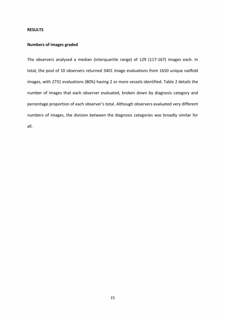
RESULTS
Numbers of images graded
The observers analysed a median (interquartile range) of 129 (117-167) images each. In
total, the pool of 10 observers returned 3401 image evaluations from 1650 unique nailfold
images, with 2731 evaluations (80%) having 2 or more vessels identified. Table 2 details the
number of images that each observer evaluated, broken down by diagnosis category and
percentage proportion of each observer’s total. Although observers evaluated very different
numbers of images, the division between the diagnosis categories was broadly similar for
all.
16
Table 2 – Number and proportion of images evaluated at baseline by observer and diagnosis
Observer
Healthy
Control
(number
[observer
percentage])
PRP (number
[observer
percentage])
SSc (number
[observer
percentage])
Total
Proportion of
total
evaluation
pool (%)
1 46 (27.2) 32 (18.9) 91 (53.8) 169 5.0
2 18 (18.4) 20 (20.4) 60 (61.2) 98 2.9
3 20 (16.7) 20 (16.7) 80 (66.7) 120 3.5
4 20 (17.5) 20 (17.5) 74 (64.9) 114 3.4
5 26 (21.8) 20 (16.8) 73 (61.3) 119 3.5
6 36 (26.3) 37 (27.0) 64 (46.7) 137 4.0
7 228 (31.3) 92 (12.6) 408 (56.0) 728 21.4
8 20 (17.2) 21 (18.1) 75 (64.7) 116 3.4
9 416 (25.4) 253 (15.4) 972 (59.2) 1641 48.3
10 33 (20.8) 37 (23.3) 89 (56) 159 4.7
Total image
evaluations
(mean
observer
percentage)
863 (22.3) 552 (18.7) 1986 (59.0) 3401 100.0
17
Of the 1650 unique, imaged nailfolds evaluated by at least one observer, 358 (22%) images
had a repeat evaluation by the same observer and 904 (57%) were evaluated by at least two
observers, with a total of 951 (59%) images contributing directly either to intra-observer or
inter-observer reliability parameter estimates for evaluability. Due to time constraints, it
was not possible to have all 1650 images assessed by 2 or more observers.
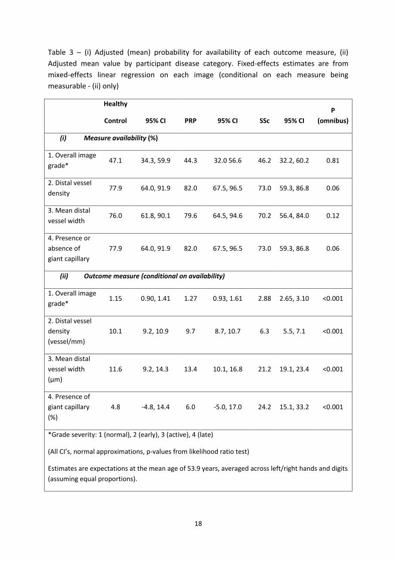
Evaluability (measure availability)
Table 3(i) shows the adjusted measure availability (as a percentage), along with 95%
confidence intervals, for each of the four outcome measures, subdivided by diagnosis. The
p-values in Table 3 relate to the omnibus test that all three groups are different, rather than
any particular pair-wise test between the three groups. Availability of measures varied
widely between observers. For outcome measures 2 and 4 (density and giant vessels), the
range was from 41.0% to 98.0% across the three diagnosis groups (P=0.06). For outcome
measure 3 (width), it was from 38.9% to 96.5% (P=0.12) and for measure 1 (grade), it was
from 18.5% to 78.1% (P=0.81). One observer was consistently and appreciably lower in
these rates than others, excluding this observer gave a minimum rate of availability for the
above figures of 34.3%, 52.0%, 51.7%, and 52.0%.
For the 3401 image evaluations producing an overall image grade, “Non-specific” was the
most frequent category: 34.6%, 28.3% and 38.9% for healthy controls, PRP and patients with
SSc respectively, with an “Ungradeable quality” given for 18.5%, 20.0% and 15.0% of image
evaluations for the respective diagnosis groups.
18
Table 3 – (i) Adjusted (mean) probability for availability of each outcome measure, (ii)
Adjusted mean value by participant disease category. Fixed-effects estimates are from
mixed-effects linear regression on each image (conditional on each measure being
measurable - (ii) only)
Healthy
Control 95% CI PRP 95% CI SSc 95% CI
P
(omnibus)
(i) Measure availability (%)
1. Overall image
grade* 47.1 34.3, 59.9 44.3 32.0 56.6 46.2 32.2, 60.2 0.81
2. Distal vessel
density 77.9 64.0, 91.9 82.0 67.5, 96.5 73.0 59.3, 86.8 0.06
3. Mean distal
vessel width 76.0 61.8, 90.1 79.6 64.5, 94.6 70.2 56.4, 84.0 0.12
4. Presence or
absence of
giant capillary
77.9 64.0, 91.9 82.0 67.5, 96.5 73.0 59.3, 86.8 0.06
(ii) Outcome measure (conditional on availability)
1. Overall image
grade* 1.15 0.90, 1.41 1.27 0.93, 1.61 2.88 2.65, 3.10 <0.001
2. Distal vessel
density
(vessel/mm)
10.1 9.2, 10.9 9.7 8.7, 10.7 6.3 5.5, 7.1 <0.001
3. Mean distal
vessel width
(µm)
11.6 9.2, 14.3 13.4 10.1, 16.8 21.2 19.1, 23.4 <0.001
4. Presence of
giant capillary
(%)
4.8 -4.8, 14.4 6.0 -5.0, 17.0 24.2 15.1, 33.2 <0.001
*Grade severity: 1 (normal), 2 (early), 3 (active), 4 (late)
(All CI’s, normal approximations, p-values from likelihood ratio test)
Estimates are expectations at the mean age of 53.9 years, averaged across left/right hands and digits
(assuming equal proportions).
19
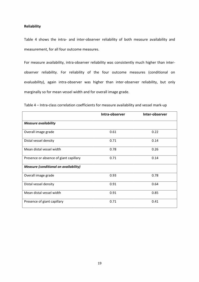
Reliability
Table 4 shows the intra- and inter-observer reliability of both measure availability and
measurement, for all four outcome measures.
For measure availability, intra-observer reliability was consistently much higher than inter-
observer reliability. For reliability of the four outcome measures (conditional on
evaluability), again intra-observer was higher than inter-observer reliability, but only
marginally so for mean vessel width and for overall image grade.
Table 4 – Intra-class correlation coefficients for measure availability and vessel mark-up
Intra-observer Inter-observer
Measure availability
Overall image grade 0.61 0.22
Distal vessel density 0.71 0.14
Mean distal vessel width 0.78 0.26
Presence or absence of giant capillary 0.71 0.14
Measure (conditional on availability)
Overall image grade 0.93 0.78
Distal vessel density 0.91 0.64
Mean distal vessel width 0.91 0.85
Presence of giant capillary 0.71 0.41
20
Outcome measures across clinical groups
Table 3(ii) shows the adjusted mean values for the four outcome measures, again
subdivided by diagnosis. These measures are conditional on their availability. After
adjustment the overall image grade (on a 1=normal to 4=late scale) was 0.11 units higher in
patients with PRP and 1.72 units higher in patients with SSc, compared to controls
(P<0.001). Mean vessel density was lower in both PRP and SSc groups by 0.4 vessels/mm
and 3.8 vessels/mm, respectively, compared to healthy controls (P<0.001). The adjusted
mean vessel width was 1.7 µm higher in patients with PRP, and 9.5 µm higher in patients
with SSc compared with healthy controls (P<0.001).
In images judged to be evaluable, the presence of giant capillaries was noted in 4.8%, 6.0%,
and 24.2% of image observations for healthy controls, and patients with PRP and SSc,
respectively (P<0.001).
There was a clear association between overall image grade and diagnosis. When image
grade was provided on the severity scale (normal, early, active or late), it was likely to be
more severe in patients with SSc (16.9% evaluated as normal) compared to healthy controls
(89.2% normal) and patients with PRP (79.4% normal). Severity was more frequently
observed as active (35.1%) or late (33.2%) in patients with SSc than as normal (16.9%) or
early (14.8%).
21
DISCUSSION
Our study involved seven different European centres supplying ten observers who examined
1650 images covering a broad spectrum of normality and abnormality. The study highlights
the challenges and complexities of nailfold capillaroscopy image assessment. A key finding
was the high intra-observer reliability (conditional on evaluability) of overall image grade,
capillary density and apex width. Inter-observer reliability was also good, although lower,
for these measures, suggesting that they have potential as outcome measures in both
single-centre (i.e. single-observer) and multi-centre studies. However, our study has also
highlighted the real-world situation that some capillaroscopy images are judged as
unevaluable, with some disagreement amongst observers about evaluability. The reliability
findings above are conditional on capillaries being sufficiently well-visualised to allow
assessment. It should be noted, however, that observers assessed density in 80.3% of
evaluations and apex width in 76.1% of evaluations, suggestive of observer confidence when
using these quantitative measures. It is likely that evaluability is to some extent dependent
upon the task given to the observer. In a recent study from our department, 84% of images
could be classified, although observers were only asked to grade capillaries as ‘normal’,
‘mildly’, ‘definitely’ or ‘grossly’ abnormal [13]. In the present study, the task was more
complex.
The study also confirmed the age and sex-adjusted differences in capillary morphology
between patients with SSc, patients with PRP, and healthy controls, although this was not a
primary objective.
Various previous studies report reliability of high magnification nailfold capillaroscopy,
highlighting the implied need for standardisation [12,13,16-24]. Some included only two
22
observers [12,16,17,20,21]. A small number of these studies included assessment of
reliability of capillary density and/or apical width. Hudson et al reported high intra- and
inter-observer (two observers) reliability of density using a widefield microscope (50x
magnification) [25]. We previously reported high inter- and intra-observer reliability of
quantitative parameters in a six-observer/three-centre study examining panoramic nailfold
mosaics: ICCs (intra- and inter-observer, for evaluable images) for capillary density were
0.92 and 0.71 respectively and for apical width 0.97 and 0.94 respectively [18]. Sekiyama et
al reported ICCs for intra- and inter-observer reliability (two observers) for number of
capillaries per mm of 0.991 and 0.952 respectively for videocapillaroscopy, and 0.993 and
0.954 respectively for widefield capillaroscopy (10-25x magnification) [21]. Each observer
acquired new images (from all eight fingers), and so, unlike our current study, reliability
incorporated both image acquisition and analysis [21]. The current study had the advantage
of being based on a large panel of observers, with analysis including finger, patient gender
and age as covariates, thereby removing differences between images and subjects that
relate to these characteristics. A clinical trial would likely also control for these
characteristics, so our estimates of reliability may be more realistic for clinical trials.
Together, the current study, and those described, support capillary density and apical width
as reliable, quantifiable outcome measures.
Smith et al studied reliability of assessment of the ‘early’, ‘active’, ‘late’ grading
(patient-level data from eight fingers) [17]. A high proportion of images (almost half by one
observer) were rated ‘non-SSc-specific', meaning images were either normal or ‘non-
specific’. Given that all patients in the study had SSc it seems likely that most were therefore
‘non-specific’, consistent with the current study findings. In this current study, again a high
23
proportion of images was graded 'non-specific', meaning that although the image was not
normal, it did not fit neatly into the ordered grading scale of 'early', 'active' or 'late' and was
therefore excluded from the analysis, which used a numerical scoring system based on
image grading (1 for 'normal' through to 4 for 'late'). For this reason, quantitative
assessment for example with capillary density or width has advantages over a qualitative
scoring system.
A very recent study from Smith et al reported that when observers were asked to assess a
single capillary (‘normal’, ‘abnormal’ or ‘not evaluable’) an unspecified proportion of the
images were ranked ‘not evaluable’ [23]. This indicates the complexities of capillaroscopic
analysis, and how challenging interpretation can be at the individual capillary level as well as
at both the ‘patient’ level (averaging across fingers) [17,21] and image/’finger’ level (as in
the current study). A further challenge is the heterogeneity of 'normal' appearances.
Although it was not within the remit of this study to examine the variation in nailfold
capillary appearances across healthy control subjects, it is worth noting, for example, that
even in healthy controls, raters subjectively marked a small proportion of capillaries as
'giants'. It is worth highlighting the importance of understanding reliability for outcome
measures derived from nailfold capillaroscopy. For clinical practice, a very high level of inter-
observer reliability is needed for a measure to be used for diagnosis. Such high levels are
often difficult to achieve and so measures are interpreted alongside other information when
assessing the patient. However, in clinical trials, high levels of inter-observer reliability are
not essential. For example if intra-observer reliability is substantially higher than inter-
observer reliability, a single observer could be used although this may lack generalisability.
24
Alternatively, the assessment process could be stratified by observer so that observer is not
confounded with treatment (i.e. similar proportions from each treatment arm are assigned
to each observer). Another option is to use a panel of observers, with the average rating
used as the outcome. This improves both generalisability and reliability. The reliability of a
panel of independent observers can be determined from the inter-observer reliability using
the Spearman-Brown formula [26]. The inter-observer reliability for vessel density in the
current study was 0.64. If the outcome was e.g. vessel density averaged across four
independent observers, reliability would be 0.88 by this formula. An increased reliability
reduces the required sample sizes for clinical trials. Using an average across four observers
would then reduce the required sample size to 0.64/0.88; a 28% reduction in participant
numbers. See Fleiss (1986) for further discussion of these matters [27].
Our study had limitations. Its aim was to assess reliability of image interpretation only (not
including image acquisition). Reliability of outcome measures could only be estimated using
data from images judged as evaluable; reflecting the reality of the clinical setting. Observers
could choose which segments of nailfold to consider, although they were expected to
analyse as much of the panoramic nailfold as possible. A key challenge of capillaroscopic
assessment is the heterogeneity of capillary morphology even within one nailfold (Figure 1),
highlighting that the same section of nailfold should be examined each time [20].
Additionally, observers received no specific training (shown to improve reliability of
capillaroscopic assessment [19]) although all had extensive clinical and research experience
with capillaroscopy. Finally, although numbers included in the PRP group were small, the
main purpose of the study was to assess reliability of assessors in grading images. The PRP
25
group was included primarily to ensure that the full spectrum of nailfold appearance was
presented to the assessors, ranging from normal to extremely abnormal.
In summary, nailfold capillaroscopic analysis is challenging, in part due to the difficulty in
obtaining good quality images from all nailfolds and the variability between observers in
determining what constitutes an evaluable nailfold. However, capillary density and apical
width, parameters likely to be helpful in quantifying changes over time, were reliable
measures in evaluable nailbeds. Quantitative capillaroscopic parameters were recently
confirmed of value in prediction of disease progression.[28-29] We are therefore optimistic
that, through a combination of standardised training and increased use of automated
analysis [30], the non-invasive technique of videocapillaroscopy will ultimately deliver as an
outcome measure of SSc-related microangiopathy.
ACKNOWLEDGMENT
The authors are grateful to Paula Pyrkotsch for her assistance with image analysis.
FUNDING
This work was supported by Scleroderma & Raynaud’s UK (grant number: MU3).

26
27
REFERENCES
1. Campbell PM, LeRoy EC. Pathogenesis of systemic sclerosis: a vascular hypothesis. Semin
Arthritis Rheum 1975: 4: 351-68.
2. Herrick AL. Vascular function in systemic sclerosis. Curr Opin Rheumatol 2000; 12: 527-
33.
3. Maricq HR, LeRoy EC. Patterns of finger capillary abnormalities in connective tissue
disease by 'wide-field' microscopy. Arthritis Rheum 1973; 16: 619-28.
4. Cutolo M, Grassi W, Matucci Cerinic M. Raynaud’s phenomenon and the role of
capillaroscopy. Arthritis Rheum 2003; 11: 3023-30.
5. Van den Hoogen F, Khanna D, Fransen J, et al. 2013 Classification criteria for systemic
sclerosis. Arthritis Rheum 2013; 65: 2737-47.
6. Van den Hoogen F, Khanna D, Fransen J, et al. 2013 Classification criteria for systemic
sclerosis. Ann Rheum Dis 2013; 72: 1747-55.
7. Koenig M, Joyal F, Fritzler MJ, et al. Autoantibodies and microvascular damage are
independent predictive factors for the progression of Raynaud's phenomenon to systemic
sclerosis: a twenty-year prospective study of 586 patients, with validation of proposed
criteria for early systemic sclerosis. Arthritis Rheum. 2008;58:3902-12.
8. Ingegnoli F, Boracchi P, Gualtierotti R, et al. Improving outcome prediction of systemic
sclerosis from isolated Raynaud’s phenomenon: role of autoantibodies and nail-fold
capillaroscopy. Rheumatol. 2010; 49, 797–805.
9. Herrick AL, Cutolo M. Clinical implications from capillaroscopic analysis in patients with
Raynaud's phenomenon and systemic sclerosis. Arthritis Rheum 2010; 62: 2595-604.
28
10. Cutolo M, Sulli A, Smith V. How to perform and interpret capillaroscopy. Best Practice
Res Clin Rheumatol 2013; 27: 237-48.
11. Allen PD, Taylor CJ, Herrick AL, et al. Image analysis of nailfold capillary patterns from
video sequences. Proceedings of Medical Image Computing and Computer-Assisted
Intervention, Springer, 1999; 698-705.
12. Anderson ME, Allen PD, Moore T, et al. Computerized nailfold video capillaroscopy - a
new tool for assessment of Raynaud’s phenomenon. J Rheumatology 2005; 32: 841-8.
13. Hughes M, Moore T, O’Leary N, et al. A study comparing videocapillaroscopy and
dermoscopy in the assessment of nailfold capillaries in patients with systemic sclerosis-
spectrum disorders. Rheumatology 2015; 54: 1435-42.
14. R Core Team. R: A Language and Environment for Statistical Computing [computer
program]. Vienna, Austria: Foundation for Statistical Computing, 2013. http://www.r-
project.org/.
15. Bates D, Mächler M, Bolker B, et al. Fitting linear mixed-effects models using lme4. J Stat
Softw 2015; 67(1), 1-48. doi:10.18637/jss.v067.i01.
16. Ingegnoli F, Gualtierotti R, Lubatti C, Zahalkova L, Meani L, Boracchi B, et al. Feasibility of
different capillaroscopic measures for identifying nailfold microvascular alterations. Semin
Arthritis Rheum 2009; 38: 289-95.
17. Smith V, Pizzorni C, De Keyser F, et al. Reliability of the qualitative and semiquantitiave
nailfold videocapillaroscopy assessment in a systemic sclerosis cohort: a two-centre study.
Ann Rheum Dis 2010; 69: 1092-6.
29
18. Hofstee HMA, Serne EH, Roberts C, et al. A multicentre study on the reliability of
qualitative and quantitative nail-fold videocapillaroscopy assessment. Rheumatol 2012; 51 :
749-55.
19. Gutierrez M, Bertolazzi C, Tardella M, et al. Interreader reliability in assessment of
nailfold capillary abnormalities by beginners: pilot study of an intensive videocapillaroscopy
training programme. J Rheumatol 2012; 39: 1248-55.
20. Murray AK, Vail A, Moore TL, et al. The influence of measurement location on reliability
of quantitative videocapillaroscopy in patients with SSc. Rheumatol 2012; 51 : 1323-30.
21. Sekiyama JY, Camargo CZ, Eduardo L, et al. Reliability of widefield nailfold capillaroscopy
and videocapillaroscopy in the assessment of patients with Raynaud’s phenomenon.
Arthritis Care Res 2013; 65: 1853-61.
22. Overbury R, Murtaugh MA, Fisher A, et al. Primary care assessment of capillary
abnormalities in patients with Raynaud’s phenomenon. Clin Rheumatol 2015; 34: 2135-40.
23. Smith V, Beeckman S, Herrick AL, et al. 1 An EULAR study group pilot study on reliability
of simple capillaroscopic definitions to describe capillary morphology in rheumatic diseases.
Rheumatol 2016; 55: 883-90.
24. Cheng C, Lee CW, Deskalakis C. A reproducible computerized method for quantitation of
capillary density using nailfold capillaroscopy. J Vis Exp 2015; 105: doi: 10.3791/53088.
25. Hudson M, Masetto A, Steele R, et al. Reliability of widefield capillary microscopy to
measure nailfold capillary density in systemic sclerosis. Clin Exp Rheumatol 2010; 28 (5
Suppl 62): S36-S41.
30
26. Spearman, CC C. Correlation calculated from faulty data. Br J Psychol 1910; 3: 271–95.
27. Fleiss, J L. Design and Analysis of Clinical Experiments. New York: John Wiley & Sons
1986.
28. Trombetta AC, Smith V, Pizzorni C et al. Quantitative alterations of capillary diameter
have a predictive value for development of the capillaroscopic systemic sclerosis pattern. J
Rheumatol. 2016; 43(3):599-606.
29. Cutolo M, Herrick AL, Distler O et al. Nailfold videocapillaroscopic and other clinical risk
factors for digital ulcers in systemic sclerosis: a multicenter, prospective cohort study.
Arthritis Rheumatol. 2016 Apr 25. doi: 10.1002/art.39718
30. Berks M, Tresadern P, Dinsdale G, et al. An automated system for detecting and
measuring nailfold capillaries. Medical Image Computing & Computer-Assisted Intervention:
MICCAI. 2014; 17: 658-65.
Recommended