Ligament Injuries to the Knee
C. Benjamin Ma, M.D.Associate Professor
Chief, UCSF Sports Medicine and ShoulderDepartment of Orthopaedic Surgery
University of California, San Francisco
Knee Ligaments
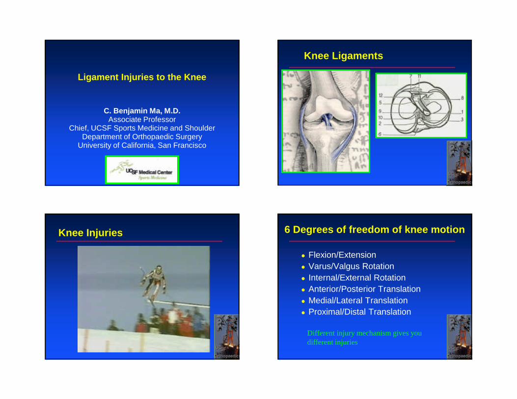
Knee Injuries 6 Degrees of freedom of knee motion
� Flexion/Extension� Varus/Valgus Rotation� Internal/External Rotation� Anterior/Posterior Translation� Medial/Lateral Translation� Proximal/Distal Translation
Different injury mechanism gives you different injuries
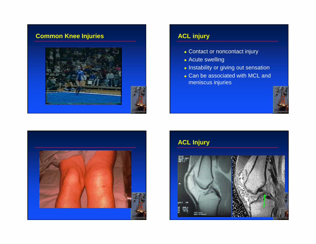
Common Knee Injuries ACL injury
� Contact or noncontact injury� Acute swelling� Instability or giving out sensation� Can be associated with MCL and
meniscus injuries
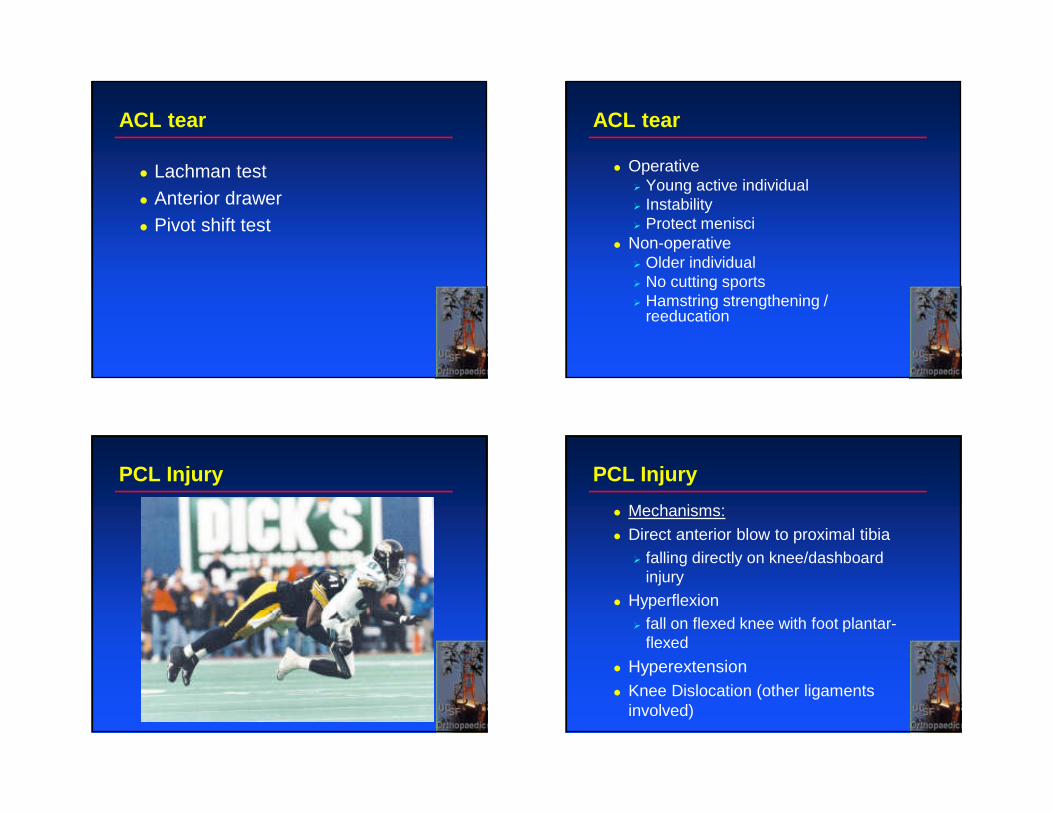
ACL Injury
� Add nml and inj MRI
ACL tear
� Lachman test� Anterior drawer� Pivot shift test
ACL tear
� Operative� Young active individual� Instability� Protect menisci
� Non-operative� Older individual� No cutting sports� Hamstring strengthening /
reeducation
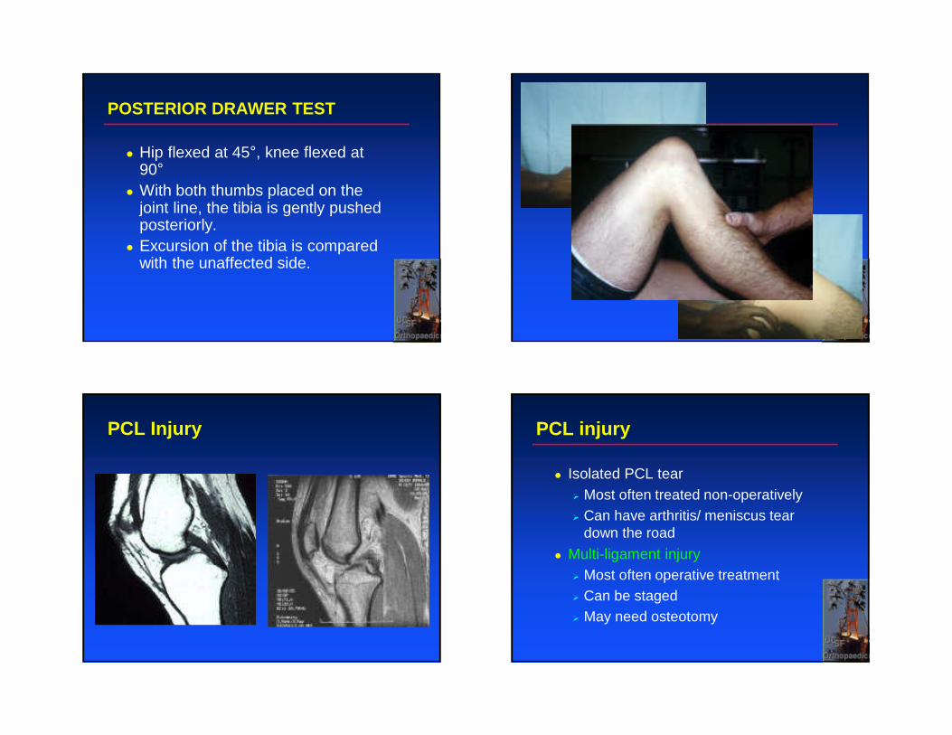
PCL Injury PCL Injury
� Mechanisms:
� Direct anterior blow to proximal tibia� falling directly on knee/dashboard
injury
� Hyperflexion� fall on flexed knee with foot plantar-
flexed
� Hyperextension� Knee Dislocation (other ligaments
involved)
POSTERIOR DRAWER TEST
� Hip flexed at 45°, knee flexed at 90°
� With both thumbs placed on the joint line, the tibia is gently pushed posteriorly.
� Excursion of the tibia is compared with the unaffected side.
PCL Injury PCL injury
� Isolated PCL tear� Most often treated non-operatively� Can have arthritis/ meniscus tear
down the road
� Multi-ligament injury� Most often operative treatment� Can be staged� May need osteotomy
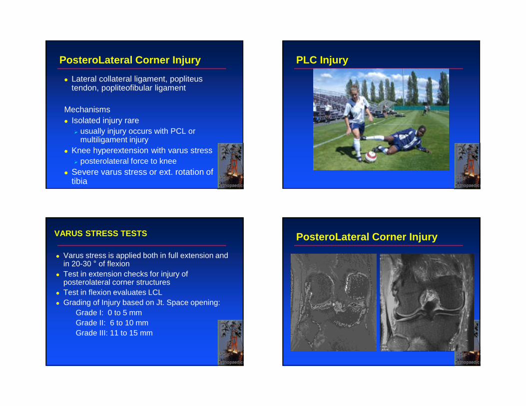
PosteroLateral Corner Injury
� Lateral collateral ligament, popliteustendon, popliteofibular ligament
Mechanisms� Isolated injury rare
� usually injury occurs with PCL or multiligament injury
� Knee hyperextension with varus stress� posterolateral force to knee
� Severe varus stress or ext. rotation of tibia
PLC Injury
VARUS STRESS TESTS
� Varus stress is applied both in full extension and in 20-30 ° of flexion
� Test in extension checks for injury of posterolateral corner structures
� Test in flexion evaluates LCL� Grading of Injury based on Jt. Space opening:
Grade I: 0 to 5 mmGrade II: 6 to 10 mmGrade III: 11 to 15 mm
PosteroLateral Corner Injury
Posterolateral Corner Injury
� For acute complete rupture� Want to treat this operatively within
3 weeks of injury
� Repair is better than reconstruction
� Reconstruction for more chronic injuries (>3 weeks) or more servere injury
Don’t want to miss this one!
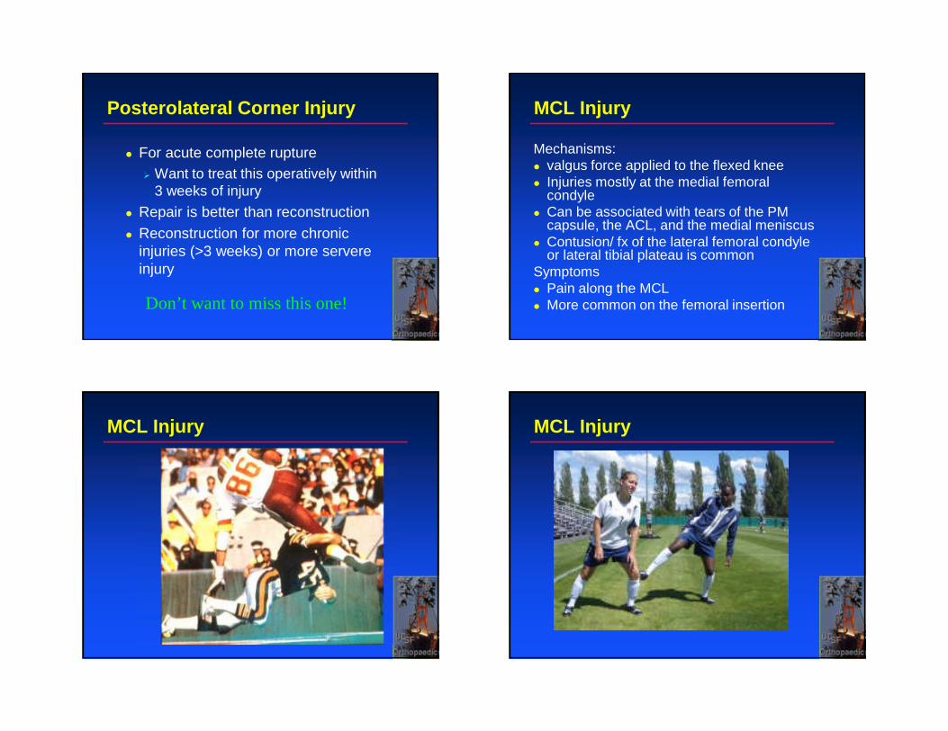
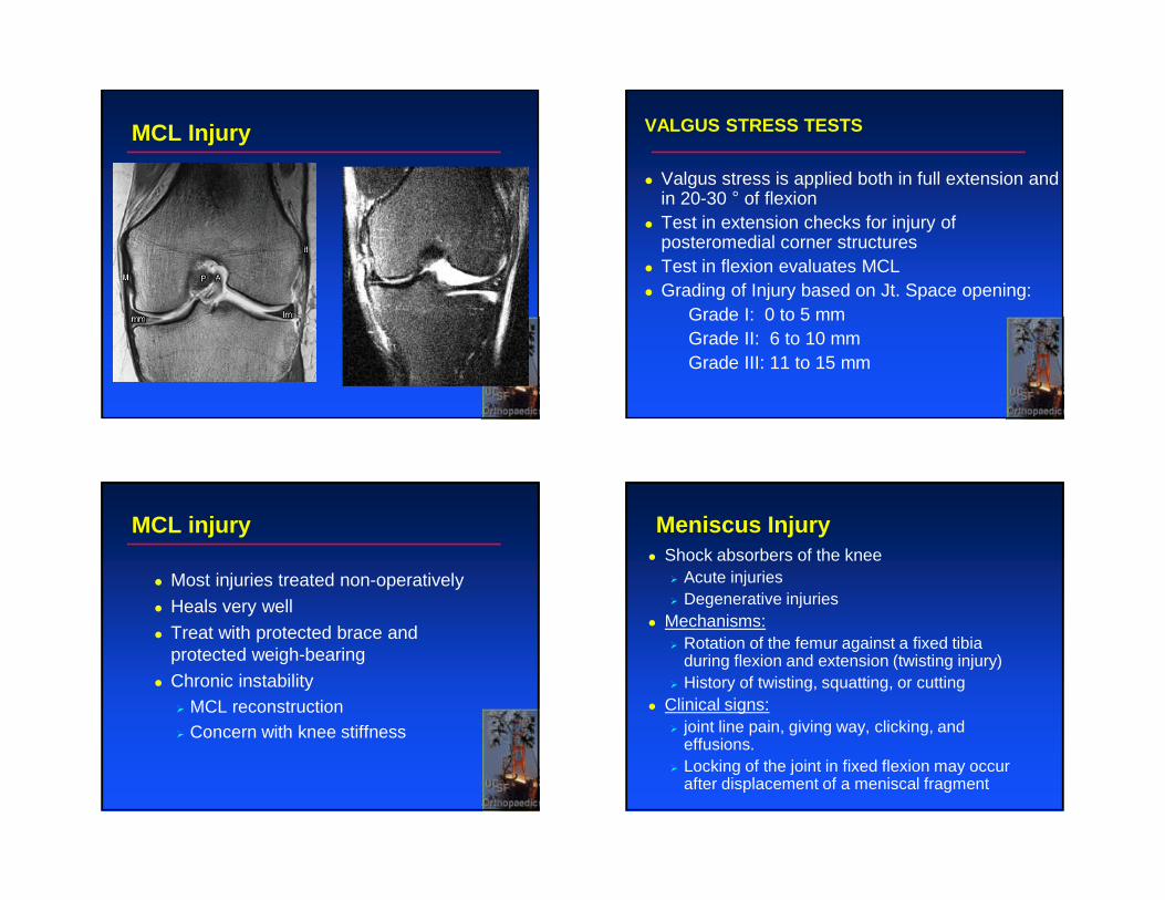
MCL Injury
Mechanisms:� valgus force applied to the flexed knee� Injuries mostly at the medial femoral
condyle� Can be associated with tears of the PM
capsule, the ACL, and the medial meniscus � Contusion/ fx of the lateral femoral condyle
or lateral tibial plateau is common Symptoms� Pain along the MCL � More common on the femoral insertion
MCL Injury MCL Injury
MCL Injury VALGUS STRESS TESTS
� Valgus stress is applied both in full extension and in 20-30 ° of flexion
� Test in extension checks for injury of posteromedial corner structures
� Test in flexion evaluates MCL� Grading of Injury based on Jt. Space opening:
Grade I: 0 to 5 mmGrade II: 6 to 10 mmGrade III: 11 to 15 mm
MCL injury
� Most injuries treated non-operatively
� Heals very well
� Treat with protected brace and protected weigh-bearing
� Chronic instability� MCL reconstruction� Concern with knee stiffness
Meniscus Injury� Shock absorbers of the knee
� Acute injuries� Degenerative injuries
� Mechanisms:� Rotation of the femur against a fixed tibia
during flexion and extension (twisting injury)� History of twisting, squatting, or cutting
� Clinical signs:� joint line pain, giving way, clicking, and
effusions. � Locking of the joint in fixed flexion may occur
after displacement of a meniscal fragment
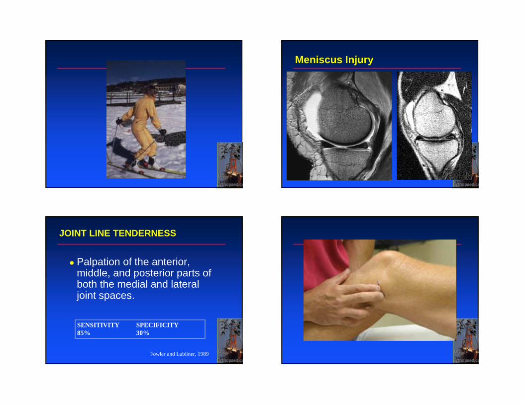
Meniscus Injury
JOINT LINE TENDERNESS
� Palpation of the anterior, middle, and posterior parts of both the medial and lateral joint spaces.
SENSITIVITY SPECIFICITY85% 30%
Fowler and Lubliner, 1989
MCMURRAY’S TEST
� Knee is flexed and placed in external rotation� Examiner applies a valgus or varus force� Knee is then extended. � (+) = Pain and/or a popping/ snapping sensation.
SENSITIVITY SPECIFICITY29% 96%
Fowler and Lubliner, 1989
MCMURRAY’S TEST
McMurray TP: The Semilunar Cartilages.
Br J Surg 29: 407-414, 1942
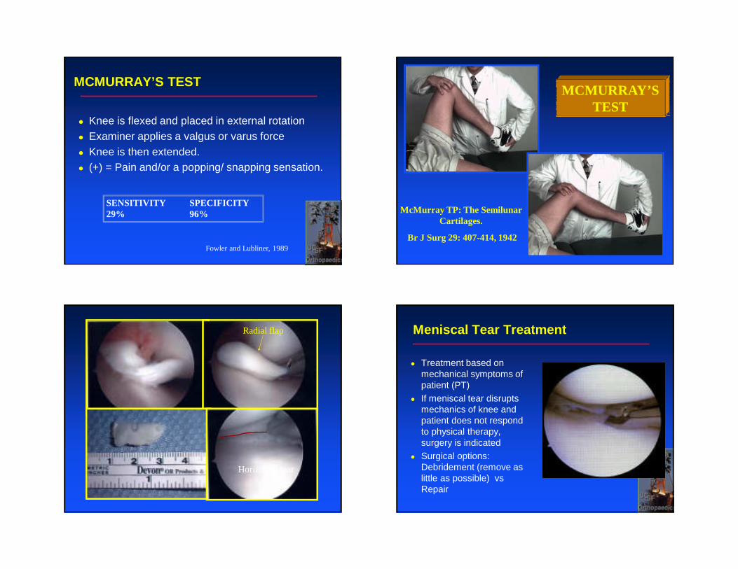
Radial flap
Horizontal tear
Meniscal Tear Treatment
� Treatment based on mechanical symptoms of patient (PT)
� If meniscal tear disrupts mechanics of knee and patient does not respond to physical therapy, surgery is indicated
� Surgical options: Debridement (remove as little as possible) vsRepair
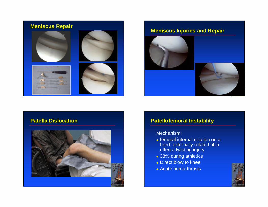
Meniscus RepairMeniscus Injuries and Repair
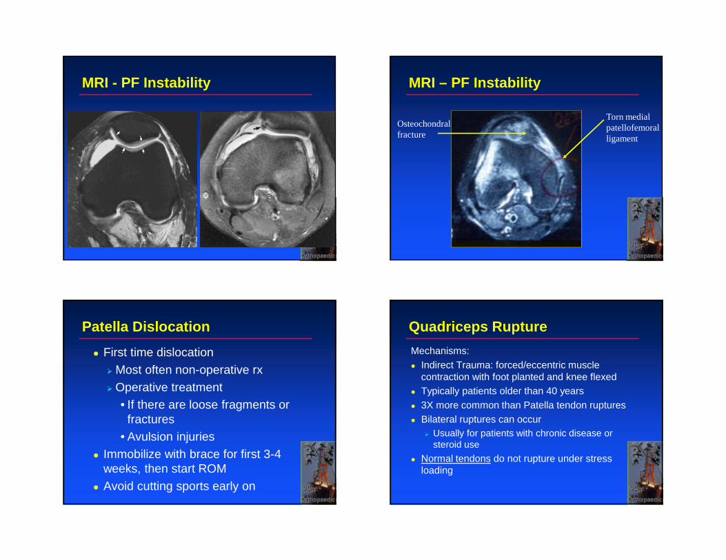
Patella Dislocation Patellofemoral Instability
Mechanism:� femoral internal rotation on a
fixed, externally rotated tibia often a twisting injury� 38% during athletics� Direct blow to knee� Acute hemarthrosis
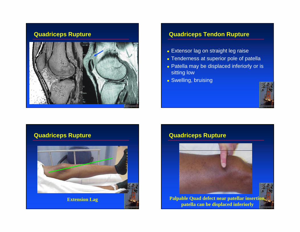
MRI - PF Instability MRI – PF Instability
Torn medialpatellofemoral ligament
Osteochondralfracture
Patella Dislocation
� First time dislocation
� Most often non-operative rx
� Operative treatment• If there are loose fragments or
fractures • Avulsion injuries
� Immobilize with brace for first 3-4 weeks, then start ROM
� Avoid cutting sports early on
Quadriceps Rupture
Mechanisms:� Indirect Trauma: forced/eccentric muscle
contraction with foot planted and knee flexed� Typically patients older than 40 years� 3X more common than Patella tendon ruptures� Bilateral ruptures can occur
� Usually for patients with chronic disease or steroid use
� Normal tendons do not rupture under stress loading
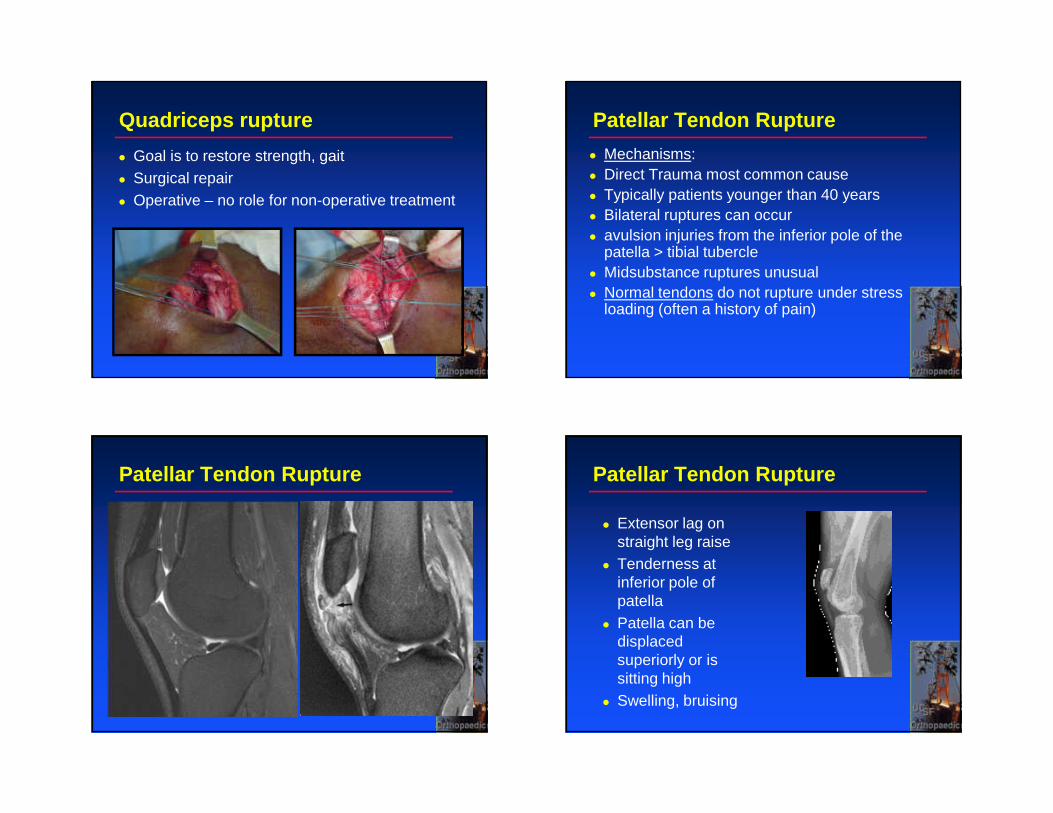
Quadriceps Rupture Quadriceps Tendon Rupture
� Extensor lag on straight leg raise� Tenderness at superior pole of patella� Patella may be displaced inferiorly or is
sitting low� Swelling, bruising
Quadriceps Rupture
Extension Lag
Quadriceps Rupture
Palpable Quad defect near patellar insertion, patella can be displaced inferiorly
Quadriceps rupture
� Goal is to restore strength, gait� Surgical repair� Operative – no role for non-operative treatment
Patellar Tendon Rupture
� Mechanisms:� Direct Trauma most common cause� Typically patients younger than 40 years� Bilateral ruptures can occur� avulsion injuries from the inferior pole of the
patella > tibial tubercle� Midsubstance ruptures unusual � Normal tendons do not rupture under stress
loading (often a history of pain)
Patellar Tendon Rupture Patellar Tendon Rupture
� Extensor lag on straight leg raise
� Tenderness at inferior pole of patella
� Patella can be displaced superiorly or is sitting high
� Swelling, bruising
Patella Tendon Rupture
� No role of non-operative treatment� Acute loss of extensor function
� Operative intervention
� Brace for 8-10 weeks� Rehabilitation
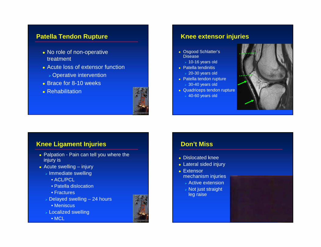
Knee extensor injuries
� Osgood Schlatter’s Disease� 10-16 years old
� Patella tendinitis� 20-30 years old
� Patella tendon rupture� 30-40 years old
� Quadriceps tendon rupture� 40-60 years old
Knee Ligament Injuries
� Palpation - Pain can tell you where the injury is
� Acute swelling – injury� Immediate swelling
• ACL/PCL• Patella dislocation• Fractures
� Delayed swelling – 24 hours• Meniscus
� Localized swelling• MCL
Don’t Miss
� Dislocated knee� Lateral sided injury� Extensor
mechanism injuries� Active extension� Not just straight
leg raise
C. Benjamin [email protected]
Recommended