See discussions, stats, and author profiles for this publication at: https://www.researchgate.net/publication/260374405
Light Influences How the Fungal Toxin Deoxynivalenol Affects Plant Cell
Death and Defense Responses
Article in Toxins · February 2014
DOI: 10.3390/toxins6020679 · Source: PubMed
CITATIONS
11READS
186
9 authors, including:
Some of the authors of this publication are also working on these related projects:
Biotechnology Ignition Grant (BIG)- Novel fermentation process for fragrant agarwood oil production View project
Exploring evolution of disease responce in wheat View project
Khairul I Ansari
City of Hope National Medical Center
81 PUBLICATIONS 2,635 CITATIONS
SEE PROFILE
Siamsa M Doyle
Umeå Plant Science Centre
24 PUBLICATIONS 439 CITATIONS
SEE PROFILE
Joanna Kacprzyk
University College Dublin
21 PUBLICATIONS 388 CITATIONS
SEE PROFILE
Mojibur R. Khan
Institute of Advanced Study in Science and Technology
95 PUBLICATIONS 1,532 CITATIONS
SEE PROFILE
All content following this page was uploaded by Stephanie Walter on 21 February 2014.
The user has requested enhancement of the downloaded file.
Toxins 2014, 6, 679-692; doi:10.3390/toxins6020679
toxins ISSN 2072-6651
www.mdpi.com/journal/toxins
Article
Light Influences How the Fungal Toxin Deoxynivalenol Affects Plant Cell Death and Defense Responses
Khairul I. Ansari 1,†, Siamsa M. Doyle 2,†, Joanna Kacprzyk 3, Mojibur R. Khan 4,
Stephanie Walter 5, Josephine M. Brennan 6, Chanemouga Soundharam Arunachalam 3,
Paul F. McCabe 3 and Fiona M. Doohan 3,*
1 Neurosurgery, Brigham and Women’s Hospital, Harvard Medical School, 4 Blackfan Circle,
Boston, MA 02115, USA; E-Mail: [email protected] 2 Department of Forest Genetics and Plant Physiology, Umeå Plant Science Centre,
Swedish University of Agricultural Sciences (SLU), Umeå 90 183, Sweden;
E-Mail: [email protected] 3 UCD Earth Institute and School of Biology and Environmental Science, College of Science,
University College Dublin, Belfield, Dublin 4, Ireland; E-Mails: [email protected] (J.K.);
[email protected] (C.S.A.); [email protected] (P.F.M.) 4 Institute of Advanced Study in Science and Technology, Guwahati-35, India;
E-Mail: [email protected] 5 Department of Integrated Pest Management, Research Centre Flakkebjerg, Forsøgsvej 1, Slagelse
DK-4200, Denmark; E-Mail: [email protected]
6 Plant Health Laboratory, Department of Agriculture and Food, Backweston, Co. Kildare, Ireland:
E-Mail: [email protected]
† These authors contributed equally to this work.
* Author to whom correspondence should be addressed; E-Mail: [email protected];
Tel.: 00353-1-7162248; Fax: 00353-1-7161102.
Received: 3 December 2013; in revised form: 6 February 2014 / Accepted: 8 February 2014 /
Published: 20 February 2014
Abstract: The Fusarium mycotoxin deoxynivalenol (DON) can cause cell death in wheat
(Triticum aestivum), but can also reduce the level of cell death caused by heat shock in
Arabidopsis (Arabidopsis thaliana) cell cultures. We show that 10 μg mL−1 DON does not
cause cell death in Arabidopsis cell cultures, and its ability to retard heat-induced cell death
is light dependent. Under dark conditions, it actually promoted heat-induced cell death.
Wheat cultivars differ in their ability to resist this toxin, and we investigated if the ability
OPEN ACCESS

Toxins 2014, 6 680
of wheat to mount defense responses was light dependent. We found no evidence that light
affected the transcription of defense genes in DON-treated roots of seedlings of two wheat
cultivars, namely cultivar CM82036 that is resistant to DON-induced bleaching of
spikelet tissue and cultivar Remus that is not. However, DON treatment of roots led to
genotype-dependent and light-enhanced defense transcript accumulation in coleoptiles.
Wheat transcripts encoding a phenylalanine ammonia lyase (PAL) gene (previously
associated with Fusarium resistance), non-expressor of pathogenesis-related genes-1
(NPR1) and a class III plant peroxidase (POX) were DON-upregulated in coleoptiles of
wheat cultivar CM82036 but not of cultivar Remus, and DON-upregulation of these
transcripts in cultivar CM82036 was light enhanced. Light and genotype-dependent
differences in the DON/DON derivative content of coleoptiles were also observed. These
results, coupled with previous findings regarding the effect of DON on plants, show that
light either directly or indirectly influences the plant defense responses to DON.
Keywords: Arabidopsis; β-1,3-glucanase; cell death; Fusarium; light; non-expressor of
pathogenesis-related genes-1 (NPR1); peroxidase; phenylalanine ammonia lyase; wheat
1. Introduction
Fusarium graminearum Schwabe [teleomorph Gibberella zeae (Schweinitz) Petch] and
F. culmorum (W.G. Smith) Saccardo cause diseases on the roots, stems and heads of cereal plants [1].
Fusarium head blight (FHB) receives significant attention because of both the yield losses and
mycotoxin contamination of grain associated with this disease. F. graminearum and F. culmorum
commonly produce the trichothecene mycotoxin deoxynivalenol (DON) in infected plant tissue and
this toxin acts as an aggressiveness factor for the pathogen during the development of root rot and
FHB disease [2,3]. DON inhibits protein synthesis and its effect on wheat (Triticum aestivum L.) head
tissue is similar to that of FHB disease, in that it bleaches the tissue [4,5]. Wheat genotypes differ in
their response to DON; resistance to DON-induced bleaching is associated with resistance to the
spread of FHB disease (type II resistance to FHB), but not with resistance to Fusarium infection (type I
resistance to FHB) [4].
Studies have shown that DON treatment induces defense gene transcription in wheat [5–7], the
production of reactive oxygen species (ROS) and, thereafter, an increase in programmed cell death
(PCD) [6]. Diamond et al. [8], however, showed that lower levels of DON (10 vs. 100–200 μg mL−1)
and a DON-producing strain of F. graminearum did not cause cell death in Arabidopsis thaliana cell
cultures, but they did reduce the level of cell death caused by heat shock. The opposing effects of DON
on cell viability and death in Arabidopsis and wheat may be due to many factors, including the
differences in DON concentrations used. We postulated that it might in part be due to light-dependent
signaling. The Arabidopsis experiments were conducted using light-grown cell cultures (that contained
mature chloroplasts), while the wheat experiments were conducted using seedlings. Therefore, the
light exposure of cells was quite different, and it is known that DON-induced bleaching of barley
tissue is light dependent [9]. The opposing effects of DON on cell death in Arabidopsis vs. wheat may

Toxins 2014, 6 681
also reflect host-dependent responses to the toxin, or a specific type of resistance to DON that is
inherited in a genotype-dependent manner. DON resistance inherent to some wheat genotypes is
associated with the capacity to convert DON to the less toxic DON-3-glucoside and co-segregated
with the QTL Fhb1 [4]. This may be in and of itself a light-dependent phenomenon, because, in
Arabidopsis, a UDP glucosyltransferase (UGT) catalyzes the glucosylation of DON [10] and an
analysis of Arabidopsis microarray experiments available in public repositories shows that the
transcription of the encoding gene is light regulated.
The first objective of this work was to try and determine if light plays a role in how DON influences
plant cell death. We show the ability of DON and DON-producing F. graminearum to retard cell death
caused by heat shock in Arabidopsis cell cultures is light dependent, and that DON actually enhanced
heat-induced cell death in dark-grown cells. The fact that the effect of DON and DON-producing
Fusarium on cell viability was light dependent, coupled with the previously reported light-dependent
bleaching of barley leaves by DON [9], indicated that light might be an important determinant of the
plant response to this toxin and its producer fungi. The second objective was to determine if light
influences the ability of wheat seedlings to mount defense in response to DON treatment. Using
seedlings whose roots were treated with DON, we show that light does enhance defense transcript
accumulation in coleoptiles and increases the DON metabolite content of coleoptiles. The extent to
which light influences defense transcript accumulation and DON metabolite translocation in
DON-treated seedlings, however, is wheat genotype-dependent. The implications of these results
are discussed.
2. Results
2.1. The Effect of DON on the Viability of Heat-Shocked Arabidopsis Cell Cultures Is Light Dependent
We used heat as an abiotic cell death inducer and determined whether light was necessary for
DON-mediated inhibition of heat-induced cell death (a phenomenon previously discovered by
Diamond et al., [8]). We compared light- and dark-incubated Arabidopsis thaliana ecotype Landsberg
erecta cell cultures with respect to the effect of 10 μg mL−1 DON on heat-induced cell death. Light-
grown cultures contained mature chloroplasts, while dark-grown cultures contained plastids, as
determined by electron microscopy [11]. Cell cultures were treated with 10 μg mL−1 DON or water
(controls) 24 h prior to heat treatment (55 °C for 10 min). Cells were treated with fluorescein diacetate
and cell fluorescence and morphology (epifluorescent microscopy) were used to distinguish between
viable and non-viable cells and to determine whether non-viable cells displayed apoptotic-like cell
death morphology or necrotic morphology (Figure S1) [8]. Results showed that the effect of DON on cell
death was light-dependent. When cells were incubated in the dark (Figure 1A, B), DON pre-treatment did
not enhance the viability of heat-shocked cells. Indeed, when comparing DON and water-pretreated
cells examined 5 h post-heat treatment, toxin treatment resulted in 2.6-fold higher numbers of cells
exhibiting apoptotic-like cell death morphology and reduced cell viability by 5.94-fold (P ≤ 0.01).
However, in light-grown cells harvested 5 h post-heat treatment, DON, as compared to water, reduced
cell mortality (6.2 and 2.8-fold reductions, respectively in cells exhibiting apoptotic-like cell death and
necrotic morphology) and enhanced the level of cell viability (by 5.0-fold; P ≤ 0.02; Figure 1C). Another
interesting observation was that the ability of DON to inhibit heat-induced cell death in light-grown

Toxins 2014, 6 682
cultures was temporal. At 24 h post-treatment, the DON (relative to water) pretreatment did not
influence the viability of heat-shocked cells (P ≥ 0.31) (Figure 1D).
Figure 1. The effect of DON on the viability of heat-stressed Arabidopsis cells. Cells were
cultured under dark conditions (A and B) or light conditions (C and D) and were treated
with DON (10 μg mL−1) or water (controls) 24 h pre-heat treatment (55 °C, 10 min) and
cells were examined at either (A and C) 5 h, or (B and D) 24 h post-heat treatment. Cells
were treated with fluorescein diacetate and examined under phase contrast microscopy
with or without UV fluorescence (490 nm) in order to determine if cells were viable, or
non-viable and exhibiting either programmed cell death (PCD) or necrotic morphology.
Results represent the mean percentage (+/− standard error) of cells in a given state, based on
five independent experiments, and in each experiment 200 cells were scored per treatment
per time point. In control, water-treated, non-heat shocked cells, ≥84% were viable and ≤14
and 3% respectively displayed PCD or necrotic morphology in dark-/light-grown cultures at
the time points analyzed.
Diamond et al. [8] showed that, like DON, pre-treatment with DON-producing F. graminearum
reduced the level of PCD caused by subsequent heat treatment in Arabidopsis cell cultures. We conducted
similar experiments, where light- and dark-incubated Arabidopsis cell cultures were pre-treated with

Toxins 2014, 6 683
conidia of wild-type DON producing F. graminearum (strain GZ3639) or its DON-minus mutant
derivative (strain GZT40), 20 h prior to heat treatment (55 °C, 10 min). Cell viability was assessed at 5 h
post-heat treatment, as described above (Figure 2). For dark-grown cultures, neither the wild type nor
mutant Fusarium significantly affected either cell viability or the morphology of dead cells (P ≥ 0.30)
(Figure 2A). This contrasted with the DON which enhanced death under dark conditions (Figure 1). In
light grown cultures, the wild-type, but not the mutant, significantly enhanced cell viability (by 2.5-fold)
and reduced cell necrosis (by 1.6-fold; P ≤ 0.05; Figure 2B). Neither fungal strain significantly
influenced the level of apoptotic-like cell death morphology observed in heat-shocked cells at this time
point (P ≥ 0.27). Therefore we concluded that the effect of toxigenic F. graminearum on plant cell
death is light dependent, possibly dependent on chloroplasts, and under light conditions, the ability of
DON to prevent cell death caused by abiotic stress is temporal.
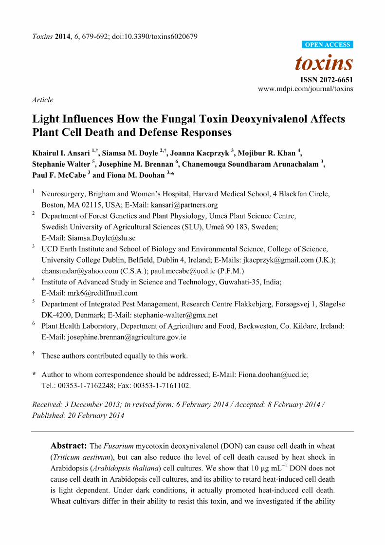
2.2. DON Induction of Defense Gene Expression in Wheat Is Light Enhanced and Genotype Dependent
The second objective was to determine if light influenced the wheat response to DON. In this study,
we used two wheat cultivars which differ with respect to the ability of their spikelets to resist DON;
cv. CM82036 is DON-resistant, while cv. Remus is susceptible [4,5]. The response analyzed was
defense gene expression, namely genes encoding POX, PAL, NPR1 and GLC1. These were analyzed
because a previous study showed that these were all DON-upregulated in wheat spikelets, and that
NPR1 transcription was more highly DON-upregulated in the toxin-treated spikelets of cv. CM82036,
as compared to cv. Remus ([12]). Furthermore, the upregulation of the PAL gene is associated with
two quantitative trait loci (QTL) that confers spikelets of cv. CM82036 with enhanced resistance to
both FHB and DON [13]. In this study, the roots of seedlings grown under light or dark conditions
were treated with DON and defense gene transcription in both roots and coleoptile was analyzed at 4
and 24 h post-treatment. The localized effect of DON on defense gene expression in roots of the two
cultivars was not light enhanced (results not shown). In coleoptiles of cv. Remus, GLC1 was the only
transcript that was significantly DON-upregulated; this phenomenon was light-enhanced (3.1-fold
higher in DON vs. water treated, light-grown cells observed 24 h post-treatment; P = 0.02) (Figure 3). In
coleoptiles of dark-grown cv. CM82036 seedlings, POX was the only transcript significantly
upregulated in response to DON treatment (2.3-fold at 24 h, P = 0.05). But, under light conditions, and
by 24 h post-root treatment, DON had transcriptionally upregulated all four defense genes in
coleoptiles of this cultivar (2.0–4.3-fold upregulation; P ≤ 0.02) (Figure 3). Moreover, the highest
transcript levels were detected at 24 as compared to 4 h post-treatment.
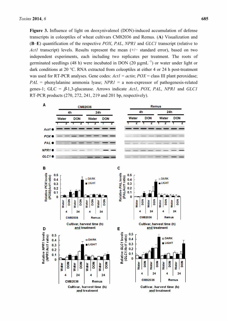
2.3. Both Genotype and Light Affect the Movement of DON Metabolites within Wheat Seedlings
Using an ELISA test, we determined the level of DON and DON derivatives in coleoptiles 24 h
post-root treatment with toxin. The test used did not discriminate between DON and DON 3-glucoside
(and thus compounds detected are described as DON metabolites). Both the incubation conditions
(light/dark) and wheat genotype influenced the level of DON metabolites detected within coleoptiles
(Figure 4). Coleoptiles of cv. CM82036 contained more DON metabolites than those of cv. Remus,
irrespective of incubation conditions (≥1.7-fold more; P = 0.003) and coleoptiles of cv. CM82036, but
not of cv. Remus, accumulated significantly more DON metabolites by this time when incubated under

Toxins 2014, 6 684
light as compared to dark conditions (1.4-fold more; P = 0.046). By 24 h post-DON treatment, neither
light nor cultivar-dependent differences in coleoptile dry weight were observed (mean = 32–35 mg;
P ≥ 0.20). However, coleoptiles of cv. CM82036 were more elongated than those of cv. Remus (results
not shown).
Figure 2. The effect of DON production by Fusarium graminearum on the viability of
heat-stressed Arabidopsis cells. Cells were cultured under dark conditions (A) or light
conditions (B) and were treated with either water, or conidia of either wild type,
DON-producing F. graminearum (strain GZ3639) or its mutant, non-DON-producing
derivative (strain GZT40). After 20 h, cells were incubated at either 23 or 55 °C for 10 min,
and thereafter at 23 °C. Cells were examined at 5 h post-heat treatment. Cells were treated
with fluorescein diacetate and examined under phase contrast microscopy with or without
UV fluorescence (490 nm) in order to determine if cells were viable, or non-viable and
exhibiting either programmed cell death (PCD) or necrotic morphology. Results represent
the mean percentage (+/− standard error) of cells in a given state, based on five
independent experiments, and in each experiment, a minimum of 200 cells were scored per
treatment. In control, water-treated, non-heat shocked cells, ≥94% were viable and ≤4.6 and
1.4% respectively displayed PCD or necrotic morphology in dark-/light-grown cultures.
Toxins 2014, 6 685
Figure 3. Influence of light on deoxynivalenol (DON)-induced accumulation of defense
transcripts in coleoptiles of wheat cultivars CM82036 and Remus. (A) Visualization and
(B–E) quantification of the respective POX, PAL, NPR1 and GLC1 transcript (relative to
Act1 transcript) levels. Results represent the mean (+/− standard error), based on two
independent experiments, each including two replicates per treatment. The roots of
germinated seedlings (48 h) were incubated in DON (20 μgmL−1) or water under light or
dark conditions at 20 °C. RNA extracted from coleoptiles at either 4 or 24 h post-treatment
was used for RT-PCR analyses. Gene codes: Act1 = actin; POX = class III plant peroxidase;
PAL = phenylalanine ammonia lyase; NPR1 = a non-expressor of pathogenesis-related
genes-1; GLC = β-1,3-glucanase. Arrows indicate Act1, POX, PAL, NPR1 and GLC1
RT-PCR products (270, 272, 241, 219 and 201 bp, respectively).
Toxins 2014, 6 686
Figure 4. The translocation of DON metabolites within seedlings of wheat cultivars
CM82036 and Remus. The roots of germinated seedlings (48 h) were incubated in DON
(20 μg mL−1). Coleoptiles were harvested 24 h post-treatment and DON was extracted and
quantified by ELISA analysis. Results represent the mean (+/− standard error), based on
two independent experiments, each including two replicates per treatment.
3. Discussion
This research has established that light is an important determinant of how plants cells respond to
the Fusarium mycotoxin DON and that wheat genotypes differ with respect to their ability to mount
light-dependent defense responses to DON. Plant defense responses against pathogens, including the
activation of PAL, the accumulation of SA, expression of PR proteins and the hypersensitive response,
are often light dependent [14,15–18]. Light can influence defense responses via its effects on chloroplast
metabolism, ROS generation and phytochrome signaling [19]. Light influences chloroplast function and
several lines of evidence point to the possible role of the chloroplast as an important determinant of the
plant response to DON. The light-grown Arabidopsis cells used in this study contained chloroplasts,
while the dark-grown did not. DON damage of chloroplasts is a light-dependent phenomenon [9]. This
could result in the accumulation of photosensitive pigments, and these can directly generate ROS in the
light [20]. DON has been shown to induce ROS production in plants [6]. Excess energy produced via the
photosynthetic electron transport chain [19] may contribute to the DON-induced ROS accumulation.
It is difficult to screen for the inhibition of cell death; such a phenomenon may be interpreted as a
null effect and would only be obvious in situations where cell death is induced by other factors, such
as heat stress. We thus used heat to induce PCD as it provided a means to activate this conserved
pathway in plants. The fact that DON and DON-production inhibits heat-induced PCD supports the
previous deduction from gene expression studies. Based on microarray studies, it was deduced that
ROS scavenging and the promotion of cell survival are key early defense strategies that are more
effectively employed by DON-resistant as compared to susceptible wheat genotypes [7]. It would be
logical for Fusarium-resistant plants to promote the survival, and hence the potential for defense, of
cells that are being attacked by necrotrophic Fusarium fungi that can colonize dead plant tissue.
Babaeizad et al. [21] showed that overexpression of a gene encoding a cell death suppressor, BAX
inhibitor-1, retarded F. graminearum colonisation of barley seedlings. While traditionally regarded as
Wheat cultivar
DO
N (μ
g m
g-1 dr
y w
eigh
t)

Toxins 2014, 6 687
a necrotroph, there is increasing evidence and belief that F. graminearum is actually a hemibiotrophic
pathogen, with a short biotrophic phase preceding the necrotrophic phase of disease spread. DON
suppression of death suggests that the role of DON may be to disable PCD during the initial biotrophic
infection stages in plant cells, with the accumulation of higher concentrations causing PCD and facilitating
nectrotrophism and disease spread. In the dark, DON enhanced the rate of apoptotic cell death. However,
the fungal DON-producing strain did not do so relative to the mutant strain. There are many possible
reasons for these contradictory results, including the variation in DON concentrations in toxin versus
fungal studies and the fact that fungal factors other than DON are also very likely to affect cell death.
This study showed that defense genes were light regulated and that NPR1, POX and PAL were more
DON responsive in seedlings of the DON-resistant cultivar CM82036, as compared to the susceptible
cultivar Remus. NPR1 and PAL genes have been associated with enhanced FHB resistance, but not
with enhanced DON resistance. NPR1 is a central regulator of plant defense responses, including
systemic acquired resistance (SAR), induced systemic resistance (ISR) and salicylic acid
(SA)/jasmonic acid (JA) cross-talk [22]. An Arabidopsis npr1-1 mutant was more susceptible to
F. graminearum and accumulated higher concentrations of DON in buds and flowers than did wild
type plants [23]. Overexpression of Arabidopsis NPR1 (AtNPR1) transcript in wheat conferred
heritable resistance to FHB disease spread, but not initial infection [24]. These data, together with the
facts that DON is associated with disease spread and that it induces the accumulation of NPR1
transcript in wheat, suggest that the NPR1 protein is linked to Type II resistance to FHB (resistance to
disease spread). In rice, overexpression of a NPR1 homolog led to the constitutive expression of
defense genes, including POX and PAL [25].
Steiner et al. [13] showed that a PAL transcript was Fusarium responsive in wheat spikelets and its
responsiveness was associated with the presence of two quantitative trait loci that confer enhanced
resistance to FHB, namely Fhb1 and Qfhs-ifa-5A. This transcript is 100% homologous to that used
herein for PCR primer design. The results of Steiner et al. [13] and the higher accumulation of
cinnamic acid, benzoic acid, and glutamine in F. graminearum-infected spikelets of the FHB resistant
wheat cv. Sumai-3 (a parent of cv. CM82036 that carries QTL Fhb1) than in spikelets of the
susceptible cv. Roblin [26] provide evidence that the phenylpropanoid pathway plays a role in host
defense against DON-producing Fusaria. QTL Fhb1 also confers wheat with enhanced resistance to
DON-induced bleaching of spikelets [4]. It is therefore likely that components of phenylpropanoid
pathway play a light-dependent role in wheat resistance to DON. This is independent of DON
conversion to DON-3-glucoside as Gunnaiah et al. [27] recently showed that Fhb1 derived from the
wheat genotype Nyubai is mainly associated with cell wall thickening due to deposition of
hydroxycinnamic acid amides, phenolic glucosides and flavonoids, but not with the conversion of
DON to less toxic DON-3-glucoside.
The cv. CM82036 differs from cv. Remus in its enhanced ability to convert DON to
DON-3-glucoside [4]. This derivative is detected by the ELISA test [28] and, based on spikelet studies [4],
it is likely to be the predominant DON metabolite in cv. CM82036, though not in cv. Remus coleoptiles. It
is possible that light affects the conversion of DON to DON-3-glucoside in a genotype-dependent manner.
The light-enhanced translocation in cv. CM82036 may be an indirect consequence of enhanced sugar
availability for the formation of the DON-3-glucoside. Whether or not translocated DON metabolites
contributed to the light-enhanced defense transcript accumulation in coleoptiles is unknown.

Toxins 2014, 6 688
4. Experimental Section
4.1. Maintenance, Growth and Treatment of Arabidopsis Cells
Arabidopsis thaliana (ecotype Landsberg erecta) cells were grown in liquid Murashige and Skoog
(MS) media ([29,30]. Cells were sub-cultured by pipetting 10 mL of culture into 100 mL of fresh
media every 7 days, and were grown on a rotary shaker at 100 rpm (5 cm rotation), a constant
temperature of 23 °C, and either in darkness or at a continuous light intensity of approximately
4 μmol photons m−2 s−1. DON (Sigma, UK) was dissolved in water at a concentration of 2000 μg mL−1
and stored at 4 °C. Conidia of Fusarium graminearum strain GZ3639 and its trichothecene-minus
mutant derivative (strain GZT40) [31] were produced as described previously [32] and adjusted to a
concentration of 5 × 10⁴ mL−1 H2O (fresh conidia were prepared for each experiment). Arabidopsis
cell samples (10 mL) were transferred to sterile 100 mL conical flasks and were treated with 0.5 mL
DON 24 h prior to heat treatment or with 2 mL of conidial inoculum 20 h prior to heat treatment.
Controls were treated with equivalent volumes of water. For heat treatment, cell culture flasks were
placed in a shaking water bath (80 rpm) that was pre-equilibrated to either 23 or 55 °C, for 10 min.
Between DON/fungal and heat treatment, and subsequent to heat treatment, samples were returned to
their prior growth conditions (as above, either light or dark). Samples were morphologically analyzed
at either 5 or 24 h after heat treatment. Five independent experiments compared the effect of DON and
water on heat/non-heat-treated cells, and another five compared the effect of F. graminearum wild
type, mutant and water on heat/non-heat-treated cells. Each experiment included one flask per
treatment per harvest time point and 200 cells were morphologically analyzed per treatment per time
point per experiment.
4.2. Morphological Analysis of Arabidopsis Cells
Cells were examined under a Leica DM LB microscope with an attached fluorescence lamp and
camera (Leica Microsystems GmBH, Wetzlar, Germany). Cells were scored as being viable or
non-viable and exhibiting either necrotic or apoptotic-like cell death morphology. The vital stain
fluorescein diacetate (FDA) was used to assay for live cells [30]. When FDA is excited by light at a
wavelength of 490 nm, a bright green fluorescence is observed in viable cells whose plasma membrane
is intact. Cells that die by necrosis do not display the protoplast retraction associated with apoptotic-like
cell death and do not fluoresce, while cells that have undergone apoptotic-like cell death show a
characteristic retraction of the protoplast away from the cell wall and cannot cleave FDA [8] (Figure S1).
4.3. Growth and Treatment of Wheat Seedlings
Wheat (Triticum aestivum) cvs. CM82036 and Remus were used in this study. Cultivar CM82036
carries a major quantitative trait locus (QTL) on the short arm of chromosome 3B that is associated
with resistance to both FHB disease and DON-induced bleaching of spikelets (Fhb1; syn. Qfhs.ndsu-3BS)
and it carries another QTL on chromosome 5A that is associated with FHB resistance but not with
DON resistance [4,33]. Cultivar Remus is susceptible to FHB and DON-induced bleaching [4,33].
Seeds were pre-germinated in the dark at 20 °C for 48 h in Petri dishes containing filter paper
moistened with 7 mL of sterile water (12 seeds per plate). Germinated seeds were then air-dried for

Toxins 2014, 6 689
10 min on filter paper and carefully placed in Petri dishes containing 7 mL of either water or DON
(20 μgmL−1 water) (12 seeds per plate) such that coleoptiles were not in contact with the treatment
solution. Wheat seedlings are less sensitive to DON than Arabidopsis cell cultures ([34]), hence the
reason for the higher concentration as compared to the Arabidopsis studies. Plates were incubated
at 20 °C under either constant darkness or constant light (~110 μmol m−2 s−1). The roots and coleoptile
were harvested at 4 or 24 h post-treatment, flash frozen in liquid N2 and stored at −70 °C prior to either
RNA or DON extraction. Seedling experiments conducted for RNA and DON analysis each included
two replica plates per treatment and each experiment was conducted twice.
4.4. Quantification of DON
Freeze-dried coleoptile tissue was homogenized as previously described [5]. DON/DON derivatives
was extracted from coleoptiles and quantified using the Ridascreen® DON Fast immunoassay
(R-Biopharm AG, Darmstadt, Germany) according to the manufacturer’s instructions. The antibodies
used in this assay detect DON and DON derivatives including the less phytotoxic DON-3-glucoside [27].
Values were based on the average obtained for two replicates per sample.
4.5. RNA Extraction and Gene-Specific RT-PCR Analyses
Freeze-dried root or coleoptile samples were homogenized and total RNA was extracted and
DNase1-treated as described by Ansari et al. [5]. Reverse transcription of total RNA was conducted as
described by Ansari et al. [5], except that the primer used was oligo dT12–18 (Life Technologies,
Paisley, UK). Both the genes of interest and wheat actin (Act1) were PCR-amplified (separately) using
gene-specific primers (Supplemental Table S1 lists GenBank accession numbers, gene-specific primer
sequences and expected product sizes); Act1 served as a control gene that was constitutively expressed
in roots, coleoptile and head tissue. RT products were diluted to 100 μL and 3 μL was PCR-amplified in
a 10 μL reaction containing 1 unit of Taq DNA polymerase and 1× PCR buffer (Life Technologies,
Paisley, UK), 1.5 mM MgCl2, 150 μM each of dATP, dGTP, dCTP and dTTP, and 100 nM each of
forward and reverse transcript-specific primers. PCR reactions were conducted in a Peltier thermal
cycler DNA engine (MJ Research, St. Bruno, Canada) and the programme constituted 30 cycles of 94 °C
for 30 s, 60 °C for 20 s and 72 °C for 45 s, with a final extension at 72 °C for 5 min. PCR products were
electrophoresed through 2% (w v−1) agarose gels containing 0.5 μg mL−1 ethidium bromide and
visualized using Imagemaster VDS and Liscap software (GE Healthcare Life Sciences,
Buckinghamshire, UK).
4.6. Data Analysis
All data analyses were conducted using Minitab (Minitab release 13©, 1994 Minitab Ltd, Coventry,
UK). No data set followed a normal distribution, as determined using the Normality test and none
could be transformed to fit a normal distribution using the Johnson Transformation tool.
Non-normally distributed data (cell viability and morphology data, gene expression data
(transcript/Act1 levels in DON relative to water-treated samples) and DON data) were analyzed using
the Mann-Whitney Rank sum test (confidence level 95%, alternatives of greater than, less than or
equal chosen as appropriate).

Toxins 2014, 6 690
5. Conclusions
We have shown that the effect of DON on the viability of abiotically stressed cells and on defense
gene expression in wheat is light enhanced. Future studies should investigate the role of cell death
suppression, cell survival pathways and light-regulated pathways in the resistance of wheat to
Fusarium fungi. The combination of DON and heat stress offers a valuable means by which to unravel
some of the complexity of plant programmed cell death.
Acknowledgements
This research was funded by Science Foundation Ireland Principal Investigator project (10-IN1-B3028)
and EU FP5 project FUCOMYR (QLRT-2000-02044). We thank Hermann Buerstmayr (IFA-Tulln,
Austria) for providing wheat seed and Robert Proctor (USDA Agricultural Research Service, Peoria,
IL, USA) for providing the Fusarium strains used in this work.
Conflicts of Interest
The authors declare no conflict of interest.
References
1. Parry, D.W.; Jenkinson, P.; McLeod, L. Fusarium ear blight (scab) in small-grain cereals—A
review. Plant Pathol. 1995, 44, 207–238.
2. Langevin, F.; Eudes, F.; Comeau, A. Effect of trichothecenes produced by Fusarium
graminearum during Fusarium head blight development in six cereal species. Eur. J. Plant Pathol.
2004, 110, 735–746.
3. Wang, H.; Hwang, S.F.; Eudes, F.; Chang, K.F.; Howard, R.J.; Turnbull, G.D. Trichothecenes
and aggressiveness of Fusarium graminearum causing seedling blight and root rot in cereals.
Plant Pathol. 2006, 55, 224–230.
4. Lemmens, M.; Scholz, U.; Berthiller, F.; Dall’Asta, C.; Koutnik, A.; Schuhmacher, R.; Adam, G.;
Buerstmayr, H.; Mesterhazy, A.; Krska, R.; et al. The ability to detoxify the mycotoxin
deoxynivalenol colocalizes with a major quantitative trait locus for Fusarium head blight
resistance in wheat. Mol. Plant. Microbe Interact. 2005, 18, 1318–1324.
5. Ansari, K.I.; Walter, S.; Brennan, J.M.; Lemmens, M.; Kessans, S.; McGahern, A.; Egan, D.;
Doohan, F.M. Retrotransposon and gene activation in wheat in response to mycotoxigenic and
non-mycotoxigenic-associated Fusarium stress. Theor. Appl. Genet. 2007, 114, 927–937.
6. Desmond, O.J.; Manners, J.M.; Stephens, A.E.; MaClean, D.J.; Schenk, P.M.; Gardiner, D.M.;
Munn, A.L.; Kazan, K. The Fusarium mycotoxin deoxynivalenol elicits hydrogen peroxide
production, programmed cell death and defence responses in wheat. Mol. Plant Pathol. 2008, 9,
435–445.
7. Walter, S.; Brennan, J.M.; Arunachalam, C.; Ansari, K.I.; Hu, X.; Khan, M.R.; Trognit, Z.F.;
Trognitz, B.; Leonard, G.; Egan, D.; et al. Components of the gene network associated with
genotype-dependent response of wheat to the Fusarium mycotoxin deoxynivalenol. Funct. Integr.
Genomics 2008, 8, 421–427.

Toxins 2014, 6 691
8. Diamond, M.; Reape, T.J.; Rocha, O.; Doyle, S.M.; Doohan, F.M.; McCabe, P.F. The mycotoxin
Deoxynivalenol produced by necrotrophic Fusarium species can inhibit plant apoptotic-like
programmed cell death. PLoS ONE 2013, doi:10.1371/journal.pone.0069542.
9. Bushnell, W.R.; Seeland, T.M.; Perkins-Veazie, P.M.; Krueger, D.E.; Collins, J.K.; Russo, V.M.
The effects of deoxynivalenol on barley leaf tissues. In Genomic and Genetic Analysis of Plant
Parasitism and Defence; Tsuyumu, S., Leach, J.E., Shiraishi, T., Wolpert, T., Eds.; APS Press:
St. Paul, MN, USA, 2004; pp. 270–284.
10. Poppenberger, B.; Berthiller, F.; Lucyshyn, D.; Sieberer, T.; Schuhmacher, R.; Krska, R.; Kuchler,
K.; Glössl, J.; Luschnig, C.; Adam, G. Detoxification of the Fusarium mycotoxin Deoxynivalenol
by a UDP-glucosyltransferase from Arabidopsis thaliana. J. Biol. Chem. 2003, 278, 47905–47914.
11. Doyle, S.M.; Diamond, M.; McCabe, P.F. Chloroplast and reactive oxygen species involvement
in apoptotic-like programmed cell death in Arabidopsis suspension cultures. J. Exp. Bot. 2010, 61,
473–482.
12. Doohan, F.M. University College Dublin, Dublin, Ireland, Unpublished data, 2008.
13. Steiner, B.; Kurz, H.; Lemmens, M.; Buerstmayr, H. Differential gene expression of related wheat
lines with contrasting levels of head blight resistance after Fusarium graminearum inoculation.
Theor. Appl. Genet. 2009, doi:10.1007/s00122-008-0935-8.
14. Chamnongpol, S.; Willekens, H.; Langebartels, C.; VanMontagu, M.; Inze, D.; VanCamp, W.
Transgenic tobacco with a reduced catalase activity develops necrotic lesions and induces
pathogenesis-related expression under high light. Plant J. 1996, 10, 491–503.
15. Genoud, T.; Buchala, A.J.; Chua, N.H.; Metraux, J.P. Phytochrome signalling modulates the
SA-perceptive pathway in Arabidopsis. Plant J. 2002, 31, 87–95.
16. Metraux, J.P.; Nawrath, C.; Genoud, T. Systemic acquired resistance. Euphytica 2002, 124, 237–243.
17. Zeier, J.; Pink, B.; Mueller, M.J.; Berger, S. Light conditions influence specific defence responses
in incompatible plant-pathogen interactions: Uncoupling systemic resistance from salicylic acid
and PR-1 accumulation. Planta 2004, 219, 673–683.
18. Griebel, T.; Zeier, J. Light regulation and daytime dependency of inducible plant defenses in
arabidopsis: Phytochrome signaling controls systemic acquired resistance rather than local
defense. Plant Physiol. 2008, 147, 790–801.
19. Roberts, M.R.; Paul, N.D. Seduced by the dark side: Integrating molecular and ecological
perspectives on the influence of light on plant defence against pests and pathogens. New Phytol.
2006, 170, 677–699.
20. Apel, K.; Hirt, H. Reactive oxygen species: Metabolism, oxidative stress, and signal transduction.
Annu. Rev. Plant Biol. 2004, 55, 373–399.
21. Babaeizad, V.; Imani, J.; Kogel, K.H.; Eichmann, R.; Hückelhoven, R. Over-expression of the
cell death regulator BAX inhibitor-1 in barley confers reduced or enhanced susceptibility to
distinct fungal pathogens. Theor. Appl. Genet. 2008, 118, 455–463.
22. Durrant, W.E.; Dong, X. Systemic acquired resistance. Annu. Rev. Phytopathol. 2004, 42, 185–209.
23. Cuzick, A.; Lee, S.; Gezan, S.; Hammond-Kosack, K.E. NPR1 and EDS11 contribute to host
resistance against Fusarium. culmorum in Arabidopsis buds and flowers. Mol. Plant Pathol. 2008,
9, 697–704.

Toxins 2014, 6 692
24. Makandar, R.; Essig, J.S.; Schapaugh, M.A.; Trick, H.N.; Shah, J. Genetically engineered
resistance to Fusarium head blight in wheat by expression of Arabidopsis NPR1. Mol. Plant
Microbe Interact. 2006, 19, 123–129.
25. Chern, M.; Fitzgerald, H.A.; Canlas, P.E.; Navarre, D.A.; Ronald, P.C. Overexpression of a rice
NPR1 homolog leads to constitutive activation of defense response and hypersensitivity to light.
Mol. Plant-Microbe Interact. 2005, 18, 511–520.
26. Hamzehzarghani, H.; Kushalappa, A.C.; Dion, Y.; Rioux, S.; Comeau, A.; Yaylayan, V.;
Marshall, W.D.; Mather, D.E. Metabolic profiling and factor analysis to discriminate quantitative
resistance in wheat cultivars against Fusarium head blight. Physiol. Mol. Plant Pathol. 2005, 66,
119–133.
27. Gunnaiah, R.; Kushalappa, A.C.; Duggavathi, R.; Fox, S.; Somers, D.J. Integrated
metabolo-proteomic approach to decipher the mechanisms by which wheat QTL (Fhb1)
contributes to resistance against Fusarium graminearum. PloS One 2012, 7, e40695.
28. Ruprich, J.; Ostry, J. Immunochemical methods in health risk assessment: Cross reactivity of
antibodies against mycotoxin deoxynivalenol with deoxynivalenol-3-glucoside. Cent. Eur. J.
Public Health 2008, 16, 34–37.
29. May, M.J.; Leaver, C.J. Oxidative stimulation of glutathione synthesis in Arabidopsis thaliana
suspension cultures. Plant Physiol. 1993, 103, 621–627.
30. McCabe, P.F.; Leaver, C.J. Programmed cell death in cell cultures. Plant Mol. Biol. 2000, 44,
359–368.
31. Proctor, R.H.; Hohn, T.M.; McCormick, S.P. Reduced virulence of Gibberella zeae caused by
disruption of a trichothecene toxin biosynthetic gene. Mol. Plant-Microbe Interact. 1995, 8, 593–601.
32. Li, W.L.; Faris, J.D.; Muthukrishnan, S.; Liu, D.J.; Chen, P.D.; Gill, B.S. Isolation and
characterization of novel cDNA clones of acidic chitinases and beta-1,3-glucanases from wheat
spikes infected by Fusarium graminearum. Theor. Appl. Genet. 2001, 102, 353–362.
33. Buerstmayr, H.; Steiner, B.; Hartl, L.; Griesser, M.; Angerer, N.; Lengauer, D.; Miedaner, T.;
Schneider, B.; Lemmens, M. Molecular mapping of QTLs for Fusarium head blight resistance in
spring wheat. II. Resistance to fungal penetration and spread. Theor. Appl. Genet. 2003, 107,
503–508.
34. Doohan, F.M. University College Dublin, Dublin, Ireland, Unpublished work, 2010.
© 2014 by the authors; licensee MDPI, Basel, Switzerland. This article is an open access article
distributed under the terms and conditions of the Creative Commons Attribution license
(http://creativecommons.org/licenses/by/3.0/).
View publication statsView publication stats
Recommended


![Deoxynivalenol (Vomitoxin) Nivalenol - FAMIC · Deoxynivalenol (Vomitoxin) Nivalenol [Methods listed in the Feed Analysis Standards] 1 Simultaneous analysis of mycotoxins by liquid](https://img.pdfslide.net/doc/110x75/5e2972e79268725bcf18f42b/deoxynivalenol-vomitoxin-nivalenol-deoxynivalenol-vomitoxin-nivalenol-methods.jpg)