Zhao, C, Yang, C, Liu, B, Lin, L, Sarker, SD, Nahar, L, Yu, H, Cao, H and Xiao, J
Bioactive compounds from marine macroalgae and their hypoglycemic benefits
http://researchonline.ljmu.ac.uk/id/eprint/7721/
Article
LJMU has developed LJMU Research Online for users to access the research output of the University more effectively. Copyright © and Moral Rights for the papers on this site are retained by the individual authors and/or other copyright owners. Users may download and/or print one copy of any article(s) in LJMU Research Online to facilitate their private study or for non-commercial research. You may not engage in further distribution of the material or use it for any profit-making activities or any commercial gain.
The version presented here may differ from the published version or from the version of the record. Please see the repository URL above for details on accessing the published version and note that access may require a subscription.
For more information please contact [email protected]
http://researchonline.ljmu.ac.uk/
Citation (please note it is advisable to refer to the publisher’s version if you intend to cite from this work)
Zhao, C, Yang, C, Liu, B, Lin, L, Sarker, SD, Nahar, L, Yu, H, Cao, H and Xiao, J (2017) Bioactive compounds from marine macroalgae and their hypoglycemic benefits. Trends in Food Science and Technology, 72. pp. 1-12. ISSN 0924-2244
LJMU Research Online
1
Bioactive compounds from marine macroalgae and their hypoglycemic benefits
Chao Zhao1,2,5, Chengfeng Yang1, Bin Liu1, Luan Lin5, Satyajit D. Sarker3, Lutfun Nahar3,
Hua Yu4, Hui Cao4, Jianbo Xiao4*
1College of Food Science, Fujian Agriculture and Forestry University, Fuzhou 350002, China
2Department of Chemistry, University of California, Davis 95616, USA
3Medicinal Chemistry and Natural Products Research Group, School of Pharmacy and Biomolecular
Sciences, Faculty of Science, Liverpool John Moores University, James Parsons Building, Byrom
Street, Liverpool L3 3AF, UK
4Institute of Chinese Medical Sciences, State Key Laboratory of Quality Control in Chinese
Medicine, University of Macau, Macau SAR, China
5Fujian Province Key Laboratory for the Development of Bioactive Material from Marine Algae,
Quanzhou Normal University, Quanzhou 362000, China
*To whom correspondence should be addressed: [email protected]
Abstract: Diabetes mellitus is a group of chronic metabolic disorders characterized by
hyperglycemia due to defects in insulin action and/or secretion. It is a worldwide problem
which has led to illness and premature mortality for many people, and the number of
diabetes cases has been rising sharply. Unluckily, many conventional antidiabetic agents
either show limited efficacy or serious mechanism-based side effects. Marine macroalgae
possess tremendous nutritional value and have been well-known to cure and prevent
diabetes. An increased interest in various bioactive natural products from marine
macroalgae, as a potential source of effective antidiabetic agents, has been observed in
recent years. The effects of macroalgae may delay the development of diabetes and alter the
metabolic abnormalities through various mechanisms of actions. This review provides an
overview of marine macroalgae used to prevent and manage diabetes and explores the
hypoglycemic properties of macroalgae-derived bioactive compounds such as polyphenol,
bromophenols, sulfated polysaccharides, fucoidan, fucosterol, phlorotannins, carotenoid
pigments and fucoxanthin with their probable mechanisms behind hypoglycemic activity.
Keywords: Phaeophyta; rhodophyta; chlorophyta; bioactive compounds; hypoglycemic
activity
2
1. Introduction
Globalization, industrialization, and changes of human environment, behavior and lifestyle
have led to increasing raising rates of both obesity and diabetes (Xiao & Högger, 2015).
Diabetes mellitus, one of the most important global health problems, was estimated as the
fifth leading cause of death globally (Roglic et al., 2005). The International Diabetes
Federation (IDF) estimated that the number of diabetes cases is expected to grow to 438
million globally in 2030 from 285 million people in 2009 (Atlas, 2009). It is a serious chronic
disease characterized by hyperglycemia due to defects in insulin action, insulin secretion, or
both of them (ADA, 2015). The main characteristic symptoms of diabetes are polyuria,
polydipsia and polyphagia (ADA, 2005). The varying degrees of insulin resistance (Pontiroli,
2004) and postprandial hyperglycemia play an important role in the development of type 2
diabetes and related complications (Lee et al., 2012). An effective control of postprandial
blood glucose level play key role in diabetes care which can improve the life quality of
patients with type 2 diabetes. A number of pharmacological approaches have been used to
control diabetes based on the different modes of action such as stimulation of insulin release,
increase in glucose transport activity, inhibition of gluconeogenesis, and reducing absorption
of glucose from the intestine (Thilagam, Parimaladevi, Kumarappan, & Mandal, 2013).
Currently available therapies, including insulin and various oral antidiabetic agents, have
been used as monotherapy or in combination to make a better glycemic regulation (Jung et al.,
2006). However, a number of those antidiabetic agents either have inadequate efficacy or
serious mechanism-based side effects (Lee et al., 2014). Thus, the search and investigation
for more effective and safer hypoglycemic agents from natural sources has continued to be an
important issue (Vinayagam, Xiao, & Xu, 2017).
Owing to the rich biodiversity, the marine environment is a vast and relatively untapped
source for new bioactive ingredients including polyunsaturated fatty acids, polyphenol,
sterols, proteins, sulfated polysaccharides, antioxidants and pigments (Lee, Ko, Kang, Lee, &
Jeon, 2016; Suleria, Gobe, Masci, & Osborne, 2016; Manikkam, Vasiljevic, Donkor, &
Mathai, 2016; Saleh, Zhang, & Shen, 2016; Ruocco, Costantini, Guariniello, & Costantini,
2016). Marine algae, the primary producers of all aquatic ecosystems, have served as
important sources of bioactive natural substances including antidiabetic, antioxidant,
3
antibacterial and antivirals agents (Choochote, Suklampoo, & Ochaikul, 2014; Zhao, Wu,
Yang, Liu, & Huang, 2015). In particular, macroalgae are well-known healthy food with
naturally rich in minerals and dietary fibers. Marine algae are consumed as a regular part of
traditional diet in the Far East and Hawaiian Islands, Japan, Korea, and China. There are
about 9,000 species macroalgae have been broadly classified into three categories according
to their composition of pigments, i.e., Phaeophyta, Rhodophyta and Chlorophyta (or the
brown, red, and green algae, respectively) (Khan et al., 2009). Diverse classes of unique
metabolites have shown numerous biological activities and potential health benefits
(Pangestuti & Kim, 2011), such as anticancer, antidiabetic, antihypertensive,
antihyperlipidemic, antioxidant, anticoagulant, anti-inflammatory, anti-estrogenic, antiviral,
antifungal, antibacterial, immunomodulatory, neuroprotective, and tissue healing properties in
vivo (Mohamed, Hashim, & Rahman, 2012). With the identification of a large number of
bioactive compounds from marine macroalgae, e.g., sulfated polysaccharides, phlorotannins
and diterpenes, an increased level of attention has been given recently to study the potential
applications of macroalgae and their components as functional ingredients for both human
and animal health (Gupta & Abu-Ghannam, 2011). Functional ingredients of macroalgae
have been found to possess antidiabetic properties and are typically used as food supplements
(Pangestuti & Kim, 2011). This review paper pay close attention to the potential applications
of marine macroalgae and/or macroalgae-derived bioactive compounds in diabetes
management (Table 1), and also discusses their possible mechanisms of action.
2. Phaeophyta (brown algae)
2.1 Pelvetia Decne. & Thur.
Pelvetia is the genus of typical marine macroalgae, and comprises only four species.
Pelvetia siliquosa C.K.Tseng & C.F.Chang has been reported to self-grow on the craggy
surfaces near the seashores of the southern area (Lee, 2003). Fucosterol (1), isolated from P.
siliquosa, was shown to decrease serum glucose levels and to inhibit glycogen degradation in
streptozotocin (STZ)-induced diabetic rats (Lee, Shin, Kim, & Lee, 2004). An extract from P.
babingtonii (Harvey) de Toni (Fucaceae) exhibited potent α-glucosidase inhibitory activity
and was effective for suppressing postprandial hyperglycemia (Ohta, Sasaki, Oohori,
4
Yoshikawa, & Kurihara, 2002). α-Glucosidase, an enzyme located in the brush-border
membranes of human intestinal cells, is involved in carbohydrate metabolism and
post-translational processing of glycoprptein (Li, Niu, Fan, Han, & Zhang, 2005). Similarly,
α-amylase is a kind of main secretory products of the pancreas and salivary glands,
constituting a family of endoamylases that plays a vital role in the digestive system and
catalyses the initial step in hydrolysis of starch to a mixture of smaller oligosaccharides
through the cleavage of α-D (1–4) glycosidic bonds (Kandra, 2003). α-Glucosidase and
α-amylase have long been recognized as preferred drug targets for the modulation of
postprandial hyperglycemia (Liu, Zhang, Wei, & Lin, 2011). Some marine macroalgae may
be considered as natural inhibitors of α-glucosidase and α-amylase and be used as auxiliary
hypoglycemic functional foods or drugs (Rengasamy, Kulkarni, Stirk, Van Staden, 2014).
2.2 Ecklonia Hornemann
Several Ecklonia species contain high levels of marine algal polyphenols (Yoon et al.,
2013). Polyphenols are one of the main classes of secondary metabolites found in terrestrial
plants and marine macroalgae, but there are fundamental differences in the chemical
structures of polyphenols found in both terrestrial and marine plants (Lee & Jeon, 2013). The
methanolic extract of Ecklonia stolonifera Okamura, a brown alga belonging to the algal
family Lessoniaceae, has rich polyphenol content, which were shown strong inhibition effect
on α-glucosidase activity in vitro as well as strong suppression of the increase in plasma
glucose level and lipid metabolism in diabetic KK-Ay mice. The bioactive compounds were
investigated to be phlorotannins (Gouveia et al., 2007; Iwai, 2008). Phlorotannins are
polyphenols which widely occur in marine organisms, especially in brown macroalgae
(Yotsu-Yamashita et al., 2013). A review have outlined various antidiabetic mechanisms
associated with phlorotannins from brown algae ( Lee & Jeon, 2013). Phlorotannins from E.
kurome Okamura showed inhibitory activities against carbohydrate-hydrolyzing enzymes in
vitro and decreased postprandial blood glucose levels in vivo (Xu et al., 2012). Before that,
Eisenia bicyclis (Kjellman) Setchell, Ecklonia stolonifera and phlorotannins isolated from
them, namely dieckol (2), eckol (3), 7-phloroeckol (4), and phlorofucofuroeckol-A (5) were
shown to possess marked α-glucosidase and protein tyrosine phosphatase 1B (PTP1B)
5
inhibitory activities (Moon et al., 2011). Moreover, the insulin receptors are back to their
original state via the activity of protein tyrosine phosphatases (PTPs) (Wälchli, Curchod,
Gobert, Arkinstall, & van Huijsduijnen, 2000). PTP1B is a member of PTPs family that have
been isolated and identified from mammalian cells, and it maintains the balance of protein
tyrosine phosphorylation with protein tyrosine kinases (PTK). Cicirelli et al. (1990) reported
that PTP1B was associated with insulin signal transduction for the first time. It has been
established that PTP1B played an important role as a negative regulator of the insulin
signalling pathway. Another study showed that a PTP1B knock-out mouse had increased
insulin sensitivity (Elchebly et al., 1999). Several clinical studies have revealed that PTP1B is
mainly responsible for dephosphorylation of the activated insulin receptor and thus down
regulates insulin signaling, which can be an effective target for the therapy of type 2 diabetes
(Zhang & Zhang, 2007).
Several known phloroglucinol derivatives isolated from E. cava Kjellman, e.g., dieckol (2),
7-phloroeckol (4), phlorofucofuroeckol-A (5), 6,6-bieckol (6), and fucodiphloroethol-G (7),
possess significant inhibitory activities against α-amylase and α-glucosidase (Lee, Karadeniz,
Kim, & Kim, 2009). Dieckol (2) not only inhibits the activities of α-glucosidase and
α-amylase but also alleviates postprandial hyperglycemia and improve insulin sensitivity in
vivo (Lee et al., 2010; Pontiroli, 2004). Dieckol (2) and the extract of E. cava can also offer
the anti-diabetic effect through activating both adenosine 5’-monophosphate (AMP)-activated
protein kinase (AMPK) and Akt kinase signal pathways (Kang et al., 2010; Kang et al., 2012).
Adiponectin activates the downstream target AMPK which is a serine/threonine kinase that
plays an important role in energy metabolism at both the cellular and whole-organism levels
(Hardie, 2008; Padmalayam & Suto, 2013). AMPK controls whole-body glucose homeostasis
by regulating metabolism in multiple peripheral tissues, and its activation induces the
expression of PPARα and carnitine palmitoyltransferase I (CPT-1) that increase fatty acid
oxidation and improve insulin sensitivity (Bijland, Mancini, & Salt, 2013; Long & Zierath,
2006). Taking all these into account, it can be assumed that E. cava may have the potential as
an AMPK activator to increase the expression of AMPK, thus controlling balance of blood
glucose. However, there are only limited number of studies have investigated the role of
macroalgae or macroalgae-derived compounds on activation of AMPK.
6
2.3 Laminaria J.V.Lamouroux
Laminaria japonica J.E.Areschoug is one of the most important marine medicinal
foodstuffs (Shirosaki & Koyama, 2011). Its rhizoid has long been applied as a traditional
medicine for diabetes mellitus in China. Butyl-isobutyl-phthalate (8), extracted from L.
japonica, exhibited hypoglycemic effect in vivo and non-competitive inhibition of
α-glucosidase in vitro (Liu, Zhang, Qin, & Lin, 2011). The synthesized
butyl-isobutyl-phthalate (8) bound with α-glucosidase and induced conformational changes of
the enzyme, thus providing a potential to develop new α-glucosidase inhibitors (Liu, Zhang,
Qiu, & Lin, 2011). However, further studies are needed to confirm those findings. Over the
past decades, L. japonica is a rich source of various functional compounds with diverse
biological properties; among those, polysaccharides including alginate, fucoidan and
laminaran are the main active components (Zha et al., 2012). Treatment with polysaccharides
from L. japonica could significantly reduce fasting blood glucose and increase the levels of
insulin and/or amylin in diabetic mice model (Li, Yu, Long, Guo, & Duan, 2012; Jia, Yang,
Wang, Liu, & Xie, 2014). High fiber intake from dried whole seaweed supplements which
consist of L. japonica and Undaria pinnatifida (48 g/day) could significantly reduce the
concentrations of fasting and postprandial blood glucose and favorably altered lipid levels in
20 obese diabetic individuals after a intervention of 4 weeks (Kim, Kim, Choi, & Lee, 2008).
The above findings indicate that Laminaria has rich antidiabetic potential, but further
investigations are required to reveal the mechanisms associated with improving diabetic
parameters, such as fasting and postprandial blood glucose concentrations.
2.4 Sargassum C.Agardh
Peroxisome proliferator-activated receptors (PPARs) are members of the nuclear receptor
superfamily of ligand-activated transcription factors (Michalik et al., 2006). There are three
isotypes of PPAR, i.e. PPARα, PPARβ/δ (PPARδ) and PPARγ (Gervois, Fruchart & Staels,
2007). Particularly, PPARα and PPARγ are regarded as important pharmacological targets for
the therapy of dyslipidemia and insulin-resistant diabetes, respectively (Pershadsingh, 2006).
PPARγ has been demonstrated to be the major functional receptor for the thiazolidinedione
7
class of insulin-sensitizing drugs (Spiegelman, 1998). Activation of PPARα, which is
predominantly expressed in the liver, could stimulate lipid consumption by enhancing the
expression of fatty acid oxidation genes (Harrity et al., 2006). Combination the action of
PPARα with PPARγ (PPARα/γ) are supposed to ameliorate both dyslipidemia and insulin
sensitivity. Sargaquinoic acid (9) and sargahydroquinoic acid (10), extracted from Sargassum
yezoense (Yamada) Yoshida & T.Konno, were identified as novel PPARα/γ dual agonists
(Kim, 2008). Sargaquinoic acid (9) and sargahydroquinoic acid (10) have beneficial effects
on glucose and lipid metabolism to improve metabolic disorders through dual activation of
PPARα/γ transcriptional activities without showing severe adverse effects as observed with
previously identified PPAR agonists (e.g., body weight gain, heart failure, renal failure,
urinary cancer and anemia) (Adeghate, Adem, Hasan, Tekes, & Kalasz, 2011; Kim, Lee, Bae,
& Kee 2012).
Sargassum ringgoldianum Harvey and S. hemiphyllum (Turner) C.Agardh extracts have
high concentration of polyphenols and fucoxanthin (11), respectively. Both of them possess
α-glucosidase and α-amylase inhibitory activities as well as property of insulin secretion
stimulation (Lee & Han, 2012; Hwang, Hung, Tsai, Chien, & Kong, 2014). Fucoidans are
complex and heterogeneous sulphated polysaccharides that usually found in brown
macroalgae, such as Fucus vesiculosus, Ecklonia kurome, and Undaria pinnatifida.
Fucoidans extracted from S. wightii Greville ex J.Agardh could inhibit α-glucosidase (Vinoth
et al., 2015). Thunberol (12), a sterol from the Chinese brown macroalga S. thunbergii
(Mertens ex Roth) Kuntze, which is one of prolific seaweed growing widely along the coast
of East China Sea, has been reported to inhibit the activity of PTP1B significantly with an
IC50 value of 2.24 mg/mL (He, Yao, Liu, & Guo, 2014). An in vivo study revealed that a
supplement of the S. coreanum J. Agardh extract could lower the blood glucose concentration
by regulating the hepatic glucose metabolic enzyme activities and improving insulin
resistance (Park, Nam, & Han, 2015). S. polycystum C. Agardh contains various nutrients and
is traditionally used against several human diseases (Motshakeri et al., 2014). Both the
alcohol and the water extracts of S. polycystum could obviously reduce the levels of blood
glucose and hemoglobin A1c (HbA1c) by increasing the response to insulin (Motshakeri,
Ebrahimi, Goh, Matanjun, & Mohamed, 2013). HbA1c was incorporated into the diagnostic
8
criteria for diabetes in updated 2010 guidelines of the American Diabetes Association (ADA,
2010; WHO, 2011). The genus Sargassum has a wide range of active substances, but only
limited studies have been performed on their antidiabetic activity.
2.5 Others
Eisenia bicyclis (Kijillman) Setchell (Lessoniaceae) is a perennial and daily consumed
edible brown alga that inhabits the middle Pacific coastlines of Korea and Japan.
Phloroglucinol derivatives isolated from E. bicyclis exhibited great potential for the effective
therapy of diabetic complications by inhibiting advanced glycation end-products (AGEs)
formation and α-amylase activity (Okada, Ishimaru, Suzuki, & Okuyama, 2004). E. bicyclis
and U. pinnatifida (Harvey) Suringar high levels of fucoxanthin (11) and was shown to
display potent inhibitory activity against AGEs formation and human recombinant aldose
reductase (HRAR), rat lens aldose reductase (RLAR) and PTP1B activity (Ah et al., 2012).
Phlorotannins extracted from Fucus vesiculosus L. (Fucaceae) inhibited the formation of
AGEs mediated by glucose and methylglyoxal in a concentration-dependent manner (Liu &
Gu, 2012). AGEs are the result of the Maillard reaction (nonenzymatic reaction), and may be
formed as a result of normal metabolism and aging (Bakker et al., 2015). The accumulation
of AGEs plays a pivotal role in the development and progression of diabetic complications
(Rigalleau et al., 2015). Therefore, it may provide a potential means to control the
development of diabetic complications by inhibiting AGEs formation.
Fucoxanthin (11), a marine carotenoid that is characteristically present in edible brown
macroalgaes such as E. bicyclis (Arame), U. pinnatifida (Wakame), was reported to improve
insulin resistance and to ameliorate blood glucose levels (D'Orazio et al., 2012; Maeda &
Dominguez, 2013). Insulin resistance is an important pathophysiological mechanism that
predicts the progression to type 2 diabetes. Also, an in vivo study on high fat diet-induced
obesity mice reflected that the fucoxanthin-rich diet could significantly suppress the body
weight and white adipose tissue weight gain induced by the high fat diet and promoted
mRNA expression of glucose transporter 4 (GLUT4) mRNA in skeletal muscle tissues
(Maeda, Hosokawa, Sashima, Murakami-Funayama, & Miyashita, 2009). The glucose uptake
in surrounding tissues is mediated by GLUT4 translocation which stimulated by Akt
9
(Ramachandran & Saravanan, 2015). Increasing the expression of GLUT4 could improve
insulin sensitivity, thus reducing or preventing insulin resistance. Fucosterol (1) constitutes
83–97% of the sterol content in brown macroalgae, and fucosterol (1) from Eisenia bicyclis
and Ecklonia stolonifera was found to be a promising candidate for the treatment of diabetes
and diabetic complications through inhibiting HRAR, RLAR, PTP1B, α-glucosidase
activities and AGEs formation (Jung et al., 2013; Sánchez-Machado, López-Hernández,
Paseiro-Losada, & López-Cervantes, 2004). Fucoidans derived from the Sporophyll of
Undaria pinnatifida were reported to substantially prevent hyperglycemia based on oral
glucose tolerance tests in non-diabetic mice and significantly reduced the levels of blood
glucose in diabetic mice (Kim, Yoon, & Lee, 2012).
Ascophyllum nodosum (L.) Le Jolis is a dominant rocky intertidal brown macroalga that
grows abundantly in the northeastern coast of North America and the northwestern coast of
Europe (Taylor, 1957). Water extracts of A. nodosum exhibited strong inhibitory activity
against α-glucosidase and its phenolic compounds could be implicated to this activity
(Apostolidis, Karayannakidis, Kwon, Chong, & Seeram, 2011). Several other studies have
also demonstrated that polyphenol-enriched extracts from A. nodosum could inhibit
α-glucosidase and α-amylase in vitro as well as have the potential to influence glycemic
control in vivo (Apostolidis & Lee, 2010; Kim, Rioux, & Turgeon, 2014; Pantidos, Boath,
Lund, Conner, & McDougall, 2014). Both Fucus vesiculosus and A. nodosum contain large
amounts of fucoidan. Interestingly, fucoidan extracted from A. nodosum has shown stronger
inhibitory activity of α-glucosidase than that of extracted from F. vesiculosus. In contrast,
fucoidan from A. nodosum decreased α-amylase activity but fucoidan extracted from F.
vesiculosus did not (Kim, Rioux, & Turgeon, 2014). This finding suggests that the ability of
fucoidan for inhibition of α-glucosidase and α-amylase varies due to different the algae
species and harvest period. Also, a double-blind experiment on healthy adults reflected that a
single ingestion of dried whole seaweed extract from A. nodosum and F. vesiculosis favorably
regulated insulin levels and sensitivity after a carbohydrate-rich meal but displayed no
significant effect on postprandial glucose response (Paradis, Couture, & Lamarche, 2011).
Their potential benefits in diabetes management should be further investigated.
Ishige okamurae Yendo is as an edible brown alga that grows on rocks in the upper and
10
middle intertidal zone on rough open coasts, and generally forms highly persistent
populations in clear waters (Zou et al., 2008). Diphlorethohydroxycarmalol (13), a kind of
phlorotannin, isolated from I. okamurae, displayed prominent inhibitory effect against
α-amylase and α-glucosidase that might provide a good way to the regulation of carbon
source, such as starch, during fermentation (Heo et al., 2009). The extracts of I. okamurae
were also shown to have the abilities to lower the blood glucose levels by regulating the
activities of hepatic glucose metabolic enzymes and improving insulin resistance in db/db
mice (Min, Kim, Jeon, & Han, 2011). Octaphlorethol A (OPA, 14), a type of phlorotannin
isolated from I. foliacea has been shown to have the potential to improve type 2 diabetes for
the first time (Lee, Ko, Kang, Lee, & Jeon, 2016). The OPA significantly improved fasting
blood glucose level and impaired glucose tolerance in type 2 diabetic db/db mice with the
mechanism of increasing in GLUT4-mediated glucose utilization via activation of AMPK in
muscle.
Overall, there is a huge knowledge gap exists between Phaeophyta bioactive compounds
and their roles in antidiabetic activities. Brown algae are rich in bioactive substances and
many in vitro studies have demonstrated the hypoglycemic potential of many of those
compounds. However, further research using in vivo studies should be conducted to offer a
better understanding of the potential mechanisms of those compounds.
3. Rhodophyta (red algae)
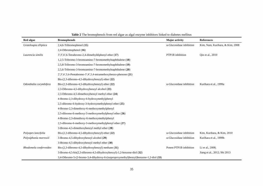
There are some red macroalgae that contain the bromophenols as algal enzyme inhibitors
linked to diabetes mellitus (Table 2), such as the family Rhodomelaceae. Grateloupia
elliptica Holmes contain two bromophenols such as 2,4,6-tribromophenol (15) and 2,4-
dibromophenol (16) with α-glucosidase inhibitory activity (Kim, Nam, Kurihara, & Kim,
2008; Kurihara, Mitani, Kawabata, & Takahashi, 1999b). Bromophenol extracts of G.
elliptica can inhibit intestinal α-glucosidase and stimulated basal glucose uptake into 3T3-L1
adipocytes (Kim, Nam, Kurihara, & Kim, 2008). Five highly brominated metabolites
compounds (17–21; Table 2) isolated from a Chinese red alga Laurencia similis showed
inhibitory activities against PTP1B (Qin et al., 2010). The compound named
bis(2,3-dibromo-4,5-dihydroxybenzyl) ether (22) was purified from Odonthalia corymbifera
11
and Polyopes lancifolia possessed strong activity against α-glucosidases. Meanwhile, six
bromophenols (23–28; Table 2) isolated from the Japanese red alga O. corymbifera also
showed α-glucosidase inhibitory activity (Kurihara et al., 1999a). The two bromophenols
such as 3-bromo-4,5-dihydroxybenzyl alcohol (29) and 3-bromo-4,5-dihydroxybenzyl methyl
ether (30) from Polysiphonia morrowii displayed activity against α-glucosidase were
identified for the first time from this species (Kurihara et al., 1999b).
Bis-(2,3-dibromo-4,5-dihydroxyphenyl)-methane (31), isolated from red macroalgae
Rhodomela confervoides (Hudson) P.C.Silva showed significant inhibition against PTP1B (Li,
Guo, Su, Han, & Shi, 2008). What’s more, an in vivo study also demonstrated the
antihyperglycemic effect of bromophenols (Shi et al., 2008). Four bromophenols namely
3-bromo-4,5-bis(2,3-dibromo-4,5-dihydroxybenzyl)-1,2-benzene-diol (32),
3,4-dibromo-5-(2-bromo-3,4-dihydroxy-6-(isopropoxymethyl)benzyl)benzene-1,2-diol (33),
2,2′,3,3′-tetrabromo-4,4′,5,5′-tetra-hydroxydiphenyl methane (34) and
2,2′,3-tribromo-3′,4,4′,5-tetrahydroxy-6′-ethyloxy-methyldiphenyl methane (35) are all
bromophenols isolated from Rhodomela confervoides which have potent PTP1B inhibition
(Jiang, Shi, Cui, & Guo, 2012; Shi et al., 2008; Shi, 2013). A series of bromophenols (36–43)
purified from red alga Symphylocladia latiuscula exhibited antidiabetic activity by inhibiting
PTP1B. Otherwise, Kurihara et al. (1999a) have reported a bromophenol
2,3,6-tribromo-4,5-dihydroxybenzyl alcohol (44) isolated from S. latiuscula wtih
α-glucosidase inhibition at a very low concentration.
Among the red seaweeds, Hypnea musciformis (Wulfen) J.V.Lamouroux extract diplayed
antihyperglycemic, antioxidant and increased plasma insulin effects in diabetic animals
(Anandakumar, Balamurugan, Rajadurai, & Vani, 2008). The edible red alga Gelidium
amansii (J.V. Lamouroux) J.V. Lamouroux is mainly distributed in northeastern Taiwan. A
mice study has shown that the plasma glucose significantly decreased in the group with oral
treatment of G. amansii ethanol extract (Choi et al., 2015). The plasma glucose, triglyceride,
and cholesterol concentrations in rats with diabetes fed the G. amansii diet for 11-week were
lower than of in rats with diabetes fed the control diet (Yang, Yao, & Chiang, 2015).
Gracilaria lemaneiformis (Bory) Greville occurs widely in the marine environment and
belongs to the family Gracilariaceae (Rhodophyta), and the sulfated polysaccharide accounts
12
for about 30% of its dry weight (Yu, Wang, Chen, Zhang, & Long, 2006). A polysaccharide
extracted from G. lemaneiformis inhibited α-glucosidase activity in vitro and the
administration of polysaccharide (200 mg/kg body weight) for 21 days significantly
decreased the blood glucose levels in diabetic mice (Liao et al., 2015).
The extract of Kappaphycus alvarezii (Doty) Doty ex Silva and the ethanol extract of fresh
Eucheuma denticulatum (N. L. Burman) Collins & Hervey demonstrated the appreciable
inhibitory activities towards α-amylase (Balasubramaniam et al., 2013; Nagarani &
Kamaguru, 2013). K. alvarezii, K. striatus (F. Schmitz) Doty ex P.C.Silva and E.
denticulatum are good sources of magnesium, which could provide 30%–90% of the daily
demand per 100 g of dried macroalgae (Balasubramaniam et al., 2013). It is highly plausible
that magnesium in red macroalgae is responsible for hypoglycaemic activity. Intracellular
free magnesium levels have been found to be closely and inversely related to the level of the
fasting blood glucose (Barbagallo et al., 2003). Magnesium , one of the most abundant ions
present in living cells, plays a pivotal role in insulin homeostasis and glucose metabolism
through multiple enzymatic reactions and its plasma concentration is remarkably constant in
endocrine (Barbagallo et al., 2003). It was shown that serum magnesium levels declined with
rise in HbA1c levels and with duration of type 2 diabetes (Ramadass, Basu, & Srinivasan,
2015). Thus, increased consumption of magnesium-rich macroalgae may reduce the risk of
type 2 diabetes. Gyeongshingangjeehwan 18 (GGEx18) is a kind of herbal drug composed of
three medicinal plants: Rheum palmatum L. (Polygonaceae), Laminaria japonica Aresch
(Laminariaceae), and Ephedra sinica Stapf (Ephedraceae). A study revealed that GGEx18
could significantly increase the expression of fatty acid oxidation genes, such as adiponectin,
AMPKs, PPARα and its target enzymes, and CPT-1, in both mesenteric adipose tissues and
3T3-L1 cells and normalized hyperglycemia and hyperinsulinemia in obese mice, thus reduce
the blood glucose levels (Oh et al., 2014). Porphyran from the red alga Porphyra yezoensis
Ueda is a water-soluble dietary fiber. A study revealed that dietary porphyran should increase
adiponectin levels thus improving glucose metabolism in diabetes (Kitano et al., 2012).
Adiponectin is an adipokine that exerts a strong insulin-sensitizing effect by binding to its
receptors like AdipoR1 and AdipoR2, resulting in activation of AMPK, PPARα, and
presumably some other unknown signaling pathways (Kadowaki et al., 2006). Therefore, the
13
adiponectin gene appears to be a promising candidate susceptibility gene for type 2 diabetes.
Most of the seaweeds contain high contents of soluble dietary fibers such as carrageenan,
agar, and alginates, which could passively retard digestion and glucose absorption. The
beneficial effects of Rhodophyta species on the prevention and management of
diabetes-related risks have clearly been indicated from in vitro and in vivo animal models.
Nevertheless, deep and systematic studies, especiallyfocusing on mechanisms of action, are
still needed. Studies on Rhodophyta sp. and Rhodophyta-derived compounds with
hypoglycemic activity are still insufficient. Thus, further research in this area is imperative to
look for more species with hypoglycemic activity and to provide strong evidence of potential
beneficial effects of hypoglycemic functional foods or drugs from macroalgae.
4. Chlorophyta (green algae)
Ulva lactuca L. is a common green macroalga in the division Chlorophyta and found
widespread in China (Tian, Yin, Zeng, Zhu, & Chen, 2015). Polysaccharides isolated from U.
lactuca could significantly decrease the blood glucose by their potential inhibitory effect on
key enzymes closely related to starch digestion and absorption in both plasma and small
intestine (Belhadj, Hentati, Elfeki, & Hamden, 2013). The Ulva rigida ethanolic extract
decreased blood glucose concentrations and micronuclei frequency in diabetic rats (Celikler
et al., 2009; Tas, Celikler, Ziyanok‐ Ayvalik, Sarandol, & Dirican, 2011). Oxidative stress is
an important factor which responsible for complications in diabetes (Sukmawati et al., 2015).
Diabetes is generally accompanied by increased production of the molecules of reactive
oxygen species and/or impaired antioxidant defense systems, which lead to oxidative damage
to biomolecules. Exposure of the genetic material to reactive oxygen species could cause
DNA damage (Evans, Dizdaroglu, & Cooke, 2004). There are some reports on the
antidiabetic activities of other Ulva species, such as U. fasciata Delile, have the abilities to
reduce blood glucose level, and restore hepatic glycogen content, carbohydrate metablic
enzymes like hexokinase, glusokinase and glucose 6-phoshatase activity in vivo (Abirami &
Kowsalya, 2013). Protein kinase C is a family of protein kinase enzymes that are involved in
controlling the intracellular signal transduction (Anderson, Mcgill, & Tuttle, 2007). The
activation of protein kinase C may occur in the organs susceptible to developing diabetic
14
complications, especially diabetic nephropathy (Kizub, Klymenko, & Soloviev, 2014).
5. Potential anti-diabetic natural products from marine algae
The WHO Expert Committee recommended that medicinal plants used in the treatment of
diabetes be further investigated as they are frequently considered to be lesser or no adverse
effects (Halberstein, 2005). Search for more safe and effective bioactive agents has continued
to be an important target in the field of diabetic research. Less than 1% of the estimated
250,000 higher plants have been screened pharmacologically and very few in regard to
diabetes (Arumugam, Manjula, & Paari, 2013). The ethnobotanical information reports state
that about 800 plants and their active extracts which may possess hypoglycemic potential
have been found. In which, about 200 pure bioactive compounds have been identified and
reported for their potential anti-diabetic effects (Alarcon-Aguilara et al., 1998; Suksomboon,
Poolsup, Boonkaew, & Suthisisang, 2011). These natural phytoconstituents showing
anti-diabetic efficacy include flavonoids, alkaloids, tannins, saponins, terpenoids, phenolics,
glycosides, steroids, chalcones, carotenoids, peptides, lipids, glycopeptides, iridoids, ursolic
acid and imidazoline (Wu, Hsieh, Lin, & Yen, 2013). The bioactive compounds are found in
many fruits, vegetables, herbs, tea, soy and beverage products, and mostly together
responsible for efficacy (Edirisinghe & Burton-Freeman, 2016).
So far, approximately 22,000 natural products of marine organisms have been discovered
whereas 131,000 terrestrial natural products exist (Blunt, Copp, Munro, Northcote, & Prinsep,
2011). According to a recent study, an estimate of 72,500 algal species has been described
throughout the world, where as most of them are marine (Guiry, 2012). To survive in various
diverse and extreme environments, marine macroalgae produce a variety of natural bioactive
compounds and metabolites (Wang, Li, Lee, & Chang, 2017). Polyphenols and
polysaccharides from marine macroalgae particularly showed very significant antidiabetic
potential against pharmacological experimental systems via interfering in carbohydrate
metabolism. Marine algae-derived functional metabolites indicate structural and functional
diversity from their terrestrial counter-part due to the differences in their metabolic pathways
(Guven, Percot, & Sezik, 2010). Algal polyphenols are derived from polymerized
phloroglucinol units, whereas polyphenols from terrestrial plants are derived from gallic and
15
ellagic acids. They are termed as phlorotannins and biosynthesized via acetate malonate
pathway (Arnold & Targett, 2002). At all events, the most active candidates will be
determined through measuring different biochemical parameters such as fasting blood
glucose, insulin, glycosylated hemoglobin, lipid profile, serum urea and creatinine, plasma
alanine and aspartate transaminases, or microscopical examinations of pancreatic sections.
6. Conclusion
Marine macroalgae and functional ingredients derived from them have increasingly been
playing a more and more important role in body health and human nutrition. Bioactive
constituents from marine macroalgae and their byproducts, like phlorotannins, fucosterol, and
carotenoid pigments including fucoxanthin can be used indirectly as functional ingredients
for the reduction of incidences of many chronic diseases in humans (Li & Kim, 2011).
Diabetes mellitus has been considered to be one of the most important global health problems
and there are many potential ways for macroalgae and macroalgae-derived bioactive
compounds to treat diabetes, including α-glucosidase and α-amylase inhibition, activation of
both AMPK and Akt signal pathways as well as HRAR, RLAR, PTP1B activities and AGE
formation inhibition etc. Marine macroalgae are usually perceived as less toxic with fewer
side-effects compared with those synthetic antidiabetic drugs. Current understanding on the
antidiabetic effects of marine macroalgae and their compounds is almost based on the data
available from in vitro and in vivo animal studies, however, these data cannot be extrapolated
into the human setting without reliable human clinical data. Further investigations are
imperative to unveil many more macroalgae and their components, which may have
antidiabetic potentials. It is also important to look in to the possible mechanisms of
antidiabetic actions of these marine macroalgae and their compounds. These antidiabetic
therapeutics from natural source are valuable lead compounds, However, they seldom can be
for direct clinical use and structural modifications are necessary. As a primary requirement
for drug development, the future potential of algal natural products used in diabetes will be
based on the modification of structures of biologically active compounds. In addition,
original alga-derived natural products is unfeasible to meet market demands and alternative
resupply approaches are being developed based on biotechnological production or chemical
16
semi-synthesis from naturally occurring precursors. Industry-scale production of complex
natural products can be harvested in the future align with the progress of the knowledge of
plant biosynthetic pathways and the development of more efficient genetic engineering
strategies and tools. It is of immense importance to gain idea on enhancement of
bioavailability and intrinsic potency with structure–activity relationship studies of algal
bioactive compounds for the treatment of diabetes. Moreover, clinical research is needed to
confirm the real efficacy of marine macroalgae to aid in diabetes prevention and
management . Pharmacists should encourage patients to seek advice about the addition of
these antidiabetic therapeutics for the treatment of diabetes. More research is needed to
identify and quantify the phytochemical compounds on diabetes, as well as the combination
therapy of algal natural products with the synthetic drugs. It is reasonable to state that marine
macroalgae seem to have great developing potential in medicinal preparation to be
sustainable nutraceutical or functional foods for complementary and alternative diabetes
therapy.
Acknowledgement
This work was financially supported by Fujian Province Key Laboratory for the
Development of Bioactive Material from Marine Algae grant (2017FZSK05), Natural
Science Foundation (2016J06009) and Marine High-tech Industrial Development Project
(MIN2014-17) of Fujian Province, China. The project was also supported by and by the
FAFU grants KXb16011A and 612014043.
References
Abirami, R. G., & Kowsalya, S. (2013). Antidiabetic activity of Ulva fasciata and its impact
on carbohydrate metabol-ism enzymes in alloxan induced diabetic rats. International
Journal of Pharmacognosy and Phytochemical Research, 3, 136–141.
ADA. (2005). Diagnosis and classification of diabetes mellitus. Diabetes Care, 28, S37–S42.
ADA. (2010). Diagnosis and classification of diabetes mellitus. Diabetes care, 33, S62–S69.
ADA. (2015). Classification and diagnosis of diabetes. Diabetes Care, 40, S11–S24.
Adeghate, E., Adem, A., Hasan, M. Y., Tekes, K., & Kalasz, H. (2011). Medicinal chemistry
17
and actions of dual and pan PPAR modulators. The Open Medicinal Chemistry Journal,
5, 93–98.
Ah, J. H., Nurul, I. M., Mee, L. C., Oh, J. H., Young, C. H., Chul, W. H., et al. (2012).
Promising antidiabetic potential of fucoxanthin isolated from the edible brown algae
Eisenia bicyclis and Undaria pinnatifida. Fisheries Science, 78, 1321–1329.
Alarcon-Aguilara, F. J., Roman-Ramos, R., Perez-Gutierrez, S., Aguilar-Contreras, A.,
Contreras-Weber, C. C., & Flores-Saenz, J. L. (1998). Study of the antihyperglycemic
effect of plants used as antidiabetics. Journal of Ethnopharmacology, 61, 101–110.
Anandakumar, S., Balamurugan M., Rajadurai, M., & Vani B. (2008). Antihyperglycemic and
antioxidant effects of red algae Hypnea musciformis in alloxan-induced diabetic rats.
Biomedicine, 28, 34–38.
Anderson, P. W., Mcgill, J. B., & Tuttle, K. R. (2007). Protein kinase C β inhibition: the
promise for treatment of diabetic nephropathy. Current Opinion in Nephrology and
Hypertension, 16, 397–402.
Apostolidis, E., & Lee, C. (2010). In vitro potential of Ascophyllum nodosum phenolic
antioxidant‐ mediated α‐ glucosidase and α‐ amylase inhibition. Journal of Food
Science, 75, H97–H102.
Apostolidis, E., Karayannakidis, P. D., Kwon, Y. I., Chong, M. L., & Seeram, N. P. (2011).
Seasonal variation of phenolic antioxidant-mediated α-glucosidase inhibition of
Ascophyllum nodosum. Plant Foods for Human Nutrition, 66, 313–319.
Arnold, T. M., & Targett, N. M. (2002). Marine tannins: The importance of a mechanistic
framework for predicting ecological roles. Journal of Chemical Ecology, 28,
1919–1934.
Arumugam, G., Manjula, P., & Paari N. (2013). A review: Anti diabetic medicinal plants used
for diabetes mellitus. Journal of Acute Disease, 196–200.
Atlas, I. D. (2009). The global burden. International Diabetes Federation. 4th Edition
Brussels, 21–27.
Bakker, S. F., Tushuizen, M. E., Gözütok, E., Çiftci, A., Gelderman, K. A., Mulder, C. J., et al.
(2015). Advanced glycation end products (AGEs) and the soluble receptor for AGE
(sRAGE) in patients with type 1 diabetes and coeliac disease. Nutrition, Metabolism and

18
Cardiovascular Diseases, 25, 230–235.
Balasubramaniam, V., Mustar, S., Khalid, N. M., Rashed, A. A., Noh, M. F. M., Wilcox, M.
D., et al. (2013). Inhibitory activities of three Malaysian edible seaweeds on lipase and
α-amylase. Journal of Applied Phycology, 25, 1405–1412.
Barbagallo, M., Dominguez, L. J., Galioto, A., Ferlisi, A., Cani, C., Malfa, L., et al. (2003).
Role of magnesium in insulin action, diabetes and cardio-metabolic syndrome X.
Molecular Aspects of Medicine, 24, 39–52.
Belhadj, S., Hentati, O., Elfeki, A., & Hamden, K. (2013). Inhibitory activities of Ulva
lactuca polysaccharides on digestive enzymes related to diabetes and obesity. Archives
of Physiology and Biochemistry, 119, 81–87.
Bijland, S., Mancini, S. J., & Salt, I. P. (2013). Role of AMP-activated protein kinase in
adipose tissue metabolism and inflammation. Clinical Science, 124, 491–507.
Blunt, J. W., Copp, B. R., Munro, M. H. G., Northcote, P. T., & Prinsep, M. R. (2011). Marine
natural products. Natural Product Reports, 28, 196–268.
Bu, T., Liu, M., Zheng, L., Guo, Y., & Lin, X. (2010). α-glucosidase inhibition and the in vivo
hypoglycemic effect of butyl-isobutyl-phthalate derived from the Laminaria japonica
rhizoid. Phytotherapy Research, 24, 1588–1591
Celikler, S., Tas, S., Vatan, O., Ziyanok-Ayvalik, S., Yildiz, G., & Bilaloglu, R. (2009).
Anti-hyperglycemic and antigenotoxic potential of Ulva rigida ethanolic extract in the
experimental diabetes mellitus. Food and Chemical Toxicology, 47, 1837–1840.
Choi, S., Oh, H., Jung, J., Park, S., Park, Y. I., Bak, S., & Lee, M. (2015). Effect of agar-free
Gelidium amansii on obesity in DIO C57BL/6J mice model. The FASEB Journal, 29,
S750.2.
Choochote, W., Suklampoo, L., & Ochaikul, D. (2014). Evaluation of antioxidant capacities
of green microalgae. Journal of Applied Phycology, 26, 43–48.
Cicirelli, M. F., Tonks, N. K., Diltz, C. D., Weiel, J. E., Fischer, E. H., & Krebs, E. (1990).
Microinjection of a protein-tyrosine-phosphatase inhibits insulin action in Xenopus
oocytes. Proceedings of the National Academy of Sciences, 87, 5514–5518.
D'Orazio, N., Gemello, E., Gammone, M. A., de Girolamo, M., Ficoneri, C., & Riccioni, G.
(2012). Fucoxantin: A treasure from the sea. Marine Drugs, 10, 604–616.
19
Edirisinghe, I., & Burton-Freeman, B. (2016). Anti-diabetic actions of Berry polyphenols –
Review on proposed mechanisms of action. Journal of Berry Research, 6, 237–250.
Elchebly, M., Payette, P., Michaliszyn, E., Cromlish, W., Collins, S., Loy, A. L., et al. (1999).
Increased insulin sensitivity and obesity resistance in mice lacking the protein tyrosine
phosphatase-1B gene. Science, 283, 1544–1548.
Evans, M. D., Dizdaroglu, M., & Cooke, M. S. (2004). Oxidative DNA damage and disease:
induction, repair and significance. Mutation Research/Reviews in Mutation Research,
567, 1–61.
Gervois, P., Fruchart, J. C., & Staels, B. (2007). Drug Insight: mechanisms of action and
therapeutic applications for agonists of peroxisome proliferator-activated receptors.
Nature Clinical Practice Endocrinology and Metabolism, 3, 145–156.
Gouveia, L., Nobre, B. P., Marcelo, F. M., Mrejen, S., Cardoso, M. T., Palavra, A. F., et al.
(2007). Functional food oil coloured by pigments extracted from microalgae with
supercritical CO2. Food Chemistry, 101, 717–723.
Guiry, M. D. (2012). How many species of algae are there? Journal of Phycology, 48,
1057–1063.
Gupta, S., & Abu-Ghannam, N. (2011). Bioactive potential and possible health effects of
edible brown seaweeds. Trends in Food Science and Technology, 22, 315–326.
Guven, K. C., Percot, A., & Sezik, E. (2010). Alkaloids in marine algae. Marine Drugs, 8,
269–284.
Halberstein, R. A. (2005). Medicinal plants: historical and cross-cultural usage patterns.
Annals of Epidemiology, 15, 686–699.
Hardie, D. (2008). AMPK: a key regulator of energy balance in the single cell and the whole
organism. International Journal of Obesity, 32, S7–S12.
Harrity, T., Farrelly, D., Tieman, A., Chu, C., Kunselman, L., Gu, L., et al. (2006).
Muraglitazar, a novel dual (alpha/gamma) peroxisome proliferator-activated receptor
activator, improves diabetes and other metabolic abnormalities and preserves beta-cell
function in db/db mice. Diabetes, 55, 240–248.
He, W. F., Yao, L. G., Liu, H. L., & Guo, Y. W. (2014). Thunberol, a new sterol from the
Chinese brown alga Sargassum thunbergii. Journal of Asian Natural Products Research,
20
16, 685–689.
Heo, S. J., Hwang, J. Y., Choi, J. I., Han, J. S., Kim, H. J., & Jeon, Y. J. (2009).
Diphlorethohydroxycarmalol isolated from Ishige okamurae, a brown algae, a potent
α-glucosidase and α-amylase inhibitor, alleviates postprandial hyperglycemia in diabetic
mice. European Journal of Pharmacology, 615, 252–256.
Hwang, P. A., Hung, Y. L., Tsai, Y. K., Chien, S. Y., & Kong, Z. L. (2014). The brown
seaweed Sargassum hemiphyllum exhibits α-amylase and α-glucosidase inhibitory
activity and enhances insulin release in vitro. Cytotechnology, 67, 653–660.
Iwai, K. (2008). Antidiabetic and antioxidant effects of polyphenols in brown alga Ecklonia
stolonifera in genetically diabetic KK-Ay mice. Plant Foods for Human Nutrition, 63(4),
163–169.
Jia, X., Yang, J., Wang, Z., Liu, R., & Xie, R. (2014). Polysaccharides from Laminaria
japonica show hypoglycemic and hypolipidemic activities in mice with experimentally
induced diabetes. Experimental Biology and Medicine, 239, 1663–1670.
Jiang, B., Shi, D., Cui, Y., & Guo, S. (2012). Design, synthesis, and biological evaluation of
bromophenol derivatives as protein tyrosine phosphatase 1B inhibitors. Archiv Der
Pharmazie, 345, 444–453.
Jung, H. A., Islam, M. N., Lee, C. M., Oh, S. H., Lee, S., Jung, J. H., et al. (2013). Kinetics
and molecular docking studies of an anti-diabetic complication inhibitor fucosterol from
edible brown algae Eisenia bicyclis and Ecklonia stolonifera. Chemico-Biological
Interactions, 206, 55–62.
Jung, M., Park, M., Lee, H. C., Kang, Y. H., Kang, E. S., & Kim, S. K. (2006). Antidiabetic
agents from medicinal plants. Current Medicinal Chemistry, 13, 1203–1218.
Kadowaki, T., Yamauchi, T., Kubota, N., Hara, K., Ueki, K., & Tobe, K. (2006). Adiponectin
and adiponectin receptors in insulin resistance, diabetes, and the metabolic syndrome.
Journal of Clinical Investigation, 116, 1784–1792.
Kandra, L. (2003). α-Amylases of medical and industrial importance. Journal of Molecular
Structure Theochem, 487–498.
Kang, C., Jin, Y. B., Lee, H., Cha, M., Sohn, E. T., Moon, J., et al. (2010). Brown alga
Ecklonia cava attenuates type 1 diabetes by activating AMPK and Akt signaling
21
pathways. Food and Chemical Toxicology, 48, 509–516.
Kang, M. C., Wijesinghe, W. A. J. P., Lee, S. H., Kang, S. M., Ko, S. C., Yang, X., et al.
(2012). Dieckol isolated from brown seaweed Ecklonia cava attenuates type II diabetes
in db/db mouse model. Food and Chemical Toxicology An International Journal
Published for the British Industrial Biological Research Association, 53, 294.
Khan, W., Rayirath, U. P., Subramanian, S., Jithesh, M. N., Rayorath, P., Hodges, D. M., et al.
(2009). Seaweed extracts as biostimulants of plant growth and development. Journal of
Plant Growth Regulation, 28, 386–399.
Kim, K. J., Yoon, K. Y., & Lee, B. Y. (2012). Fucoidan regulate blood glucose homeostasis in
C57BL/KSJ m+/+ db and C57BL/KSJ db/db mice. Fitoterapia, 83, 1105–1109.
Kim, K. T., Rioux, L. E., & Turgeon, S. L. (2014). Alpha-amylase and alpha-glucosidase
inhibition is differentially modulated by fucoidan obtained from Fucus vesiculosus and
Ascophyllum nodosum. Phytochemistry, 98, 27–33.
Kim, K. Y., Kurihara, H., & Kim, S. M. (2010). α-Glucosidase inhibitory activity of
bromophenol purified from the red alga Polyopes lancifolia. Journal of Food Science,
75, H145–H150.
Kim, K., Nam, K., Kurihara, H., & Kim, S. (2008). Potent α-glucosidase inhibitors purified
from the red alga Grateloupia elliptica. Phytochemistry, 69, 2820–2825.
Kim, M. S., Kim, J. Y., Choi, W. H., & Lee, S. S. (2008). Effects of seaweed supplementation
on blood glucose concentration, lipid profile, and antioxidant enzyme activities in
patients with type 2 diabetes mellitus. Nutrition Research and Practice, 2, 62–67.
Kim, S. N. (2008). Sargaquinoic acid and sargahydroquinoic acid from Sargassum yezoense
stimulate adipocyte differentiation through PPAR alpha/gamma activation in 3T3-L1
cells. FEBS Letters, 582, 3465–3472.
Kim, S. N., Lee, W., Bae, G. U., & Kee, Y. K. (2012). Anti-diabetic and hypolipidemic effects
of Sargassum yezoense in db/db mice. Biochemical and Biophysical Research
Communications, 424, 675–680.
Kitano, Y., Murazumi, K., Duan, J., Kurose, K., Kobayashi, S., Sugawara, T., et al. (2012).
Effect of dietary porphyran from the red alga, Porphyra yezoensis, on glucose
metabolism in diabetic KK-Ay mice. Journal of Nutritional Science and Vitaminology,
22
58, 14–19.
Kizub, I. V., Klymenko, K. I., & Soloviev, A. I. (2014). Protein kinase C in enhanced vascular
tone in diabetes mellitus. International Journal of Cardiology, 174, 230–242.
Kurihara, H., Mitani, T., Kawabata, J., & Takahashi, K. (1999a). Two new bromophenols
from the red alga Odonthalia corymbifera. Journal of Natural Products, 62, 882–884.
Kurihara, H., Mitani, T., Kawabata, J., & Takahashi, K. (1999b). Inhibitory potencies of
bromophenols from Rhodomelaceae algae against α-glucosidase activity. Fisheries
Science, 65, 300–303.
Lee, C. W., & Han, J. S. (2012). Hypoglycemic effect of Sargassum ringgoldianum extract in
STZ-induced diabetic mice. Preventive Nutrition and Food Science, 17, 8–13.
Lee, S. (2003). Anti-oxidant activities of fucosterol from the marine algae Pelvetia siliquosa.
Archives of Pharmacal Research, 26, 719–722.
Lee, S. H., & Jeon, Y. J. (2013). Anti-diabetic effects of brown algae derived phlorotannins,
marine polyphenols through diverse mechanisms. Fitoterapia, 86, 129–136.
Lee, S. H., Kang, N., Kim, E. A., Heo, S. J., Moon, S. H., Jeon, B. T., et al. (2014).
Antidiabetogenic and antioxidative effects of octaphlorethol a isolated from the brown
algae Ishige foliacea in streptozotocin-induced diabetic mice. Food Science and
Biotechnology, 23, 1261–1266.
Lee, S. H., Karadeniz, F., Kim, M. M., & Kim, S. K. (2009). α‐ Glucosidase and α‐ amylase
inhibitory activities of phloroglucinal derivatives from edible marine brown alga,
Ecklonia cava. Journal of the Science of Food and Agriculture, 89, 1552–1558.
Lee, S. H., Ko, S. C., Kang, M. C., Lee, D. H., & Jeon, Y. J. (2016). Octaphlorethol a, a
marine algae product, exhibits antidiabetic effects in type 2 diabetic mice by activating
amp-activated protein kinase and upregulating the expression of glucose transporter 4.
Food and Chemical Toxicology, 91, 58–64.
Lee, S. H., Min, K. H., Han, J. S., Lee, D. H., Park, D. B., Jung, W. K., et al. (2012). Effects
of brown alga, Ecklonia cava on glucose and lipid metabolism in C57BL/KsJ- db/db
mice, a model of type 2 diabetes mellitus. Food and Chemical Toxicology, 50, 575–582.
Lee, S. H., Park, M. H., Heo, S. J., Kang, S. M., Ko, S. C., Han, J. S., et al. (2010). Dieckol
isolated from Ecklonia cava inhibits α-glucosidase and α-amylase in vitro and alleviates
23
postprandial hyperglycemia in streptozotocin-induced diabetic mice. Food and
Chemical Toxicology, 48, 2633–2637.
Lee, S. H., Ko, S. C., Kang, M. C., Lee, D. H., Jeon, Y. J. (2016). Octaphlorethol A, a marine
algae product, exhibits antidiabetic effects in type 2 diabetic mice by activating
AMP-activated protein kinase and upregulating the expression of glucose transporter 4.
Food and Chemical Toxicology, 91, 58-64.
Lee, Y. S., Shin, K. H., Kim, B. K., & Lee, S. (2004). Anti-diabetic activities of fucosterol
from Pelvetia siliquosa. Archives of Pharmacal Research, 27, 1120–1122.
Li, J., Guo, S. J., Su, H., Han, L. J., & Shi, D. Y. (2008). Total synthesis of
bis-(2,3-dibromo-4,5-dihydroxyphenyl)-methane as potent PTP1B inhibitor. Chinese
Chemical Letters, 19, 1290–1292.
Li, X. C, Niu, R. L., Fan, X., Han, L. J., & Zhang, L. X. (2005). Macroalage as a source of
alpha-glucosidase inhibitors. Chinese Journal of Oceanology and Limnology, 23,
354–356.
Li, X., Yu, Z., Long, S., Guo, Y., & Duan, D. (2012). Hypoglycemic effect of Laminaria
japonica polysaccharide in a type 2 diabetes mellitus mouse model. Isrn Endocrinology,
2012, 507462.
Li, Y. X., & Kim, S. K. (2011). Utilization of seaweed derived ingredients as potential
antioxidants and functional ingredients in the food industry: An overview. Food Science
and Biotechnology, 20, 1461–1466.
Liao, X., Yang, L., Chen, M., Yu, J., Zhang, S., & Ju, Y. (2015). The hypoglycemic effect of a
polysaccharide (GLP) from Gracilaria lemaneiformis and its degradation products in
diabetic mice. Food and Function, 6, 2542–2549.
Liu, H., & Gu, L. (2012). Phlorotannins from brown algae (Fucus vesiculosus) inhibited the
formation of advanced glycation endproducts by scavenging reactive carbonyls. Journal
of Agricultural and Food Chemistry, 60, 1326–1334.
Liu, M., Zhang, W., Qiu, L., & Lin, X. K. (2011). Synthesis of butyl-isobutyl-phthalate and
its interaction with α-glucosidase in vitro. Journal of Biochemistry, 149, 27–33.
Liu, M., Zhang, W., Wei, J. T., & Lin, X. K. (2011). Synthesis and α-glucosidase inhibitory
mechanisms of bis (2,3-dibromo-4,5-dihydroxybenzyl) ether, a potential marine
24
bromophenol α-glucosidase inhibitor. Marine Drugs, 9, 1554–1565.
Liu, X., Li, X., Gao, L., Cui, C., Li, C., Li, J., et al. (2011). Extraction and PTP1B inhibitory
activity of bromophenols from the marine red alga Symphyocladia latiuscula. Chinese
Journal of Oceanology and Limnolog, 29, 686–690.
Long, Y. C., & Zierath, J. R. (2006). AMP-activated protein kinase signaling in metabolic
regulation. Journal of Clinical Investigation, 116, 1776.
Maeda, H., & Dominguez, H. (2013). Anti-obesity and anti-diabetic activities of algae.
Functional Ingredients from Algae for Foods and Nutraceuticals, 256, 453–476.
Maeda, H., Hosokawa, M., Sashima, T., Murakami-Funayama, K., & Miyashita, K. (2009).
Anti-obesity and anti-diabetic effects of fucoxanthin on diet-induced obesity conditions
in a murine model. Molecular Medicine Reports, 2, 897–902.
Manikkam, V., Vasiljevic, T., Donkor, O. N., Mathai, M. L. (2016). A review of potential
marine-derived hypotensive and anti-obesity peptides. Critical Reviews in Food Science
and Nutrition, 56, 92-112.
Michalik, L., Auwerx, J., Berger, J. P., Chatterjee, V. K., Glass, C. K., Gonzalez, F. J., et al.
(2006). International Union of Pharmacology. LXI. Peroxisome proliferator-activated
receptors. Pharmacological Reviews, 58, 726–741.
Min, K. H., Kim, H. J., Jeon, Y. J., & Han, J. S. (2011). Ishige okamurae ameliorates
hyperglycemia and insulin resistance in C57BL/KsJ- db/db mice. Diabetes Research and
Clinical Practice, 93, 70–76.
Mohamed, S., Hashim, S. N., & Rahman, H. A. (2012). Seaweeds: A sustainable functional
food for complementary and alternative therapy. Trends in Food Science and Technology,
23, 83–96.
Moon, H. E., Islam, M. N., Ahn, B. R., Chowdhury, S. S., Sohn, H. S., Jung, H. A., et al.
(2011). Protein tyrosine phosphatase 1B and α-glucosidase inhibitory phlorotannins
from edible brown algae, Ecklonia stolonifera and Eisenia bicyclis. Bioscience
Biotechnology and Biochemistry, 75, 1472–1480.
Motshakeri, M., Ebrahimi, M., Goh, Y. M., Matanjun, P., & Mohamed, S. (2013). Sargassum
polycystum reduces hyperglycaemia, dyslipidaemia and oxidative stress via increasing
insulin sensitivity in a rat model of type 2 diabetes. Journal of the Science of Food and
25
Agriculture, 93, 1772–1778.
Motshakeri, M., Ebrahimi, M., Goh, Y. M., Othman, H. H., Hair-Bejo, M., & Mohamed, S.
(2014). Effects of brown seaweed (Sargassum polycystum) extracts on kidney, liver, and
pancreas of type 2 diabetic rat model. Evidence-based Complementary and Alternative
Medicine: eCAM, 2014, 68–78.
Nagarani, N., & Kamaguru, A. (2013). Evaluation of anti-inflammatory, antidiabetic,
cytotoxic activity of Kappaphycus alvarezii. International Journal of Pharma and Bio
Sciences, 4, 921–929.
Oh, J., Lee, H., Lim, H., Woo, S., Shin, S. S., & Yoon, M. (2014). The herbal composition
GGEx18 from Laminaria japonica, Rheum palmatum, and Ephedra sinica inhibits
visceral obesity and insulin resistance by upregulating visceral adipose genes involved
in fatty acid oxidation. Pharmaceutical Biology, 53, 301–312.
Ohta, T., Sasaki, S., Oohori, T., Yoshikawa, S., & Kurihara, H. (2002). α-Glucosidase
inhibitory activity of a 70% methanol extract from Ezoishige (Pelvetia babingtonii de
Toni) and its effect on the elevation of blood glucose level in rats. Bioscience,
Biotechnology, and Biochemistry, 66, 1552–1554.
Okada, Y., Ishimaru, A., Suzuki, R., & Okuyama, T. (2004). A new phloroglucinol derivative
from the brown alga Eisenia bicyclis: Potential for the effective treatment of diabetic
complications. Journal of Natural Products, 67, 103–105.
Padmalayam, I., & Suto, M. (2013). Role of adiponectin in the metabolic syndrome: current
perspectives on its modulation as a treatment strategy. Current Pharmaceutical Design,
19, 5755–5763.
Pangestuti, R., & Kim, S. K. (2011). Biological activities and health benefit effects of natural
pigments derived from marine algae. Journal of Functional Foods, 3, 255–266.
Pantidos, N., Boath, A., Lund, V., Conner, S., & McDougall, G. J. (2014). Phenolic-rich
extracts from the edible seaweed, ascophyllum nodosum, inhibit α-amylase and
α-glucosidase: Potential anti-hyperglycemic effects. Journal of Functional Foods, 10,
201–209.
Paradis, M. E., Couture, P., & Lamarche, B. (2011). A randomised crossover
placebo-controlled trial investigating the effect of brown seaweed (Ascophyllum
26
nodosum and Fucus vesiculosus) on postchallenge plasma glucose and insulin levels in
men and women. Applied Physiology, Nutrition, and Metabolism, 36, 913–919.
Park, M. H., Nam, Y. H., & Han, J. S. (2015). Sargassum coreanum extract alleviates
hyperglycemia and improves insulin resistance in db/db diabetic mice. Nutrition
Research and Practice, 9, 472–479.
Pershadsingh, H. A. (2006). Dual peroxisome proliferator-activated receptor-α/γ agonists.
Treatments in Endocrinology, 5, 89-99.
Pontiroli, A. E. (2004). Type 2 diabetes mellitus is becoming the most common type of
diabetes in school children. Acta Diabetologica, 41, 85–90.
Qin, J., Su, H., Zhang, Y., Gao, J., Zhu, L., Wu, X., et al. (2010). Highly brominated
metabolites frommarine red alga Laurencia similis inhibit protein tyrosine phosphatase
1B. Bioorganic and Medicinal Chemistry Letters, 20, 7152–7154.
Ramachandran, V., & Saravanan, R. (2015). Glucose uptake through translocation and
activation of GLUT4 in PI3K/Akt signaling pathway by asiatic acid in diabetic rats.
Human and Experimental Toxicology, 34, 884–893.
Ramadass, S., Basu, S., & Srinivasan, A. (2015). SERUM magnesium levels as an indicator
of status of Diabetes Mellitus type 2. Diabetes & Metabolic Syndrome: Clinical
Research and Reviews, 9, 42–45.
Rengasamy, K. R., Kulkarni, M. G., Stirk, W. A., Van Staden, J. (2014). Advances in algal
drug research with emphasis on enzyme inhibitors. Biotechnology Advances, 32,
1364–1381.
Renner, S., & Ricklefs, E. (1995). Dioecy and its correlates in the flowering plants. American
Journal of Botany, 82, 596–606.
Rigalleau, V., Cougnard-Gregoire, A., Nov, S., Gonzalez, C., Maury, E., Lorrain, S., et al.
(2015). Association of advanced glycation end products and chronic kidney disease with
macroangiopathy in type 2 diabetes. Journal of Diabetes and Its Complications, 29,
270–274.
Roglic, G., Unwin, N., Bennett, P. H., Mathers, C., Tuomilehto, J., Nag, S., et al. (2005). The
burden of mortality attributable to diabetes: realistic estimates for the year 2000.
Diabetes Care, 28, 2130–2135.
27
Ruocco, N., Costantini, S., Guariniello, S., Costantini, M. (2016). Polysaccharides from the
marine environment with pharmacological, cosmeceutical and nutraceutical potential.
Molecules, 21, 551.
Sánchez-Machado, D. I., López-Hernández, J., Paseiro-Losada, P., & López-Cervantes, J.
(2004). An HPLC method for the quantification of sterols in edible seaweeds.
Biomedical Chromatography, 18, 183–190.
Saleh, A. S. M., Zhang, Q., & Shen, Q. (2016). Recent research in antihypertensive activity
of food protein-derived hydrolyzates and peptides. Critical Reviews in Food Science and
Nutrition, 56, 760-787.
Shi, D. (2013). HPN, a synthetic analogue of bromophenol from red alga Rhodomela
confervoides: Synthesis and anti-diabetic effects in C57BL/KsJ-db/db mice. Marine
Drugs, 11, 350–362.
Shi, D. Y., Xu, F., He, J., Li, J., Xiao, F., & Han, L. J. (2008). Inhibition of bromophenols
against PTP1B and anti-hyperglycemic effect of Rhodomela confervoides extract in
diabetic rats. Chineseence Bulletin, 53, 2476–2479.
Shirosaki, M., & Koyama, T. (2011). Laminaria japonica as a food for the prevention of
obesity and diabetes. Advances in Food and Nutrition Research, 64, 199–212.
Spiegelman, B. M. (1998). PPAR-gamma: adipogenic regulator and thiazolidinedione
receptor. Diabetes, 47, 507–514.
Suleria, H. Á. R., Gobe, G., Masci, P., Osborne, S. A. (2016). Marine bioactive compounds
and health promoting perspectives; innovation pathways for drug discovery. Trends in
Food Science & Technology, 50, 44-55.
Sukmawati, D., Fujimura, S., Jitsukawa, S., Ito-Hirano, R., Ishii, T., Sato, T., et al. (2015).
Oxidative stress tolerance of early stage diabetic endothelial progenitor cell.
Regenerative Therapy, 1, 38–44.
Suksomboon, N., Poolsup, N., Boonkaew, S., & Suthisisang, C. C. (2011). Meta-analysis of
the effect of herbal supplement on glycemic control in type 2 diabetes. Journal of
Ethnopharmacology, 137, 1328–1333.
Tas, S., Celikler, S., Ziyanok‐ Ayvalik, S., Sarandol, E., & Dirican, M. (2011). Ulva rigida
improves carbohydrate metabolism, hyperlipidemia and oxidative stress in
28
streptozotocin‐ induced diabetic rats. Cell Biochemistry and Function, 29, 108–113.
Taylor, W. R. (1957). Book reviews: marine algae of the northeastern coast of north america.
Science, 126.
Thilagam, E., Parimaladevi, B., Kumarappan, C., Mandal, S. C. (2013). α-Glucosidase and
α-amylase inhibitory activity of Senna surattensis. Journal of Acupuncture and Meridian
Studies, 6, 24–30.
Tian, H., Yin, X., Zeng, Q., Zhu, L., & Chen, J. (2015). Isolation, structure, and surfactant
properties of polysaccharides from Ulva lactuca L. from South China Sea. International
Journal of Biological Macromolecules, 79, 577–582.
Vinayagam, R., Xiao, J.B., & Xu, B.J. (2017). An insight into anti-diabetic properties of
dietary phytochemicals. Phytochemistry Reviews, 16, 535–553.
Vinoth, K. T., Lakshmanasenthil, S., Geetharamani, D., Marudhupandi, T., Suja, G., &
Suganya, P. (2015). Fucoidan: A α-D-glucosidase inhibitor from Sargassum wightii with
relevance to type 2 diabetes mellitus therapy. International Journal of Biological
Macromolecules, 72C, 1044–1047.
Wälchli, S., Curchod, M. L., Gobert, R. P., Arkinstall, S., & van Huijsduijnen, R. H. (2000).
Identification of Tyrosine Phosphatases that dephosphorylate the insulin receptor a Brute
Force approch based on “Substrate-Trapping” mutants. Journal of Biological Chemistry,
275, 9792–9796.
Wang, H. D., Li, X. C., Lee D. J., & Chang J. S. (2017). Potential biomedical applications of
marine algae. Bioresource Technology, http://dx.doi.org/10.1016/j.biortech.2017.05.198.
WHO. (2011). Use of glycated haemoglobin (HbA1c) in diagnosis of diabetes mellitus:
abbreviated report of a WHO consultation.
Wu, C. H., Hsieh, H. T., Lin, J. A., & Yen, G. C. (2013). Alternanthera paronychioides
protects pancreatic β-cells from glucotoxicity by its antioxidant, antiapoptotic and
insulin secretagogue actions. Food Chemistry, 139, 362–370.
Xiao, J. B., Högger P. (2015). Dietary polyphenols and type 2 diabetes: current insights and
future perspectives. Current Medicinal Chemistry, 22, 23–38.
Xu, H. L., Kitajim, C., Ito, H., Miyazaki, T., Bab, M., Okuyam, T., & Ok, Y. (2012).
Antidiabetic effect of polyphenols from brown alga Ecklonia kurome in genetically
29
diabetic KK-Ay mice. Pharmaceutical Biology, 50, 393–400.
Yang, T. H., Yao, H. T., & Chiang, M. T. (2015). Red algae (Gelidium amansii) reduces
adiposity via activation of lipolysis in rats with diabetes induced by
streptozotocin-nicotinamide. Journal of Food and Drug Analysis, 23, 758–765.
Yoon, J. S., Yadunandam, A. K., Kim, S. J., Woo, H. C., Kim, H. R., & Kim, G. D. (2013).
Dieckol, isolated from Ecklonia stolonifera, induces apoptosis in human hepatocellular
carcinoma Hep3B cells. Journal of Natural Medicines, 67, 519–527.
Yotsu-Yamashita, M., Kondo, S., Segawa, S., Lin, Y. C., Toyohara, H., Ito, H., et al. (2013).
Isolation and structural determination of two novel phlorotannins from the brown alga
Ecklonia kurome Okamura, and their radical scavenging activities. Marine Drugs, 11,
165–183.
Yu, J., Wang, X., Chen, M. Z., Zhang, Y. Y., & Long, Z. J. (2006). Analysis on nutritional
components and polysaccharide composition of gracilaria lemaneiformis from Chaoshan
Coast. Food Science, 27, 93–97.
Zha, X. Q., Xiao, J. J., Zhang, H. N., Wang, J. H., Pan, L. H., Yang, X. F., et al. (2012).
Polysaccharides in Laminaria japonica (LP): Extraction, physicochemical properties
and their hypolipidemic activities in diet-induced mouse model of atherosclerosis. Food
Chemistry, 134, 244–252.
Zhang, S., & Zhang, Z. Y. (2007). PTP1B as a drug target: recent developments in PTP1B
inhibitor discovery. Drug Discovery Today, 12, 373–381.
Zhao, C., Wu, Y. J., Yang, C. F., Liu, B., & Huang, Y, F. (2015). Hypotensive, hypoglycemic
and hypolipidemic effects of bioactive compounds from microalgae and marine
microorganisms. International Journal of Food Science and Technology, 50, 1705–1717.
Zou, Y., Qian, Z. J., Li, Y., Kim, M. M., Lee, S. H., & Kim, S. K. (2008). Antioxidant effects
of phlorotannins isolated from Ishige okamurae in free radical mediated oxidative
systems. Journal of Agricultural and Food Chemistry, 56, 7001–7009.
30
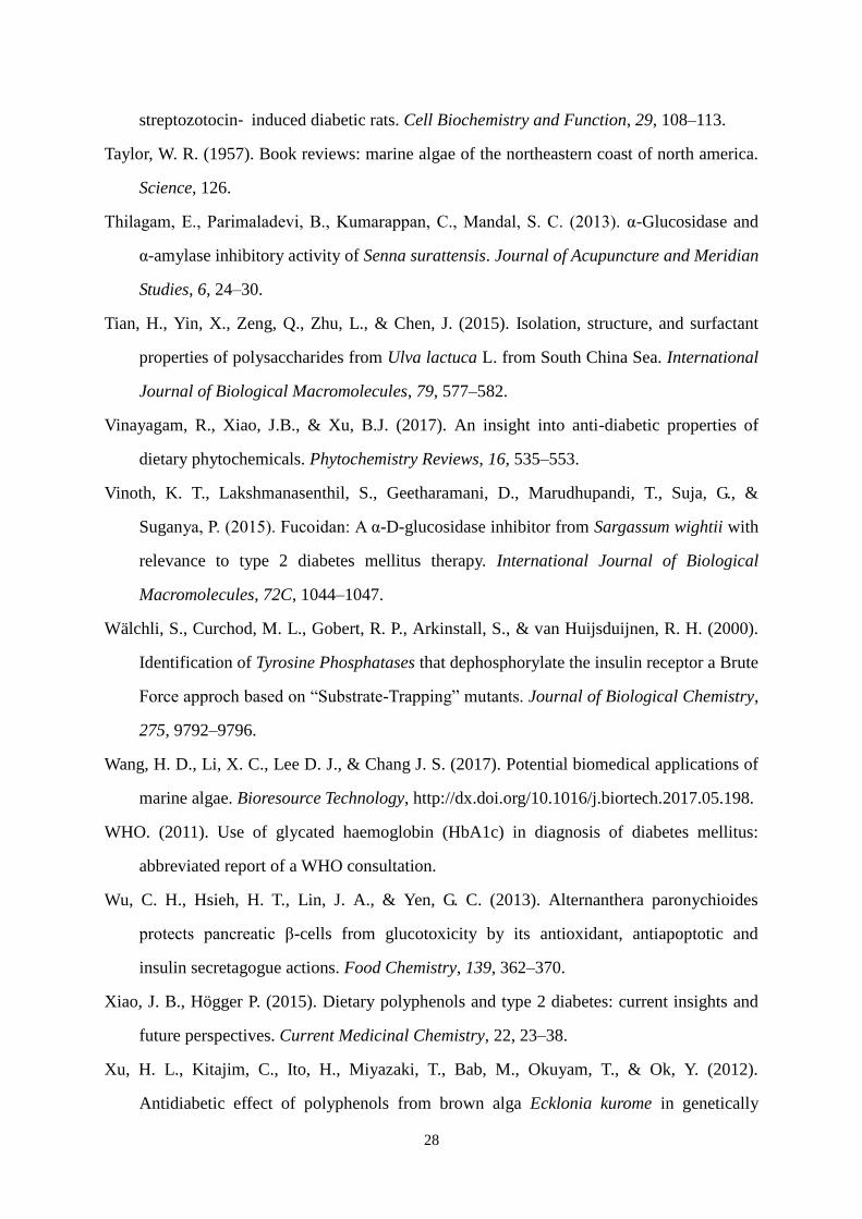
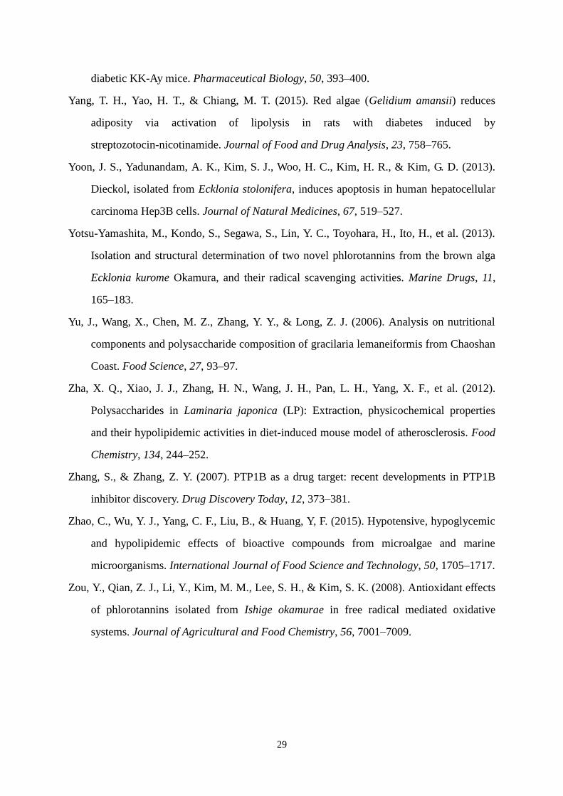
Fig. 1 Chemical structures of bioactive compounds from marine macroalgae (references seen in Table 1
and Table 2)
1 Fucosterol 2 Dieckol
3 Eckol 4 7-Phloroeckol
5 Phlorofucofuroeckol-A 6 6,6-Bieckol
7 Fucodiphloroethol-G 8 Butyl-isobutyl-phthalate
31
9 Sargaquinoic acid 10 Sargahydroquinoic acid
11 Fucoxanthin
12 Thunberol 13 Diphlorethohydroxycarmalol
14 Octaphlorethol A
15 2,4,6-Tribromophenol 16 2,4- Dibromophenol 17 3′,5′,6′,6-Tetrabromo-2,4-dimethyldiphenyl ether
18 19 20 21
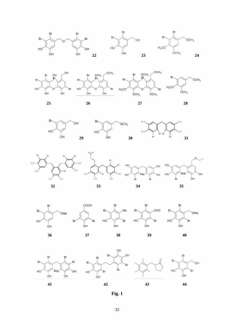
32
22 23 24
25 26 27 28
29 30 31
32 33 34 35
36 37 38 39 40
41 42 43 44
Fig. 1
33
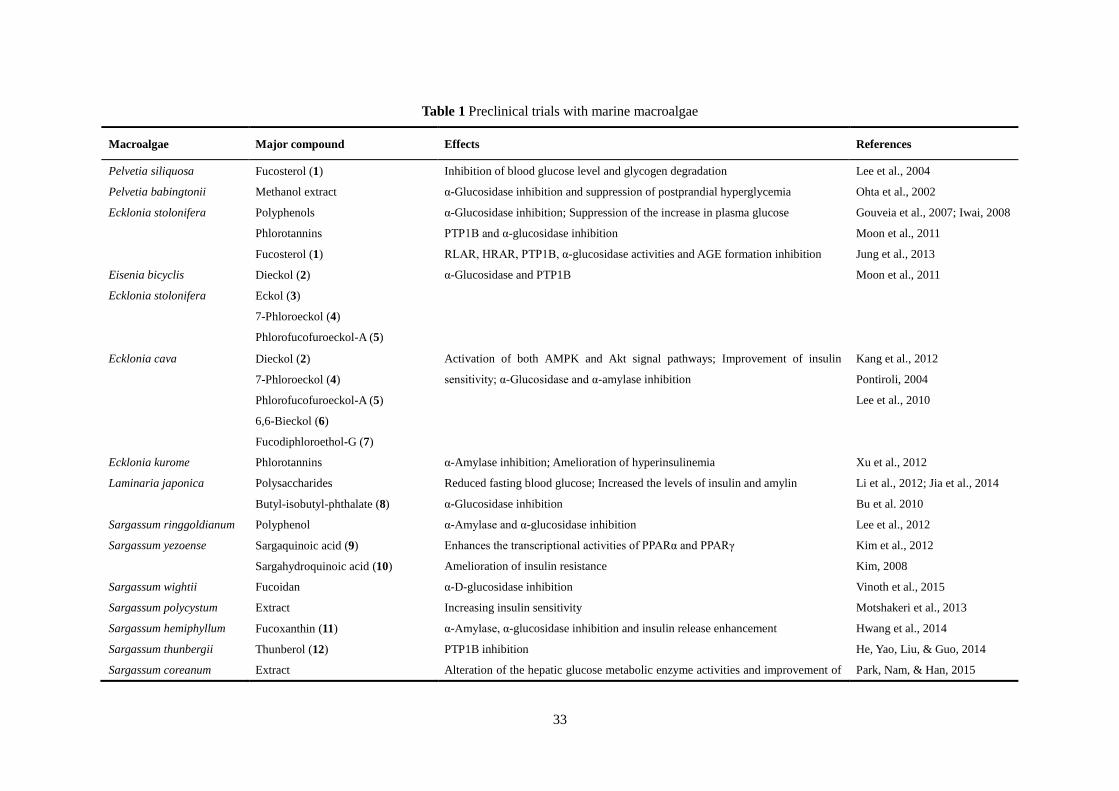
Table 1 Preclinical trials with marine macroalgae
Macroalgae Major compound Effects References
Pelvetia siliquosa Fucosterol (1) Inhibition of blood glucose level and glycogen degradation Lee et al., 2004
Pelvetia babingtonii Methanol extract α-Glucosidase inhibition and suppression of postprandial hyperglycemia Ohta et al., 2002
Ecklonia stolonifera Polyphenols α-Glucosidase inhibition; Suppression of the increase in plasma glucose Gouveia et al., 2007; Iwai, 2008
Phlorotannins PTP1B and α-glucosidase inhibition Moon et al., 2011
Fucosterol (1) RLAR, HRAR, PTP1B, α-glucosidase activities and AGE formation inhibition Jung et al., 2013
Eisenia bicyclis
Ecklonia stolonifera
Dieckol (2)
Eckol (3)
7-Phloroeckol (4)
Phlorofucofuroeckol-A (5)
α-Glucosidase and PTP1B Moon et al., 2011
Ecklonia cava Dieckol (2)
7-Phloroeckol (4)
Phlorofucofuroeckol-A (5)
6,6-Bieckol (6)
Fucodiphloroethol-G (7)
Activation of both AMPK and Akt signal pathways; Improvement of insulin
sensitivity; α-Glucosidase and α-amylase inhibition
Kang et al., 2012
Pontiroli, 2004
Lee et al., 2010
Ecklonia kurome Phlorotannins α-Amylase inhibition; Amelioration of hyperinsulinemia Xu et al., 2012
Laminaria japonica Polysaccharides Reduced fasting blood glucose; Increased the levels of insulin and amylin Li et al., 2012; Jia et al., 2014
Butyl-isobutyl-phthalate (8) α-Glucosidase inhibition Bu et al. 2010
Sargassum ringgoldianum Polyphenol α-Amylase and α-glucosidase inhibition Lee et al., 2012
Sargassum yezoense Sargaquinoic acid (9)
Sargahydroquinoic acid (10)
Enhances the transcriptional activities of PPARα and PPARγ Kim et al., 2012
Amelioration of insulin resistance Kim, 2008
Sargassum wightii Fucoidan α-D-glucosidase inhibition Vinoth et al., 2015
Sargassum polycystum Extract Increasing insulin sensitivity Motshakeri et al., 2013
Sargassum hemiphyllum Fucoxanthin (11) α-Amylase, α-glucosidase inhibition and insulin release enhancement Hwang et al., 2014
Sargassum thunbergii Thunberol (12) PTP1B inhibition He, Yao, Liu, & Guo, 2014
Sargassum coreanum Extract Alteration of the hepatic glucose metabolic enzyme activities and improvement of Park, Nam, & Han, 2015
34
insulin resistance
Undaria pinnatifida Fucoxanthin (11) HRAR, RLAR, PTP1B inhibition, and AGE formation Ah et al., 2012
Improve insulin signaling Maeda et al., 2013
Eisenia bicyclis Phlorotannins Inhibition of AGEs and α-amylase Okada et al., 2004
Fucoxanthin (11) Inhibition of RLAR, HRAR, PTP1B activities and AGE formation Ah et al., 2012
Fucosterol (1) Inhibition of RLAR, HRAR, PTP1B, α-glucosidase activities and AGE formation Jung et al., 2013
Ascophyllum nodosum Phlorotannins
Fucoidan
α-Amylase and α-glucosidase inhibition Apostolidis et al., 2011; Kim et
al., 2014; Pantidos et al., 2014
Ishige okamurae Diphlorethohydroxycarmalol (13) α-Amylase and α-glucosidase inhibition Heo et al., 2009
Ishige okamurae Extract Altering the hepatic glucose metabolic enzyme activities and improves insulin
resistance.
Min et al., 2011
Ishige foliacea Octaphlorethol A (14) Increasing in GLUT4-mediated glucose utilization via activation of AMPK in
muscle.
Lee, Ko, Kang, Lee, & Jeon,
2016
Kappaphycus alvarezii
Eucheuma denticulatum
Extract Inhibitory activity towards α-amylase Nagarani & Kamaguru 2013;
Balasubramaniam et al., 2013
Gracilaria lemaneiformis Polysaccharide Inhibitory to the α-glucosidase activity; decrease in blood glucose levels Liao et al., 2015
Gelidium amansiithe Ethanol extract Plasma glucose significantly decreased Choi et al., 2015
Porphyra yezoensis Porphyran Increasing adiponectin levels Kitano et al., 2012
Ulva rigida Ethanolic extract Regeneration of β-cells and/or potentiating the insulin release Celikler et al., 2009; Tas et al.,
2011
Ulva fasciata Sulfated polysaccharides Reduce blood glucose level, and restore hepatic glycogen content and carbohydrate
metablic enzymes
Abirami & Kowsalya, 2013
Ulva lactuca Polysaccharides α-Amylase, maltase and sucrase inhibition; Delay glucose absorption Belhadj et al., 2013
35
Table 2 The bromophenols from red algae as algal enzyme inhibitors linked to diabetes mellitus
Red algae Bromophenols Major activity References
Grateloupia elliptica 2,4,6-Tribromophenol (15) α-Glucosidase inhibition Kim, Nam, Kurihara, & Kim, 2008
2,4-Dibromophenol (16)
Laurencia similis 3′,5′,6′,6-Tetrabromo-2,4-dimethyldiphenyl ether (17) PTP1B inhibition Qin et al., 2010
1,2,5-Tribromo-3-bromoamino-7-bromomethylnaphthalene (18)
2,5,8-Tribromo-3-bromoamino-7-bromomethylnaphthalene (19)
2,5,6-Tribromo-3-bromoamino-7-bromomethylnaphthalene (20)
2′,5′,6′,5,6-Pentabromo-3′,4′,3,4-tetramethoxybenzo-phenone (21)
Bis-(2,3-dibromo-4,5-dihydroxybenzyl) ether (22)
Odonthalia corymbifera Bis-(2,3-dibromo-4,5-dihydroxybenzyl) ether (22) α-Glucosidase inhibition Kurihara et al., 1999a
2,3-Dibromo-4,5-dihydroxybenzyl alcohol (23)
2,3-Dibromo-4,5-dimethoxybenzyl methyl ether (24)
4-Bromo-2,3-dihydroxy-6-hydroxymethylphenyl
2,5-dibromo-6-hydroxy-3-hydroxymethylphenyl ether (25)
4-Bromo-2,3-dimethoxy-6-methoxymethylphenyl
2,5-dibromo-6-methoxy-3-methoxymethylphenyl ether (26)
4-Bromo-2,3-dimethoxy-6-methoxymethylphenyl
2,5-dibromo-6-methoxy-3-methoxymethylphenyl ether (27)
3-Bromo-4,5-dimethoxybenzyl methyl ether (28)
Polyopes lancifolia Bis-(2,3-dibromo-4,5-dihydroxybenzyl) ether (22) α-Glucosidase inhibition Kim, Kurihara, & Kim, 2010
Polysiphonia morrowii 3-Bromo-4,5-dihydroxybenzyl alcohol (29) α-Glucosidase inhibition Kurihara et al., 1999b
3-Bromo-4,5-dihydroxybenzyl methyl ether (30)
Rhodomela confervoides Bis-(2,3-dibromo-4,5-dihydroxybenzyl) methane (31) Potent PTP1B inhibition Li et al., 2008;
3-Bromo-4,5-bis(2,3-dibromo-4,5-dihydroxybenzyl)-1,2-benzene-diol (32) Jiang et al., 2012; Shi 2013
3,4-Dibromo-5-(2-bromo-3,4-dihydroxy-6-(isopropoxymethyl)benzyl)benzene-1,2-diol (33)
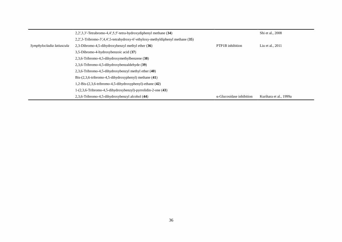
36
2,2′,3,3′-Tetrabromo-4,4′,5,5′-tetra-hydroxydiphenyl methane (34) Shi et al., 2008
2,2′,3-Tribromo-3′,4,4′,5-tetrahydroxy-6′-ethyloxy-methyldiphenyl methane (35)
Symphylocladia latiuscula 2,3-Dibromo-4,5-dihydroxybenzyl methyl ether (36) PTP1B inhibition Liu et al., 2011
3,5-Dibromo-4-hydroxybenzoic acid (37)
2,3,6-Tribromo-4,5-dihydroxymethylbenzene (38)
2,3,6-Tribromo-4,5-dihydroxybenzaldehyde (39)
2,3,6-Tribromo-4,5-dihydroxybenzyl methyl ether (40)
Bis-(2,3,6-tribromo-4,5-dihydroxyphenyl) methane (41)
1,2-Bis-(2,3,6-tribromo-4,5-dihydroxyphenyl)-ethane (42)
1-(2,3,6-Tribromo-4,5-dihydroxybenzyl)-pyrrolidin-2-one (43)
2,3,6-Tribromo-4,5-dihydroxybenzyl alcohol (44) α-Glucosidase inhibition Kurihara et al., 1999a
Recommended