Research Article
Loss of Mpdz impairs ependymal cell integrityleading to perinatal-onset hydrocephalus in miceAnja Feldner1, M Gordian Adam1,†, Fabian Tetzlaff1, Iris Moll1, Dorde Komljenovic2, Felix Sahm3,4,
Tobias Bäuerle2,‡, Hiroshi Ishikawa5, Horst Schroten6, Thomas Korff7, Ilse Hofmann8,9,
Hartwig Wolburg10, Andreas von Deimling3,4 & Andreas Fischer1,9,11,*
Abstract
Hydrocephalus is a common congenital anomaly. LCAM1 andMPDZ (MUPP1) are the only known human gene loci associatedwith non-syndromic hydrocephalus. To investigate functions ofthe tight junction-associated protein Mpdz, we generated mousemodels. Global Mpdz gene deletion or conditional inactivation inNestin-positive cells led to formation of supratentorial hydro-cephalus in the early postnatal period. Blood vessels, epithelialcells of the choroid plexus, and cilia on ependymal cells, whichline the ventricular system, remained morphologically intact inMpdz-deficient brains. However, flow of cerebrospinal fluidthrough the cerebral aqueduct was blocked from postnatal day3 onward. Silencing of Mpdz expression in cultured epithelialcells impaired barrier integrity, and loss of Mpdz in astrocytesincreased RhoA activity. In Mpdz-deficient mice, ependymal cellshad morphologically normal tight junctions, but expression ofthe interacting planar cell polarity protein Pals1 was diminishedand barrier integrity got progressively lost. Ependymal denuda-tion was accompanied by reactive astrogliosis leading to aque-ductal stenosis. This work provides a relevant hydrocephalusmouse model and demonstrates that Mpdz is essential tomaintain integrity of the ependyma.
Keywords aqueductal stenosis; cerebrospinal fluid; ependymal cells;
hydrocephalus; tight junction
Subject Category Neuroscience
DOI 10.15252/emmm.201606430 | Received 21 March 2016 | Revised 11 April
2017 | Accepted 12 April 2017 | Published online 12 May 2017
EMBO Mol Med (2017) 9: 890–905
Introduction
Congenital hydrocephalus, an abnormal accumulation of cere-
brospinal fluid (CSF) in brain cavities, is diagnosed in ~1 of 2,000
newborns (Schrander-Stumpel & Fryns, 1998; Garne et al, 2010).
Increasing amounts of CSF in the ventricular system raise the
intracranial pressure leading to compression of brain tissue and
enlargement of the head circumference. The most common treat-
ment is surgical insertion of a catheter to drain excess CSF into
another body cavity. However, this does not reverse 80–90% of the
neurological impairment of neonates with fetal onset hydro-
cephalus. Therefore, hydrocephalus may not only be a disorder of
CSF dynamics, but also a brain disorder (Guerra et al, 2015).
Disturbance of CSF formation, flow, or absorption can cause
congenital hydrocephalus (Jimenez et al, 2014). Overproduction of
CSF has been described in rare cases, in particular choroid plexus
hyperplasia or papilloma (Fujimoto et al, 2004). Brain malforma-
tions (e.g., Arnold–Chiari or Dandy–Walker syndrome) can obstruct
CSF flow (Schrander-Stumpel & Fryns, 1998), which is driven by
pressure gradients generated, for example, by blood pulsations.
However, it is assumed that motile, water-propelling cilia on
ependymal cells lining the ventricular system are also required for
proper CSF flow (Lee, 2013; Jimenez et al, 2014). This hypothesis is
supported by the fact that some ciliopathies are associated with
hydrocephalus and that various rodent models carrying mutations
in genes required for cilia function develop postnatal hydrocephalus
(Davy & Robinson, 2003; Ibanez-Tallon et al, 2004; Banizs et al,
2005; Lechtreck et al, 2008; Jacquet et al, 2009; Wodarczyk et al,
2009; Tissir et al, 2010; Liu et al, 2014; Koschutzke et al, 2015;
1 Vascular Signaling and Cancer, German Cancer Research Center (DKFZ), Heidelberg, Germany2 Division of Medical Physics in Radiology, German Cancer Research Center (DKFZ), Heidelberg, Germany3 Department of Neuropathology, Institute of Pathology, Ruprecht-Karls-University Heidelberg, Heidelberg, Germany4 Clinical Cooperation Unit Neuropathology, German Consortium for Translational Cancer Research (DKTK), German Cancer Research Center (DKFZ), Heidelberg, Germany5 Department of NDU Life Sciences, School of Life Dentistry, Nippon Dental University, Chiyoda-ku, Tokyo, Japan6 Pediatric Infectious Diseases, University Children’s Hospital Mannheim, Heidelberg University, Mannheim, Germany7 Department of Cardiovascular Research, Institute of Physiology and Pathophysiology, Heidelberg University, Heidelberg, Germany8 Vascular Oncology and Metastasis, German Cancer Research Center (DKFZ), Heidelberg, Germany9 Vascular Biology, CBTM, Medical Faculty Mannheim, Heidelberg University, Mannheim, Germany
10 Department of Pathology and Neuropathology, University of Tuebingen, Tuebingen, Germany11 Medical Clinic I, Endocrinology and Clinical Chemistry, Heidelberg University Hospital, Heidelberg, Germany
*Corresponding author. Tel: +49 6221 424150; E-mail: [email protected]†Present address: Immunocore Limited, Abingdon, Oxon, UK‡Present address: Institute of Radiology, University Medical Center Erlangen, Friedrich-Alexander-Universität Erlangen-Nürnberg, Erlangen, Germany
EMBO Molecular Medicine Vol 9 | No 7 | 2017 ª 2017 The Authors. Published under the terms of the CC BY 4.0 license890
Published online: May 12, 2017
Rachel et al, 2015). Lastly, congenital hydrocephalus can also be
acquired due to prenatal cerebral infections, injuries, or hemor-
rhages, which either inhibit CSF flow or CSF reabsorption (Tully &
Dobyns, 2014).
A genetic etiology is assumed for a large proportion of patients
suffering from congenital hydrocephalus. Up to date, mutations that
cause non-syndromic congenital hydrocephalus in humans have
been detected in only two genes. (i) Mutations in L1CAM on chro-
mosome Xq28 are responsible for X-linked recessive congenital
hydrocephalus (HSAS, OMIM #307000). L1CAM mutations can also
lead to syndromic disorders (L1 syndrome, CRASH syndrome) asso-
ciated with hydrocephalus (Tully & Dobyns, 2014). (ii) Homozygous
MPDZ loss-of-function mutations on chromosome 9p23 are respon-
sible for autosomal recessive non-syndromic hydrocephalus (HYC2,
OMIM #615219). Fetuses carrying truncating MPDZ mutations
developed macrocephaly, extreme dilation of the lateral ventricles
with dangling of the choroids and thinning of the cerebral cortex
(Al-Dosari et al, 2013).
Interestingly, both L1CAM and the multi-PDZ domain protein
(MPDZ, also known as MUPP1) are involved in cell–cell adhesion.
In mice, loss of the junctional adhesion molecule C (Jam3) also
causes hydrocephalus (Wyss et al, 2012). It was therefore hypothe-
sized that disruption of the junctions between cells of the ventricu-
lar zone may be the common cause (Al-Dosari et al, 2013; Jimenez
et al, 2014; Guerra et al, 2015). Mechanistically, this could occur
due to reactive astrogliosis following ependymal cell injury (Wagner
et al, 2003) to re-establish a surface between brain tissue and CSF
(Sarnat, 1995). In particular, astrogliosis within the cerebral aque-
duct would then obstruct CSF flow.
MPDZ contains 13 PDZ domains, which mediate multiple
protein–protein interactions, and thereby acts as a scaffold protein
for tight junction-associated proteins (JAM-A, claudin-1, ZO-3,
Pals1, Par6), adherens junction proteins (Nectins), transmembrane
receptors (Adachi et al, 2009), and the RhoA-specific guanine
exchange factor Syx (PLEKHG) (Estevez et al, 2008). Mpdz is
expressed ubiquitously in the brain of mouse embryos with high-
est mRNA levels in the ependymal lining of the ventricular
system, and the choroid plexus (Ullmer et al, 1998; Becamel et al,
2001; Sitek et al, 2003). The role of MPDZ for restricting epithe-
lial permeability is unclear. Silencing of MPDZ expression did not
alter paracellular permeability of EpH4 breast carcinoma cells
(Adachi et al, 2009). However, loss of MPDZ may be compen-
sated by increased expression of INADL (also known as InaD-like
or PATJ), a structural paralogue of the MPDZ protein (Adachi
et al, 2009; Assemat et al, 2013).
In this study, we generated global and conditional mouse models
to elucidate the pathogenesis of autosomal recessive non-syndromic
hydrocephalus by targeting the murine Mdpz locus.
Results
Targeting Mpdz in mice
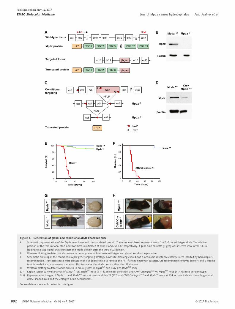
A homozygous MPDZ nonsense mutation in exon 6 which truncates
the MPDZ protein after the first PDZ domain was found in patients
suffering from autosomal recessive hydrocephalus (Al-Dosari et al,
2013). To target Mpdz in mice, we used embryonic stem cells, in
which a stop cassette had been inserted into intron 11–12 (clone
XG734; BayGenomics), and injected into mouse blastocysts. This
strategy leads to a premature stop codon and truncates the Mpdz
protein after the 3rd PDZ domain (Fig 1A). After germline transmis-
sion, we obtained heterozygous MpdzGt(XG734)Byg(+/�)1AFis mice
(Mpdz+/� mice), which were viable and fertile, and backcrossed
into the C57BL/6 strain. Western blotting revealed that intact Mpdz
protein could no longer be detected in brain lysates derived from
neonatal Mpdz�/� mice (Fig 1B).
In a second approach, loxP sequences were inserted into introns
3–4 and 5–6 by homologous recombination in ES cells. This allows
recombination of the floxed allele by Cre recombinase to delete exons
4 and 5 (MpdzD) resulting in a truncating frameshift mutation remov-
ing all 13 PDZ domains (Fig 1C). Cre recombinase was expressed
under control of the ubiquitously active CMV promoter (Schwenk
et al, 1995), leading to a strong reduction of Mpdz protein expression
levels in astrocytes isolated from neonatal mice (Fig 1D) Homozygous
floxed Mpdzfl/fl mice and heterozygous CMV-Cre;Mpdz+/D were
viable, fertile, and indistinguishable from control littermates.
Homozygous Mpdz mutations cause postnatal lethality
Mdpz+/� mice were interbred and we obtained offspring in the
expected Mendelian ratio (1:2:1) when animals were genotyped in
the embryo–fetal period. Genotyping between postnatal days 1 and
10 (P1–P10) revealed 88 (24%) wild-type, 205 (56%) heterozygous,
73 (20%) knockout mice, indicating a non-Mendelian distribution
[P = 0.038; chi-square test (Montoliu, 2012)]. Indeed, knockout
animals died in the postnatal period and median survival of Mpdz�/�
mice was 20 days (Fig 1E). No significant sex-specific differences
in survival were detectable. Similar data were obtained with CMV-
Cre;MpdzD/D mice. After crossing CMV-Cre;Mpdzwt/D with Mpdzfl/fl
mice, offspring CMV-Cre;MpdzD/D mice were born at expected
Mendelian frequency of 25% [n = 167; P = 0.02; chi-square test
(Montoliu, 2012)] and died during the first month of life (median
survival 25 days) (Fig 1F).
Mpdz�/� mouse pups looked unremarkable at P1 and P2. There
were no obvious phenotypic differences compared to littermate
controls. At P3, some of the homozygous mice could already be recog-
nized due to a slightly enlarged and dome-shaped skull. Brains isolated
from Mpdz�/� mice at P4 were enlarged (Fig 1G). From P5 onward,
Mpdz�/� mice showed growth retardation and increased frequency
and severity of neurological symptoms (decreased alertness, lethargy,
movement disorders, muscle weakness, apathy). Therefore, all mice
were sacrificed upon exhibition of movement disorders or lethargy.
Clinical autopsy and histopathological examination of Mpdz�/�
mice at P7 revealed macrocephaly, enlarged hemispheres, and
massive CSF accumulation in lateral brain ventricles. Hydro-
cephalus was never detected in any of the wild-type or heterozygous
mice. CSF was clear and there were no signs of brain hemorrhage.
Compared to wild-type littermates, no obvious alterations in heart,
liver, kidney, and gut were detectable. This resembles findings from
patients with homozygous MPDZ mutations (Al-Dosari et al, 2013).
CMV-Cre;MpdzD/D mice also developed macrocephaly and severe
CSF accumulation in lateral brain ventricles, indicating that trunca-
tion of Mpdz acts as a null allele (Fig 1H). In older mice (P21) with
advanced hydrocephalus, brain hemorrhages were frequently
observed.
ª 2017 The Authors EMBO Molecular Medicine Vol 9 | No 7 | 2017
Anja Feldner et al Loss of Mpdz causes hydrocephalus EMBO Molecular Medicine
891
Published online: May 12, 2017
A B
C
E F
G H
D
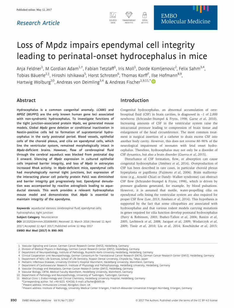
Figure 1. Generation of global and conditional Mpdz knockout mice.
A Schematic representation of the Mpdz gene locus and the translated protein. The numbered boxes represent exons 1–47 of the wild-type allele. The relativeposition of the translational start and stop sites is indicated at exon 2 and exon 47, respectively. A gene-trap cassette (b-geo) was inserted into intron 11–12leading to a stop signal that truncates the Mpdz protein after the third PDZ domain.
B Western blotting to detect Mpdz protein in brain lysates of littermate wild-type and global knockout Mpdz mice.C Schematic drawing of the conditional Mpdz gene targeting strategy. LoxP sites flanking exon 4 and a neomycin resistance cassette were inserted by homologous
recombination. Transgenic mice were crossed with Flp deleter mice to remove the FRT-flanked neomycin cassette. Cre recombinase removes exons 4 and 5 leadingto a frameshift and a nonsense mutation. This truncates the Mpdz protein after the L27 domain.
D Western blotting to detect Mpdz protein in brain lysates of Mpdzfl/fl and CMV-Cre;MpdzD/D mice.E, F Kaplan–Meier survival analysis of Mpdz�/� vs. Mpdz+/+ mice (n > 41 mice per genotype) and CMV-Cre;MpdzD/D vs. Mpdzfl/fl mice (n > 48 mice per genotype).G, H Representative images of Mpdz�/� and Mpdz+/+ mice at postnatal day 27 (P27) and CMV-Cre;MpdzD/D and Mpdzfl/fl mice at P24. Arrows indicate the enlarged and
dome-shaped skull and the enlarged brain hemispheres.
Source data are available online for this figure.
EMBO Molecular Medicine Vol 9 | No 7 | 2017 ª 2017 The Authors
EMBO Molecular Medicine Loss of Mpdz causes hydrocephalus Anja Feldner et al
892
Published online: May 12, 2017
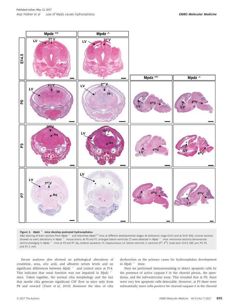
Mpdz�/� mice develop hydrocephalus after birth
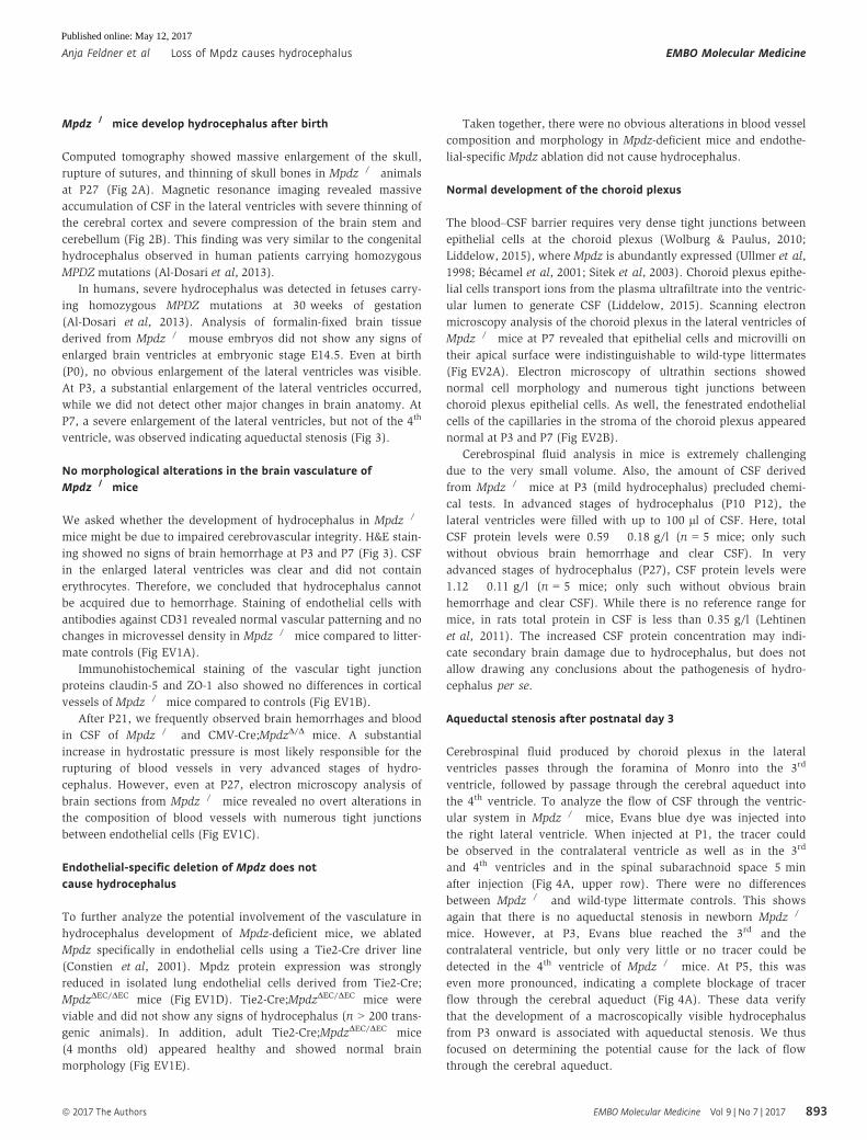
Computed tomography showed massive enlargement of the skull,
rupture of sutures, and thinning of skull bones in Mpdz�/� animals
at P27 (Fig 2A). Magnetic resonance imaging revealed massive
accumulation of CSF in the lateral ventricles with severe thinning of
the cerebral cortex and severe compression of the brain stem and
cerebellum (Fig 2B). This finding was very similar to the congenital
hydrocephalus observed in human patients carrying homozygous
MPDZ mutations (Al-Dosari et al, 2013).
In humans, severe hydrocephalus was detected in fetuses carry-
ing homozygous MPDZ mutations at 30 weeks of gestation
(Al-Dosari et al, 2013). Analysis of formalin-fixed brain tissue
derived from Mpdz�/� mouse embryos did not show any signs of
enlarged brain ventricles at embryonic stage E14.5. Even at birth
(P0), no obvious enlargement of the lateral ventricles was visible.
At P3, a substantial enlargement of the lateral ventricles occurred,
while we did not detect other major changes in brain anatomy. At
P7, a severe enlargement of the lateral ventricles, but not of the 4th
ventricle, was observed indicating aqueductal stenosis (Fig 3).
No morphological alterations in the brain vasculature ofMpdz�/� mice
We asked whether the development of hydrocephalus in Mpdz�/�
mice might be due to impaired cerebrovascular integrity. H&E stain-
ing showed no signs of brain hemorrhage at P3 and P7 (Fig 3). CSF
in the enlarged lateral ventricles was clear and did not contain
erythrocytes. Therefore, we concluded that hydrocephalus cannot
be acquired due to hemorrhage. Staining of endothelial cells with
antibodies against CD31 revealed normal vascular patterning and no
changes in microvessel density in Mpdz�/� mice compared to litter-
mate controls (Fig EV1A).
Immunohistochemical staining of the vascular tight junction
proteins claudin-5 and ZO-1 also showed no differences in cortical
vessels of Mpdz�/� mice compared to controls (Fig EV1B).
After P21, we frequently observed brain hemorrhages and blood
in CSF of Mpdz�/� and CMV-Cre;MpdzD/D mice. A substantial
increase in hydrostatic pressure is most likely responsible for the
rupturing of blood vessels in very advanced stages of hydro-
cephalus. However, even at P27, electron microscopy analysis of
brain sections from Mpdz�/� mice revealed no overt alterations in
the composition of blood vessels with numerous tight junctions
between endothelial cells (Fig EV1C).
Endothelial-specific deletion of Mpdz does notcause hydrocephalus
To further analyze the potential involvement of the vasculature in
hydrocephalus development of Mpdz-deficient mice, we ablated
Mpdz specifically in endothelial cells using a Tie2-Cre driver line
(Constien et al, 2001). Mpdz protein expression was strongly
reduced in isolated lung endothelial cells derived from Tie2-Cre;
MpdzDEC/DEC mice (Fig EV1D). Tie2-Cre;MpdzDEC/DEC mice were
viable and did not show any signs of hydrocephalus (n > 200 trans-
genic animals). In addition, adult Tie2-Cre;MpdzDEC/DEC mice
(4 months old) appeared healthy and showed normal brain
morphology (Fig EV1E).
Taken together, there were no obvious alterations in blood vessel
composition and morphology in Mpdz-deficient mice and endothe-
lial-specific Mpdz ablation did not cause hydrocephalus.
Normal development of the choroid plexus
The blood–CSF barrier requires very dense tight junctions between
epithelial cells at the choroid plexus (Wolburg & Paulus, 2010;
Liddelow, 2015), where Mpdz is abundantly expressed (Ullmer et al,
1998; Becamel et al, 2001; Sitek et al, 2003). Choroid plexus epithe-
lial cells transport ions from the plasma ultrafiltrate into the ventric-
ular lumen to generate CSF (Liddelow, 2015). Scanning electron
microscopy analysis of the choroid plexus in the lateral ventricles of
Mpdz�/� mice at P7 revealed that epithelial cells and microvilli on
their apical surface were indistinguishable to wild-type littermates
(Fig EV2A). Electron microscopy of ultrathin sections showed
normal cell morphology and numerous tight junctions between
choroid plexus epithelial cells. As well, the fenestrated endothelial
cells of the capillaries in the stroma of the choroid plexus appeared
normal at P3 and P7 (Fig EV2B).
Cerebrospinal fluid analysis in mice is extremely challenging
due to the very small volume. Also, the amount of CSF derived
from Mpdz�/� mice at P3 (mild hydrocephalus) precluded chemi-
cal tests. In advanced stages of hydrocephalus (P10�P12), the
lateral ventricles were filled with up to 100 ll of CSF. Here, totalCSF protein levels were 0.59 � 0.18 g/l (n = 5 mice; only such
without obvious brain hemorrhage and clear CSF). In very
advanced stages of hydrocephalus (P27), CSF protein levels were
1.12 � 0.11 g/l (n = 5 mice; only such without obvious brain
hemorrhage and clear CSF). While there is no reference range for
mice, in rats total protein in CSF is less than 0.35 g/l (Lehtinen
et al, 2011). The increased CSF protein concentration may indi-
cate secondary brain damage due to hydrocephalus, but does not
allow drawing any conclusions about the pathogenesis of hydro-
cephalus per se.
Aqueductal stenosis after postnatal day 3
Cerebrospinal fluid produced by choroid plexus in the lateral
ventricles passes through the foramina of Monro into the 3rd
ventricle, followed by passage through the cerebral aqueduct into
the 4th ventricle. To analyze the flow of CSF through the ventric-
ular system in Mpdz�/� mice, Evans blue dye was injected into
the right lateral ventricle. When injected at P1, the tracer could
be observed in the contralateral ventricle as well as in the 3rd
and 4th ventricles and in the spinal subarachnoid space 5 min
after injection (Fig 4A, upper row). There were no differences
between Mpdz�/� and wild-type littermate controls. This shows
again that there is no aqueductal stenosis in newborn Mpdz�/�
mice. However, at P3, Evans blue reached the 3rd and the
contralateral ventricle, but only very little or no tracer could be
detected in the 4th ventricle of Mpdz�/� mice. At P5, this was
even more pronounced, indicating a complete blockage of tracer
flow through the cerebral aqueduct (Fig 4A). These data verify
that the development of a macroscopically visible hydrocephalus
from P3 onward is associated with aqueductal stenosis. We thus
focused on determining the potential cause for the lack of flow
through the cerebral aqueduct.
ª 2017 The Authors EMBO Molecular Medicine Vol 9 | No 7 | 2017
Anja Feldner et al Loss of Mpdz causes hydrocephalus EMBO Molecular Medicine
893
Published online: May 12, 2017
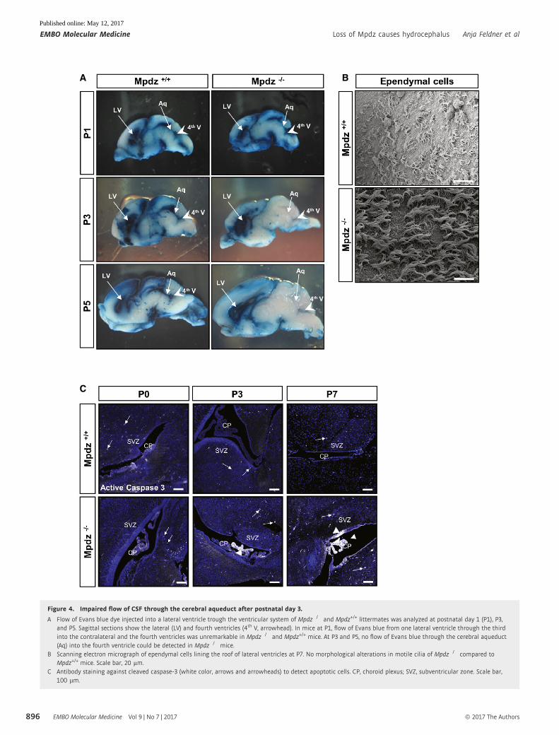
No overt cilia dysfunction in Mpdz-deficient brains
In humans, CSF flow is commonly ascribed to a pressure gradient
generated by blood pulsations between the sites of production and
absorption. However, it is also assumed that CSF movement
through the ventricular system is facilitated by motile 9 + 2 cilia at
the apical cell membrane of ependymal cells (Lee, 2013; Jimenez
et al, 2014). In mice, motile cilia generate CSF flow from P8 onward
(Tissir et al, 2010; Siyahhan et al, 2014), and cilia dysfunction
causes postnatal hydrocephalus (Davy & Robinson, 2003; Ibanez-
Tallon et al, 2004; Banizs et al, 2005; Lechtreck et al, 2008; Jacquet
et al, 2009; Wodarczyk et al, 2009; Tissir et al, 2010; Liu et al,
2014; Koschutzke et al, 2015; Rachel et al, 2015).
Scanning electron microscopy revealed normal numbers of cilia
on the ependymal cell surface within the lateral ventricles of
Mpdz�/� mice at P7 (Fig 4B). In accordance with normal cilia
morphology, we could not detect typical signs of a ciliopathy in
Mpdz�/� mice, such as cysts in liver or kidney (Fig EV2C).
A B
Figure 2. Mpdz-deficient mice develop hydrocephalus.
A At postnatal day 27 (P27), Mpdz�/� and wild-type littermates were subjected to a computed tomography. Three-dimensional reconstruction shows macrocephaly andthinning of skull bones (arrows).
B T2-weighted coronal and sagittal magnetic resonance images of the head of Mpdz�/� vs. Mpdz+/+ mice at P27. CSF in the enlarged lateral ventricles appearshyperintense (asterisks).
EMBO Molecular Medicine Vol 9 | No 7 | 2017 ª 2017 The Authors
EMBO Molecular Medicine Loss of Mpdz causes hydrocephalus Anja Feldner et al
894
Published online: May 12, 2017
Serum analyses also showed no pathological alterations of
creatinine, urea, uric acid, and albumin serum levels and no
significant differences between Mpdz�/� and control mice at P14.
This indicates that renal function was not impaired in Mpdz�/�
mice. Taken together, the normal cilia morphology and the fact
that motile cilia generate significant CSF flow in mice only from
P8 and onward (Tissir et al, 2010) dismisses the idea of cilia
dysfunction as the primary cause for hydrocephalus development
in Mpdz�/� mice.
Next we performed immunostaining to detect apoptotic cells by
the presence of active caspase-3 in the choroid plexus, the epen-
dyma, and the subventricular zone. This revealed that at P0, there
were very few apoptotic cells detectable. However, at P3 there were
substantially more cells positive for cleaved caspase-3 in the choroid
Figure 3. Mpdz�/� mice develop postnatal hydrocephalus.H&E staining of brain sections from Mpdz�/� and littermate Mpdz+/+ mice at different developmental stages. At embryonic stage E14.5 and at birth (P0), coronal sectionsshowed no overt alterations in Mpdz�/� mouse brains. At P3 and P7, enlarged lateral ventricles (*) were detected in Mpdz�/� mice. Horizontal sections demonstrateventriculomegaly in Mpdz�/� mice at P3 and P7. Aq, cerebral aqueduct; Hi, hippocampus; LV, lateral ventricle; V, ventricle (3rd, 4th). Scale bars: E14.5, 500 lm; P0, P3,and P7, 1 mm.
ª 2017 The Authors EMBO Molecular Medicine Vol 9 | No 7 | 2017
Anja Feldner et al Loss of Mpdz causes hydrocephalus EMBO Molecular Medicine
895
Published online: May 12, 2017
A
C
B
Figure 4. Impaired flow of CSF through the cerebral aqueduct after postnatal day 3.
A Flow of Evans blue dye injected into a lateral ventricle trough the ventricular system of Mpdz�/� and Mpdz+/+ littermates was analyzed at postnatal day 1 (P1), P3,and P5. Sagittal sections show the lateral (LV) and fourth ventricles (4th V, arrowhead). In mice at P1, flow of Evans blue from one lateral ventricle through the thirdinto the contralateral and the fourth ventricles was unremarkable in Mpdz�/� and Mpdz+/+ mice. At P3 and P5, no flow of Evans blue through the cerebral aqueduct(Aq) into the fourth ventricle could be detected in Mpdz�/� mice.
B Scanning electron micrograph of ependymal cells lining the roof of lateral ventricles at P7. No morphological alterations in motile cilia of Mpdz�/� compared toMpdz+/+ mice. Scale bar, 20 lm.
C Antibody staining against cleaved caspase-3 (white color, arrows and arrowheads) to detect apoptotic cells. CP, choroid plexus; SVZ, subventricular zone. Scale bar,100 lm.
EMBO Molecular Medicine Vol 9 | No 7 | 2017 ª 2017 The Authors
EMBO Molecular Medicine Loss of Mpdz causes hydrocephalus Anja Feldner et al
896
Published online: May 12, 2017
plexus and the ependyma of Mpdz�/� mice compared to controls.
At P7, we detected strong caspase-3 activity within the ependymal
cell layer and throughout the choroid plexus (Fig 4C).
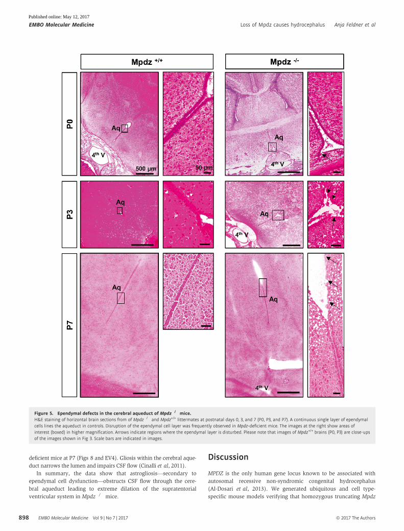
Loss of Mpdz leads to defects in the ependyma
The ventricular system is lined by the ependyma, a single layer of
simple cuboidal to columnar epithelium with microvilli and motile
cilia on the apical surface. Mpdz expression is very pronounced in
this cell layer (Becamel et al, 2001; Sitek et al, 2003), which sepa-
rates CSF from brain tissue. Immunostaining indicated higher rates
of cell death in the ependyma of Mpdz�/� compared to wild-type
littermate controls (Fig 4C). H&E staining revealed that at P0, there
were some sporadic defects within the ependymal layer of the
lateral ventricles of Mpdz�/� mice. Most notably, we found severe
defects in the ependymal lining of the cerebral aqueduct. Here single
cells or a whole stretch of ependymal cells was missing. At P3 and
P7, this was even more severe. In parts of the cerebral aqueduct,
ependymal denudation had occurred (Fig 5). This indicates that loss
of tight junction-associated protein Mpdz disturbs the integrity of
the ependymal layer leading to partial ependymal denudation.
Inactivation of Mpdz with Nestin-Cre leads tohydrocephalus formation
To further test this hypothesis, we inactivated the Mpdz gene in
radial glia cells, the precursors for ependymal cells, and neuronal
precursors using the Nestin-Cre driver line (Tronche et al, 1999).
Indeed, Nestin-Cre+/+;MpdzD/D mice developed hydrocephalus in
the early postnatal period. Very similar to the global Mpdz�/� mice,
there was severe enlargement of the lateral ventricles (Fig EV3).
Loss of Mpdz leads to impaired epithelial barrier function
As the loss of Mpdz induced major changes in the ependyma, we
were wondering whether Mpdz might have a function in epithe-
lial barrier control. It is important to note that presence and
normal morphological appearance of tight junctions do not
exclude a barrier defect. This had been shown, for example, in
occludin (Ocln)- or claudin-5 (Cldn5)-deficient mice, in which
tight junctions are still formed but are not fully functional (Saitou
et al, 2000; Nitta et al, 2003). Therefore, we analyzed cellular
permeability after silencing of Mpdz expression. Due to a lack of
suitable ependymal cell lines and the difficulty to culture suitable
amounts of primary ependymal cells from mice, we employed
human MCF7 cells as surrogate model to study cellular barrier
functions. MPDZ expression was silenced by transfection with
siRNAs or by transduction with lentiviral particles expressing
shRNAs by 80 � 7%. Cellular permeability was determined by
measuring transepithelial electrical resistance (TER) and corre-
sponding capacitance (Ccl). Once cells had reached full conflu-
ence, as indicated by low Ccl, we observed severely reduced TER
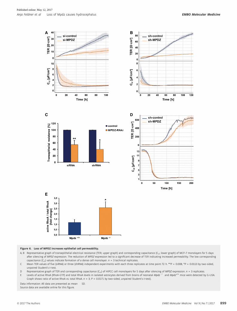
in MCF7 cells, indicating impaired barrier properties (Fig 6A–C).
These findings could be verified with human choroid plexus
epithelial papilloma (HIBCPP) cells. HIBCPP cells form tight junc-
tions, develop a high electrical resistance and minimal levels of
macromolecular flux when grown on transwell filters, and
thereby represent an excellent model system for the blood–
cerebrospinal fluid barrier (Schwerk et al, 2012). Silencing of
MPDZ expression led to lower TER indicating impaired barrier
function of HIBCPP cells (Fig 6D).
Mpdz binds to the RhoA-specific guanine exchange factor Syx
(PLEKHG) (Estevez et al, 2008), and recruits it to the Pals1 polarity
complex. Thereby, Mpdz is involved in controlling the activity of
RhoA. This small G protein can remodel the cytoskeleton and cell–cell
junctions leading to increased endothelial permeability (Wu et al,
2011; Ngok et al, 2012). In oligodendrocytes, NG2 stimulates RhoA
activity at the cell periphery via Mpdz (MUPP1) and Syx1 (Biname
et al, 2013). Notably, increased Rho kinase activity after deletion of
myosin IXa can lead to hydrocephalus in mice (Abouhamed et al,
2009). To test whether Mpdz also controls RhoA activity in cells of the
CNS, we analyzed its activity in primary astrocytes isolated from
neonatal mouse brains. Astrocyte lysates derived from Mpdz�/� mice
had significantly higher RhoA activity levels compared to those
derived from wild-type littermate controls (Fig 6E).
Taken together, the data showed that Mpdz is critical to maintain
an intact ependymal cell layer and epithelial barrier integrity.
Disturbed Pals1 expression in Mpdz�/� mice
Mpdz is associated with components of the planar cell polarity
complex via binding to Pals1 (Wu et al, 2011), and disturbance of
planar cell polarity can lead to hydrocephalus in mouse models
(Tissir et al, 2010; Ohata et al, 2014). We detected that the onset of
Pals1 expression in the ependymal cell layer is delayed in neonatal
Mpdz�/� mice. Whereas wild-type littermate controls showed
robust Pals1 expression in ependymal cells of the lateral ventricles
already at P0, this was almost absent in Mpdz�/� mice. Only from
P7 on there was Pals1 expression detectable in ependymal cells but
still not continuous through the ependymal layer (Fig 7A).
However, the expression levels and dynamic expression pattern of
the planar cell polarity protein Crb3 were unremarkable in Mpdz�/�
mice (Fig 7B). Also, the expression pattern of adherens junction
protein E-cadherin was not altered in Mpdz�/� mice (Fig 7C).
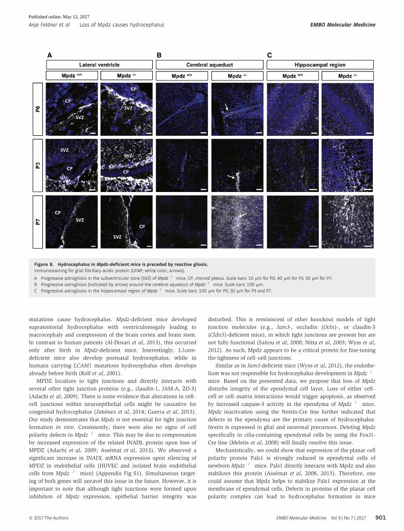
Hydrocephalus development is preceded by astrogliosis
Defects of the ependyma lead to reactive astrogliosis, which can
obstruct the cerebral aqueduct (Wagner et al, 2003). Therefore, we
tested whether ependymal defects in Mpdz�/� mice are accompa-
nied by astrogliosis. This repair mechanism is characterized by
enhanced glial fibrillary acidic protein (GFAP) expression, cellular
hypertrophy, and astrocyte proliferation (Fawcett & Asher, 1999). At
P0, when no hydrocephalus was present, GFAP expression was
prominent in the ependyma of the lateral ventricles and in the
subependymal zone of Mpdz�/� mice. Strong GFAP expression was
also observed in the hippocampal region of Mpdz�/� but not in
control mice. Most importantly, we observed astrogliosis within the
cerebral aqueduct of Mpdz�/� mice (Figs 8 and EV4). This clearly
indicates that these cells are subjected to stress or damage prior to
the development of a hydrocephalus.
At P3, we detected much stronger GFAP expression in the epen-
dyma and the subependymal zone in Mpdz�/� mice compared to
control, which was even more pronounced at P7. In particular, the
cerebral aqueduct was affected. In addition, strong astrogliosis in
Mpdz�/� mice was observed in the hippocampal region of Mpdz-
ª 2017 The Authors EMBO Molecular Medicine Vol 9 | No 7 | 2017
Anja Feldner et al Loss of Mpdz causes hydrocephalus EMBO Molecular Medicine
897
Published online: May 12, 2017
deficient mice at P7 (Figs 8 and EV4). Gliosis within the cerebral aque-
duct narrows the lumen and impairs CSF flow (Cinalli et al, 2011).
In summary, the data show that astrogliosis—secondary to
ependymal cell dysfunction—obstructs CSF flow through the cere-
bral aqueduct leading to extreme dilation of the supratentorial
ventricular system in Mpdz�/� mice.
Discussion
MPDZ is the only human gene locus known to be associated with
autosomal recessive non-syndromic congenital hydrocephalus
(Al-Dosari et al, 2013). We generated ubiquitous and cell type-
specific mouse models verifying that homozygous truncating Mpdz
Figure 5. Ependymal defects in the cerebral aqueduct of Mpdz�/� mice.H&E staining of horizontal brain sections from of Mpdz�/� and Mpdz+/+ littermates at postnatal days 0, 3, and 7 (P0, P3, and P7). A continuous single layer of ependymalcells lines the aqueduct in controls. Disruption of the ependymal cell layer was frequently observed in Mpdz-deficient mice. The images at the right show areas ofinterest (boxed) in higher magnification. Arrows indicate regions where the ependymal layer is disturbed. Please note that images of Mpdz+/+ brains (P0, P3) are close-upsof the images shown in Fig 3. Scale bars are indicated in images.
EMBO Molecular Medicine Vol 9 | No 7 | 2017 ª 2017 The Authors
EMBO Molecular Medicine Loss of Mpdz causes hydrocephalus Anja Feldner et al
898
Published online: May 12, 2017
A B
C
E
D
Figure 6. Loss of MPDZ increases epithelial cell permeability.
A, B Representative graph of transepithelial electrical resistance (TER; upper graph) and corresponding capacitance (Ccl; lower graph) of MCF-7 monolayers for 5 daysafter silencing of MPDZ expression. The reduction of MPDZ expression led to a significant decrease of TER indicating increased permeability. The low correspondingcapacitance (Ccl) values indicate formation of a dense cell monolayer. n = 3 technical replicates.
C Mean TER values of five (siRNA) or three (shRNA) independent experiments with each three replicates at time point 72 h. **P = 0.008; *P = 0.0113 by two-sided,unpaired Student’s t-test.
D Representative graph of TER and corresponding capacitance (Ccl) of HIPCC cell monolayers for 5 days after silencing of MPDZ expression. n = 3 replicates.E Levels of active RhoA (RhoA-GTP) and total RhoA levels in isolated astrocytes derived from brains of neonatal Mpdz�/� and Mpdz+/+ mice were detected by G-LISA.
Graph shows ratio of active RhoA vs. total RhoA. n = 3; P = 0.0171 by two-sided, unpaired Student’s t-test).
Data information: All data are presented as mean � SD.Source data are available online for this figure.
ª 2017 The Authors EMBO Molecular Medicine Vol 9 | No 7 | 2017
Anja Feldner et al Loss of Mpdz causes hydrocephalus EMBO Molecular Medicine
899
Published online: May 12, 2017
A
C
B
Figure 7. Diminished expression of Pals1 in ependymal cells of Mpdz�/� mice.Immunostaining for the planar cell polarity proteins Pals1 and Crb3 (white color) and the adherens junction protein E-cadherin (brown color) in brains ofMpdz-deficient miceat postnatal days 0, 3, and 7 (P0, P3, and P7).
A Expression of Pals1 (white color, arrows) is absent (*) or diminished in Mpdz�/� mice. Scale bars: 10 lm for P0, 20 lm for P3, 10 lm for P7.B Expression of Crb3 is not altered in Mpdz�/� compared to control mice. Scale bars: 10 lm for P0, 20 lm for P3, 10 lm for P7.C The dynamic expression pattern of E-cadherin is not altered in Mpdz�/� compared to control mice. Scale bars: 100 lm and for zoom-ins 10 lm.
Data information: CP, choroid plexus; EP, ependymal cells; LV, lateral ventricle.
EMBO Molecular Medicine Vol 9 | No 7 | 2017 ª 2017 The Authors
EMBO Molecular Medicine Loss of Mpdz causes hydrocephalus Anja Feldner et al
900
Published online: May 12, 2017
mutations cause hydrocephalus. Mpdz-deficient mice developed
supratentorial hydrocephalus with ventriculomegaly leading to
macrocephaly and compression of the brain cortex and brain stem.
In contrast to human patients (Al-Dosari et al, 2013), this occurred
only after birth in Mpdz-deficient mice. Interestingly, L1cam-
deficient mice also develop postnatal hydrocephalus, while in
humans carrying LCAM1 mutations hydrocephalus often develops
already before birth (Rolf et al, 2001).
MPDZ localizes to tight junctions and directly interacts with
several other tight junction proteins (e.g., claudin-1, JAM-A, ZO-3)
(Adachi et al, 2009). There is some evidence that alterations in cell–
cell junctions within neuroepithelial cells might be causative for
congenital hydrocephalus (Jimenez et al, 2014; Guerra et al, 2015).
Our study demonstrates that Mpdz is not essential for tight junction
formation in vivo. Consistently, there were also no signs of cell
polarity defects in Mpdz�/� mice. This may be due to compensation
by increased expression of the related INADL protein upon loss of
MPDZ (Adachi et al, 2009; Assemat et al, 2013). We observed a
significant increase in INADL mRNA expression upon silencing of
MPDZ in endothelial cells (HUVEC and isolated brain endothelial
cells from Mpdz�/� mice) (Appendix Fig S1). Simultaneous target-
ing of both genes will unravel this issue in the future. However, it is
important to note that although tight junctions were formed upon
inhibition of Mpdz expression, epithelial barrier integrity was
disturbed. This is reminiscent of other knockout models of tight
junction molecules (e.g., Jam3-, occludin (Ocln)-, or claudin-5
(Cldn5)-deficient mice), in which tight junctions are present but are
not fully functional (Saitou et al, 2000; Nitta et al, 2003; Wyss et al,
2012). As such, Mpdz appears to be a critical protein for fine-tuning
the tightness of cell–cell junctions.
Similar as in Jam3-deficient mice (Wyss et al, 2012), the endothe-
lium was not responsible for hydrocephalus development inMpdz�/�
mice. Based on the presented data, we propose that loss of Mpdz
disturbs integrity of the ependymal cell layer. Loss of either cell–
cell or cell–matrix interactions would trigger apoptosis, as observed
by increased caspase-3 activity in the ependyma of Mpdz�/� mice.
Mpdz inactivation using the Nestin-Cre line further indicated that
defects in the ependyma are the primary cause of hydrocephalus.
Nestin is expressed in glial and neuronal precursors. Deleting Mpdz
specifically in cilia-containing ependymal cells by using the FoxJ1-
Cre line (Meletis et al, 2008) will finally resolve this issue.
Mechanistically, we could show that expression of the planar cell
polarity protein Pals1 is strongly reduced in ependymal cells of
newborn Mpdz�/� mice. Pals1 directly interacts with Mpdz and also
stabilizes this protein (Assemat et al, 2008, 2013). Therefore, one
could assume that Mpdz helps to stabilize Pals1 expression at the
membrane of ependymal cells. Defects in proteins of the planar cell
polarity complex can lead to hydrocephalus formation in mice
A B C
Figure 8. Hydrocephalus in Mpdz-deficient mice is preceded by reactive gliosis.Immunostaining for glial fibrillary acidic protein (GFAP; white color, arrows).
A Progressive astrogliosis in the subventricular zone (SVZ) of Mpdz�/� mice. CP, choroid plexus. Scale bars: 10 lm for P0, 40 lm for P3, 50 lm for P7.B Progressive astrogliosis (indicated by arrow) around the cerebral aqueduct of Mpdz�/� mice. Scale bars: 100 lm.C Progressive astrogliosis in the hippocampal region of Mpdz�/� mice. Scale bars: 100 lm for P0, 50 lm for P3 and P7.
ª 2017 The Authors EMBO Molecular Medicine Vol 9 | No 7 | 2017
Anja Feldner et al Loss of Mpdz causes hydrocephalus EMBO Molecular Medicine
901
Published online: May 12, 2017
(Tissir et al, 2010; Ohata et al, 2014). The expression of another cell
polarity protein Crb3 was not affected in the ependyma of Mpdz�/�
mice, indicating not a general impairment of planar cell polarity.
Pals1 is linked to the RhoA-specific guanine exchange factor Syx via
the adaptor protein Mpdz (Estevez et al, 2008). As such, Mpdz is
involved in the control of RhoA activity (Wu et al, 2011; Ngok et al,
2012; Biname et al, 2013). RhoA-dependent kinases play a major
role in controlling the stability of cell–cell junctions. Increased RhoA
activity after deletion of myosin IXa (Myo9a), a motor molecule with
a Rho GTPase-activating (GAP) domain, leads to hydrocephalus
formation in mice (Abouhamed et al, 2009). The Myo9a knockout
model shows many similarities with Mpdz�/� mice with distortion
of the ependymal cell layer, stenosis of the aqueduct, and dilation of
the lateral and third ventricles. Similar as observed after silencing of
Mpdz expression, there was increased RhoA activity in cultured
epithelial cells after silencing of Myo9a (Abouhamed et al, 2009).
Disturbance of ependymal integrity induces hydrocephalus in
other mouse models, for example, mice deficient for Myo9a, Pkcl,
Numbl, Ophn1, Myh4, Dlg5, or Mdnah5 (Ibanez-Tallon et al, 2004;
Imai et al, 2006; Kuo et al, 2006; Khelfaoui et al, 2007; Ma et al,
2007; Nechiporuk et al, 2007; Abouhamed et al, 2009). Ependymal
damage most likely impairs the brain–CSF barrier and disturbs CSF
homeostasis. We suggest that this subsequently results in the initia-
tion of repair processes, in particular reactive astrogliosis. The data
indicate that at birth, when no signs of hydrocephalus are yet visi-
ble, ependymal defects and reactive gliosis are present in the epen-
dyma and subventricular zone of Mpdz�/� mice. Astrogliosis is, in
principle, beneficial; however, in the narrow aqueduct, astrogliosis
can rapidly obstruct CSF flow into the 4th ventricle. As such, this
work showed that slight impairment of ependymal integrity triggers
astrogliosis in the subependymal zone, resulting in aqueductal
stenosis and ventriculomegaly in Mpdz�/� mice.
Materials and Methods
Animal experiments
Mice were kept under pathogen-free barrier conditions, and animal
procedures were performed in accordance with the institutional and
national regulations and approved by the local committees for
animal experimentation (Heidelberg University and DKFZ) and the
local government (Regierungsprasidium Karlsruhe, Germany).
Murine embryonic stem cells containing a gene trap in intron 11–12
of the Mpdz gene were obtained from the Mutant Mouse Resource &
Research Center (MMRRC) and injected into blastocysts. Anesthesia
was administered by intraperitoneal injection of 100 mg/kg keta-
mine (Ketavet, Pfizer) and 20 mg/kg xylazine (Rompun 2%, Bayer).
Chimeric mice were crossed to C57BL/6 mice. Heterozygous
MpdzGt(XG734)Byg(+/�)1AFis mice (Mpdz+/� mice) were further back-
crossed to C57BL/6 mice for at least eight generations.
Floxed Mpdz mice were generated by homologous recombination
in C57Bl/6 × 129/SvEv hybrid ES cells (inGenious Targeting Labo-
ratory, Ronkonkoma, NY, USA) and crossed with mice expressing
Cre recombinase under control of the CMV promoter for ubiquitous
gene ablation (Schwenk et al, 1995). Tie2-Cre mice allowed gene
deletion primarily in endothelial cells and a subset of hematopoietic
cells (Constien et al, 2001) and Nestin-Cre for deletion in radial glia
(Tronche et al, 1999). All Cre lines were backcrossed into C57Bl/6
for more than 10 generations.
Magnetic resonance imaging and volumetriccomputed tomography
Mice were anaesthetized using a mixture of isofluran (1.5%) and
oxygen (0.5 l/min). Magnetic resonance (MR) images were acquired
on a 1.5-T clinical MR scanner (Symphony, Siemens, Germany)
using a homebuilt coil for radiofrequency excitation and detection,
designed as a cylindrical volume resonator (n = 3 mice per geno-
type). Morphological T2-weighted images were acquired using a
turbo spin-echo sequence (orientation axial, TR 3,240 ms, TE
81 ms, matrix 152 × 256, resolution 0.35 × 0.35 × 1.5 mm3, 3 aver-
ages, 15 images, scan time 3:40 min).
Volumetric computed tomography (VCT) imaging was obtained
using the following parameters: tube voltage 80 kV, tube current
50 mA, scan time 51 s, rotation speed 10 s, frames per second 120,
and slice thickness 0.2 mm. Image reconstructions were performed
using a modified FDK (Feldkamp Davis Kress) cone beam recon-
struction algorithm (kernel H80a; Afra, Erlangen, Germany). Unen-
hanced VCT images and MRI-acquired T2-weighted images were
analyzed using Osirix Imaging Software.
Antibodies
For immunofluorescence analysis, the following primary antibodies
were used: rabbit anti-ZO-1 (1:100 dilution, Thermo Fisher, #61-
7300), rabbit anti-claudin-5 (1:400, Abcam, #ab53765), rabbit anti-
GFAP (1:500, DAKO, #Z0334), rat anti-CD31 (clone Mec13.3, BD
Biosciences), Pals1 (1:100, Merck, #7-708), rat anti-Crb3 (14F9,
Abcam, ab180835), mouse anti-E-cadherin (1:200, BD Bioscience,
#610181), and rabbit anti-cleaved caspase-3 (Asp175, 1:100 Cell
Signaling, #9661). Secondary antibodies were 3 mg/ml Alexa 546-
conjugated goat anti-rabbit IgG and 3 mg/ml Alexa 488-conjugated
goat anti-rat IgG (all from Invitrogen). For Western blotting, rabbit
anti-MPDZ (1:500, Thermo Scientific, 42-2700) and mouse anti-beta
actin (1:2,500, Sigma-Aldrich #A5441) were used.
Immunofluorescence and histology
Freshly dissected brains were embedded in Tissue-Tek (Sakura,
Netherlands), frozen, and stored at�80°C. Sections (7 lm) were fixed
in methanol for 20 min at �20°C. For GFAP staining, mice were
perfused with 4% paraformaldehyde in PBS through the left ventricle
of the heart and tissue was embedded in paraffin. Paraffin was
removed from fresh sections (4 lm), and antigen retrieval was carried
out in citrate buffer (pH 6.0). Blocking solution was 10% goat serum.
Primary and secondary antibodies were diluted in blocking solution.
Primary antibodies were incubated overnight at 4°C and secondary
antibodies for 1 h at room temperature. Sections were washed with
TBS-T between incubation steps and finally mounted with Fluoro-
mount (Dako). Confocal images were obtained using a LSM 700
microscope (Carl Zeiss) and analyzed using the Fiji software.
For histology, paraffin-embedded tissue sections were stained
with hematoxylin and eosin. Images were taken with a CellObserver
microscope (Carl Zeiss) and analyzed using the ZEN Blue software
(Carl Zeiss).
EMBO Molecular Medicine Vol 9 | No 7 | 2017 ª 2017 The Authors
EMBO Molecular Medicine Loss of Mpdz causes hydrocephalus Anja Feldner et al
902
Published online: May 12, 2017
Electron microscopy
Newborn Mpdz-deficient and wild-type littermates (n = 3 for each
age and genotype) were euthanized and perfused with PBS and
subsequently with 4% glutaraldehyde and 4% formaldehyde in
0.1 M cacodylate buffer through the left ventricle of the heart.
Dissected brains were fixed in 2.0% glutaraldehyde in 0.05 M
cacodylate buffer for 2 h and stained with 1% OsO4 in cacodylate
buffer for another 2 h followed by contrasting in 0.5% uranyl-
acetate. After dehydration in an ascending series of ethanol and
propylene oxide, the samples were flat-embedded in Epon (Serva,
Germany). Using an ultramicrotome (Ultracut, Leica, Bensheim,
Germany), 0.5-lm- and 50-nm-thin sections were cut. Ultrathin
sections were stained with 2% uranyl-acetate for 15 min and
contrasted in lead citrate for 5 min, mounted on copper grids, and
finally analyzed with a Zeiss-EM910 electron microscope.
For scanning electron microscopy, glutaraldehyde-fixed brains
were dehydrated, transferred into hexamethyldisilazane, and slowly
air-dried. Samples were cut and coated with gold using the agar
sputter coater (Agar Scientific, England). Images were taken using
the Hitachi S4500 and analyzed with the digital image processing
2.6 software (Point Electronic, Halle).
Ventricular dye injections
For intracranial dye injection, mice (n = 5 per genotype) were anes-
thetized by intraperitoneal injection of ketamine and xylazine
according to approved experimental protocols. Evans blue dye
(5 ll, 1% in PBS) was injected slowly into the lateral ventricle using
a 0.1-ml syringe. The syringe was left in the injection site to prevent
reflux of fluid. Mice were well euthanized 5 min after the injection
and the heads were immediately fixed in 4% paraformaldehyde
overnight. Brains were dissected, and photographed with an
SMZ800 stereo microscope (Nikon).
Analysis of serum creatinine, urea, and albumin
Plasma and CSF metabolites were measured in the central labora-
tory of the University Hospital Heidelberg by the same procedures
used and validated for routine diagnostic analysis (n = 5 mice).
Creatinine, urea, and uric acid were measured enzymatically, and
total protein in CSF was measured by precipitation with pyrogallol
red on an ADVIA 2400 clinical chemistry XPT analyzer (Siemens
Healthcare Diagnostics, Eschborn, Germany).
Cell culture
MCF7 cells were cultured in DMEM containing 10% fetal calf serum
(FCS), 100 units/ml penicillin, and 100 lg/ml streptomycin. Human
choroid plexus epithelial papilloma HIBCPP cells were cultured
in DMEM/F12 supplemented with 10% FCS, 5 lg/ml insulin,
100 U/ml penicillin, and 100 lg/ml streptomycin. For experiments,
cells grown in the standard and the inverted cell culture insert
system were used as previously described (Schwerk et al, 2012).
Three different lentiviral shRNA vectors for silencing MPDZ were
obtained from Biocat (V2LHS_3656, 16945 16946). Transduced
MCF7 cells were selected with 10 lM blasticidin-S. Transient trans-
fections were performed using Oligofectamine (Life Technologies).
Subsequently, MCF7 cells were seeded onto transwell inserts
(0.4 lm pore diameter, Greiner BioOne) and used for transepithelial
resistance measurements (cellZscope, nanoAnalytics). Medium was
exchanged every second day. Astrocytes from neonatal mice (P2)
were isolated and cultured as described (Reischl et al, 2014).
Standardized multiplex cell contamination and cell line authentica-
tion testing (Multiplexion, Germany) were conducted on a regular basis.
Quantitative real-time PCR
Total RNA was isolated with the innuPREP RNA Mini Kit (Jena
Analytics) and transcribed into cDNA (High Capacity cDNA Reverse
Transcription Kit; Applied Biosystems). cDNA was mixed with
POWER SYBR Green Master Mix and qPCR performed using an ABI
StepOnePlus cycler (Applied Biosystems). The housekeeping genes
GAPDH, RPL32, and OAZ1 were used for normalization. Primer
sequences are provided upon request. All experiments included two
technical and three biological replicates.
RhoA activity analysis
RhoA activation (ratio RhoA-GTP/total RhoA) was determined using
the RhoA G-LISA Activation Assay (Cytoskeleton Inc., BK124).
Statistical analyses
Results are expressed as means plus/minus standard deviation.
Comparisons between groups were made by a two-sided, unpaired
t-test. Comparison between multiple groups was made by ANOVA.
Alterations in Mendelian distribution were calculated with the soft-
ware tool Mendel.xls based on a chi-square test (Montoliu, 2012).
P-values < 0.05 were considered as significant.
Study approval
All animal work was approved by the local committees for animal
experimentation (Heidelberg University and DKFZ) and the local
The paper explained
ProblemCongenital hydrocephalus, an abnormal accumulation of CSF in braincavities, is diagnosed in ~1 of 2,000 newborns. A genetic etiology isassumed for a large proportion of patients. Mutations that cause non-syndromic congenital hydrocephalus in humans have been detectedin only two genes: L1CAM and MPDZ. The function of MPDZ is not fullyunderstood.
ResultsMouse models to inactivate the Mpdz gene were generated, and thesemimic the human pathology. Mpdz is needed to maintain the integ-rity of the ependymal cell layer in the brain that forms the cere-brospinal fluid–brain barrier. Loss of Mpdz leads to detachment ofependymal cells resulting in astrogliosis, a repair process that leads toobstruction of CSF transport through the cerebral aqueduct.
ImpactThe work provides a fully penetrant and clinical relevant model tostudy the pathogenesis of non-syndromic congenital hydrocephalus.
ª 2017 The Authors EMBO Molecular Medicine Vol 9 | No 7 | 2017
Anja Feldner et al Loss of Mpdz causes hydrocephalus EMBO Molecular Medicine
903
Published online: May 12, 2017
government (Regierungsprasidium Karlsruhe, Germany). This work
is not considered “Human Subjects Research”.
Expanded View for this article is available online.
AcknowledgementsWe thank Katharina Neumeier, Christian Clappier, and Sonja Reidenbach for
technical assistance, Ulrich Kloz and Frank van der Hoeven for generating
transgenic mice, Dr. Damir Krunic (DKFZ microscopy core facility) for help with
FIJI software data analysis, Dr. Manfred Ruppel (Frankfurt University) for
performing scanning electron microscopy, and members of the DKFZ Labora-
tory Animal Facility for support. We are grateful to Gabi Frommer-Kästle for
skillful help with the ultrathin sectioning, to Dr. Bernd Arnold (DFKZ Heidel-
berg) for providing Tie2-Cre mice, and to Dr. Hai-Kun Liu (DKFZ Heidelberg) for
providing Nestin-Cre mice. This work was supported by the Deutsche
Forschungsgemeinschaft (DFG, SFB-TR23 Vascular Differentiation and Remod-
eling) and the Chica and Heinz Schaller Foundation. A.F. is supported by the
Helmholtz Society.
Author contributionsAFe and MGA performed the majority of experiments. FT performed Western
blotting and characterized the Nestin-Cre;flox-Mpdz line, IM performed
immunohistochemistry, DK and TB performed CT scans and MRI, FS and AvD
performed histopathological examination, IH and HW performed electron
microscopy, and HI, HS, and TK provided essential material and methodology
and assisted with experimental design. All authors analyzed data. AFe and AFi
wrote the manuscript. AFi conceived and directed the project.
Conflict of interestThe authors declare that they have no conflict of interest.
References
Abouhamed M, Grobe K, San IVLC, Thelen S, Honnert U, Balda MS, Matter K,
Bähler M (2009) Myosin IXa regulates epithelial differentiation and its
deficiency results in hydrocephalus. Mol Biol Cell 20: 5074 – 5085
Adachi M, Hamazaki Y, Kobayashi Y, Itoh M, Tsukita S, Furuse M, Tsukita S
(2009) Similar and distinct properties of MUPP1 and PATJ, two
homologous PDZ domain-containing tight-junction proteins. Mol Cell Biol
29: 2372 – 2389
Al-Dosari MS, Al-Owain M, Tulbah M, Kurdi W, Adly N, Al-Hemidan A,
Masoodi TA, Albash B, Alkuraya FS (2013) Mutation in MPDZ causes severe
congenital hydrocephalus. J Med Genet 50: 54 – 58
Assémat E, Bazellières E, Pallesi-Pocachard E, Le Bivic A, Massey-Harroche D
(2008) Polarity complex proteins. Biochim Biophys Acta 1778: 614 – 630
Assémat E, Crost E, Ponserre M, Wijnholds J, Le Bivic A, Massey-Harroche D
(2013) The multi-PDZ domain protein-1 (MUPP-1) expression regulates
cellular levels of the PALS-1/PATJ polarity complex. Exp Cell Res 319:
2514 – 2525
Banizs B, Pike MM, Millican CL, Ferguson WB, Komlosi P, Sheetz J, Bell PD,
Schwiebert EM, Yoder BK (2005) Dysfunctional cilia lead to altered
ependyma and choroid plexus function, and result in the formation of
hydrocephalus. Development 132: 5329 – 5339
Bécamel C, Figge A, Poliak S, Dumuis A, Peles E, Bockaert J, Lübbert H, Ullmer
C (2001) Interaction of serotonin 5-hydroxytryptamine type 2C receptors
with PDZ10 of the multi-PDZ domain protein MUPP1. J Biol Chem 276:
12974 – 12982
Biname F, Sakry D, Dimou L, Jolivel V, Trotter J (2013) NG2 regulates
directional migration of oligodendrocyte precursor cells via Rho GTPases
and polarity complex proteins. J Neurosci 33: 10858 – 10874
Cinalli G, Spennato P, Nastro A, Aliberti F, Trischitta V, Ruggiero C, Mirone G,
Cianciulli E (2011) Hydrocephalus in aqueductal stenosis. Childs Nerv Syst
27: 1621 – 1642
Constien R, Forde A, Liliensiek B, Gröne H-J, Nawroth P, Hämmerling G,
Arnold B (2001) Characterization of a novel EGFP reporter mouse to
monitor Cre recombination as demonstrated by a Tie2Cre mouse line.
Genesis 30: 36 – 44
Davy BE, Robinson ML (2003) Congenital hydrocephalus in hy3 mice is
caused by a frameshift mutation in Hydin, a large novel gene. Hum Mol
Genet 12: 1163 – 1170
Estévez MA, Henderson JA, Ahn D, Zhu XR, Poschmann G, Lübbert H, Marx R,
Baraban JM (2008) The neuronal RhoA GEF, Tech, interacts with the
synaptic multi-PDZ-domain-containing protein, MUPP1. J Neurochem 106:
1287 – 1297
Fawcett JW, Asher RA (1999) The glial scar and central nervous system repair.
Brain Res Bull 49: 377 – 391
Fujimoto Y, Matsushita H, Plese JP, Marino R (2004) Hydrocephalus due to
diffuse villous hyperplasia of the choroid plexus: case report and review of
the literature. Pediatr Neurosurg 40: 32 – 36
Garne E, Loane M, Addor MC, Boyd PA, Barisic I, Dolk H (2010) Congenital
hydrocephalus—prevalence, prenatal diagnosis and outcome of pregnancy
in four European regions. Eur J Paediatr Neurol 14: 150 – 155
Guerra MM, Henzi R, Ortloff A, Lichtin N, Vio K, Jimenez AJ, Dominguez-Pinos
MD, Gonzalez C, Jara MC, Hinostroza F et al (2015) Cell junction
pathology of neural stem cells is associated with ventricular zone
disruption, hydrocephalus, and abnormal neurogenesis. J Neuropathol Exp
Neurol 74: 653 – 671
Ibañez-Tallon I, Pagenstecher A, Fliegauf M, Olbrich H, Kispert A, Ketelsen
UP, North A, Heintz N, Omran H (2004) Dysfunction of axonemal
dynein heavy chain Mdnah5 inhibits ependymal flow and reveals a
novel mechanism for hydrocephalus formation. Hum Mol Genet 13:
2133 – 2141
Imai F, Hirai S, Akimoto K, Koyama H, Miyata T, Ogawa M, Noguchi S,
Sasaoka T, Noda T, Ohno S (2006) Inactivation of aPKClambda results in
the loss of adherens junctions in neuroepithelial cells without affecting
neurogenesis in mouse neocortex. Development 133: 1735 – 1744
Jacquet BV, Salinas-Mondragon R, Liang H, Therit B, Buie JD, Dykstra M,
Campbell K, Ostrowski LE, Brody SL, Ghashghaei HT (2009) FoxJ1-
dependent gene expression is required for differentiation of radial glia
into ependymal cells and a subset of astrocytes in the postnatal brain.
Development 136: 4021 – 4031
Jiménez AJ, Domínguez-Pinos M-D, Guerra MM, Fernández-Llebrez P, Pérez-
Fígares J-M (2014) Structure and function of the ependymal barrier and
diseases associated with ependyma disruption. Tissue Barriers 2: e28426
Khelfaoui M, Denis C, van Galen E, de Bock F, Schmitt A, Houbron C, Morice
E, Giros B, Ramakers G, Fagni L et al (2007) Loss of X-linked mental
retardation gene oligophrenin1 in mice impairs spatial memory and leads
to ventricular enlargement and dendritic spine immaturity. J Neurosci 27:
9439 – 9450
Koschützke L, Bertram J, Hartmann B, Bartsch D, Lotze M, von Bohlen und
Halbach O (2015) SrGAP3 knockout mice display enlarged lateral ventricles
and specific cilia disturbances of ependymal cells in the third ventricle.
Cell Tissue Res 361: 645 – 650
Kuo CT, Mirzadeh Z, Soriano-Navarro M, Ra�sin M, Wang D, Shen J, �Sestan N,
Garcia-Verdugo J, Alvarez-Buylla A, Jan LY et al (2006) Postnatal deletion
EMBO Molecular Medicine Vol 9 | No 7 | 2017 ª 2017 The Authors
EMBO Molecular Medicine Loss of Mpdz causes hydrocephalus Anja Feldner et al
904
Published online: May 12, 2017
of numb/numblike reveals repair and remodeling capacity in the
subventricular neurogenic niche. Cell 127: 1253 – 1264
Lechtreck KF, Delmotte P, Robinson ML, Sanderson MJ, Witman GB (2008)
Mutations in Hydin impair ciliary motility in mice. J Cell Biol 180: 633 – 643
Lee L (2013) Riding the wave of ependymal cilia: genetic susceptibility to
hydrocephalus in primary ciliary dyskinesia. J Neurosci Res 91: 1117 – 1132
Lehtinen MK, Zappaterra MW, Chen X, Yang YJ, Hill AD, Lun M, Maynard T,
Gonzalez D, Kim S, Ye P et al (2011) The cerebrospinal fluid provides a
proliferative niche for neural progenitor cells. Neuron 69: 893 – 905
Liddelow SA (2015) Development of the choroid plexus and blood-CSF barrier.
Front Neurosci 9: 32
Liu B, Chen S, Johnson C, Helms JA (2014) A ciliopathy with hydrocephalus,
isolated craniosynostosis, hypertelorism, and clefting caused by deletion of
Kif3a. Reprod Toxicol 48: 88 – 97
Ma X, Bao J, Adelstein RS (2007) Loss of cell adhesion causes hydrocephalus
in nonmuscle myosin II-B-ablated and mutated mice. Mol Biol Cell 18:
2305 – 2312
Meletis K, Barnabé-Heider F, Carlén M, Evergren E, Tomilin N, Shupliakov O,
Frisén J (2008) Spinal cord injury reveals multilineage differentiation of
ependymal cells. PLoS Biol 6: 1494 – 1507
Montoliu L (2012) Mendel: a simple excel workbook to compare the observed
and expected distributions of genotypes/phenotypes in transgenic and
knockout mouse crosses involving up to three unlinked loci by means of a
v2 test. Transgenic Res 21: 677 – 681
Nechiporuk T, Fernandez TE, Vasioukhin V (2007) Failure of epithelial tube
maintenance causes hydrocephalus and renal cysts in Dlg5�/� mice. Dev
Cell 13: 338 – 350
Ngok SP, Geyer R, Liu M, Kourtidis A, Agrawal S, Wu C, Seerapu HR, Lewis-
Tuffin LJ, Moodie KL, Huveldt D et al (2012) VEGF and angiopoietin-1 exert
opposing effects on cell junctions by regulating the Rho GEF Syx. J Cell
Biol 199: 1103 – 1115
Nitta T, Hata M, Gotoh S, Seo Y, Sasaki H, Hashimoto N, Furuse M, Tsukita S
(2003) Size-selective loosening of the blood-brain barrier in claudin-5-
deficient mice. J Cell Biol 161: 653 – 660
Ohata S, Nakatani J, Herranz-Pérez V, Cheng J, Belinson H, Inubushi T, Snider
WD, García-Verdugo JM, Wynshaw-Boris A, Álvarez-Buylla A (2014) Loss of
dishevelleds disrupts planar polarity in ependymal motile cilia and results
in hydrocephalus. Neuron 83: 558 – 571
Rachel RA, Yamamoto EA, Dewanjee MK, May-Simera HL, Sergeev YV, Hackett
AN, Pohida K, Munasinghe J, Gotoh N, Wickstead B et al (2015) CEP290
alleles in mice disrupt tissue-specific cilia biogenesis and recapitulate
features of syndromic ciliopathies. Hum Mol Genet 24: 3775 – 3791
Reischl S, Li L, Walkinshaw G, Flippin LA, Marti HH, Kunze R (2014) Inhibition
of HIF prolyl-4-hydroxylases by FG-4497 reduces brain tissue injury and
edema formation during ischemic stroke. PLoS One 9: e84767
Rolf B, Kutsche M, Bartsch U (2001) Severe hydrocephalus in L1-deficient
mice. Brain Res 891: 247 – 252
Saitou M, Furuse M, Sasaki H, Schulzke JD, Fromm M, Takano H, Noda T,
Tsukita S (2000) Complex phenotype of mice lacking occludin, a
component of tight junction strands. Mol Biol Cell 11: 4131 – 4142
Sarnat HB (1995) Ependymal reactions to injury. A review. J Neuropathol Exp
Neurol 54: 1 – 15
Schrander-Stumpel C, Fryns JP (1998) Congenital hydrocephalus: nosology
and guidelines for clinical approach and genetic counselling. Eur J Pediatr
157: 355 – 362
Schwenk F, Baron U, Rajewsky K (1995) A cre-transgenic mouse strain for the
ubiquitous deletion of loxP-flanked gene segments including deletion in
germ cells. Nucleic Acids Res 23: 5080 – 5081
Schwerk C, Papandreou T, Schuhmann D, Nickol L, Borkowski J, Steinmann U,
Quednau N, Stump C, Weiss C, Berger J et al (2012) Polar invasion and
translocation of Neisseria meningitidis and Streptococcus suis in a novel
human model of the blood-cerebrospinal fluid barrier. PLoS One 7: e30069
Sitek B, Poschmann G, Schmidtke K, Ullmer C, Maskri L, Andriske M, Stichel
CC, Zhu XR, Luebbert H (2003) Expression of MUPP1 protein in mouse
brain. Brain Res 970: 178 – 187
Siyahhan B, Knobloch V, de Zélicourt D, Asgari M, Schmid Daners M,
Poulikakos D, Kurtcuoglu V (2014) Flow induced by ependymal cilia
dominates near-wall cerebrospinal fluid dynamics in the lateral ventricles.
J R Soc Interface 11: 20131189
Tissir F, Qu Y, Montcouquiol M, Zhou L, Komatsu K, Shi D, Fujimori T, Labeau
J, Tyteca D, Courtoy P et al (2010) Lack of cadherins Celsr2 and Celsr3
impairs ependymal ciliogenesis, leading to fatal hydrocephalus. Nat
Neurosci 13: 700 – 707
Tronche F, Kellendonk C, Kretz O, Gass P, Anlag K, Orban PC, Bock R, Klein R,
Schutz G (1999) Disruption of the glucocorticoid receptor gene in the
nervous system results in reduced anxiety. Nat Genet 23: 99 – 103
Tully HM, Dobyns WB (2014) Infantile hydrocephalus: a review of
epidemiology, classification and causes. Eur J Med Genet 57: 359 – 368
Ullmer C, Schmuck K, Figge A, Lübbert H (1998) Cloning and characterization
of MUPP1, a novel PDZ domain protein. FEBS Lett 424: 63 – 68
Wagner C, Batiz LF, Rodríguez S, Jiménez AJ, Páez P, Tomé M, Pérez-Fígares
JM, Rodríguez EM (2003) Cellular mechanisms involved in the stenosis
and obliteration of the cerebral aqueduct of hyh mutant mice developing
congenital hydrocephalus. J Neuropathol Exp Neurol 62: 1019 – 1040
Wodarczyk C, Rowe I, Chiaravalli M, Pema M, Qian F, Boletta A (2009) A
novel mouse model reveals that polycystin-1 deficiency in ependyma and
choroid plexus results in dysfunctional cilia and hydrocephalus. PLoS One
4: e7137
Wolburg H, Paulus W (2010) Choroid plexus: biology and pathology. Acta
Neuropathol 119: 75 – 88
Wu C, Agrawal S, Vasanji A, Drazba J, Sarkaria S, Xie J, Welch CM, Liu M,
Anand-Apte B, Horowitz A (2011) Rab13-dependent trafficking of RhoA is
required for directional migration and angiogenesis. J Biol Chem 286:
23511 – 23520
Wyss L, Schäfer J, Liebner S, Mittelbronn M, Deutsch U, Enzmann G, Adams
RH, Aurrand-Lions M, Plate KH, Imhof BA et al (2012) Junctional adhesion
molecule (JAM)-C deficient C57BL/6 mice develop a severe hydrocephalus.
PLoS One 7: e45619
License: This is an open access article under the
terms of the Creative Commons Attribution 4.0
License, which permits use, distribution and reproduc-
tion in any medium, provided the original work is
properly cited.
ª 2017 The Authors EMBO Molecular Medicine Vol 9 | No 7 | 2017
Anja Feldner et al Loss of Mpdz causes hydrocephalus EMBO Molecular Medicine
905
Published online: May 12, 2017
Recommended