MAGNETIC RESONANCE IMAGING
2003 Noble Prize Laureates in Physiology or Medicine
Paul C. Lauterbur and Peter Mansfield
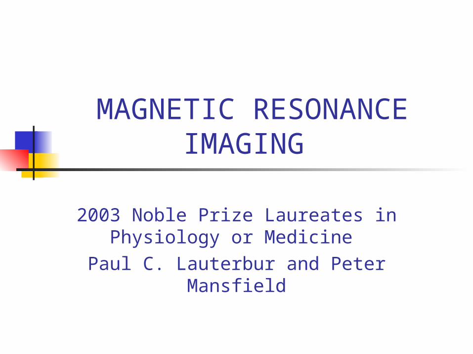
Noble Prize
6 October 2003Press Release
The Nobel Assembly at Karolinska Institute has today decided to award
The Nobel Prize in Physiology or Medicine for 2003 jointly to
Paul C. Lauterbur and Peter Mansfieldfor their discoveries concerning“magnetic resonance imaging”
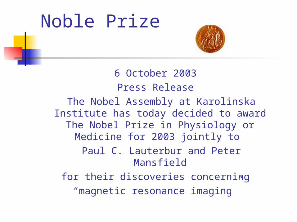
“for their discoveries concerning magnetic resonance imaging”
Paul C. Lauterbur Peter Mansfield½ of the prize USA ½ of the prize United KindomUniversity of Illinois University of Notingham Urbana, IL, USA. United Kingdom. b. 1929 b. 1933
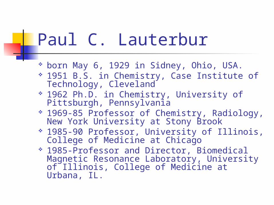
Paul C. Lauterbur born May 6, 1929 in Sidney, Ohio, USA. 1951 B.S. in Chemistry, Case Institute of
Technology, Cleveland 1962 Ph.D. in Chemistry, University of
Pittsburgh, Pennsylvania 1969-85 Professor of Chemistry, Radiology,
New York University at Stony Brook 1985-90 Professor, University of Illinois,
College of Medicine at Chicago 1985-Professor and Director, Biomedical
Magnetic Resonance Laboratory, University of Illinois, College of Medicine at Urbana, IL.
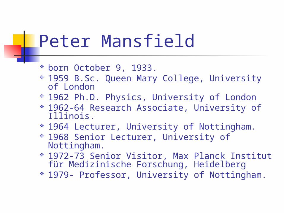
Peter Mansfield born October 9, 1933. 1959 B.Sc. Queen Mary College, University of
London 1962 Ph.D. Physics, University of London 1962-64 Research Associate, University of
Illinois. 1964 Lecturer, University of Nottingham. 1968 Senior Lecturer, University of
Nottingham. 1972-73 Senior Visitor, Max Planck Institut
für Medizinische Forschung, Heidelberg 1979- Professor, University of Nottingham.
History of MRI Late 1800’s November 5, 1895. William Roentgen
discovered X-rays. Roentgen discovered that: X-rays travel in straight lines, could not be refracted or reflected did not respond to magnetic or electric
field. February, 1896, X-rays were being used
clinically in the United States.
History of MRI In the 1930’s, a physics phenomenon was
discovered, called nuclear magnetic resonance or NMR.
Felix Bloch, working at Stanford University, and Edward Purcell, from Harvard University, discovered NMR.
In NMR nuclei were placed in a magnetic field, they absorbed energy in the radiofrequency range of the electromagnetic spectrum, and re-emitted this energy when the nuclei transferred to their original state.
History of MRI This phenomenon was termed NMR as follows: "Nuclear" as only the nuclei of certain atoms reacted in that way; "Magnetic" as a magnetic field was
required; "Resonance" because of the direct frequency
dependence of the magnetic and radiofrequency fields.
History of MRI For their discovery of NMR Bloch and Purcell
were awarded the Nobel Prize for Physics in 1952.
Use of NMR to investigate the chemical composition and physical structure of matter.
Relaxation times, T1 and T2. T1: Time taken by nuclei in test samples to
return to their natural alignment T2: Duration of the magnetic signal from the
sample.
History of MRI In 1970s Raymond Damadian, proposed
that each tissue in the body has a different relaxation time, but cancerous tissue has an abnormally long relaxation time.
He believed that the NMR could be used as an “external probe for the internal detection of cancer”
Damadian presented first commercial NMR scanner at the annual meeting of the American Roentgen Ray Society in 1980.
History of MRI Paul C. Lauterbur determined the
origin of the radio waves by analysis of their characteristics.
Discovered the possibility to create a two-dimensional picture by introducing gradients in the magnetic field.
In 1972, obtained the first MRI.
History of MRI Pater Mansfield further developed the
utilization of gradients in the magnetic field.
Signals could be mathematically analyzed. Showed how extremely fast imaging could
be achievable. In 1976, he and his colleagues created the
first MRI of a human body part, a finger.
What is an MRI? Magnetic Resonance Imaging (MRI) :safe and
noninvasive test. Diagnostic technique :uses strong magnetic
field and pulses of radio waves. Produces pictures of structures inside the
body. Images :slices of an organ or part of body. MRI’s computer: 3-D images.
How it works? Body :strong magnetic field. Machine uses :strong magnetic field and
pulses of radio waves. Machine creates an image :how
hydrogen atoms react. Usually images are created as single
slices of organs or structures. MRI computer combine them to give a 3
D image.
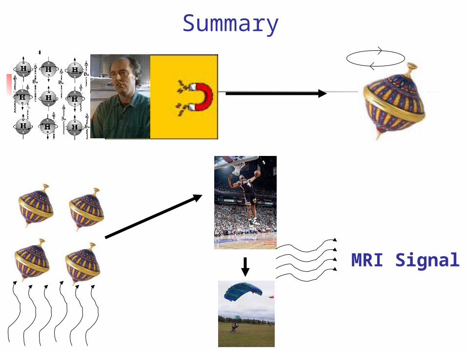
Using Our Body’s Magnets Because of predictions from
physics and math we know there are very weak magnets in all living tissues
These magnets are atoms with unpaired numbers of protons and electrons like hydrogen 1H
There are billions and billions of hydrogens in your body
Using Our Body’s Magnets
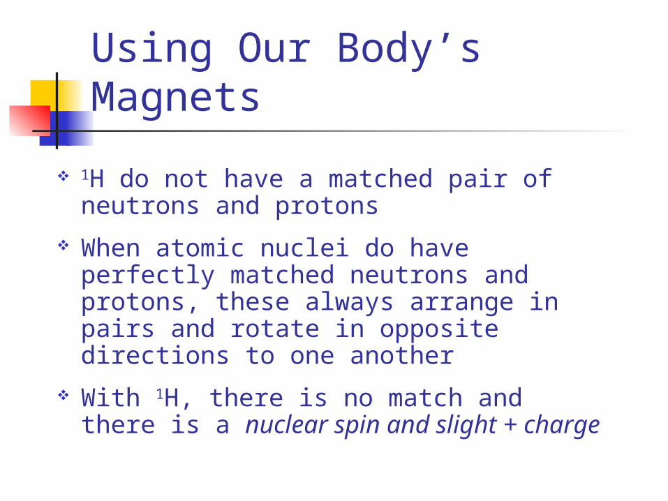
1H do not have a matched pair of neutrons and protons
When atomic nuclei do have perfectly matched neutrons and protons, these always arrange in pairs and rotate in opposite directions to one another
With 1H, there is no match and there is a nuclear spin and slight + charge
Using Our Body’s Magnets
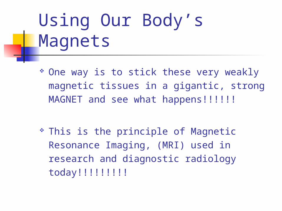
One way is to stick these very weakly magnetic tissues in a gigantic, strong MAGNET and see what happens!!!!!!
This is the principle of Magnetic Resonance Imaging, (MRI) used in research and diagnostic radiology today!!!!!!!!!
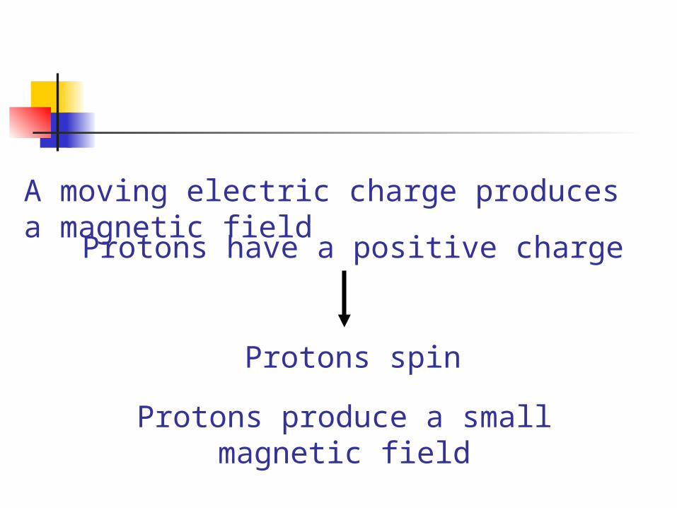
A moving electric charge produces a magnetic field
Protons have a positive charge
Protons spin
Protons produce a small magnetic field
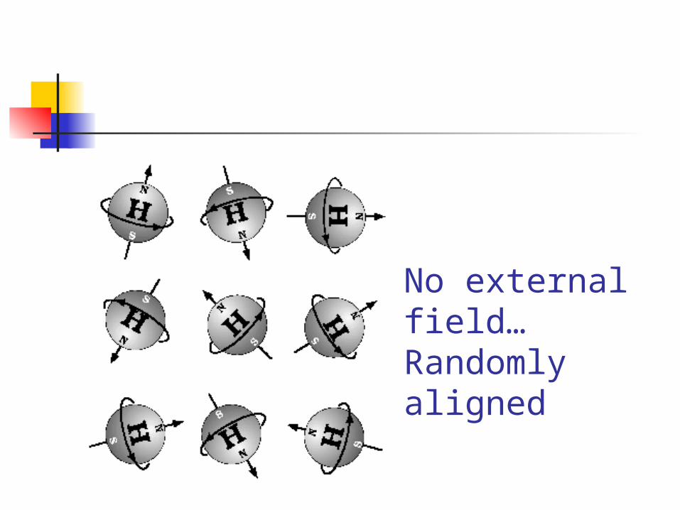
No external field…Randomly aligned
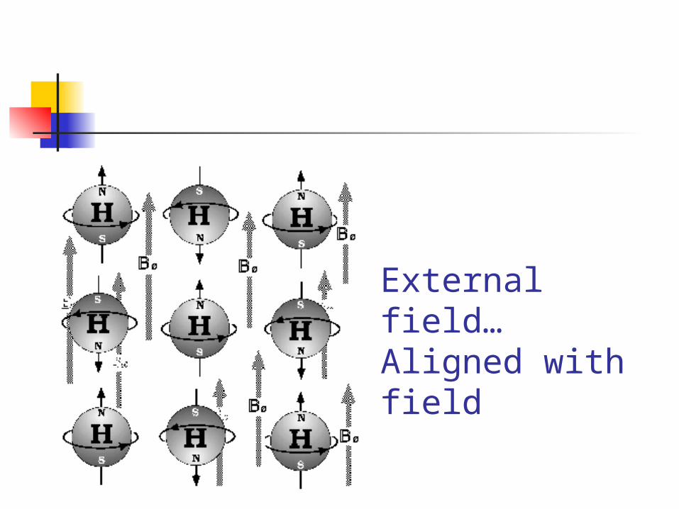
External field…Aligned with field
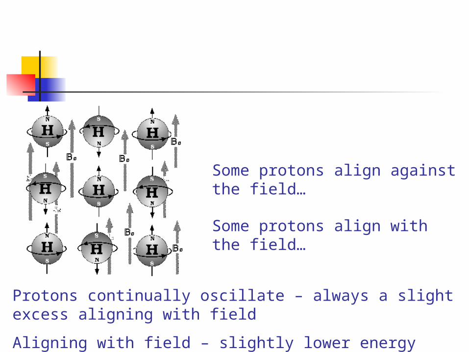
Some protons align with the field…
Some protons align against the field…
Protons continually oscillate – always a slight excess aligning with field
Aligning with field – slightly lower energy state
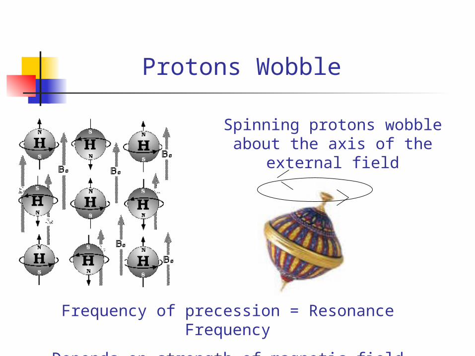
Protons Wobble
Spinning protons wobble about the axis of the external field
Frequency of precession = Resonance Frequency
Depends on strength of magnetic field
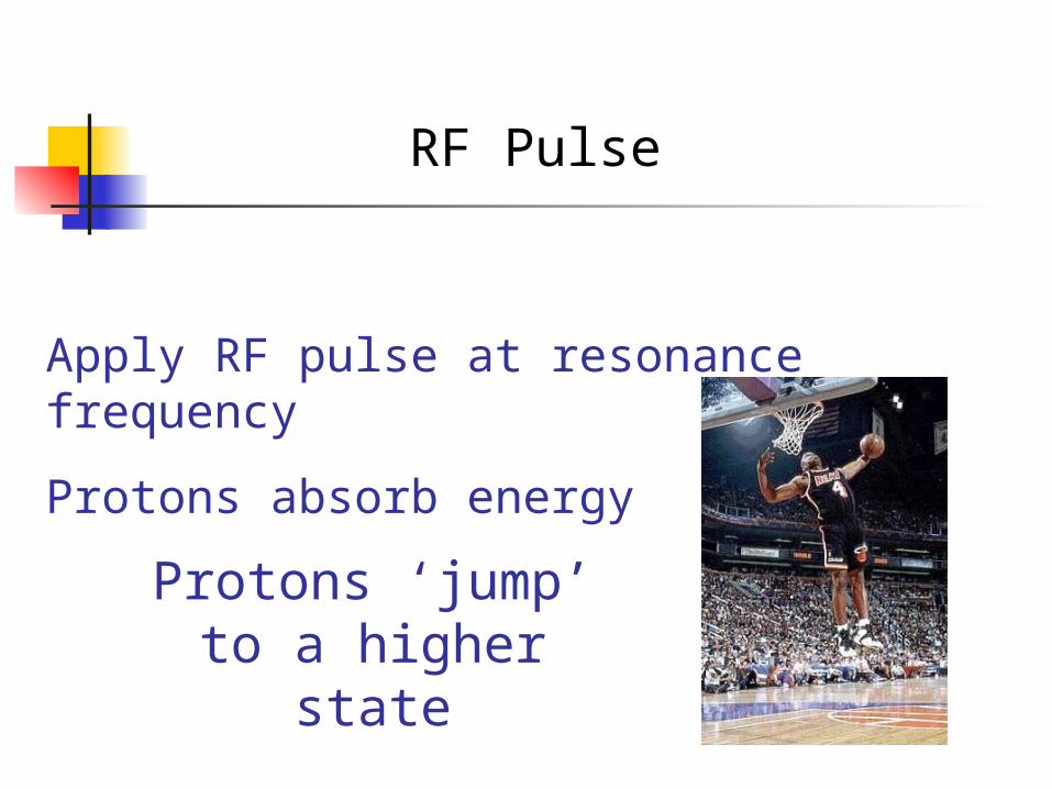
RF Pulse
Apply RF pulse at resonance frequency
Protons absorb energy
Protons ‘jump’ to a higher state
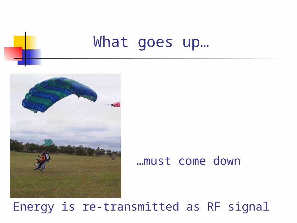
What goes up…
…must come down
Energy is re-transmitted as RF signal
Summary
MRI Signal
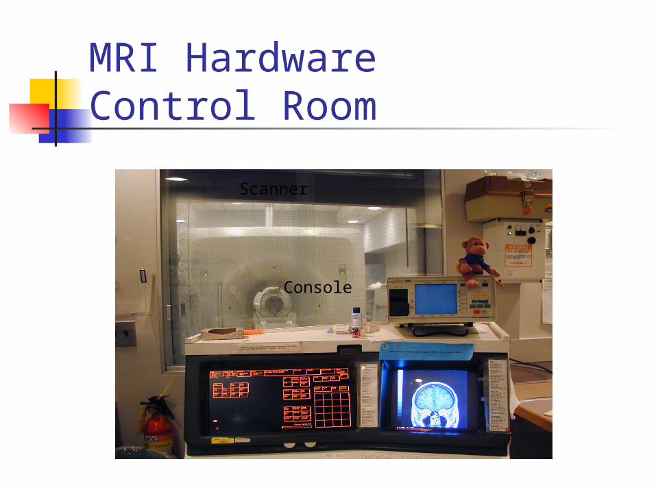
MRI HardwareControl Room
Scanner
Console
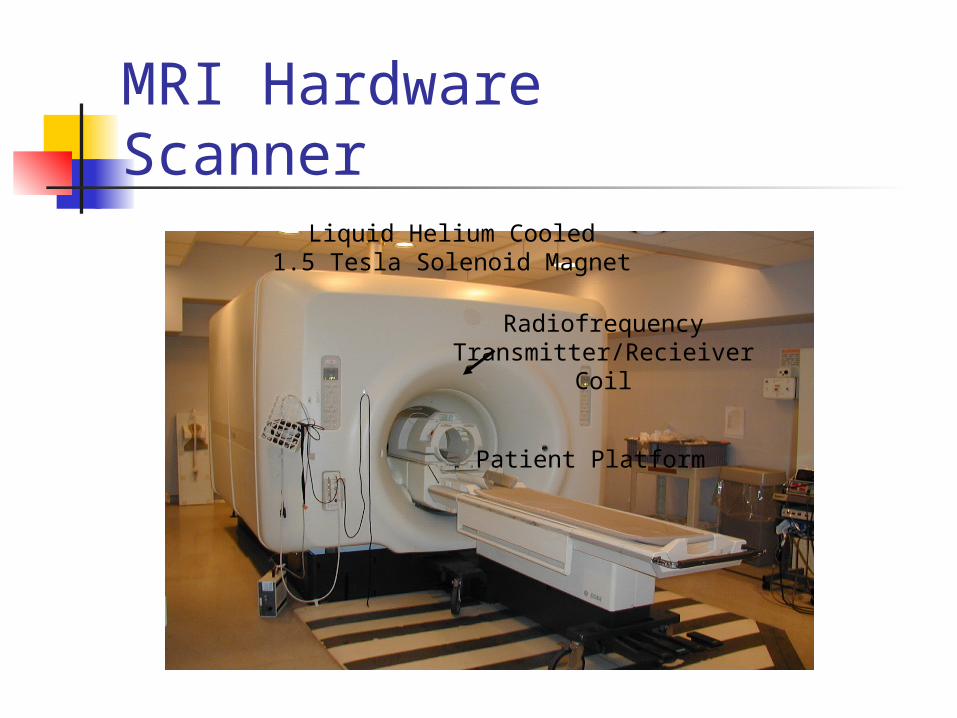
MRI HardwareScanner
Liquid Helium Cooled1.5 Tesla Solenoid Magnet
Patient Platform
RadiofrequencyTransmitter/Recieiver
Coil
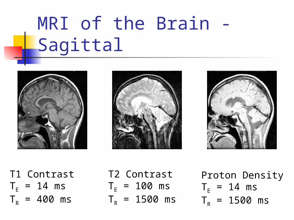
MRI of the Brain - Sagittal
T1 ContrastTE = 14 msTR = 400 ms
T2 ContrastTE = 100 msTR = 1500 ms
Proton DensityTE = 14 msTR = 1500 ms
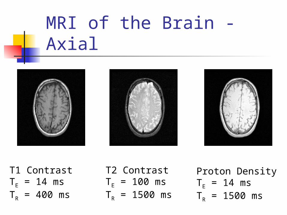
MRI of the Brain - Axial
T1 ContrastTE = 14 msTR = 400 ms
T2 ContrastTE = 100 msTR = 1500 ms
Proton DensityTE = 14 msTR = 1500 ms
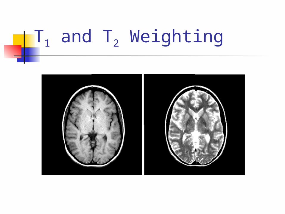
T1 and T2 Weighting
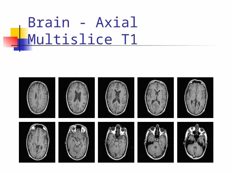
Brain - Axial Multislice T1
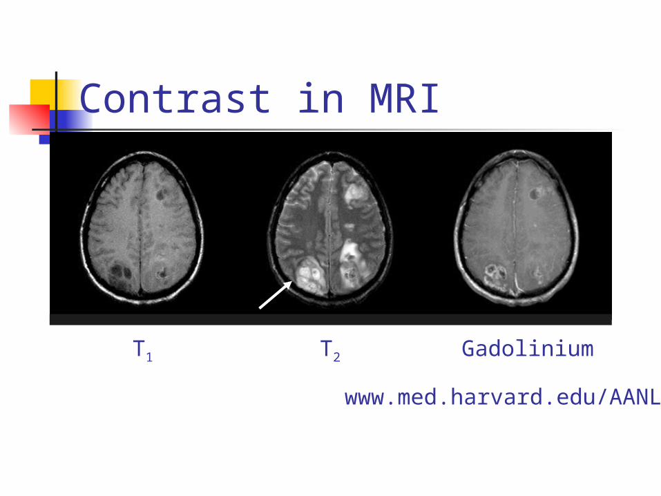
Contrast in MRI
T1 T2 Gadolinium
The Whole Brain Atlas: http://www.med.harvard.edu/AANLIB/
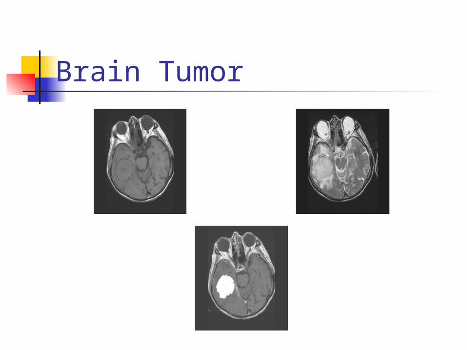
Brain Tumor
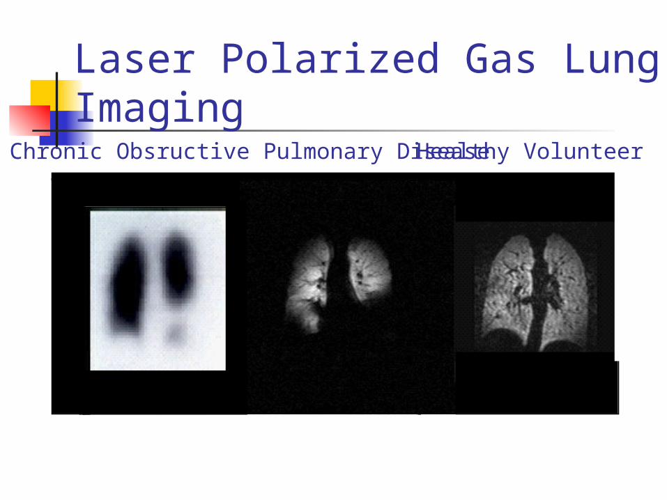
Laser Polarized Gas Lung ImagingChronic Obsructive Pulmonary Disease Healthy Volunteer
Advantages of MRI Diagnosing multiple sclerosis (MS) Diagnosing tumors of the pituitary
gland and brain. Diagnosing infections in the brain,
spine or joints Visualizing torn ligaments in the
wrist, knee and ankle
Advantages of MRI Visualizing shoulder injuries Diagnosing tendonitis Evaluating masses in the soft tissues of
the body Evaluating bone tumors, cysts and
bulging or herniated discs in the spine
Diagnosing strokes in their earliest stages.
Disadvantages of MRI Not for everybody. machine makes a tremendous amount
of noise. require patients to hold very still for
extended periods of time. Orthopedic hardware (screws, plates,
artificial joints) in the area of a scan can cause severe artifacts (distortions) on the images.
very expensive.
Future of MRI Very small scanners. Functional brain mapping. Ventilation dynamics of the lungs
through the use of hyperpolarized helium-3 gas.
Image strokes in their earliest stages. Limitless future
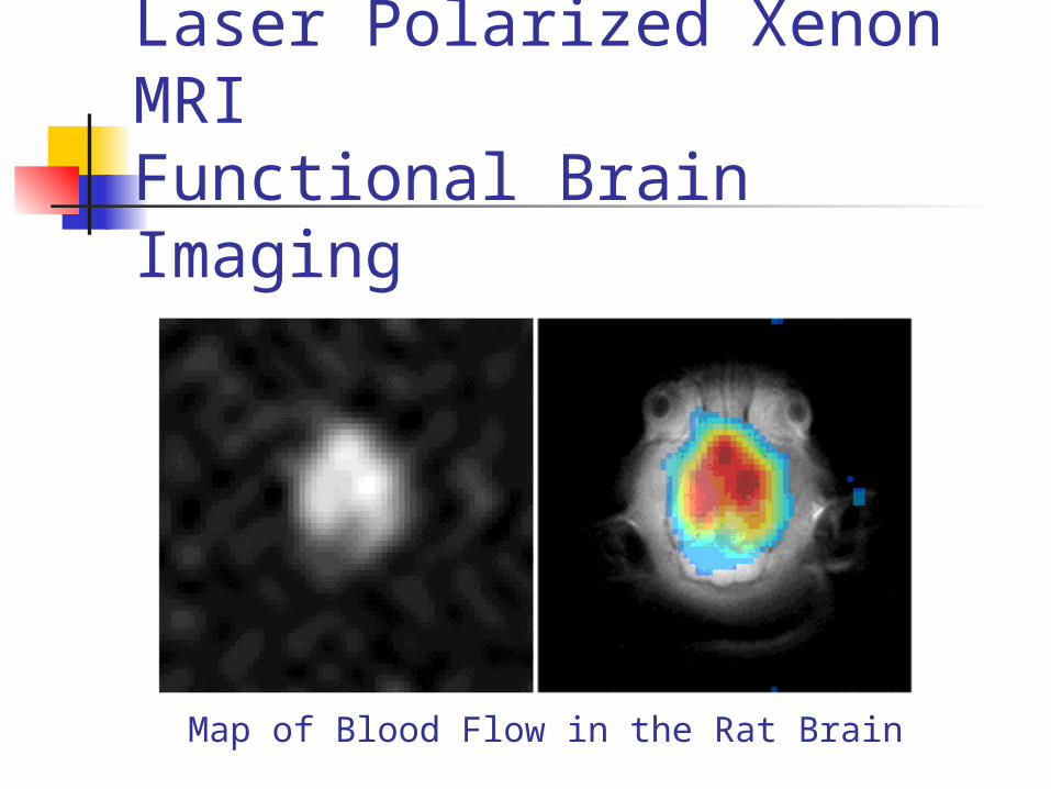
Laser Polarized Xenon MRIFunctional Brain Imaging
Map of Blood Flow in the Rat Brain
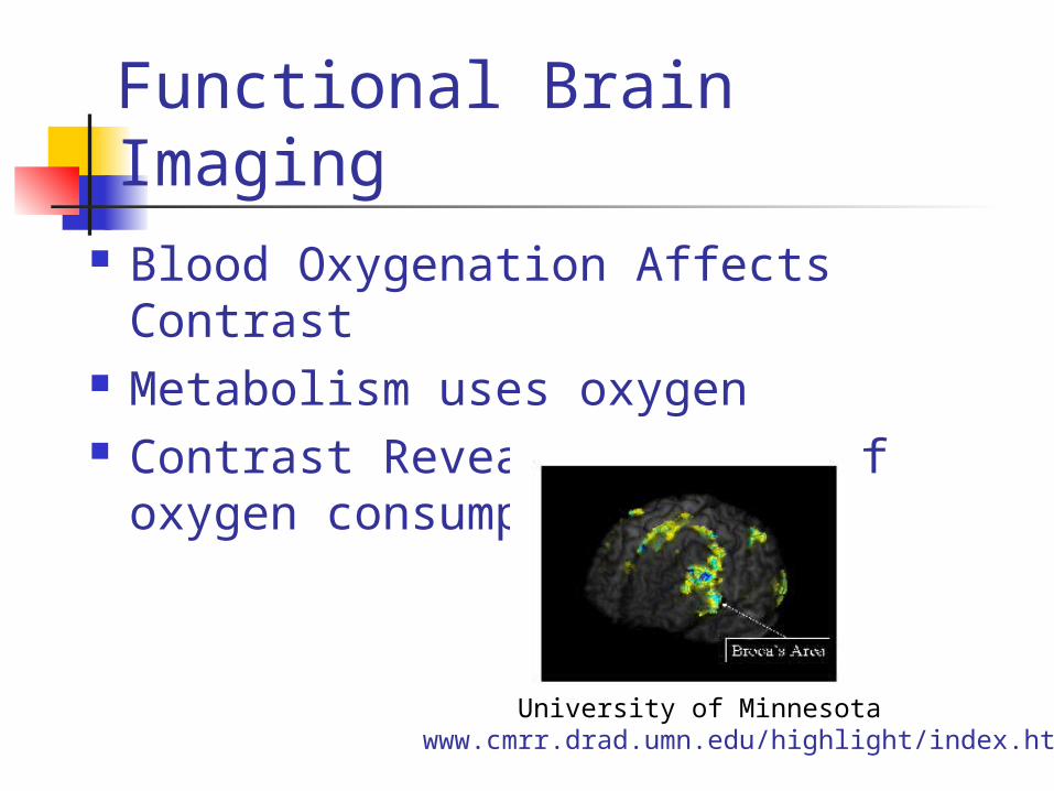
Functional Brain Imaging Blood Oxygenation Affects Contrast Metabolism uses oxygen Contrast Reveals regions of oxygen
consumption
University of Minnesotahttp://www.cmrr.drad.umn.edu/highlight/index.html
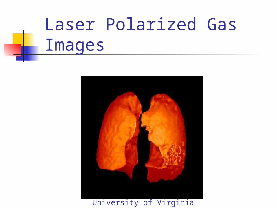
Laser Polarized Gas Images
University of Virginia

Sources used:
http://www.nobel.se/medicine/laureates/2003/
http://inventors.about.com/ http://www.bae.ncsu.edu/ http://www.isbe.man.ac.uk/ www.cmrr.drad.umn.edu/ Slides provided by Dr. Vankley.
Recommended