TH
EJ
OU
RN
AL
OF
CE
LL
BIO
LO
GY
JCB: ARTICLE
© The Rockefeller University Press $30.00The Journal of Cell Biology, Vol. 179, No. 7, December 31, 2007 1497–1510http://www.jcb.org/cgi/doi/10.1083/jcb.200708107
JCB 1497
IntroductionEffi cient synaptic vesicle (SV) endocytosis is essential to main-
tain the ability of the synapse to release neurotransmitter under
sustained stimulation. Strong evidence suggests that the clathrin-
based endocytic machinery is required to recognize and prop-
erly sort SV proteins during the generation of functional SVs
(Brodin et al., 2000; Murthy and De Camilli, 2003; Galli and
Haucke, 2004; Sudhof, 2004), even at small central synapses
(Granseth et al., 2006). In spite of this, the precise molecular
mechanism of SV cargo recognition has remained elusive.
Although conventional endocytic sorting signals, such as tyro-
sine- or dileucine-based motifs (Bonifacino and Traub, 2003),
may target cargo proteins for constitutive internalization by the
heterotetrameric adaptor assembly protein complex 2 (AP-2;
comprising α, β2, μ2, and σ2 subunits), no common endocytic
sorting signals have been identifi ed for SV proteins. One possi-
bility is that SV proteins remain preassembled (Bennett et al.,
1992; Willig et al., 2006), possibly by cholesterol-rich micro-
domains (Jia et al., 2006). Alternatively, SV proteins may undergo
rapid postfusion reclustering to allow effi cient recapturing by
the endocytic machinery. Both of these scenarios would alleviate
the need for an endocytic sorting signal within each individual
SV protein. Instead, SV recycling may depend on the specifi c
recognition of one (Voglmaier et al., 2006) or a few SV proteins
by endocytic adaptors.
A likely candidate to connect the exo- and endocytic
limbs of the SV cycle is synaptotagmin 1. Genetic, antibody-
mediated, or chemical perturbation of synaptotagmin 1 function
leads to partial vesicle depletion and SV recycling defects in
the squid giant synapse (Llinas et al., 2004), at the Drosophila melanogaster neuromuscular junction (DiAntonio et al., 1993;
Poskanzer et al., 2003), in Caenorhabditis elegans (Jorgensen
et al., 1995), and in mammalian central nervous system synapses
(Nicholson-Tomishima and Ryan, 2004). Synaptotagmin 1 also
appears to infl uence the recycling effi ciency of other SV pro-
teins (Poskanzer et al., 2003; Nicholson-Tomishima and Ryan,
2004) through specifi c regions in its cytoplasmic tail (Poskanzer
et al., 2006). The molecular mechanism underlying the function
of synaptotagmin in SV endocytosis has remained uncertain
but may involve its ability to associate with phosphoino-
sitides (Bai et al., 2004), AP-2μ (Zhang et al., 1994; Chapman
Molecular basis of synaptic vesicle cargo recognition by the endocytic sorting adaptor stonin 2
Nadja Jung,1 Martin Wienisch,2 Mingyu Gu,3 James B. Rand,4 Sebastian L. Müller,5 Gerd Krause,5
Erik M. Jorgensen,3 Jürgen Klingauf,2 and Volker Haucke1
1Department of Membrane Biochemistry, Institute of Chemistry and Biochemistry, Freie Universität Berlin, 14195 Berlin, Germany2Department of Membrane Biophysics, Max-Planck-Institute for Biophysical Chemistry, 37077 Göttingen, Germany3Department of Biology, University of Utah, Salt Lake City, UT 841124Program in Molecular, Cell and Developmental Biology, Oklahoma Medical Research Foundation, Oklahoma City, OK 731045Leibniz-Institut für Molekulare Pharmakologie, 13125 Berlin, Germany
Synaptic transmission depends on clathrin-mediated
recycling of synaptic vesicles (SVs). How select
SV proteins are targeted for internalization has
remained elusive. Stonins are evolutionarily conserved
adaptors dedicated to endocytic sorting of the SV pro-
tein synaptotagmin. Our data identify the molecular de-
terminants for recognition of synaptotagmin by stonin 2
or its Caenorhabditis elegans orthologue UNC-41B. The
interaction involves the direct association of clusters of
basic residues on the surface of the cytoplasmic domain of
synaptotagmin 1 and a β strand within the μ–homology
domain of stonin 2. Mutation of K783, Y784, and E785
to alanine within this stonin 2 β strand results in failure
of the mutant stonin protein to associate with synaptotag-
min, to accumulate at synapses, and to facilitate synapto-
tagmin internalization. Synaptotagmin-binding–defective
UNC-41B is unable to rescue paralysis in C. elegans
stonin mutant animals, suggesting that the mechanism of
stonin-mediated SV cargo recognition is conserved from
worms to mammals.
Correspondence to Volker Haucke: [email protected]
Abbreviations used in this paper: AP-2, assembly protein complex 2; CLASP, clathrin-associated sorting protein; HEK, human embryonic kidney; μHD, μ–homology domain; NIE, neuroimmune endocrine; SV, synaptic vesicle; WT, wild type.
The online version of this paper contains supplemental material.
on January 2, 2008 w
ww
.jcb.orgD
ownloaded from
http://www.jcb.org/cgi/content/full/jcb.200708107/DC1Supplemental Material can be found at:
JCB • VOLUME 179 • NUMBER 7 • 2007 1498
et al., 1998; Grass et al., 2004), or the endocytic adaptor stonin 2
(termed stoned B in fl ies and UNC-41 in C. elegans; Fergestad
and Broadie, 2001; Martina et al., 2001; Stimson et al., 2001;
Walther et al., 2004).
Stonin 2 represents the fi rst, and so far only, endocytic pro-
tein specifi cally dedicated to SV recycling by acting as a sorting
adaptor for synaptotagmin 1 (Diril et al., 2006). Expression of
stonin 2 in fi broblasts is suffi cient to rescue clathrin/AP-2–mediated
internalization of surface-stranded synaptotagmin 1 (Jarousse and
Kelly, 2001) and facilitates synaptotagmin 1 redistribution into
SVs in primary neurons (Diril et al., 2006). Stonin 2 is linked
to the endocytic machinery by direct interactions with AP-2
(Walther et al., 2004) and eps15 or intersectin (Martina et al., 2001),
and can therefore be classifi ed as a clathrin-associated sorting
protein (CLASP; Traub, 2005). CLASPs compose a set of adaptors,
including β-arrestins (Lefkowitz and Whalen, 2004), autosomal
recessive hypercholesterolemia (Edeling et al., 2006), Dab2
(Mishra et al., 2002), Huntingtin interacting protein 1/1R, and
numb (Santolini et al., 2000), that target specifi c cargo to clathrin-
coated pits or subsets thereof for regulated internalization.
In contrast to other CLASPs, we lack detailed molecular
information on how stonin 2 and its orthologues recognize SV
cargo during exo- and endocytic vesicle cycling. We demon-
strate that an evolutionarily conserved β strand within the
μ–homology domain (μHD) of stonin 2 recognizes basic patches
on the surface of the cytoplasmic domain of synaptotagmin 1.
Mutation of critical residues within this β strand results in
failure of the mutant stonin protein to physically associate with
synaptotagmin and to facilitate synaptotagmin internalization
in fi broblasts or primary neurons. Furthermore, synaptotagmin-
binding–defective UNC-41B is unable to rescue paralysis in
C. elegans unc-41/stonin mutant animals, suggesting that the
mechanism of stonin-mediated SV cargo recognition is conserved
from worms to mammals.
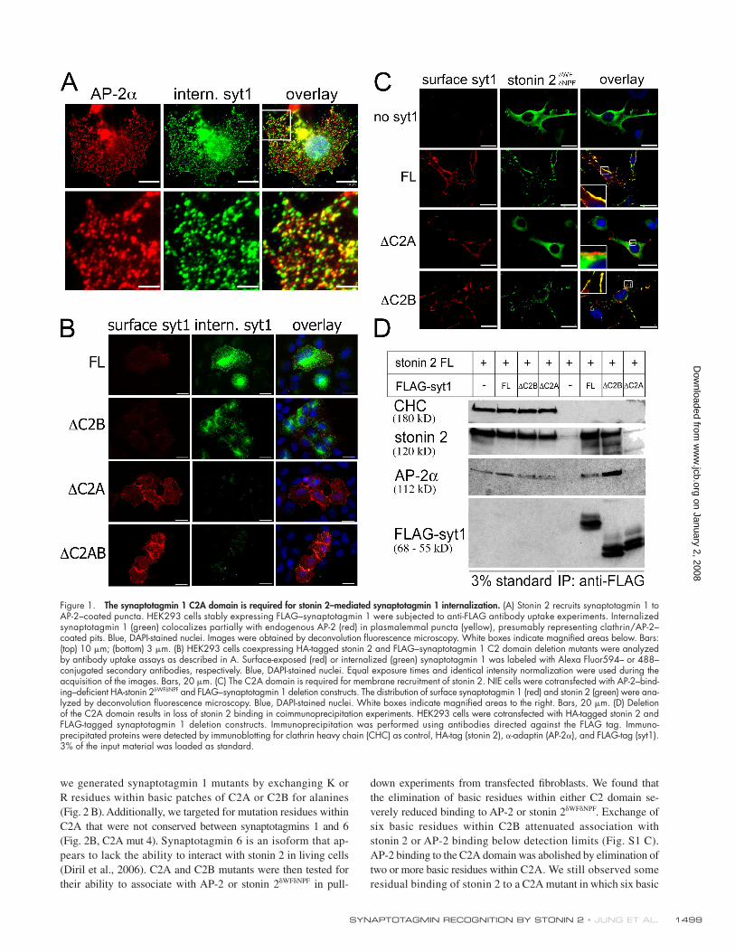
ResultsStonin 2 physically and functionally interacts with the C2A domain of synaptotagmin 1 to facilitate internalizationGiven the importance of functional SV recycling and the role
of synaptotagmin 1 in coupling exo- and endocytosis in neuro
secretory cells, we first set out to identify the domains re-
quired for its internalization. Expression of stonin 2 in human
embryonic kidney (HEK) 293 cells stably transfected with
FLAG–synaptotagmin 1 leads to redistribution of synaptotag-
min 1 from the plasma membrane to internal compartments
(Diril et al., 2006). Internalization assays based on anti-FLAG
antibody uptake showed that endocytosed synaptotagmin local-
izes to a subset of AP-2–coated puncta and to a perinuclear en-
dosomal compartment (Fig. 1 A; Diril et al., 2006), suggesting
that stonin 2 targets synaptotagmin 1 to clathrin/AP-2–coated
pits. To confine the region within synaptotagmin 1 required
for stonin 2–mediated internalization deletion, constructs lack-
ing one or both of the C2 domains were generated and ana-
lyzed by antibody uptake experiments. After antibody chase for
20 min at 37°C, surface-stranded synaptotagmin 1 was detected
by Alexa Fluor 594–labeled secondary antibodies under non-
permeabilizing conditions, remaining surface immunoreactivity
was blocked, and internalized synaptotagmin 1 was revealed
using Alexa Fluor 488–labeled secondary antibodies. Surpris-
ingly, we found that synaptotagmin 1 lacking the C2B domain
(syt1∆C2B) was internalized, albeit with reduced effi ciency,
whereas mutant proteins lacking either the C2A (syt1∆C2A) or
both C2 domains (syt1∆C2AB) were not endocytosed (Fig. 1 B).
Thus, the C2A domain contributes the major internalization
signal for stonin 2–dependent synaptotagmin 1 endocytosis.
This interpretation is supported by membrane recruitment ex-
periments in N1E neuroblastoma cells. Overexpression of wild-
type (WT) synaptotagmin 1 or syt1∆C2B, but not syt1∆C2A,
caused a redistribution of WT stonin 2 (stonin 2WT; Diril et al.,
2006) or an AP-2 and Eps15 homology domain–binding–defective
stonin 2 mutant (stonin 2δWFδNPF, used to exclude indirect ef-
fects mediated via AP-2 or, e.g., eps15) from the cytosol to the
plasmalemma (Fig. 1 C). Stonin 2 coimmunoprecipitated with WT
synaptotagmin 1 or syt1∆C2B but not with syt1∆C2A, which
is consistent with the microscopic data (Fig. 1 D). AP-2 was found
in the same complex, presumably because of its direct inter-
action with stonin 2. This complex was also seen in affi nity
chromatography experiments using extracts from transfected
HEK cells incubated with GST-fused C2 domains (Figs. S1 A
and S2 B, available at http://www.jcb.org/cgi/content/full/
jcb.200708107/DC1). Stonin 2WT or stonin 2δWFδNPF preferen-
tially associated with the C2A domain of synaptotagmin 1.
AP-2 effi ciently copurifi ed with either C2A or C2B from cell
extracts containing stonin 2WT but was predominantly retained
by C2B if lysates from stonin 2δWFδNPF–expressing cells were
used (Fig. S1, A and B), which is in agreement with published
data (Chapman et al., 1998; Haucke et al., 2000). To further
corroborate the direct association of stonin 2 with synapto-
tagmin 1–C2A, we performed in vitro binding experiments using
GST-fused synaptotagmin 1 and His6-tagged stonin 2 purifi ed
from HEK293 fi broblasts (Fig. S2 B). Stonin 2 most effi ciently
bound to GST-C2A or -C2AB, whereas a much weaker inter-
action with the C2B domain was observed (Fig. 2 A). Stonin 2 did
not bind to GST control beads. These results were confi rmed by
experiments using 35S-labeled stonin 2 synthesized by coupled
transcription/translation in vitro (Fig. S2 A). We conclude that
stonin 2 directly associates with synaptotagmin 1, mainly via
determinants in the C2A domain.
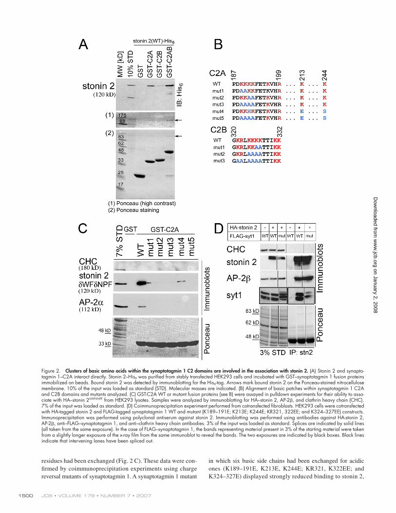
Stonin 2 recognizes basic patches within the synaptotagmin 1 C2 domainsCoimmunoprecipitation experiments from transfected fi bro-
blasts (Fig. S3, available at http://www.jcb.org/cgi/content/
full/jcb.200708107/DC1) indicated that the major binding site
for synaptotagmin 1 comprised the carboxy-terminal μHD of
stonin 2. The μHD exhibits 30% amino acid identity with
the μ2 subunit of AP-2, which associates with a stretch of basic
amino acid residues within the C2B domain of synaptotagmin 1
(Chapman et al., 1998; Grass et al., 2004). Considering the
homology between AP-2μ and the stonin 2 μHD and the exis-
tence of basic patches in both C2 domains, we rationalized that
basic residues might play a role in stonin 2 binding. To test this,
on January 2, 2008 w
ww
.jcb.orgD
ownloaded from
SYNAPTOTAGMIN RECOGNITION BY STONIN 2 • JUNG ET AL. 1499
we generated synaptotagmin 1 mutants by exchanging K or
R residues within basic patches of C2A or C2B for alanines
(Fig. 2 B). Additionally, we targeted for mutation residues within
C2A that were not conserved between synaptotagmins 1 and 6
(Fig. 2B, C2A mut 4). Synaptotagmin 6 is an isoform that ap-
pears to lack the ability to interact with stonin 2 in living cells
(Diril et al., 2006). C2A and C2B mutants were then tested for
their ability to associate with AP-2 or stonin 2δWFδNPF in pull-
down experiments from transfected fi broblasts. We found that
the elimination of basic residues within either C2 domain se-
verely reduced binding to AP-2 or stonin 2δWFδNPF. Exchange of
six basic residues within C2B attenuated association with
stonin 2 or AP-2 binding below detection limits (Fig. S1 C).
AP-2 binding to the C2A domain was abolished by elimination of
two or more basic residues within C2A. We still observed some
residual binding of stonin 2 to a C2A mutant in which six basic
Figure 1. The synaptotagmin 1 C2A domain is required for stonin 2–mediated synaptotagmin 1 internalization. (A) Stonin 2 recruits synaptotagmin 1 to AP-2–coated puncta. HEK293 cells stably expressing FLAG–synaptotagmin 1 were subjected to anti-FLAG antibody uptake experiments. Internalized synaptotagmin 1 (green) colocalizes partially with endogenous AP-2 (red) in plasmalemmal puncta (yellow), presumably representing clathrin/AP-2–coated pits. Blue, DAPI-stained nuclei. Images were obtained by deconvolution fl uorescence microscopy. White boxes indicate magnifi ed areas below. Bars: (top) 10 μm; (bottom) 3 μm. (B) HEK293 cells coexpressing HA-tagged stonin 2 and FLAG–synaptotagmin 1 C2 domain deletion mutants were analyzed by antibody uptake assays as described in A. Surface-exposed (red) or internalized (green) synaptotagmin 1 was labeled with Alexa Fluor594– or 488–conjugated secondary antibodies, respectively. Blue, DAPI-stained nuclei. Equal exposure times and identical intensity normalization were used during the acquisition of the images. Bars, 20 μm. (C) The C2A domain is required for membrane recruitment of stonin 2. NIE cells were cotransfected with AP-2–bind-ing–defi cient HA-stonin 2δWFδNPF and FLAG–synaptotagmin 1 deletion constructs. The distribution of surface synaptotagmin 1 (red) and stonin 2 (green) were ana-lyzed by deconvolution fl uorescence microscopy. Blue, DAPI-stained nuclei. White boxes indicate magnifi ed areas to the right. Bars, 20 μm. (D) Deletion of the C2A domain results in loss of stonin 2 binding in coimmunoprecipitation experiments. HEK293 cells were cotransfected with HA-tagged stonin 2 and FLAG-tagged synaptotagmin 1 deletion constructs. Immunoprecipitation was performed using antibodies directed against the FLAG tag. Immuno-precipitated proteins were detected by immunoblotting for clathrin heavy chain (CHC) as control, HA-tag (stonin 2), α-adaptin (AP-2α), and FLAG-tag (syt1). 3% of the input material was loaded as standard.
on January 2, 2008 w
ww
.jcb.orgD
ownloaded from
JCB • VOLUME 179 • NUMBER 7 • 2007 1500
residues had been exchanged (Fig. 2 C). These data were con-
fi rmed by coimmunoprecipitation experiments using charge
reversal mutants of synaptotagmin 1. A synaptotagmin 1 mutant
in which six basic side chains had been exchanged for acidic
ones (K189–191E, K213E, K244E; KR321, K322EE; and
K324–327E) displayed strongly reduced binding to stonin 2,
Figure 2. Clusters of basic amino acids within the synaptotagmin 1 C2 domains are involved in the association with stonin 2. (A) Stonin 2 and synapto-tagmin 1–C2A interact directly. Stonin 2–His6 was purifi ed from stably transfected HEK293 cells and incubated with GST–synaptotagmin 1 fusion proteins immobilized on beads. Bound stonin 2 was detected by immunoblotting for the His6-tag. Arrows mark bound stonin 2 on the Ponceau-stained nitrocellulose membrane. 10% of the input was loaded as standard (STD). Molecular masses are indicated. (B) Alignment of basic patches within synaptotagmin 1 C2A and C2B domains and mutants analyzed. (C) GST-C2A WT or mutant fusion proteins (see B) were assayed in pulldown experiments for their ability to asso-ciate with HA–stonin 2δWFδNPF from HEK293 lysates. Samples were analyzed by immunoblotting for HA–stonin 2, AP-2β, and clathrin heavy chain (CHC). 7% of the input was loaded as standard. (D) Coimmunoprecipitation experiment performed from cotransfected fi broblasts. HEK293 cells were cotransfected with HA-tagged stonin 2 and FLAG-tagged synaptotagmin 1 WT and mutant (K189–191E; K213E; K244E; KR321, 322EE; and K324–327EE) constructs. Immunoprecipitation was performed using polyclonal antiserum against stonin 2. Immunoblotting was performed using antibodies against HA-stonin 2, AP-2β, anti–FLAG–synaptotagmin 1, and anti–clathrin heavy chain antibodies. 3% of the input was loaded as standard. Splices are indicated by solid lines (all taken from the same exposure). In the case of FLAG–synaptotagmin 1, the bands representing material present in 3% of the starting material were taken from a slightly longer exposure of the x-ray fi lm from the same immunoblot to reveal the bands. The two exposures are indicated by black boxes. Black lines indicate that intervening lanes have been spliced out.
on January 2, 2008 w
ww
.jcb.orgD
ownloaded from

SYNAPTOTAGMIN RECOGNITION BY STONIN 2 • JUNG ET AL. 1501
although the interaction was not abolished completely (Fig. 2 D),
indicating that additional, perhaps co operative, mechanisms might
be at work. In summary, we identify basic patches within both
C2 domains of synaptotagmin 1 as the major determinants for
association with stonin 2.
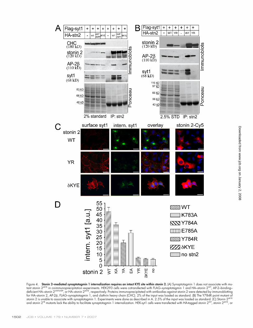
The KYE site within the stonin 2 𝛍HD is required for synaptotagmin 1 interaction and internalizationWe observed that stonin 2 associated via its μHD (Fig. S3,
A and B) with basic residues within the synaptotagmin 1 C2
domains, which is similar to the interaction between AP-2μ
and synaptotagmin 1 C2B. Hence, we hypothesized that these
interactions might be structurally related. Previous studies had
suggested that a β strand comprising residue Y343 within
subdomain B of AP-2μ is part of the synaptotagmin 1 bind-
ing site (Haucke et al., 2000). Structural data are available for
AP-2 (Owen and Evans, 1998; Collins et al., 2002) including its
μ2 sub unit, which displays high sequence homology (52%)
to the car boxy-terminal region of stonin 2. We used μ2 as
a template to generate a molecular homology model of the
stonin 2 μHD (Fig. S3 D). The stonin 2 μHD model displays an
overall β-fold structure very similar to that of AP-2μ, except for
a few loops that differ from the template. Conserved features
include the β strand suggested to harbor the synaptotagmin
binding site within AP-2μ, which contains the aforementioned
tyrosine residue (Y343 in AP-2μ). This tyrosine (corresponding
to Y784 in human stonin 2), as well as several fl anking residues,
is also evolutionarily conserved within stonin family members
from humans to nematodes (Fig. 3 A), which is suggestive of its
functional importance.
We hypothesized that Y784 and its neighbors form part
of the binding site for synaptotagmin 1. To test this, we gener-
ated a stonin 2 mutant in which residues K783, Y784, and E785
(Fig. 3 A, red; and Fig. S3 D, right, red) had been exchanged
by alanines (stonin 2δKYE). We transfected fibroblasts with
stonin 2δWFδNPF, the KYE mutant stonin 2 (stonin 2δKYE), or a mutant
of stonin 2 combining these mutations (stonin 2δWFδNPFδKYE) and
performed affi nity chromatography experiments using immobi-
lized GST-fused synaptotagmin 1 C2 domains. Although stonin
2δWFδNPF readily copurifi ed with synaptotagmin 1 C2A or AB, the
double mutant of stonin 2 (stonin 2δWFδNPFδKYE) had completely
Figure 3. Evolutionarily conserved residues within a 𝛃 strand of its 𝛍HD are required for association of stonin 2 with synaptotagmin 1. (A) The KYE site is evolutionarily conserved. Multiple protein sequence alignment of stonin 2 and orthologues from rat, mouse, human, cattle, zebrafi sh, D. melanogaster, and C. elegans. Numbers refer to the last amino acid residue within the predicted β strand. Conserved resi-dues K783, Y784, and E785 are colored in red. (B) Mutation of conserved residues K783A, Y784A, and E785A within stonin 2 abolishes its ability to bind to synaptotagmin 1. GST–synaptotagmin 1 fusion proteins immobilized on beads were incubated with cell extracts de-rived from HEK293 cells transfected with AP-2–binding–defi cient HA–stonin 2δWFδNPF, HA–stonin 2δKYE, or HA–stonin 2δWFδNPFδKYE, respectively. Bound proteins were detected by immunoblotting for clathrin heavy chain (CHC), HA–stonin 2, and AP-2μ. 8% of the input was loaded as standard (STD). (C) In vitro binding experiment using purifi ed recombinant proteins. GST-C2AB immobilized on beads was incu-bated with purifi ed stonin 2–His6 WT or δKYE. Bound stonin 2–His6 was detected by immuno-blotting for the His6-tag. The arrow marks the stonin 2–His6 band on the Ponceau S–stained nitrocellulose membrane. 10% of the input was loaded as standard.
on January 2, 2008 w
ww
.jcb.orgD
ownloaded from
JCB • VOLUME 179 • NUMBER 7 • 2007 1502
Figure 4. Stonin 2–mediated synaptotagmin 1 internalization requires an intact KYE site within stonin 2. (A) Synaptotagmin 1 does not associate with mu-tant stonin 2δKYE in coimmunoprecipitation experiments. HEK293 cells were cotransfected with FLAG–synaptotagmin 1 and HA–stonin 2WT, AP-2–binding–defi cient HA–stonin 2δWFδNPF, or HA–stonin 2δKYE, respectively. Proteins immunoprecipitated with antibodies against stonin 2 were detected by immunoblotting for HA–stonin 2, AP-2β, FLAG–synaptotagmin 1, and clathrin heavy chain (CHC). 2% of the input was loaded as standard. (B) The Y784R point mutant of stonin 2 is unable to associate with synaptotagmin 1. Experiments were done as described in A. 2.5% of the input was loaded as standard. (C) Stonin 2δKYE and stonin 2YR mutants lack the ability to facilitate synaptotagmin 1 internalization. HEK-syt1 cells were transfected with HA-tagged stonin 2WT, stonin 2δKYE, or
on January 2, 2008 w
ww
.jcb.orgD
ownloaded from

SYNAPTOTAGMIN RECOGNITION BY STONIN 2 • JUNG ET AL. 1503
lost its ability to associate with synaptotagmin (Fig. 3 B). Trace
amounts of stonin 2δKYE were retained on GST-C2B– or -C2AB–
containing beads. This is owed to the fact that this mutant re-
tains the ability to bind to AP-2 (Fig. 4 A) and, thus, indirectly
associates with the C2B domain of synaptotagmin 1 via AP-2.
Similar results were obtained in direct binding experiments
using His6-tagged stonin 2δKYE purifi ed from HEK cells (Fig. 3 C)
or synthesized by coupled transcription/translation in vi tro
(Fig. S3 C).
To further corroborate these findings, we performed co-
immunoprecipitation experiments. Fibroblasts were cotransfected
with synaptotagmin 1 and stonin 2WT, stonin 2δWFδNPF, or the
δKYE mutant of stonin 2. As expected, synaptotagmin 1 was
found in immunoprecipitates containing stonin 2WT or stonin
2δWFδNPF but not stonin 2δKYE. Conversely, stonin 2δKYE, but not
stonin 2δWFδNPF, retained its ability to associate with AP-2 (Fig. 4 A).
Similar results were seen for a stonin 2 mutant in which Y784
had been exchanged for R (stonin 2YR; Fig. 4 B). This was cor-
roborated in direct binding assays using 35S-labeled stonin 2YR
(Fig. S3 C). To exclude the possibility that the introduced muta-
tions negatively impact protein folding and, therefore, non-
specifi cally affect synaptotagmin binding, we probed the structure
of the generated stonin 2 mutants by limited proteolysis. The di-
gestion patterns of stonin 2WT, stonin 2δKYE, and stonin 2YR were
not found to be signifi cantly different (Fig. S4, available at
http://www.jcb.org/cgi/content/full/jcb.200708107/DC1), ex-
cluding gross structural changes induced by the mutations.
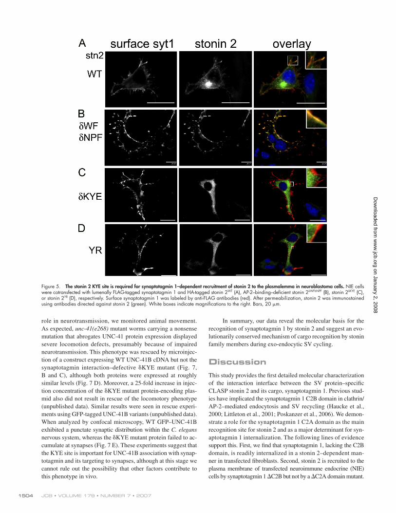
Synaptotagmin-binding–defective stonin 2 is unable to facilitate synaptotagmin 1 internalization in fi broblasts or neurons and fails to accumulate at synapsesTo study the functional importance of the interaction between
stonin 2 and synaptotagmin 1, we performed endocytosis ex-
periments in HEK–synaptotagmin 1 cells coexpressing either
stonin 2WT or synaptotagmin 1–binding–defi cient mutants thereof.
Neither stonin 2δKYE nor stonin 2YR mutants were able to facili-
tate AP-2–dependent synaptotagmin 1 internalization (Fig. 4,
C and D). Stonin 2δKYE and stonin 2YR also failed to become re-
cruited to the plasmalemma by overexpressed synaptotagmin 1
in N1E neuroblastoma cells (Fig. 5). Quantitative fl uorescence-
based analysis of synaptotagmin 1 internalization indicated that
mutations of individual residues within the KYE site to alanines
reduced the effi ciency of endocytosis and that these effects were
additive (Fig. 4 D). Based on these fi ndings, we conclude that the
direct association between stonin 2 and synaptotagmin 1 is neces-
sary for the physiological function of stonin 2 as a synaptotagmin-
specifi c endocytic sorting adaptor.
In primary neurons, stonin 2 localizes to pre-SV clusters
(Diril et al., 2006). We hypothesized that this localization might be
caused by its interaction with synaptotagmin 1, at least in part.
We transfected primary rat hippocampal neurons with HA-tagged
stonin 2WT, stonin 2δKYE, or stonin 2YR, respectively, and studied their
localization by indirect immunofl uorescence microscopy. As ex-
pected, stonin 2WT colocalized with synaptotagmin 1 in pre-SV clus-
ters. In contrast, we were unable to detect such colocalization in
the case of the synaptotagmin-binding–defective stonin 2 mutants,
stonin 2δKYE, or stonin 2YR (Fig. 6 A). Thus, stonin 2 represents
the fi rst example of an endocytic protein known to be targeted
to synapses by interaction with a SV protein, further emphasiz-
ing its important role as a specialized adaptor dedicated to
SV recycling.
We then quantitatively analyzed the effect of stonin 2WT or
the synaptotagmin-binding–defective KYE mutant on the parti-
tioning of synaptotagmin 1 between the presynaptic plasma-
lemma or an internal SV-localized pool in primary hippocampal
neurons in culture. To this aim, we used a previously described
approach using ecliptic pHluorin-tagged synaptotagmin 1 (syt-
pHluorin). SytpHluorin is properly targeted to synapses where
it undergoes activity-dependent exo-endocytic cycling (Diril
et al., 2006). The pH dependence of the pHluorin fl uorescence
allows quantitative monitoring of the distribution of the synap-
totagmin chimera (Wienisch and Klingauf, 2006). Fluorescence
analysis after acid quenching and ammonium dequenching
(Fig. 6, B and C) revealed that coexpression of stonin 2WT signifi -
cantly decreased the relative steady-state plasmalemmal frac-
tion of sytpHluorin at presynaptic boutons, resulting in a strong
increase of the vesicular/surface stranded pool ratio, as reported
previously (Diril et al., 2006). In contrast, stonin 2δKYE had lost
the ability to facilitate targeting of sytpHluorin to the recycling
vesicle pool but instead displayed a modest, albeit statistically
insignifi cant, dominant-negative effect, i.e., led to a small de-
crease in the vesicular/surface stranded pool ratio of sytpHluorin
(Fig. 6 D). Thus, the ability of stonin 2 to associate with synap-
totagmin 1 is essential for its role as a synaptotagmin-specifi c
sorting adaptor in neurons.
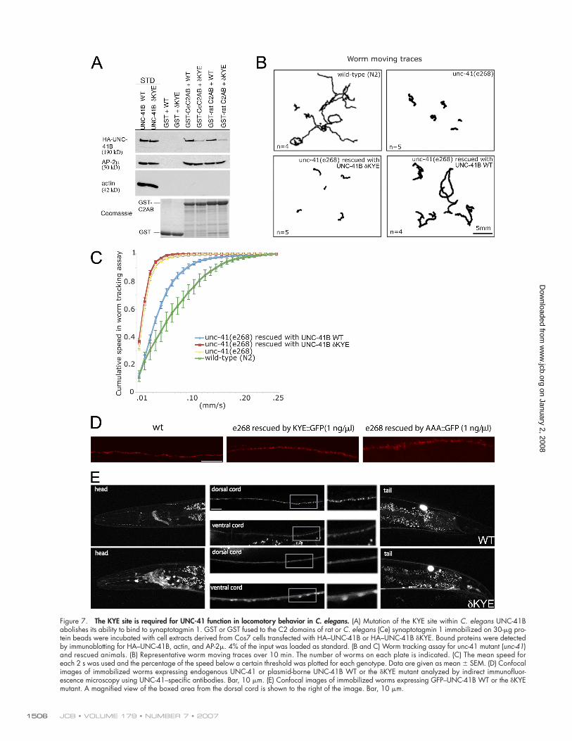
Synaptotagmin-binding–defective UNC-41B is unable to rescue paralysis in C. elegans mutant animalsAs mentioned in the preceding paragraph, the μHD of stonins,
and in particular the KYE site responsible for its association
with synaptotagmin 1, is highly conserved between differ-
ent stonin family members from mice to worms (Fig. 3 A).
The C. elegans genome contains a single member of the stonin
family encoded by unc-41, expressed as two alternative transcripts
A and B (unpublished data). UNC-41B expressed in fi broblasts,
like its mammalian counterpart stonin 2, was able to associate
with the C2 domains of synaptotagmin 1, which is consistent
with its putative role in SV recycling. Mutation of the KYE site
within UNC-41B strongly impaired its synaptotagmin 1– binding
ability (Fig. 7 A). To investigate whether UNC-41 plays a
stonin 2YR, respectively. Synaptotagmin 1 internalization was assayed as described in the Fig. 1 legend. Red, surface synaptotagmin 1; green, internal synaptotagmin 1; blue, DAPI-stained nuclei. Stonin 2 expression was verifi ed using Cy5-labeled secondary antibodies (right). Equal exposure times and identical intensity normalization were used during image acquisition. Bars, 20 μm. (D) Quantifi cations of synaptotagmin internalization experiments using stonin 2 mutants. Fluorescence intensities were quantifi ed by applying the Mask function of the Slidebook 4.0.8 Digital Microscopy Software (Intelligent Im-aging Innovations) on the green channel (internalized synaptotagmin 1) in stonin 2–transfected cells (red). Error bars represent SEM.
on January 2, 2008 w
ww
.jcb.orgD
ownloaded from
JCB • VOLUME 179 • NUMBER 7 • 2007 1504
role in neurotransmission, we monitored animal movement.
As expected, unc-41(e268) mutant worms carrying a nonsense
mutation that abrogates UNC-41 protein expression displayed
severe locomotion defects, presumably because of impaired
neurotransmission. This phenotype was rescued by microinjec-
tion of a construct expressing WT UNC-41B cDNA but not the
synaptotagmin interaction–defective δKYE mutant (Fig. 7,
B and C), although both proteins were expressed at roughly
simi lar levels (Fig. 7 D). Moreover, a 25-fold increase in injec-
tion concentration of the δKYE mutant protein-encoding plas-
mid also did not result in rescue of the locomotory phenotype
(unpublished data). Similar results were seen in rescue experi-
ments using GFP-tagged UNC-41B variants (unpublished data).
When analyzed by confocal microscopy, WT GFP–UNC-41B
exhibited a punctate synaptic distribution within the C. elegans
nervous system, whereas the δKYE mutant protein failed to ac-
cumulate at synapses (Fig. 7 E). These experiments suggest that
the KYE site is important for UNC-41B association with synap-
totagmin and its targeting to synapses, although at this stage we
cannot rule out the possibility that other factors contribute to
this pheno type in vivo.
In summary, our data reveal the molecular basis for the
recognition of synaptotagmin 1 by stonin 2 and suggest an evo-
lutionarily conserved mechanism of cargo recognition by stonin
family members during exo-endocytic SV cycling.
DiscussionThis study provides the fi rst detailed molecular characterization
of the interaction interface between the SV protein–specifi c
CLASP stonin 2 and its cargo, synaptotagmin 1. Previous stud-
ies have implicated the synaptotagmin 1 C2B domain in clathrin/
AP-2–mediated endocytosis and SV recycling (Haucke et al.,
2000; Littleton et al., 2001; Poskanzer et al., 2006). We demon-
strate a role for the synaptotagmin 1 C2A domain as the main
recognition site for stonin 2 and as a major determinant for syn-
aptotagmin 1 internalization. The following lines of evidence
support this. First, we fi nd that synaptotagmin 1, lacking the C2B
domain, is readily internalized in a stonin 2–dependent man-
ner in transfected fi broblasts. Second, stonin 2 is recruited to the
plasma membrane of transfected neuroimmune endocrine (NIE)
cells by synaptotagmin 1 ∆C2B but not by a ∆C2A domain mutant.
Figure 5. The stonin 2 KYE site is required for synaptotagmin 1–dependent recruitment of stonin 2 to the plasmalemma in neuroblastoma cells. NIE cells were cotransfected with lumenally FLAG-tagged synaptotagmin 1 and HA-tagged stonin 2WT (A), AP-2–binding–defi cient stonin 2δWFδNPF (B), stonin 2δKYE (C), or stonin 2YR (D), respectively. Surface synaptotagmin 1 was labeled by anti-FLAG antibodies (red). After permeabilization, stonin 2 was immunostained using antibodies directed against stonin 2 (green). White boxes indicate magnifi cations to the right. Bars, 20 μm.
on January 2, 2008 w
ww
.jcb.orgD
ownloaded from

SYNAPTOTAGMIN RECOGNITION BY STONIN 2 • JUNG ET AL. 1505
Figure 6. The stonin 2 KYE site is required for synaptic localization of stonin 2 and facilitates targeting of synaptotagmin 1 to recycling vesicles in primary neurons. (A) Primary hippocampal neurons at DIV9 were transfected with HA–stonin 2WT, –stonin 2YR, or –stonin 2δKYE, respectively. Monoclonal antibodies against synaptotagmin 1 are used to decorate presynaptic sites (red). Low-power views (top) exemplify the overall distribution of HA-tagged stonin 2 or mu-tants (green; insets, threefold magnifi ed images of boxed area). High-power views (bottom) represent the localization of HA–stonin 2 or mutants in selected neurites. Bars, 10 μm. (B–D) Stonin 2WT but not stonin 2δKYE mutant enhances targeting of sytpHluorin to recycling vesicles in primary hippocampal neurons. (B) To assess the surface and vesicular pools of sytpHluorin, the relative fl uorescence (F) values were measure after acid quenching and ammonium de-quenching. Bar, 10 μm. (C) Quantitative analysis of n = 7 experiments, each comprising >50 boutons as in B. Mean time course of sytpHluorin at synaptic boutons normalized to initial F values is shown. (D) Ratios of vesicular/surface stranded pools of sytpHluorin (n = 7 experiments; error bars represent SEM; control, 3.05 ± 0.3; stonin 2WT, 4.28 ± 0.19; stonin 2δKYE, 2.6 ± 0.15; **, P < 0.01). Expression of WT stonin 2 results in a signifi cant increase in the vesicular/surface pool ratio, whereas stonin 2δKYE has lost this ability.
on January 2, 2008 w
ww
.jcb.orgD
ownloaded from
JCB • VOLUME 179 • NUMBER 7 • 2007 1506
Figure 7. The KYE site is required for UNC-41 function in locomotory behavior in C. elegans. (A) Mutation of the KYE site within C. elegans UNC-41B abolishes its ability to bind to synaptotagmin 1. GST or GST fused to the C2 domains of rat or C. elegans (Ce) synaptotagmin 1 immobilized on 30-μg pro-tein beads were incubated with cell extracts derived from Cos7 cells transfected with HA–UNC-41B or HA–UNC-41B δKYE. Bound proteins were detected by immunoblotting for HA–UNC-41B, actin, and AP-2μ. 4% of the input was loaded as standard. (B and C) Worm tracking assay for unc-41 mutant (unc-41) and rescued animals. (B) Representative worm moving traces over 10 min. The number of worms on each plate is indicated. (C) The mean speed for each 2 s was used and the percentage of the speed below a certain threshold was plotted for each genotype. Data are given as mean ± SEM. (D) Confocal images of immobilized worms expressing endogenous UNC-41 or plasmid-borne UNC-41B WT or the δKYE mutant analyzed by indirect immunofl uor-escence microscopy using UNC-41–specifi c antibodies. Bar, 10 μm. (E) Confocal images of immobilized worms expressing GFP–UNC-41B WT or the δKYE mutant. A magnifi ed view of the boxed area from the dorsal cord is shown to the right of the image. Bar, 10 μm.
on January 2, 2008 w
ww
.jcb.orgD
ownloaded from
SYNAPTOTAGMIN RECOGNITION BY STONIN 2 • JUNG ET AL. 1507
Third, direct binding, as well as coimmunoprecipitation experi-
ments, reveals that conserved basic residues within C2A form
the main interaction site for stonin 2. In contrast, AP-2 predomi-
nantly associates with the C2B domain, which is consistent with
earlier data (Chapman et al., 1998; Haucke et al., 2000). We hy-
pothesize that the identifi ed basic patch within the C2A domain
is part of an endocytosis signal. However, synaptotagmin 1 mu-
tants displaying reduced affi nity for stonin 2 (Fig. 2 D) appeared
to retain the ability to undergo stonin 2–dependent internaliza-
tion (not depicted), suggesting that additional determinants and/
or cooperative effects are likely to be involved. It is worthwhile to
note that both C2 domains contain additional positively charged
surface-exposed side chains that could well serve as additional
interaction sites for stonin 2.
Site-directed mutagenesis paired with structure-based
homology modeling has allowed us to unravel the cognate rec-
ognition site for synaptotagmin 1 within the μHD of stonin 2.
Mutational analysis reveals the functional importance of a
β strand, including residues K783–E785, that is evolutionarily
conserved between different members of the stonin/stoned B
family of adaptors from nematodes to mammals (Fig. 3 A).
We propose that the function of stonin 2 as a synaptotagmin-specifi c
endocytic sorting adaptor dedicated to SV recycling is based on
the ability of residues outlined by the KYE site to interact with
patches of basic side chains within the synaptotagmin C2 do-
mains. This proposal is based on the observations that stonin 2δKYE
is unable to interact with synaptotagmin directly in vitro or in
living cells, to facilitate synaptotagmin 1 endocytosis in fi bro-
blasts, to become enriched at presynaptic sites in primary neu-
rons, or to target synaptotagmin 1 to the recycling vesicle pool
at synapses. As with all mutant proteins, a valid concern is
that the mutations may affect protein structure and/or stability.
We feel that this is unlikely for several reasons. When expressed
in fi broblasts or primary neurons, stonin 2δKYE appeared to be
expressed at levels comparable to those of its WT counterpart.
Moreover, it retained its ability to associate with AP-2 in vitro
and in living cells or to become targeted to clathrin-coated pits in
primary astrocytes (unpublished data). When probed for struc-
tural integrity by limited proteolysis using different proteases,
two independent mutants of stonin 2 (δKYE and Y784R) gave
rise to fragmentation patterns virtually identical to those seen
for the WT. A possible, yet speculative model for the recogni-
tion of synaptotagmin 1–C2A by the μHD of stonin 2 based on
our collective mapping data is shown in Fig. S5 (available
at http://www.jcb.org/cgi/content/full/jcb.200708107/DC1).
Considering the high degree of sequence conservation with re-
gard to synaptotagmin 1–C2 domains and stonin/stoned B family
members, we consider it likely that other stonins, such as stoned B
in D. melanogaster and UNC-41 in C. elegans, use a similar mode
of cargo recognition.
One of the remaining puzzles is the observation that
synaptotagmin 1 displays binding affinity for the ubiquitous
adaptor AP-2μ and, in this respect, joins a growing number of
pre- and postsynaptic proteins that use similar modes of recogni-
tion by AP-2μ for regulated endocytosis, including AMPA-type
glutamate (Kastning et al., 2007) and GABAA receptors (Kittler
et al., 2005). However, in vivo synaptotagmin endocytosis is
strongly facilitated by its direct association with stonin 2 (Figs. 5,
6, and 7). This phenotype is even more pronounced in D. melano-gaster, where stonin 2/stoned B is encoded by an essential gene
(Fergestad and Broadie, 2001; Stimson et al., 2001). One pos-
sibility to explain the pivotal roles of stoned proteins could be
the need for sorting during SV cycling. SV recycling requires
constitutive cargo to be excluded from the forming vesicle and,
thus, might benefi t from the presence of a specifi c stonin fam-
ily sorting adaptor. The presence of stonin 2 allows concentra-
tion of SV proteins, including synaptotagmin 1 and its partners,
independently of the requirements of AP-2 for recognition of
constitutive cargo (i.e., activation by kinases such as adaptor-
associated kinase 1 and cyclin G–associated kinase), such as
transferrin or EGF receptors. In addition, the use of C2A as an
interaction interface for stonin 2 may alleviate constraints on
SV cargo recognition imposed by the multiplicity of binding
partners targeting the C2B domain of synaptotagmin 1. Multiple
mechanisms of regulation of SV endocytosis by synaptotagmin 1
have recently been observed in D. melanogaster (Poskanzer
et al., 2006). Mechanistically, the action of stonin 2 and its or-
thologues stoned B (D. melanogaster) and UNC-41B (C. elegans) may therefore resemble that of other CLASPs, including
β-arrestins (Traub, 2005) or dishevelled-2 (Yu et al., 2007), in
targeting cargo to subsets of clathrin-coated vesicles (Puthenveedu
and von Zastrow, 2006). In the case of the presynaptic compart-
ment, a precise fi ne tuning of the endocytic process is required
to maintain the exact composition of SV proteins and lipids
(Takamori et al., 2006) and to ensure release competence.
The present study could form a fi rst basis for the mechanistic
understanding of this process.
Materials and methodsCell culture and transfectionsHEK293 and NIE-115 cells were cultured in DME (Invitrogen) containing 4.5 g/liter glucose and NIE cells in DME containing 1 g/liter glucose, sup-plemented with 10% FCS, penicillin, and streptomycin. Culturing of primary hippocampal neurons has been previously described (Mueller et al., 2004). Cell lines as well as primary neurons were transfected using Lipofectamine 2000 (Invitrogen). Calcium phosphate transfection was used for sytpHluorin recycling assays. Doxycycline-regulable HEK293 cell lines were generated using the T-Rex system (HEKTR-stn2WT, HEKTR-stn2δKYE; Invitrogen). HEK293 cells stably expressing lumenally FLAG-tagged synaptotagmin 1 (HEK-syt1) have been previously described (Diril et al., 2006). For morphological ex-periments, cells were grown on Matrigel-coated glass coverslips.
Plasmids and DNA constructsThe following amino terminally HA-tagged human stonin 2 constructs were made by inserting PCR products into EcoRV–XbaI restriction sites in pcHA2: HA–stonin 2 (aa 421–898), HA–stonin 2 (aa 1–555), and HA–stonin 2δWWW (W15A, W102A, and W232A). We generated plasmid construct, allowing for the expression of lumenally FLAG-tagged rat synapto-tagmin 1 by introducing the cDNA into the BamHI–XhoI restriction sites into the pcFLAG vector. By application of PCR site-directed mutagenesis, we pre-pared several synaptotagmin 1 mutation constructs: ∆C2B (1–265), ∆C2A (∆140–270), ∆C2AB (1–139), and syt1mut (K189–192E; K213E; K244E; and KR321,322EE, K324–327E). The following rat synaptotagmin 1 GST fusion constructs were generated using pGEX4T-1 (BamHI–XhoI): C2A (140–265), C2B (271–421), C2AB (140–421), C2Amut1 (KK189, 190AA), C2Amut2 (KK191, 192AA), C2Amut3 (K189–192A), C2Amut4 (K191H, K213E, and K244S), C2Amut5 (K189–192A, K213E, and K244S), C2Bmut1 (KK326, 327AA), C2Bmut2 (K324–327A), and C2Bmut3 (KR321, 322AA; and K324–327A). Constructs allowing for the expression of HA-tagged human stonin 2 were generated as previously described (Walther et al., 2004; Diril et al. 2006). The following mutants were prepared by PCR
on January 2, 2008 w
ww
.jcb.orgD
ownloaded from

JCB • VOLUME 179 • NUMBER 7 • 2007 1508
site-directed mutagenesis: HA-stonin 2δWWW (W15A, W102A, and W232A), HA-stonin 2δWFδNPF (WF15, 18AA; WF102, 105AA; WF232, 235AA; NPF313–315NAV; and NPF329–313NAV), HA-stonin 2δKYE (KYE783–785AAA), HA-stonin 2δWFδNPFδKYE (WF15, 18AA; WF102, 105AA; WF232, 235AA; NPF313–315NAV; NPF329–313NAV; and KYE783–785AAA), HA-stonin 2KA (K783A), HA-stonin 2YA (Y784A), HA-stonin 2EA (E785A), and HA-stonin 2YR (Y784A). C. elegans synaptotagmin 1 C2AB (worm base: F31E8.2a [WS]; aa 158–441) was cloned into pGEX4T-1.
The GFP–UNC-41B WT (pMG13) C. elegans expression plasmid con-sisted of the 2.5-kb unc-41 promoter (from RM536), 0.85-kb GFP fragment (from fi re lab vector 95.77), and 5.4-kb unc-41b cDNA plus 3′UTR (from RM 536) inserted into the EcoRI–SalI restriction sites of pGEM-3zf(+) using the following primers: 5′-A G G A G A A T T C C T C C C G G C A A T T C G T A A T A C G T C -3′; 5′-G G G T C C T G A A A A T G T T C T A T G -3′; 5′-A C A T T C C C G G G A T G G A A C A A G -C A G A A A A A G C A -3′; 5′-A C T T G T C G A C C A T G T G T C A G A G G T T T T C A C C G T C -3′; 5′-A G A T C C C G G G A G A A C C T C C G C C T C C T T T G T A T A G T T C A T C C A T G C C-A T G -3′; and 5′-A C C G C C C G G G A T G A G T A A A G G A G A A G A A C T T T T C -3′.
An analogous construct was made for expression of the GFP–UNC-41B δKYE mutant (pMG14, UNC-41 KYE→AAA translational GFP).
AntibodiesPolyclonal anti–stonin 2 antiserum was generated as described previously (Walther et al., 2004). Monoclonal antibodies against the α subunit of AP-2 (clone AP6) and clathrin heavy chain (clone TD1) were a gift from P. De Camilli (Yale University, New Haven, CT). Mono- (clone M2) or poly-clonal anti-FLAG, as well as anti–β-actin antibodies, were obtained from Sigma-Aldrich, monoclonal anti-HA antibodies were obtained from Babco, polyclonal anti-HA antibodies (Y11) were obtained from Santa Cruz Bio-technology, Inc., monoclonal antibodies directed against the His6-tag were obtained from EMD, monoclonal anti-α, -β2, and -μ2 antibodies were obtained from BD Biosciences, and monoclonal anti–synaptotagmin 1 anti-bodies (clone 41.1) were obtained from Synaptic Systems GmbH. Fluorescent dye–conjugated secondary antibodies were obtained from Invitrogen and horseradish peroxidase–labeled secondary antibodies, as well as unlabeled goat anti–mouse and goat anti–rabbit antibodies were obtained from Dianova GmbH.
Protein expression and purifi cationGST–synaptotagmin 1 fusion proteins were expressed in Escherichia coli (ER2566) at 25°C for 3 h after induction with 0.5 mM IPTG. Bacterial pel-lets obtained from 1-liter cultures were resuspended in 100 ml PBS. Cells were lysed using lysozyme, 1% Triton X-100, benzonase (to remove possi-ble nucleic acid contaminants), and sonifi cation. The bacterial extract was cleared by centrifugation at 39,000 g for 15 min and GST fusion proteins were affi nity purifi ed using GST-bind resin (EMD).
Stonin 2 WT and δKYE mutant proteins were affi nity purifi ed from HEKTR-stn2WT and HEKTR-stn2δKYE cells using Ni-NTA Agarose (QIAGEN). Protein expression was induced by addition of 1 μg/ml doxycycline to the growth medium for at least 16 h before cells were lysed in homogenization buffer (20 mM Hepes, pH 7.4, 150 mM NaCl, 2 mM MgCl2, 1 mM PMSF, and 0.1% mammalian protease inhibitor cocktail [Sigma-Aldrich]) using a ball-bearing cell cracker with a clearance of 12 μm. The cell extract was cleared by consecutive centrifugation at 20,000 g for 5 min and 180,000 g for 15 min. The supernatant was supplemented with 320 mM sucrose, 500 mM NaCl, 1% CHAPS, 1 mM DTT, 10 mM imidazole, and 1 mM PMSF before application on Ni-NTA Agarose for 2 h at 4°C on a rotating wheel. The beads were washed twice in washing buffer (20 mM Hepes, pH 7.4, 500/150 mM NaCl, 2 mM MgCl2, 320 mM sucrose, 1% CHAPS, 1 mM DTT, and 10 mM imidazole) containing 500 mM NaCl and once with washing buffer containing 150 mM NaCl. Bound protein was eluted in washing buffer containing 150 mM NaCl and 120 mM imidazole.
Affi nity chromatography, in vitro binding, and immunoprecipitation experiments24–48 h after transfection, transiently transfected HEK293 cells were lysed in 20 mM Hepes, 100 mM KCl, 2 mM MgCl2, 1% Triton X-100, 1 mM PMSF, 0.3% mammalian protease inhibitor cocktail for 10 min on ice. Cleared cell extracts were incubated with GST fusion proteins on a rotating wheel (1 mg of cell extract at 1 mg/ml) for 2 h at 4°C. After extensive washes, bound proteins were eluted with 80 μl of sample buffer. For immuno-precipitation experiments, transfected HEK293 cells were lysed in the buffer specifi ed in the preceding paragraph. Monoclonal antibodies immobilized on protein A/G–Sepharose (Santa Cruz Biotechnology, Inc.) were incubated with 1 mg of cell extracts at 1 mg/ml for 4 h at 4°C under gentle agitation. Beads were washed extensively and eluted with 60 μl of sample buffer.
Samples were analyzed by SDS-PAGE and immunoblotting. For in vitro binding experiments, 2–3 μg of immobilized GST–synaptotagmin 1 fusion proteins were incubated with 600 ng to 1 μg stonin 2–His6 in 100 μl of binding buffer (20 mM Hepes, pH 7.4, 150 mM NaCl, 2 mM MgCl2, 320 mM sucrose, 1% CHAPS, 1 mM DTT, and 10 mM imidazole) for 2 h at 4°C. After three washes in binding buffer, the beads were eluted in 50 μl of sample buffer. Samples were analyzed by SDS-PAGE and immunoblotting.
For some experiments, radioactively labeled 35S-labeled stonin 2WT, 35S-labeled stonin 2YR, or 35S-labeled stonin 2δKYE, synthesized by the cou-pled TNT in vitro transcription/translation kit (Promega), was incubated with 2 μg GST–synaptotagmin 1 fusion proteins in 100 μl of binding buffer for 2 h at 4°C on a rotating wheel. After three washes in binding buffer, the beads were eluted in 50 μl SDS-PAGE sample buffer and the entire sample was applied to SDS PAGE. Bound 35S-labeled stonin 2 was detected by autoradiography using the Cyclone PhosphoImager system (PerkinElmer).
Tryptic in gel digest and mass spectrometryThe SDS polyacrylamide gel, containing the proteins of interest, was stained using the freshly prepared colloidal Coomassie and destained ac-cording to the manufacturer’s instructions (Roth). Gel bands were excised under clean conditions with new razor blades and cut into 1-mm3 pieces. Gel fragments were transferred to a 500-μl reaction tube and incubated in a shaker in 20 μl of a 1:1 solution of acetonitrile/100 mM NH4HCO3 for 15 min. Samples were centrifuged for a short time and supernatant was exchanged for 100% acetonitrile and incubated for 5 min or until the gel pieces turned white. Acetonitrile was removed and gel pieces lyophilized for 10 min. For reduction of disulfi de bridges, the lyophilized gel pieces were incubated in 20 μl of 100 mM DTT in 100 mM NH4HCO3 for 30 min at 56°C. After incubation, samples were centrifuged for a short time, super-natant was removed, and volume was measured. Gel fragments were again dehydrated twice by the addition of 20 μl of 100% acetonitrile. Cysteine residues were covalently modifi ed by carbamidomethylation by addition of 20 μl of 55 mM iodacetamide in 100 mM NH4HCO3 and incubation for 20 min at room temperature in the dark. Supernatant was removed and exchanged for 100 mM NH4HCO3 and incubated for 15 min at room temperature. Gel pieces were incubated in 20 μl of 100% aceto-nitrile until they turned white and lyophilized for 10 min. 12.5 μg/ml tryp-sin in 25 mM NH4HCO3 was prepared and added to the lyophilized gel pieces (volume = 20 μl − volume of supernatant, measured after reduction in DTT + 3 μl). Samples were placed for 30 min on ice before incubation at 37°C overnight. Samples were centrifuged and again incubated at 37°C for 30 min before 3 μl of the supernatant was removed and sub-jected to mass spectrometric analyses.
Limited proteolysisWe performed limited proteolysis to assess the folding properties of stonin 2 mutants by digestion of stonin 2–His6 purifi ed from stable HEK cells or in vitro–transcribed/translated stonin 2. 0–20 μg/ml Trypsin and 0–80 ng/ml proteinase K were chosen for this purpose. Proteolysis reactions were performed in 20 mM Hepes, pH 7.4, 100 mM KCl, and 2 mM MgCl2 for in vitro–translated stonin 2 and in 20 mM Hepes, pH 7.4, 150 mM NaCl, 2 mM MgCl2, 320 mM sucrose, 1% CHAPS, 1 mM DTT, and 10 mM imid-azole for purifi ed stonin 2–His6 for 10 min at 37°C. In vitro–translated digested protein was analyzed by SDS-PAGE and autoradiography and stonin 2–His6 by immunoblotting for the His6-tag.
Antibody internalization and membrane recruitment assaysFor indirect immunofl uorescence microscopy, HEK293 cells stably express-ing FLAG–synaptotagmin 1 were cooled on ice and preincubated with polyclonal anti-FLAG antibodies in Optimem (Invitrogen) for 30 min at 10°C. After chase at 37°C for 20 min, cells were fi xed in 4% PFA for 10 min. Surface-bound anti-FLAG antibodies were blocked with unlabeled goat anti–rabbit IgG (1:5) overnight at 4°C. Cells were permeabilized and pro-cessed for immunostaining using Alexa Fluor 488– or 594–labeled sec-ondary antibodies. Synaptotagmin internalization in transfected HEK-syt1 cells was essentially done as previously described (Diril et al., 2006). Membrane recruitment experiments in NIE-115 cells were performed as in Diril et al. (2006).
Images were taken at room temperature by a charge-coupled device camera (AxioCam; Carl Zeiss, Inc.) mounted on an inverted microscope (Axiovert 200M; Carl Zeiss, Inc.) with an oil-immersion objective (63×1.4 NA; Carl Zeiss, Inc.) illuminated and controlled by the Stallion Ra-tio Imaging system (Intelligent Imaging Innovations, Inc.). Imaging data were digitized, analyzed, and processed by nearest neighbor deconvolu-tion with Slidebook 4.0.10 software (Intelligent Imaging Innovations, Inc.).
on January 2, 2008 w
ww
.jcb.orgD
ownloaded from

SYNAPTOTAGMIN RECOGNITION BY STONIN 2 • JUNG ET AL. 1509
Slidebook 4.0.10 was used for quantifi cations based on data obtained from at least three independent experiments. Using equal exposure times, at least three low-magnifi cation images were acquired from each experiment, yielding a minimum of nine datasets (each containing 5–20 transfected cells). The Mask function was applied on the fl uorescence channel represent-ing stonin 2 (stonin 2 mask). Fluorescence values representing internalized synaptotagmin 1 within the stonin 2 mask were calculated, and mean internal-ized synaptotagmin 1 fl uorescence values (±SEM) per cell were estimated and plotted in a histogram. Background fl uorescence was subtracted.
SytpHluorin recycling assays in living neuronsPublished procedures were used to assay sytpHluorin recycling in neurons (Diril et al., 2006). In brief, hippocampal neurons from 1–3-d-old Wistar rats were transfected by calciumphosphate DNA coprecipitation and used after 11–14 d in vitro. Synaptic boutons were stimulated by electric fi eld stimulation (platinum electrodes, 10-mm spacing, 200 pulses of 50mA and alternating polarity, 10 μM CNQX, and 50 μM AP-5 to prevent recurrent action potentials) at room temperature. Fast solution exchanges were achieved by a piezo-controlled stepper device (SF77B; Warner Instru-ments). Images were taken by a cooled slow-scan charge-coupled device camera (PCO; SensiCam-QE) mounted on an inverted microscope (Axio-vert S100TV; Carl Zeiss, Inc.) with a water-immersion objective(63×1.2 NA; Carl Zeiss, Inc.) Imaging data were digitized and preanalyzed with Till Vision Software (Till Photonics) by using regions of interest to delimit puncta. For further analysis, the data were collected, normalized, and av-eraged using self-written macros in Igor Pro (Wavemetrics).
C. elegans strains, microinjection, and confocal imagingThe following strains were used for rescue experiments: WT C. ele-gans (Bristol N2); CB268: unc-41(e268); EG4499: unc-41(e268); oxEx920[Punc-41A::unc-41B(unc-41B WT cDNA), Pcc::GFP]; and EG4501: unc-41(e268); oxEx922[Punc-41A::unc-41B(unc-41B KYE→AAA cDNA), Pcc::GFP]. Strains used for confocal imaging were: EG4741: oxEx1050[Punc-41A::GFP::unc-41B(cDNA WT), Pcc::GFP]; EG4746: oxEx1055[Punc-41A::GFP::unc-41B(cDNA KYE→AAA mutant), Pcc::GFP]; EG4751: unc-41(e268), oxEx1059[Punc-41A::GFP::unc-41B(cDNA WT), Pcc::GFP]; and EG4753: unc-41(e268), oxEx1061[Punc-41A::GFP::unc-41B(cDNA KYE→AAA mutant), Pcc::GFP]; EG4774: snt-1(md290), oxEx1071[Punc-41A::GFP::unc-41B(cDNA WT), Pcc::GFP].
For rescue experiments, 50 ng/μl Punc-122::GFP used as an injec-tion marker was mixed with 1 kb DNA ladder (Fermentas) as carrier DNA to give a fi nal DNA concentration of 100 ng/μl. RM#536p (Punc-41A::unc-41B(cDNA)), WT, or KYE→AAA mutant constructs or analogous GFP fusion proteins (pMG13/14: Punc-41A::gfp:: unc-41B) were injected at 1 ng/ml into unc-41(e268) mutant or WT animals.
Worms were immobilized with 2% phenoxyl propanol, and GFP fl uorescence was imaged at room temperature on a confocal laser-scanning microscope (LSM5; Pascal) using an oil-immersion objective (plan-Neofl uar 40× 1.3 NA; Carl Zeiss, Inc.) and analyzed by confocal software (Carl Zeiss, Inc.).
Worm-tracking assay1% unseeded 50-mm agar plates containing 0.004% Bromphenol blue were prepared. Right before the assay, 200 ml 2% OP50 E. coli were spread onto the assay plates to analyze worm feeding behavior. Four to fi ve worms were put on a single plate. Worm positions were recorded every 2 s for 10 min. Images were analyzed by worm tracker 06 (an ImageJ plug-in developed by J. White, University of Utah, Salt Lake City, UT). 300 mean speed values were collected for each worm. 6–10 worms were analyzed per genotype.
Molecular modeling and multiple sequence alignmentsMultiple protein sequence alignments were performed using the ClustalW program (available at http://www.ebi.ac.uk/clustalw/). The x-ray struc-ture of μ2 was used as a structural template for the model of stonin 2–μHD (G563-E875; 1GW5: chain M, G165-C435). Additional fragments with sequence homology to other known Protein Database structures (1BWU and 2FEA) were used to complete the model for residues 622–638 and 736–772 of stonin 2 for which corresponding loop regions in μ2 were not resolved or no homologous sequence was available in the μ2 template. The x-ray structure of synaptotagmin 1–C2A (1BYN: E140-K267) was used to create the interaction model shown in Fig. S5. Interaction models considering complementary shape, electrostatic potentials, and data from mutational analysis were generated by manual docking using the bio-polymer module of SYBYL7.2 and minimized with an AMBER 7.0 force fi eld within SYBYL (TRIPOS, Inc.).
Online supplemental materialFig. S1 shows association of stonin 2 with synaptotagmin 1. Fig. S2 shows that stonin 2 directly binds to synaptotagmin 1 in vitro. Fig. S3 shows that the interaction between stonin 2 and synaptotagmin 1 depends on residues within the carboxy-terminal μHD of stonin 2. Fig. S4 shows that limited pro-teolytic digests of stonin 2 WT, δKYE, and YR mutants indicate structural integrity of the mutated proteins. Fig. S5 shows a hypothetical model of the interaction between stonin 2 (gray) and the C2A domain of synaptotagmin 1 (yellow) via two parallel β strands. Online supplemental material is avail-able at http://www.jcb.org/cgi/content/full/jcb.200708017/DC1.
This work was supported by grants from the Deutsche Forschungsgemeinschaft (SFB449, TP A11, and HA2686/1-1, 1-2 to V. Haucke; and SFB523 and TP B10 to J. Klingauf), the German Federal Ministry of Science (BioDISC-2/ RENTRAFF), and the Human Frontier Science Program (to J. Klingauf). N. Jung received initial support from the Lichtenberg Foundation and was a student of the International PhD Program in Molecular Biology at the University of Göttingen. M. Wienisch was a student of the International PhD Programin Neuroscience at the University of Göttingen and received support from the Boehringer Ingel-heim Fonds. E. Jorgensen is an Investigator of the Howard Hughes Medical Institute, and this work was supported by a grant from the National Institutes of Health (NS034307).
Submitted: 15 August 2007Accepted: 25 November 2007
ReferencesBai, J., W.C. Tucker, and E.R. Chapman. 2004. PIP2 increases the speed of re-
sponse of synaptotagmin and steers its membrane-penetration activity toward the plasma membrane. Nat. Struct. Mol. Biol. 11:36–44.
Bennett, M.K., N. Calakos, T. Kreiner, and R.H. Scheller. 1992. Synaptic vesicle membrane proteins interact to form a multimeric complex. J. Cell Biol. 116:761–775.
Bonifacino, J.S., and L.M. Traub. 2003. Signals for sorting of transmem-brane proteins to endosomes and lysosomes. Annu. Rev. Biochem. 72:395–447.
Brodin, L., P. Low, and O. Shupliakov. 2000. Sequential steps in clathrin-medi-ated synaptic vesicle endocytosis. Curr. Opin. Neurobiol. 10:312–320.
Chapman, E.R., R.C. Desai, A.F. Davis, and C.K. Tornehl. 1998. Delineation of the oligomerization, AP-2 binding, and synprint binding region of the C2B domain of synaptotagmin. J. Biol. Chem. 273:32966–32972.
Collins, B.M., A.J. McCoy, H.M. Kent, P.R. Evans, and D.J. Owen. 2002. Molecular architecture and functional model of the endocytic AP2 complex. Cell. 109:523–535.
DiAntonio, A., K.D. Parfi tt, and T.L. Schwarz. 1993. Synaptic transmission per-sists in synaptotagmin mutants of Drosophila. Cell. 73:1281–1290.
Diril, M.K., M. Wienisch, N. Jung, J. Klingauf, and V. Haucke. 2006. Stonin 2 is an AP-2-dependent endocytic sorting adaptor for synaptotagmin internal-ization and recycling. Dev. Cell. 10:233–244.
Edeling, M.A., S.K. Mishra, P.A. Keyel, A.L. Steinhauser, B.M. Collins, R. Roth, J.E. Heuser, D.J. Owen, and L.M. Traub. 2006. Molecular switches involving the AP-2 beta2 appendage regulate endocytic cargo selection and clathrin coat assembly. Dev. Cell. 10:329–342.
Fergestad, T., and K. Broadie. 2001. Interaction of stoned and synaptotagmin in synaptic vesicle endocytosis. J. Neurosci. 21:1218–1227.
Galli, T., and V. Haucke. 2004. Cycling of synaptic vesicles: how far? How fast! Sci. STKE. 2004:re19.
Granseth, B., B. Odermatt, S.J. Royle, and L. Lagnado. 2006. Clathrin-mediated endocytosis is the dominant mechanism of vesicle retrieval at hippocam-pal synapses. Neuron. 51:773–786.
Grass, I., S. Thiel, S. Honing, and V. Haucke. 2004. Recognition of a basic AP-2 binding motif within the C2B domain of synaptotagmin is dependent on multimerization. J. Biol. Chem. 279:54872–54880.
Haucke, V., M.R. Wenk, E.R. Chapman, K. Farsad, and P. De Camilli. 2000. Dual interaction of synaptotagmin with mu2- and alpha-adaptin facili-tates clathrin-coated pit nucleation. EMBO J. 19:6011–6019.
Jarousse, N., and R.B. Kelly. 2001. The AP2 binding site of synaptotagmin 1 is not an internalization signal but a regulator of endocytosis. J. Cell Biol. 154:857–866.
Jia, J.Y., S. Lamer, M. Schumann, M.R. Schmidt, E. Krause, and V. Haucke. 2006. Quantitative proteomics analysis of detergent-resistant membranes from chemical synapses: evidence for cholesterol as spatial organizer of synaptic vesicle cycling. Mol. Cell. Proteomics. 5:2060–2071.
on January 2, 2008 w
ww
.jcb.orgD
ownloaded from

JCB • VOLUME 179 • NUMBER 7 • 2007 1510
Jorgensen, E.M., E. Hartwieg, K. Schuske, M.L. Nonet, Y. Jin, and H.R. Horvitz. 1995. Defective recycling of synaptic vesicles in synaptotagmin mutants of Caenorhabditis elegans. Nature. 378:196–199.
Kastning, K., V. Kukhtina, J.T. Kittler, G. Chen, A. Pechstein, S. Enders, S.H. Lee, M. Sheng, Z. Yan, and V. Haucke. 2007. Molecular determinants for the interaction between AMPA receptors and the clathrin adaptor com-plex AP-2. Proc. Natl. Acad. Sci. USA. 104:2991–2996.
Kittler, J.T., G. Chen, S. Honing, Y. Bogdanov, K. McAinsh, I.L. Arancibia-Carcamo, J.N. Jovanovic, M.N. Pangalos, V. Haucke, Z. Yan, and S.J. Moss. 2005. Phospho-dependent binding of the clathrin AP2 adaptor complex to GABAA receptors regulates the effi cacy of inhibitory synap-tic transmission. Proc. Natl. Acad. Sci. USA. 102:14871–14876.
Lefkowitz, R.J., and E.J. Whalen. 2004. beta-arrestins: traffi c cops of cell signal-ing. Curr. Opin. Cell Biol. 16:162–168.
Littleton, J.T., J. Bai, B. Vyas, R. Desai, A.E. Baltus, M.B. Garment, S.D. Carlson, B. Ganetzky, and E.R. Chapman. 2001. synaptotagmin mutants reveal essential functions for the C2B domain in Ca2+-triggered fusion and recycling of synaptic vesicles in vivo. J. Neurosci. 21:1421–1433.
Llinas, R.R., M. Sugimori, K.A. Moran, J.E. Moreira, and M. Fukuda. 2004. Vesicular reuptake inhibition by a synaptotagmin I C2B domain antibody at the squid giant synapse. Proc. Natl. Acad. Sci. USA. 101:17855–17860.
Martina, J.A., C.J. Bonangelino, R.C. Aguilar, and J.S. Bonifacino. 2001. Stonin 2: an adaptor-like protein that interacts with components of the endocytic machinery. J. Cell Biol. 153:1111–1120.
Mishra, S.K., P.A. Keyel, M.J. Hawryluk, N.R. Agostinelli, S.C. Watkins, and L.M. Traub. 2002. Disabled-2 exhibits the properties of a cargo-selective endocytic clathrin adaptor. EMBO J. 21:4915–4926.
Mueller, V.J., M. Wienisch, R.B. Nehring, and J. Klingauf. 2004. Monitoring clathrin-mediated endocytosis during synaptic activity. J. Neurosci. 24:2004–2012.
Murthy, V.N., and P. De Camilli. 2003. Cell biology of the presynaptic terminal. Annu. Rev. Neurosci. 26:701–728.
Nicholson-Tomishima, K., and T.A. Ryan. 2004. Kinetic effi ciency of endo-cytosis at mammalian CNS synapses requires synaptotagmin I. Proc. Natl. Acad. Sci. USA. 101:16648–16652.
Owen, D.J., and P.R. Evans. 1998. A structural explanation for the recognition of tyrosine-based endocytotic signals. Science. 282:1327–1332.
Poskanzer, K.E., K.W. Marek, S.T. Sweeney, and G.W. Davis. 2003. Synapto-tagmin I is necessary for compensatory synaptic vesicle endocytosis in vivo. Nature. 426:559–563.
Poskanzer, K.E., R.D. Fetter, and G.W. Davis. 2006. Discrete residues in the c(2)b domain of synaptotagmin I independently specify endocytic rate and synaptic vesicle size. Neuron. 50:49–62.
Puthenveedu, M.A., and M. von Zastrow. 2006. Cargo regulates clathrin-coated pit dynamics. Cell. 127:113–124.
Santolini, E., C. Puri, A.E. Salcini, M.C. Gagliani, P.G. Pelicci, C. Tacchetti, and P.P. Di Fiore. 2000. Numb is an endocytic protein. J. Cell Biol. 151:1345–1352.
Stimson, D.T., P.S. Estes, S. Rao, K.S. Krishnan, L.E. Kelly, and M. Ramaswami. 2001. Drosophila stoned proteins regulate the rate and fi delity of syn-aptic vesicle internalization. J. Neurosci. 21:3034–3044.
Sudhof, T.C. 2004. The synaptic vesicle cycle. Annu. Rev. Neurosci. 27:509–547.
Takamori, S., M. Holt, K. Stenius, E.A. Lemke, M. Gronborg, D. Riedel, H. Urlaub, S. Schenck, B. Brugger, P. Ringler, et al. 2006. Molecular anat-omy of a traffi cking organelle. Cell. 127:831–846.
Traub, L.M. 2005. Common principles in clathrin-mediated sorting at the Golgi and the plasma membrane. Biochim. Biophys. Acta. 1744:415–437.
Voglmaier, S.M., K. Kam, H. Yang, D.L. Fortin, Z. Hua, R.A. Nicoll, and R.H. Edwards. 2006. Distinct endocytic pathways control the rate and extent of synaptic vesicle protein recycling. Neuron. 51:71–84.
Walther, K., M.K. Diril, N. Jung, and V. Haucke. 2004. Functional dissection of the interactions of stonin 2 with the adaptor complex AP-2 and synapto-tagmin. Proc. Natl. Acad. Sci. USA. 101:964–969.
Wienisch, M., and J. Klingauf. 2006. Vesicular proteins exocytosed and sub-sequently retrieved by compensatory endocytosis are nonidentical. Nat. Neurosci. 9:1019–1027.
Willig, K.I., S.O. Rizzoli, V. Westphal, R. Jahn, and S.W. Hell. 2006. STED microscopy reveals that synaptotagmin remains clustered after synaptic vesicle exocytosis. Nature. 440:935–939.
Yu, A., J.F. Rual, K. Tamai, Y. Harada, M. Vidal, X. He, and T. Kirchhausen. 2007. Association of dishevelled with the clathrin ap-2 adaptor is re-quired for frizzled endocytosis and planar cell polarity signaling. Dev. Cell. 12:129–141.
Zhang, J.Z., B.A. Davletov, T.C. Sudhof, and R.G. Anderson. 1994. Synapto-tagmin I is a high affi nity receptor for clathrin AP-2: implications for membrane recycling. Cell. 78:751–760.
on January 2, 2008 w
ww
.jcb.orgD
ownloaded from

Supplemental data
Figure S1. Association of stonin 2 with synaptotagmin 1. (A) 6 µg GST–synaptotagmin 1 cytoplasmic domain (C2A, C2B, or C2AB) fusion proteins (bottom) were analyzed in pulldown experiments for their ability to associate with HA–stonin 2WT from HEK293 cell lysates (Materials and methods). Samples were analyzed by immunoblotting (top) for HA–stonin 2, AP-2µ, or clathrin heavy chain (CHC). 8% standard (STD), 8% of the total input (cell extract). (B) Same as in A, but with an AP-2–binding–deficient HA–stonin 2δWFδNPF mutant. (C) GST-C2B wild-type or mutant proteins (Fig. 2 B) were analyzed in pulldown experiments for their ability to associate with HA–stonin 2δWFδNPF from HEK293 lysates. Samples were analyzed by immunoblotting for HA–stonin 2, AP-2β, and clathrin heavy chain. 3% of the input was loaded as standard.
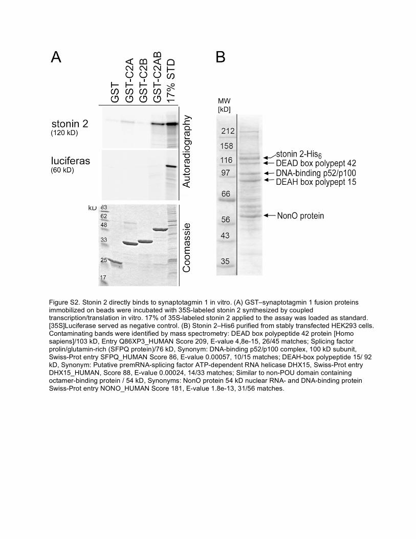
Figure S2. Stonin 2 directly binds to synaptotagmin 1 in vitro. (A) GST–synaptotagmin 1 fusion proteins immobilized on beads were incubated with 35S-labeled stonin 2 synthesized by coupled transcription/translation in vitro. 17% of 35S-labeled stonin 2 applied to the assay was loaded as standard. [35S]Luciferase served as negative control. (B) Stonin 2–His6 purified from stably transfected HEK293 cells. Contaminating bands were identified by mass spectrometry: DEAD box polypeptide 42 protein [Homo sapiens]/103 kD, Entry Q86XP3_HUMAN Score 209, E-value 4,8e-15, 26/45 matches; Splicing factor prolin/glutamin-rich (SFPQ protein)/76 kD, Synonym: DNA-binding p52/p100 complex, 100 kD subunit, Swiss-Prot entry SFPQ_HUMAN Score 86, E-value 0.00057, 10/15 matches; DEAH-box polypeptide 15/ 92 kD, Synonym: Putative premRNA-splicing factor ATP-dependent RNA helicase DHX15, Swiss-Prot entry DHX15_HUMAN, Score 88, E-value 0.00024, 14/33 matches; Similar to non-POU domain containing octamer-binding protein / 54 kD, Synonyms: NonO protein 54 kD nuclear RNA- and DNA-binding protein Swiss-Prot entry NONO_HUMAN Score 181, E-value 1.8e-13, 31/56 matches.

Figure S3. The interaction between stonin 2 and synaptotagmin 1 depends on residues within the carboxy-terminal µHD of stonin 2. (A) Stonin 2 constructs tested for synaptotagmin 1 interaction in B. (B) Coimmunoprecipitation experiments from fibroblast cell extracts. HEK293 cells stably transfected with FLAG-tagged synaptotagmin 1 (HEK-syt1) were transfected with HA-tagged stonin 2 constructs shown in A. Immunoprecipitation was performed using an antiserum directed against stonin 2. Coimmunoprecipitated proteins were detected by immunoblotting against FLAG-tag (syt1), β2-adaptin (AP-2β), HA-tag (stonin 2), and clathrin heavy chain as negative control. 10% of the input was loaded as standard. WVXF: AP-2–binding motif (δWWW: WVXF→AVXF). (C) Stonin 2 δKYE and YR mutations result in loss of interaction with synaptotagmin 1. 2 µg GST-C2AB fusion proteins were incubated with 35S-labeled stonin 2 synthesized by coupled transciption/translation in vitro. Bound stonin 2 was detected by autoradiography. 20% of the input material was loaded as standard. (D) Molecular homology model of the stonin 2 µHD (right) based on the crystal structure (1GW5_M) of AP-2µ as a main template (left). Modeled loops within stonin 2 that differ from the template are colored in cyan. The KYE site (783–785) within stonin 2 is depicted in red (arrow).

Figure S4. Limited proteolytic digests of stonin 2 wild-type, δKYE, and YR mutants indicate structural integrity of the mutated proteins. (A) Limited tryptic digests of carboxy-terminally His6-tagged stonin 2 purified from HEK293 cells or synthesized by coupled transcription/translation in vitro (in vitro t/t stonin 2). The digest was performed for 10 min at 37°C with the indicated concentrations of trypsin. (B) Limited tryptic digest of 35S-labeled stonin 2WT, 35S-labeled stonin 2δKYE, and 35S-labeled stonin 2YR result in similar digestion patterns. Proteins were digested as described in A. (C) Limited proteolysis experiment as described in B using proteinase K at the indicated concentrations.

Figure S5. Hypothetical model of the interaction between stonin 2 (gray) and the C2A domain of synaptotagmin 1 (yellow) via two parallel β-strands. (A and B) B shows details of the possible interaction site marked by a rectangle in A. Purple dashed lines refer to putative hydrogen bonds between backbone atoms of β-strands and side chains within both protein domains. The KYE site (red arrow) within stonin 2 and basic side chains K191 and K213 (purple) within synaptotagmin 1–C2A are sensitive to mutation in protein–protein interaction assays.
Recommended