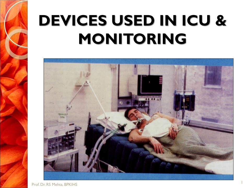
DEVICES USED IN ICU &
MONITORING
1 Prof. Dr. RS Mehta, BPKIHS

INTRODUCTION
Intensive care unit (ICU) equipment
includes patient monitoring, respiratory
and cardiac support, pain management,
emergency resuscitation devices, and
other life support equipment .
2 Prof. Dr. RS Mehta, BPKIHS

Contd…
They are designed to care for patients who are
seriously injured,
have a critical or life-threatening illness, or
have undergone a major surgical procedure thereby requiring 24-hour care and monitoring.
3 Prof. Dr. RS Mehta, BPKIHS

PURPOSE
An ICU may be designed and equipped to
provide care to patients with a range of
conditions, or it may be designed and
equipped to provide specialized care to
patients with specific conditions.
4 Prof. Dr. RS Mehta, BPKIHS

Contd…
Neuromedical ICU cares for patients with
acute conditions involving the nervous
system or patients who have just had
neurosurgical procedures and require
equipment for monitoring and assessing
the brain and spinal cord.
5 Prof. Dr. RS Mehta, BPKIHS

Contd…
A neonatal ICU is designed and
equipped to care for infants who are ill,
born prematurely, or have a condition
requiring constant monitoring.
A trauma/burn ICU provides specialized
injury and wound care for patients
involved in auto accidents and patients
who have gunshot injuries or burns.
6 Prof. Dr. RS Mehta, BPKIHS

7 Prof. Dr. RS Mehta, BPKIHS

TYPES OF DEVICES
Intensive care unit equipment includes
Patient monitoring devices
Life support and emergency resuscitation
devices, and
Diagnostic devices.
8 Prof. Dr. RS Mehta, BPKIHS

PATIENT MONITORING EQUIPMENT
Arterial line
Bed side monitor
Blood pressure device (sphygmomanometer)
Blood pressure monitor
Electrocardiograph(ECG or EKG machine)
Electroencephalograph(EEG machine)
Intracranial pressure monitor
Pulse Oximeter
Glucometer
9 Prof. Dr. RS Mehta, BPKIHS

LIFE SUPPORT AND EMERGENCY
RESUSCITATION DEVICES
Mechanical Ventilator
Laryngoscope
Airway
Infusion pump
Crash cart(Resuscitation cart)
Intra aortic ballon pump
Continuous positive air pressure machine (CPAP)
Defibrillator
10 Prof. Dr. RS Mehta, BPKIHS

DIAGNOSTIC EQUIPMENT
Mobile x-ray units
portable clinical laboratory devices,
Bronchoscope
Colonoscope
Endoscope
Gastroscope
11 Prof. Dr. RS Mehta, BPKIHS
OTHER ICU EQUIPMENT
Disposable ICU equipment includes
Urinary catheter
Urinary drainage collector
Suction catheter
Nasogastric (NG) tube
Intravenous(IV) line or catheter
Feeding tube
Breathing tube( Endotracheal tube)
12 Prof. Dr. RS Mehta, BPKIHS

Hemodynamic Monitoring
13 Prof. Dr. RS Mehta, BPKIHS

Prof. Dr. RS Mehta, BPKIHS

Overview
Blood pressure monitoring
◦ NIBP
◦ IBP
Central venous pressure monitoring
Pulmonary artery pressure
monitoring
Mixed venous oxygen monitoring
Cardiac output
15 Prof. Dr. RS Mehta, BPKIHS

Why monitor BP?
◦ Alterations inherent
◦ Provides data for interpretation/therapeutic
decisions
◦ Important for determining organ perfusion
(MAP most important, except with the heart)
16 Prof. Dr. RS Mehta, BPKIHS

Noninvasive Hemodynamic Monitoring
Noninvasive BP
Heart Rate, pulses
Mental Status
Skin Temperature
Capillary Refill
Urine Output
17 Prof. Dr. RS Mehta, BPKIHS

Indications for
Arterial Blood Pressure
Frequent titration of vasoactive drips
Major surgery involving large fluid shifts
CVP
Aortic surgery
Unstable blood pressures
Frequent ABGs or labs
Unable to obtain Non-invasive BP
18 Prof. Dr. RS Mehta, BPKIHS

Supplies to Gather
Arterial Catheter
Pressure Tubing
Pressure Cable
Sterile Gown
Sterile Towels
Sterile Gloves
Pressure Bag
Flush – 500cc NS
Suture (silk 2.0)
Chlorhexidine Swabs
Mask
19 Prof. Dr. RS Mehta, BPKIHS

Potential Complications
Associated With Arterial Lines
Hemorrhage
Air Emboli
Infection
Altered Skin Integrity
Impaired Circulation
20 Prof. Dr. RS Mehta, BPKIHS

ARTERIAL LINE DEFINITION:
It is the method of direct continuous monitoring of systemic arterial pressure by inserting a catheter into peripheral artery either in arm or in leg. The catheter is connected with a transducer with electrical signals.
21 Prof. Dr. RS Mehta, BPKIHS

PURPOSE:
The arterial line provides a way to
constantly measure a patient's blood
pressure and may be essential to the
stabilization of the patient.
Continuous measurement of arterial blood
pressure in case of open heart surgery.
22 Prof. Dr. RS Mehta, BPKIHS

Arterial lines may be useful in patients
with very high or low blood pressures.
The arterial line also provides access for
frequent blood sampling.
23 Prof. Dr. RS Mehta, BPKIHS

COMPLICATIONS:
The major complications associated with
the arterial line are bleeding, infection, and
rarely, a lack of blood flow to the tissue
supplied by the artery.
24 Prof. Dr. RS Mehta, BPKIHS

NURSES ROLE / NURSING CARE
Never give any medication through an arterial
line.
Always check the pressure of the pressurized
bag and maintain a pressure of 300mm of hg.
Cover the cannula cap with adhesive tape.
Flush properly the arterial line every hour and
every time after a blood sample is drawn.
Always compress the site after removal of
arterial line for 10 min.
25 Prof. Dr. RS Mehta, BPKIHS

BEDSIDE MONITOR
A bedside monitor is a
display of major body
functions on a device
that looks like a
television screen or
computer monitor.
26 Prof. Dr. RS Mehta, BPKIHS

It is a comprehensive patient monitoring
systems that can be configured to
continuously measure and display a
number of parameters via electrodes and
sensors that are connected to the patient.
27 Prof. Dr. RS Mehta, BPKIHS

These may include the electrical activity
of the heart via an EKG, respiration rate
(breathing), blood pressure, body
temperature, cardiac output, and amount
of oxygen and carbon dioxide in the
blood.
28 Prof. Dr. RS Mehta, BPKIHS

Each patient bed in an ICU has a
physiologic monitor that measure these
body activities. All monitors are
networked to a central nurses' station.
29 Prof. Dr. RS Mehta, BPKIHS

PURPOSES:
The monitor is typically used when the
doctor wants to measure functions like
the heart rate, respiratory rate, blood
pressure and temperature. In addition,
special functions such as capnography,
oximetry, electroencephalography and
pulmonary artery catheter readings are
also used in certain situations.
30 Prof. Dr. RS Mehta, BPKIHS

The bedside monitor has alarms that signal
the nurse if a body function needs
attention.
31 Prof. Dr. RS Mehta, BPKIHS

NURSES ROLE:
Check properly each connection so as to
get a desired reading.
Any abnormality in a reading is signalled by
an alarm so inform doctor immediately.
32 Prof. Dr. RS Mehta, BPKIHS

Central Venous Line or Catheter
A central venous catheter is a special IV line that is inserted into a large vein in the body. Several veins are used for central venous catheters including those located in the shoulder (subclavian vein), neck (jugular vein), and groin (femoral vein)
33 Prof. Dr. RS Mehta, BPKIHS

34 Prof. Dr. RS Mehta, BPKIHS

Common sites for central venous
catheter insertion
1
Prof. Dr. RS Mehta, BPKIHS

PROCEDURE
The most
common used
method is
seldinger
technique.
Prof. Dr. RS Mehta, BPKIHS

In some patients, a central venous
catheter may be inserted into the
elbow vein (anticubital vein) and
advanced into the subclavian vein.
37 Prof. Dr. RS Mehta, BPKIHS

38 Prof. Dr. RS Mehta, BPKIHS
PURPOSE
These special IVs are used when the
patient either does not have adequate
veins in the arms or needs special
medications and/or nutrition that cannot
be given through the smaller arm veins.
Serve as a guide of fluid balance in
critically ill patients.
Determine the function of the right side
of the heart
39 Prof. Dr. RS Mehta, BPKIHS

complication
Bleeding and infection are complications
associated with IV catheters. As previously
mentioned, collapse of a lung is a rare
complication of central venous catheters.
If this occurs, a chest tube (thoracostomy
tube) may be required to re-expand the
lung.
40 Prof. Dr. RS Mehta, BPKIHS

Arterial puncture, cardiac puncture
Pneumothorax, Hemomothorax
Air emboli, Thrombosis
Cardiac temponade
Cardiac arrhythmias
Carotid Artery Puncture
Perforation of SVC or R. Atrium/Ventricle
Pleural Effusion
41 Prof. Dr. RS Mehta, BPKIHS

NURSES ROLE
Monitor for the signs of complications.
Assess for patency of the CVP line.
Sterile dressing should be done to
prevent infection( CVP care per the
hospital protocol)
The length of the indwelling catheter
should be recorded and regularly
monitored.
42 Prof. Dr. RS Mehta, BPKIHS

ICP monitor
ICU patients who have sustained head
trauma, brain hemorrhage, brain surgery,
or conditions in which the brain may
swell might require intracranial pressure
monitoring.
43 Prof. Dr. RS Mehta, BPKIHS

PURPOSE
The purpose of ICP monitoring is to
continuously measure the pressure
surrounding the brain. If the pressure
surrounding the brain gets too high, it can
cause decreased blood flow to the brain
and potentially lead to brain damage.
44 Prof. Dr. RS Mehta, BPKIHS

The ICP monitor is usually inserted by a
neurosurgeon while the patient is in the
ICU or operating room. After using
numbing medicine (local anesthetics), the
neurosurgeon makes a skin incision and
inserts the ICP monitor into the brain
through a very small hole created in the
skull.
45 Prof. Dr. RS Mehta, BPKIHS

The ICP monitor is usually inserted in the
left or right top-front part of the brain.
Some ICP monitors can drain spinal fluid if
necessary.
46 Prof. Dr. RS Mehta, BPKIHS

complication
Potential complications associated with
ICP monitoring include infection and brain
hemorrhage, which are very infrequent.
47 Prof. Dr. RS Mehta, BPKIHS

Nurses role
Optimizing cerebral tissue perfusion.
Preventing infection.
Maintaining patient airway.
Maintaining negative fluid balance.
Prevent infection( dressing)
48 Prof. Dr. RS Mehta, BPKIHS

PULSE OXIMETER
A pulse oximeter is the device that
measures and displays the oxygen arterial
saturation. The study is called pulse
oxymetry.
The pulse oximeter is a small device that
has to be in contact with the skin to detect
the oxygen saturation.
49 Prof. Dr. RS Mehta, BPKIHS

The device is usually place on the
patient's finger, earlobe, toe or nose. The
pulse oximeter gives off light that
determines the oxygen saturation of the
blood.
50 Prof. Dr. RS Mehta, BPKIHS

Breathing Machine (Mechanical
Ventilator
A breathing machine
helps the patient
breathe. It is designed
to help patients who
cannot breathe
adequately on their
own. The breathing
machine does not fix
any problems of the
lungs.
51 Prof. Dr. RS Mehta, BPKIHS

It is a device that simply pushes air and
oxygen into the lungs and withdraws
carbon dioxide from the lungs. The lungs
must function in order for the breathing
machine to be effective.
52 Prof. Dr. RS Mehta, BPKIHS

PURPOSE
A breathing machine is
used whenever a
patient cannot breathe
without assistance.
Doctors, nurses and
respiratory therapists
all work to make sure
a breathing machine is
not used any longer
than necessary.
53 Prof. Dr. RS Mehta, BPKIHS

The goal when a breathing machine is
first used is to get the patient to be able
to breathe on their own, so that the
breathing machine can be removed.
54 Prof. Dr. RS Mehta, BPKIHS

complications
Patients who require breathing machine
support are at increased risk to develop
pneumonia. Occasionally, patients may
develop a collapsed lung. Both of these
complications require treatment
55 Prof. Dr. RS Mehta, BPKIHS

NURSES ROLE
Promoting effective airway clearance.
preventing trauma and infection.
Check
Ventilator functioning properly
Blockage of air passage
Too much sputum, secretions
When sedation drugs are used
ABG, hypoxia
56 Prof. Dr. RS Mehta, BPKIHS
b. Suction periodically as per need
c. Change the mode setup as adviced.
d. Give sedatives as adviced.
57 Prof. Dr. RS Mehta, BPKIHS

INFUSION PUMP
An intravenous (IV) infusion pump is a
machine that carefully controls the rate at
which IV fluids and/or IV medications are
given.
58 Prof. Dr. RS Mehta, BPKIHS

PURPOSE
Under some circumstances, the rate at
which IV fluids and/or IV medications are
given needs to be closely controlled.
59 Prof. Dr. RS Mehta, BPKIHS
These pumps are very reliable. Mechanical
problems are possible, but very rare. If the
IV infusion pump does not work correctly,
an alarm will sound.
60 Prof. Dr. RS Mehta, BPKIHS

NURSES ROLE
Using aseptic technique and universal
precautions, iv infusion should be set.
Set the flow rate as prescribed calculating
the amount of fluid.
Observe for the signs of infiltration or
other complications such as
thrombophlebitis. Fluid or electrolyte
overload and embolism before
administration.
61 Prof. Dr. RS Mehta, BPKIHS

Resuscitation Cart (Crash Cart)
The resuscitation cart
contains all of the
equipment and
medications needed
for advanced life
support and CPR
(cardiopulmonary
resuscitation).
62 Prof. Dr. RS Mehta, BPKIHS

purpose
This emergency equipment is used only if
the patient's heart or lungs stop working.
The cart is brought to the patient's
bedside when the patient's heart or lungs
are failing or have failed.
63 Prof. Dr. RS Mehta, BPKIHS
NURSES ROLE
Keep the resuscitation cart ready all the
time.
Check the devices and ensure that the
devices are kept in charging.
Check for the emergency (life saving)
medication for their expiry date.
64 Prof. Dr. RS Mehta, BPKIHS

DEFIBRILLATOR
A defibrillator is a device that is designed
to pass electrical current through a
patient’s heart. The passing of electrical
current through the heart is called
defibrillation. A defibrillation is done
through pads placed on the patient’s chest.
65 Prof. Dr. RS Mehta, BPKIHS

purpose
A defibrillation is used to restore a
patient’s heart rhythm to normal.
Abnormal heart rhythms may be treated
with medications while other rhythms
need to be treated with defibrillation.
66 Prof. Dr. RS Mehta, BPKIHS

Life threatening heart rhythms need
defibrillation immediately while other heart
rhythms may be defibrillated in a scheduled
fashion.
Defibrillation may be done using the manual
defibrillator or the automatic external
defibrillator (AED).
67 Prof. Dr. RS Mehta, BPKIHS
Complication
The defibrillator pads may cause a skin
irritation and leave a temporary redden
area where they contacted the chest.
Unfortunately defibrillation does not
always return the patient’s heart rhythm
back to normal.
68 Prof. Dr. RS Mehta, BPKIHS

NURSES ROLE
Keep the patient in comfortable position
and obtain 12 lead ECG.
Give the patient 100 % oxygen by
inhalation.
Apply electrode paste on the DC paddle,
rub it and apply the paste at the patient’s
chest in the second intercostal space at the
right side of breast line and at the apex of
the heart.
69 Prof. Dr. RS Mehta, BPKIHS

TURN OFF the oxygen to the patient as a
spark from paddle could blow the oxygen
on the fire.
Be sure to say “ ALL CLEAR”. No one
should touch the patient or the bed during
cardioversion.
Check the rhythm on ECG monitor.
Keep the patient in comfortable position
and give 100% oxygen by inhalation.
70 Prof. Dr. RS Mehta, BPKIHS

Report and record the procedure and
clean the paddle area with spirit swab.
Keep the difibrilator on continue electrical
charging.
71 Prof. Dr. RS Mehta, BPKIHS

MAINTENANCE OF ICU
EQUIPMENTS
Since ICU equipment is used continuously
on critically ill patients, it is essential that
equipment be properly maintained,
particularly devices that are used for life
support and resuscitation.
72 Prof. Dr. RS Mehta, BPKIHS

Contd…
Staff in the ICU should perform daily checks on equipment and inform biomedical engineering staff when equipment needs maintenance, repair, or replacement.
For mechanically complex devices, service and preventive maintenance contracts are available from the manufacturer or third-party servicing companies, and should be kept current at all times.
73 Prof. Dr. RS Mehta, BPKIHS

Health care team roles
Equipment in the ICU is used by a team
specialized in their use. The team usually
comprises a critical care attending
physician (also called an intensivist), critical
care nurses, an infectious disease team,
critical care respiratory therapists,
pharmacologists, physical therapists, and
dietitians.
74 Prof. Dr. RS Mehta, BPKIHS

Radiologic technologists perform mobile x
ray examinations (bedside radiography).
Either nurses or clinical laboratory
personnel perform point-of-care blood
analysis. Equipment in the ICU is
maintained and repaired by hospital
biomedical engineering staff and/or the
equipment manufacturer.
75 Prof. Dr. RS Mehta, BPKIHS

Thank you
76 Prof. Dr. RS Mehta, BPKIHS

3.CARDIAC PACING
Cardiac Pacing is
the repetitive
delivery of very
low electrical
energies to the
heart to initiate
and maintain
cardiac rhythm.
77 Prof. Dr. RS Mehta, BPKIHS

METHODS
Percussive pacing
Transcutaneous
Epicardial
Transvenous
Permanent pacing
78 Prof. Dr. RS Mehta, BPKIHS

Types of PA catheters
1. The thermo dilution catheter:
is the one described above; using this
catheter, thermo dilution cardiac output &
other divided haemodynamic parameters
may be measured
2. Pacing:
Some PAC’s have the capacity to provide
intra cardiac pacing
79 Prof. Dr. RS Mehta, BPKIHS

3. Mixed venous oxygen saturation:
Special fiber-optic PAC can be used to monitor
mixed venous oxygen saturation SVO2
continuously by the principle of absorption and
reflectance of light through blood
The normal SVO2 is 75% and a 5– 10 %
increase or decrease is considered significant
A significant decrease in SVO2 may be due to: (a) a decrease in the cardiac output
(b) increase in metabolic rate
(c) decrease in arterial oxygen saturation.
80 Prof. Dr. RS Mehta, BPKIHS

4. Ejection fraction catheter :
New-catheters with faster thermistor response times
can be used to determine the right ventricular ejection
fraction in addition to the cardiac output
5. Continuous cardiac output measurement
:
Continuous cardiac output measuring PACs contain
an integrated thermal filament at level of the RV
This filament is activated in a programmed sequence
to provide small amounts of heat, which is then
detected in the PA by a thermistor
The data by the device yields a rapidly updated, near
continuous value for cardiac output
81 Prof. Dr. RS Mehta, BPKIHS

82 Prof. Dr. RS Mehta, BPKIHS

PULMONARY ARTERY CATHETER
83 Prof. Dr. RS Mehta, BPKIHS

Components of a Pulmonary Artery
Catheter
84 Prof. Dr. RS Mehta, BPKIHS

85 Prof. Dr. RS Mehta, BPKIHS
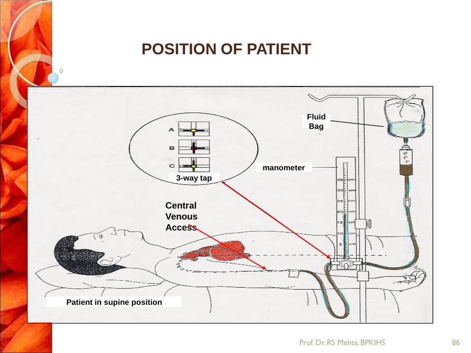
POSITION OF PATIENT
3-way tap
manometer
Fluid
Bag
Patient in supine position
Central
Venous
Access
86 Prof. Dr. RS Mehta, BPKIHS

87 Prof. Dr. RS Mehta, BPKIHS
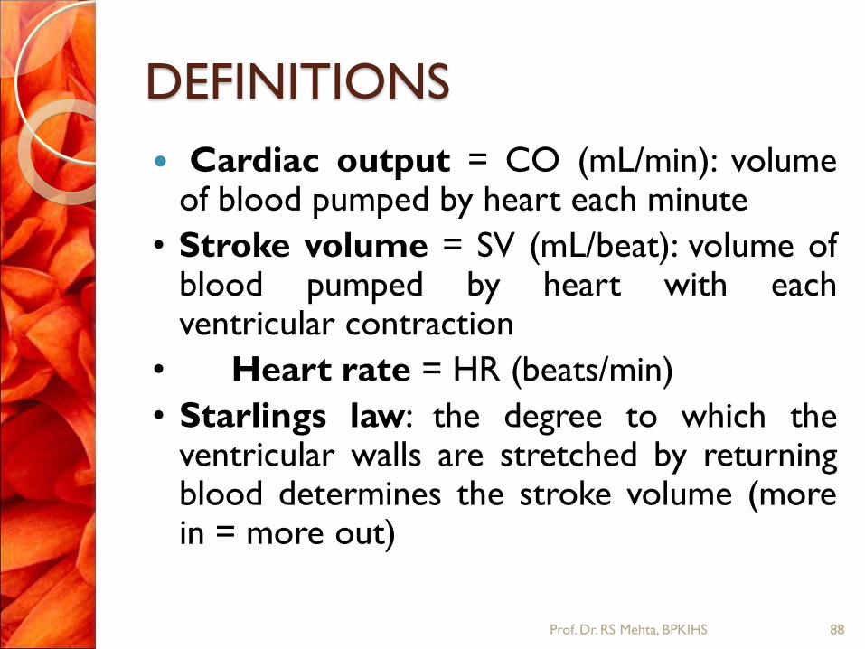
DEFINITIONS
Cardiac output = CO (mL/min): volume of blood pumped by heart each minute
• Stroke volume = SV (mL/beat): volume of blood pumped by heart with each ventricular contraction
• Heart rate = HR (beats/min)
• Starlings law: the degree to which the ventricular walls are stretched by returning blood determines the stroke volume (more in = more out)
88 Prof. Dr. RS Mehta, BPKIHS

Regulation of Cardiodynamics
Intrinsic: Within the heart (SV) – force of contraction related to degree of stretch of myocardium
Lots of stretch = increased force production
Extrinsic: outside the heart (NS: Autonomic or Hormonal) – Heart rate influenced by both sympathetic and parasympathetic (autonomic) nervous system – Stroke volume influenced by blood pressure
89 Prof. Dr. RS Mehta, BPKIHS

90 Prof. Dr. RS Mehta, BPKIHS

Definition
Hemodynamic regulation is known as
optimization of heart rate, preload, afterlo
ad, and contractility.
Heart rate, or heart pulse, is the speed
of the heartbeat measured by the number
of poundings of the heart per unit
of time — typically beats per
minute (bpm).
91 Prof. Dr. RS Mehta, BPKIHS

Contd…
Preload is the end diastolic pressure
that stretches the right or left ventricle of
the heart to its greatest geometric
dimensions under variable physiologic
demand
Afterload is the tension or stress
developed in the wall of the left ventricle
during ejection.
92 Prof. Dr. RS Mehta, BPKIHS

MONITORING
Oxygenation
Ventilation
Circulation
Temperature
Monitoring patients on ventilator
Monitoring patients with raised ICP
93 Prof. Dr. RS Mehta, BPKIHS

MONITORING CIRCULATION
Clinical parameters
◦ Pulse- Rate, Rhythm,
Volume, Character
◦ NIBP
◦ Pulse Oximeter-
Plethysomography
◦ Cyanosis
◦ Temperature
◦ Capillary Refill
◦ Urine Output
◦ Peripheral
Temperature
◦ JVP
◦ Pedal Edema
◦ Basal Rales
94 Prof. Dr. RS Mehta, BPKIHS

Role of
Nurses in
ICU
95 Prof. Dr. RS Mehta, BPKIHS

ICU nurses play a vital role in the patient’s care, including the following:
◦ Taking regular blood tests
◦ Changing the patient’s treatment in line with test results
◦ Giving the patient the drugs and fluids that the doctors have prescribed
◦ Recording a patient’s blood pressure, heart rate and oxygen levels
◦ Clearing fluid and mucus from the patient’s chest using a suction tube
◦ Turning the patient in his or her bed every few hours to prevent sores on the skin
96 Prof. Dr. RS Mehta, BPKIHS

Contd…
◦ Cleaning the patient’s teeth and moistening
the mouth with a wet sponge
◦ Washing the patient in bed
◦ Changing the sheets
◦ Changing a patient’s surgical stockings, which
help circulation when he or she is inactive
(lying still) for a long time
◦ Putting drops in the patient’s eyes to make it
easier to blink
97 Prof. Dr. RS Mehta, BPKIHS

Nurses role to patient with CVP
Position the patient in Semi Fowler
position.
Removes clothing that could constrict the
neck or upper chest
Provide adequate lightening to visualize
effectively the external jugular veins.
Prevent the infection from the ports by
change dressing.
98 Prof. Dr. RS Mehta, BPKIHS

Contd…
Label the date of insertion and change.
Observe for complication such as
pneumothorax, hemothorax, hematoma,
cardiac tamponade, air embolism and
colonization of micro-organism.
99 Prof. Dr. RS Mehta, BPKIHS

THANK YOU!
100 Prof. Dr. RS Mehta, BPKIHS
Recommended