MORPHOLOGY OF BACTERIA
Prepared by ,Mr. Snehal Patel,Assistant Professor,Sumandeep Vidyapeeth,Vadodara, Gujarat, India.

CONTENTS
• INTRODUCTION
• SIZE OF BACTERIA
• SHAPE OF BACTERIA
• ARRANGEMENTS OF BACTERIAL CELLS
• STRUCTURE OF BACTERIAL CELL

INTRODUCTION• Bacteria is unicellular, free-living, microscopic
microorganisms capable of performing all the essentialfunctions of life.
• They possess both deoxyribonucleic acid (DNA) andRibonucleic acid (RNA).
• Bacteria are prokaryotic microorganisms that do notcontain chlorophyll.
• They occur in water, soil, air, food, and all naturalenvironment.
• They can survive extremes of temperature, pH, oxygen, andatmospheric pressure.

SIZE OF BACTERIA
• Bacteria are very small microorganisms whichare visible under the microscope.
• They are having the size range in microns.
• Bacteria are stained by staining reagents andthen visualised under high power ofmagnification (1000X) of compoundmicroscope.
• An electron microscope is used for clearvisualization of internal structure of bacteria.

SHAPE OF BACTERIA
On the basis of shape bacteria are classified as
1. Cocci
2. Bacilli
3. Vibrios
4. Spirilla
5. Spirochetes
6. Actinomycetes
7. Mycoplasma
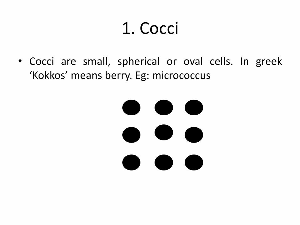
1. Cocci
• Cocci are small, spherical or oval cells. In greek‘Kokkos’ means berry. Eg: micrococcus

2. Bacilli• They are rod shaped cells. Eg: Bacillus anthracis.
• It is derived from greek word “ Bacillus” meaning stick.
• In some of the bacilli the length of cell may be equal towidth. Such bacillary forms are known as coccobacilli.Eg: Bracella.

3. Vibrios
• They are comma shaped curved rods. Eg: Vibriocomma.
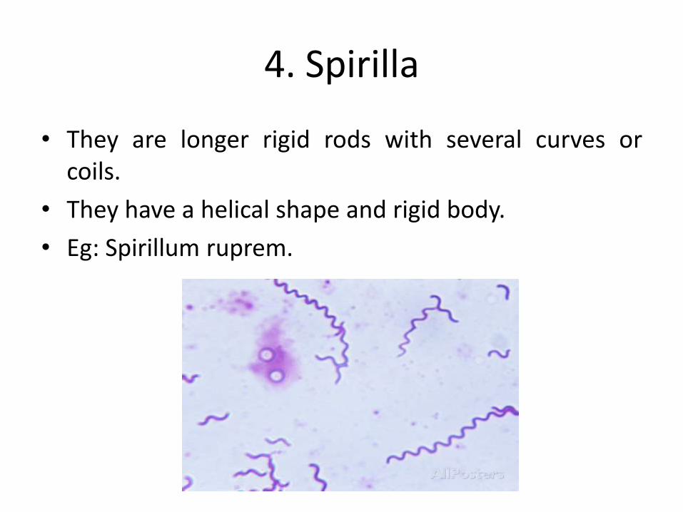
4. Spirilla
• They are longer rigid rods with several curves orcoils.
• They have a helical shape and rigid body.
• Eg: Spirillum ruprem.

5. Spirochetes
• They are slender and flexuous spiral forms.
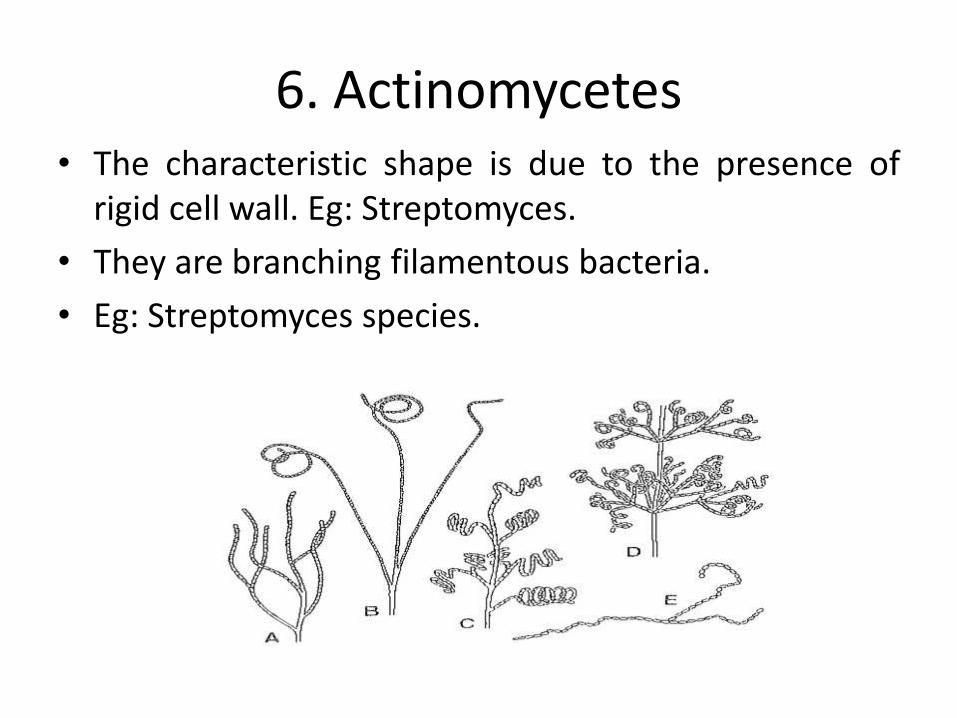
6. Actinomycetes• The characteristic shape is due to the presence of
rigid cell wall. Eg: Streptomyces.
• They are branching filamentous bacteria.
• Eg: Streptomyces species.
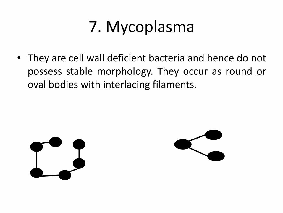
7. Mycoplasma
• They are cell wall deficient bacteria and hence do notpossess stable morphology. They occur as round oroval bodies with interlacing filaments.

ARRANGEMENT OF BACTERIAL CELLS
Cocci appears as several characteristicsarrangement or grouping.
1.Diplococci
2.Streptococci
3.Tetracocci
4.Staphylococci
5.Sarcinae
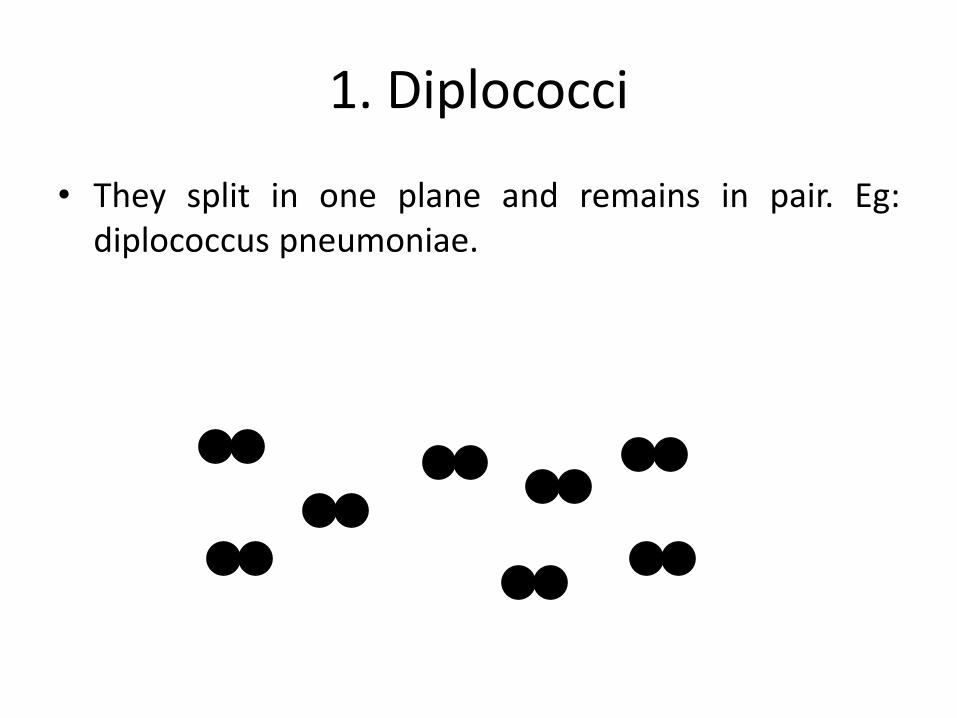
1. Diplococci
• They split in one plane and remains in pair. Eg:diplococcus pneumoniae.

2. Streptococci
These cells divide in one planes and remain attached ,to form chains. Eg: streptococcus lactis.
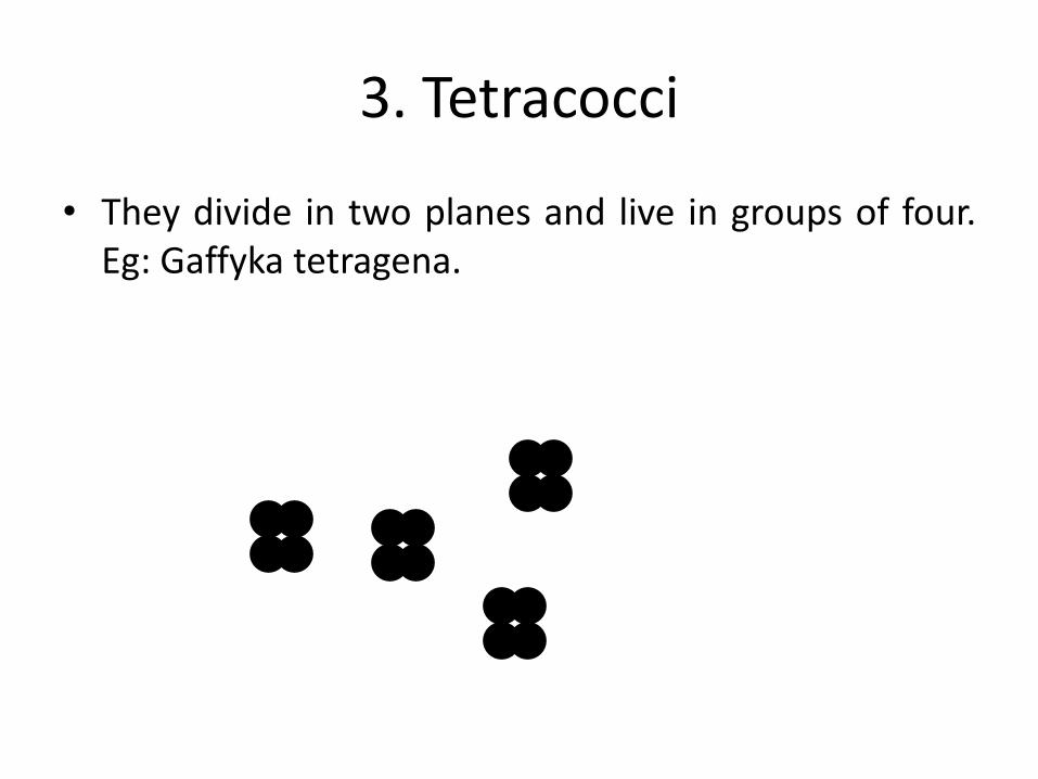
3. Tetracocci
• They divide in two planes and live in groups of four.Eg: Gaffyka tetragena.

4. Staphylococci • Cocci cells divide in three planes in an irregular
pattern. These cells produce bunches of cocci as ingrapes. Eg: staphylococcus aureus, staphylococcusalbus.
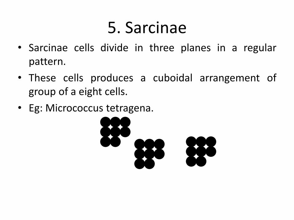
5. Sarcinae• Sarcinae cells divide in three planes in a regular
pattern.
• These cells produces a cuboidal arrangement ofgroup of a eight cells.
• Eg: Micrococcus tetragena.

Arrangement of grouping formed by bacilli species
1. Diplobacilli
2. Streptobacilli
3. Trichomes
Trichomes
Diplobacilli
streptobacilli
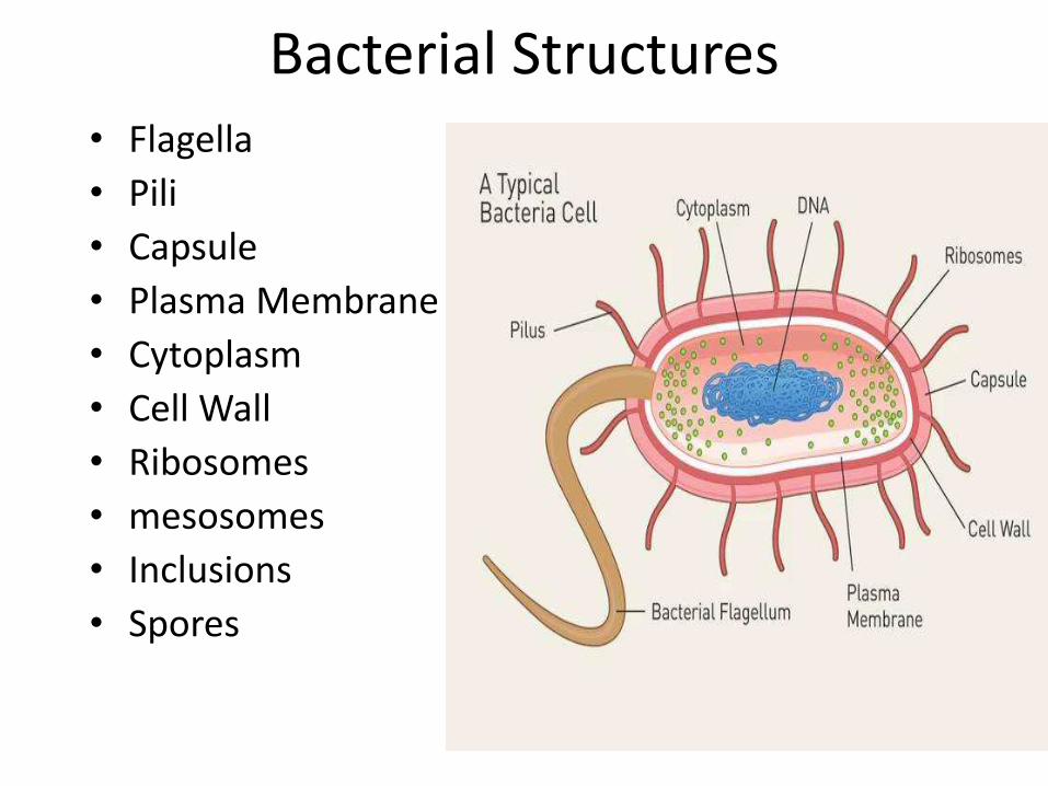
Bacterial Structures• Flagella
• Pili
• Capsule
• Plasma Membrane
• Cytoplasm
• Cell Wall
• Ribosomes
• mesosomes
• Inclusions
• Spores
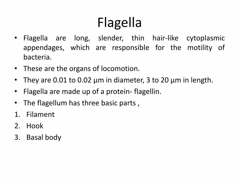
Flagella• Flagella are long, slender, thin hair-like cytoplasmic
appendages, which are responsible for the motility ofbacteria.
• These are the organs of locomotion.
• They are 0.01 to 0.02 µm in diameter, 3 to 20 µm in length.
• Flagella are made up of a protein- flagellin.
• The flagellum has three basic parts ,
1. Filament
2. Hook
3. Basal body
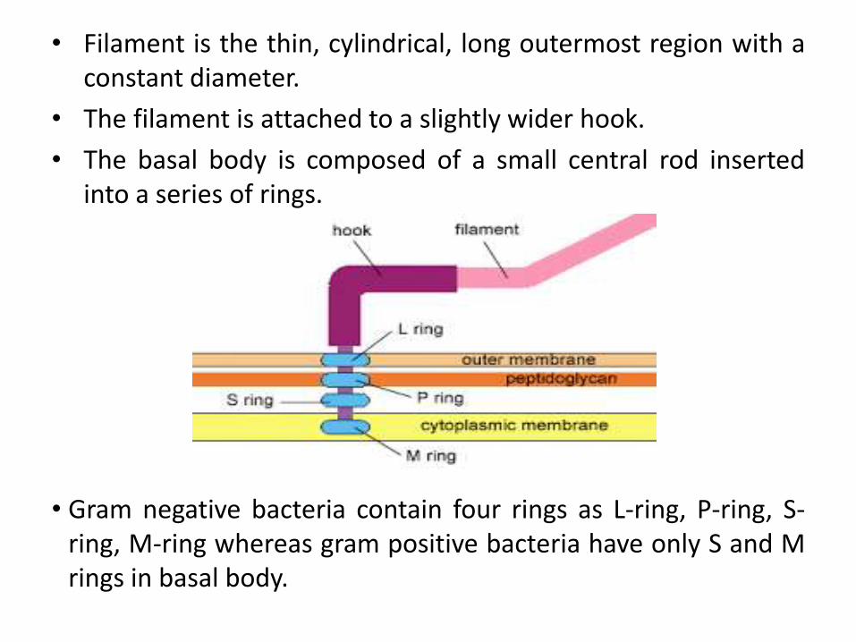
• Filament is the thin, cylindrical, long outermost region with aconstant diameter.
• The filament is attached to a slightly wider hook.
• The basal body is composed of a small central rod insertedinto a series of rings.
• Gram negative bacteria contain four rings as L-ring, P-ring, S-ring, M-ring whereas gram positive bacteria have only S and Mrings in basal body.

Flagella may be seen on bacterial body in following manner.1. Monotrichous: These bacteria have single polar flagellum.
Eg: vibrio cholera2. Lophotrichous: These bacteria have two or more flagella
only at one end of the cell. Eg: pseudomonas fluorescence.3. Amphitrichous: These bacteria have single polar flagella or
tuft of flagella at both poles. Eg :Aquaspirillum serpens.4. Peritrichous: Several flagella present all over the surface of
bacteria. Eg: Escherichia coli, Salmonella typhi.
Pili or fimbriae• Pili are hair-like microfibrils, 0.5 to 2 µm in length and 5 to 7 nm in
diameter.• They are thinner, shorter and more numerous than flagella.• They are present only on gram negative cells.• They are composed of protein known as pillin.• They are unrelated to motility and are found on motile and non-motile
cells.• Fimbriae and pili, these two terms are used interchangeably but they
can be distinguished.• Fimbriae can be evenly distributed over the entire surface of the cell or
they occurs at the poles of the bacterial cell. Each bacteria possess 100to 200 fimbriae.
• Pili are usually longer than fimbriae and number only one or two percell.Function:
• Pili play an important role in attachment to surfaces. Hence pili is alsocalled organ of adhesion.

Plasma membrane• The cytoplasmic (plasma) membrane is a thin ( 5 to 10 nm).
• It separates the cell wall and cytoplasm.
• It composed of phospholipids (20 to 30 %) and proteins ( 60 to70 %).
• Prokaryotic plasma membranes are less rigid than eukaryoticmembrane due to lack of sterols.
Functions:
1. It acts as a semipermeable membrane controlling the inflowand outflow of metabolites to and from the protoplasm.
2. It provides the mechanical strength to the bacterial cell.
3. It helps in DNA replication.
4. It contains enzyme, permease, which plays an important rolein the passage of selective nutrients through the membranes.

Cytoplasm• The bacterial cytoplasm is a suspension of
organic, inorganic solutes in a viscous watersolution.
• The cytoplasm of bacteria differs from that ofhigher eukaryotic microorganisms in notcontaining endoplasmic reticulum, Golgiapparatus, mitochondria and lysosomes.
• It contains the ribosomes, proteins and otherwater soluble components and reserve material.
• In most bacterial, extrachromosal DNA ( plasmidDNA ) is also present.
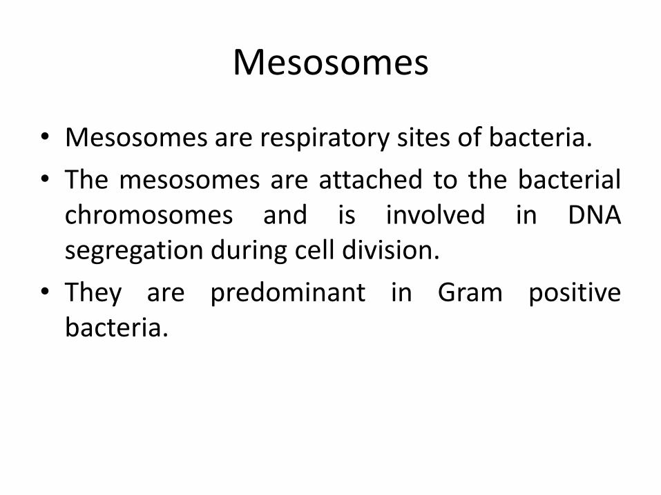
Mesosomes
• Mesosomes are respiratory sites of bacteria.
• The mesosomes are attached to the bacterialchromosomes and is involved in DNAsegregation during cell division.
• They are predominant in Gram positivebacteria.
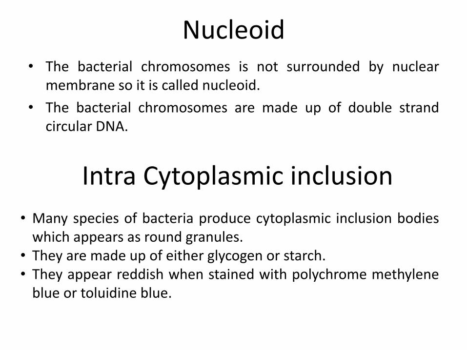
Nucleoid• The bacterial chromosomes is not surrounded by nuclear
membrane so it is called nucleoid.
• The bacterial chromosomes are made up of double strandcircular DNA.
Intra Cytoplasmic inclusion
• Many species of bacteria produce cytoplasmic inclusion bodieswhich appears as round granules.
• They are made up of either glycogen or starch.• They appear reddish when stained with polychrome methylene
blue or toluidine blue.

Capsule• Bacteria synthesize loose amorphous organic exopolymer
which is deposited outside and tightly to cell wall calledcapsules.
• Capsules may be composed of complex polypeptides orpolysaccharides. Water (98%) is the main component ofbacterial capsule.
• Some times the capsular material is loosely associated withthe bacterium, it can be easily washed away. The loose layer iscalled slime layer.
• Capsulated bacteria produces smooth colonies and noncapsulated bacteria produces rough colonies on the surface ofagar media.
Functions
1. They protect the cell from drying.
2. They protects the bacterial cell against anti-bacterial agentsand phages.
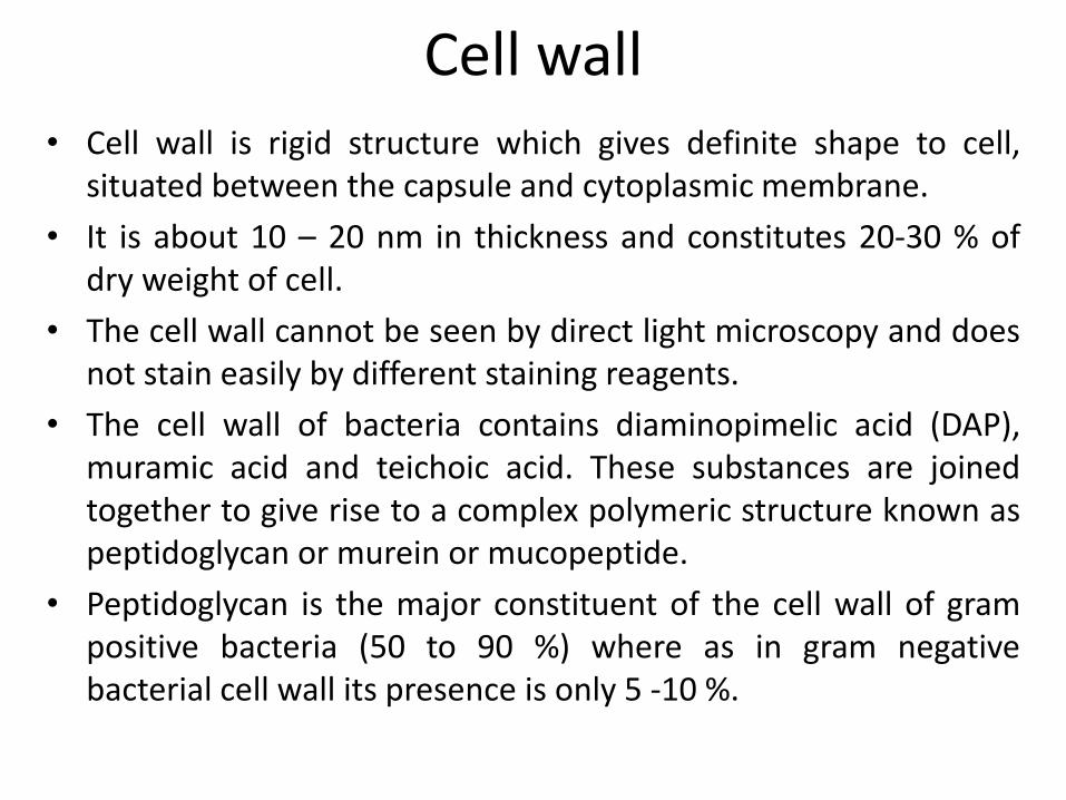
Cell wall• Cell wall is rigid structure which gives definite shape to cell,
situated between the capsule and cytoplasmic membrane.
• It is about 10 – 20 nm in thickness and constitutes 20-30 % ofdry weight of cell.
• The cell wall cannot be seen by direct light microscopy and doesnot stain easily by different staining reagents.
• The cell wall of bacteria contains diaminopimelic acid (DAP),muramic acid and teichoic acid. These substances are joinedtogether to give rise to a complex polymeric structure known aspeptidoglycan or murein or mucopeptide.
• Peptidoglycan is the major constituent of the cell wall of grampositive bacteria (50 to 90 %) where as in gram negativebacterial cell wall its presence is only 5 -10 %.

Chemical structure of a bacterial cell wall
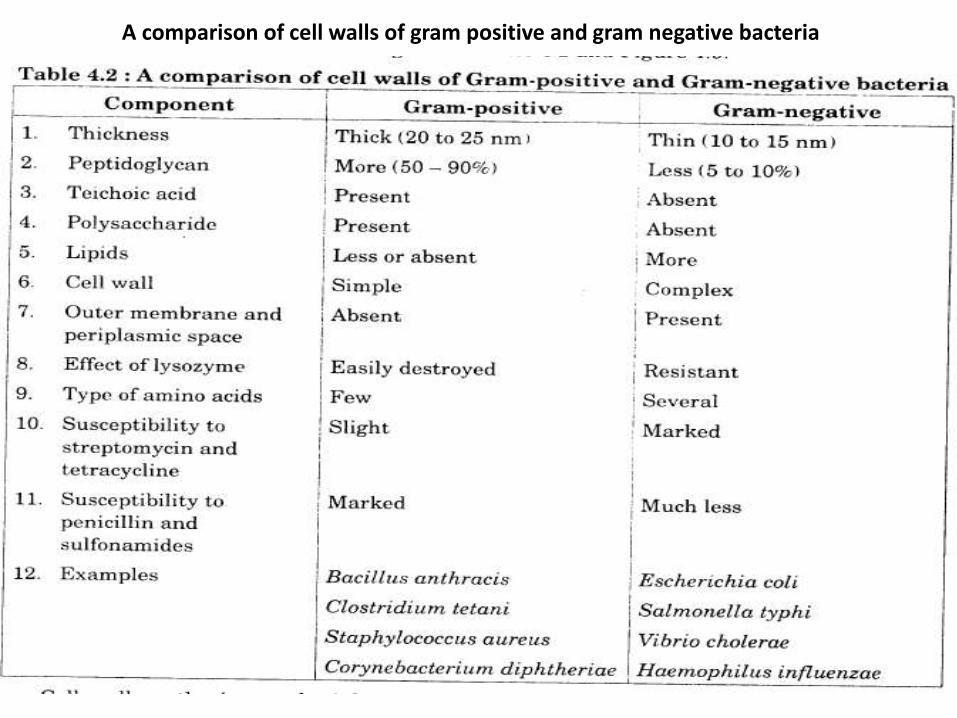
A comparison of cell walls of gram positive and gram negative bacteria
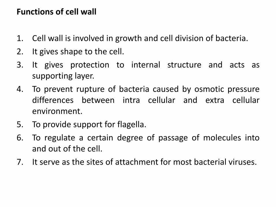
Functions of cell wall
1. Cell wall is involved in growth and cell division of bacteria.
2. It gives shape to the cell.
3. It gives protection to internal structure and acts assupporting layer.
4. To prevent rupture of bacteria caused by osmotic pressuredifferences between intra cellular and extra cellularenvironment.
5. To provide support for flagella.
6. To regulate a certain degree of passage of molecules intoand out of the cell.
7. It serve as the sites of attachment for most bacterial viruses.

Ribosomes• Ribosomes are the center of protein synthesis.
• They are slightly smaller than eukaryotic ribosomes.
• The sedimentation constant is 70s.
• This 70s ribosomes are made up of two subunits namely alarge subunits 50s and a small subunit 30s.
30s
50s
• During active protein synthesis the ribosomes are associatedwith mRNA and such associations are called polysomes.
mRNA
Polysomes
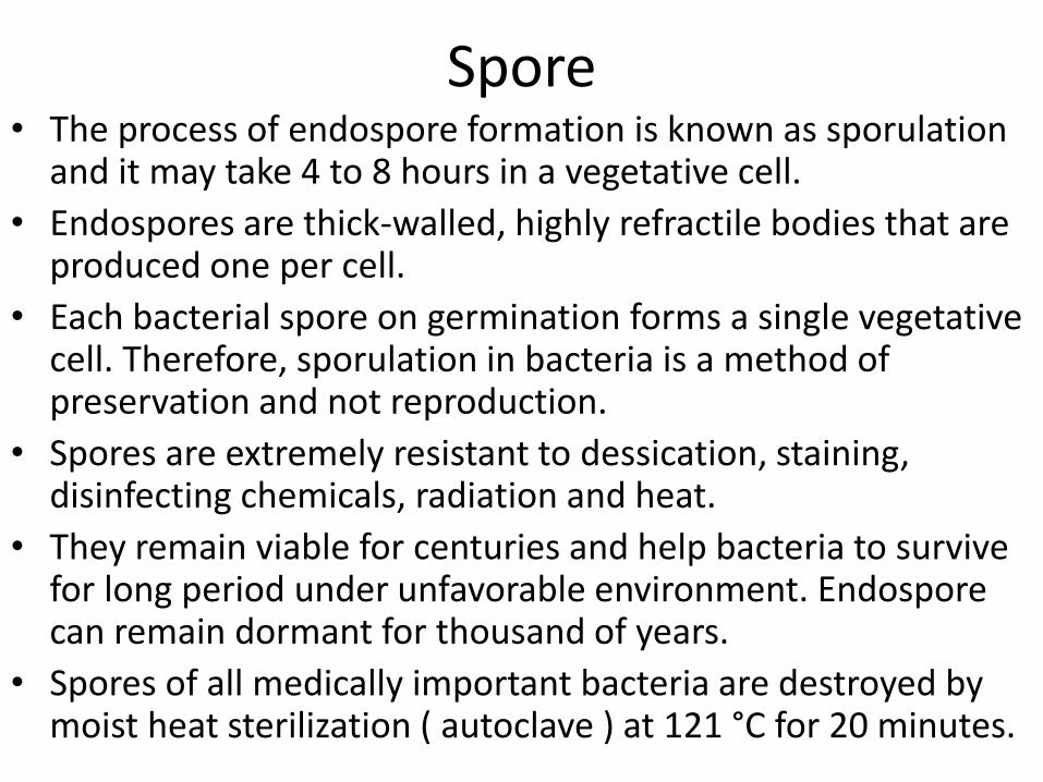
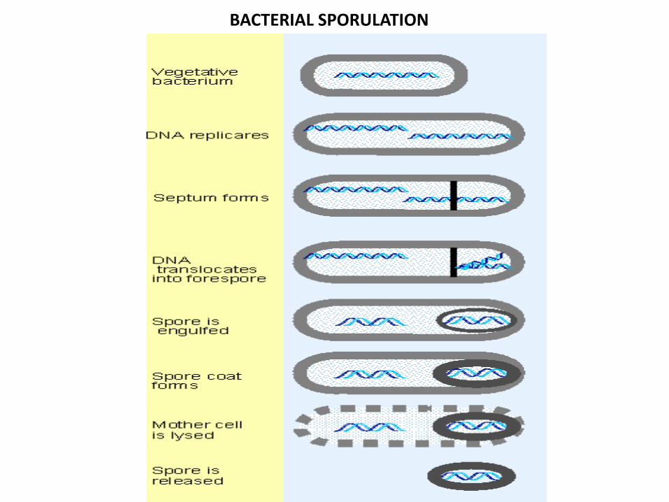
Spore • The process of endospore formation is known as sporulation
and it may take 4 to 8 hours in a vegetative cell.
• Endospores are thick-walled, highly refractile bodies that are produced one per cell.
• Each bacterial spore on germination forms a single vegetative cell. Therefore, sporulation in bacteria is a method of preservation and not reproduction.
• Spores are extremely resistant to dessication, staining, disinfecting chemicals, radiation and heat.
• They remain viable for centuries and help bacteria to survive for long period under unfavorable environment. Endosporecan remain dormant for thousand of years.
• Spores of all medically important bacteria are destroyed by moist heat sterilization ( autoclave ) at 121 °C for 20 minutes.
BACTERIAL SPORULATION
Recommended